Uni Internationa! - Home - The University of Arizona...
57
EVALUATION OF ION-EXCHANGE RESINS FOR THE TREATMENT OF ENTERIC HYPEROXALURIA. Item Type text; Thesis-Reproduction (electronic) Authors Detlefs, Corey Lane. Publisher The University of Arizona. Rights Copyright © is held by the author. Digital access to this material is made possible by the University Libraries, University of Arizona. Further transmission, reproduction or presentation (such as public display or performance) of protected items is prohibited except with permission of the author. Download date 15/06/2018 10:52:03 Link to Item http://hdl.handle.net/10150/274889
Transcript of Uni Internationa! - Home - The University of Arizona...
EVALUATION OF ION-EXCHANGE RESINS FORTHE TREATMENT OF ENTERIC HYPEROXALURIA.
Item Type text; Thesis-Reproduction (electronic)
Authors Detlefs, Corey Lane.
Publisher The University of Arizona.
Rights Copyright © is held by the author. Digital access to this materialis made possible by the University Libraries, University of Arizona.Further transmission, reproduction or presentation (such aspublic display or performance) of protected items is prohibitedexcept with permission of the author.
Download date 15/06/2018 10:52:03
Link to Item http://hdl.handle.net/10150/274889

INFORMATION TO USERS
This reproduction was made from a copy of a document sent to us for microfilming. While the most advanced technology has been used to photograph and reproduce this document, the quality of the reproduction is heavily dependent upon the quality of the material submitted.
The following explanation of techniques is provided to help clarify markings or notations which may appear on this reproduction.
1.The sign or "target" for pages apparently lacking from the document photographed is "Missing Page(s)". If it was possible to obtain the missing page(s) or section, they are spliced into tiie film along with adjacent pages. This may have necessitated cutting through an image and duplicating adjacent pages to assure complete continuity.
2. When an image on the film is obliterated with a round black mark, it is an indication of either blurred copy because of movement during exposure, duplicate copy, or copyrighted materials that should not have been filmed. For blurred pages, a good image of the page can be found in the adjacent frame. If copyrighted materials were deleted, a target note will appear listing the pages in the adjacent frame,
3. When a map, drawing or chart, etc., is part of the material being photographed, a definite method of "sectioning" the material has been followed. It is customary to begin filming at the upper left hand corner of a large sheet and to continue from left to right in equal sections with small overlaps. If necessary, sectioning is continued again—beginning below the first row and continuing on until complete.
4. For illustrations that cannot be satisfactorily reproduced by xerographic means, photographic prints can be purchased at additional cost and inserted into your xerographic copy. These prints are available upon request from the Dissertations Customer Services Department.
5. Some pages in any document may have indistinct print. In all cases the best available copy has been filmed.
Uni
Internationa! 300 N. Zeeb Road Ann Arbor, Ml 48106

1321801
DETLEFS, COREY LANE
EVALUATION OF ION-EXCHANGE RESINS FOR THE TREATMENT OF ENTERIC HYPEROXALURIA
THE UNIVERSITY OF ARIZONA M.S. 1983
University Microfilms
International 300 N. Zeeb Road, Ann Arbor. Ml 48106

PLEASE NOTE:
In all cases this material has been filmed in the best possible way from the available copy. Problems encountered with this document have been identified here with a check mark V .
1. Glossy photographs or pages
2. Colored illustrations, paper or print
3. Photographs with dark background ^
4. Illustrations are poor copy
5. Pages with black marks, not original copy
6. Print shows through as there is text on both sides of page
7. Indistinct, broken or small print on several pages
8. Print exceeds margin requirements
9. Tightly bound copy with print lost in spine
10. Computer printout pages with indistinct print
11. Page(s) lacking when material received, and not available from school or author.
12. Page(s) seem to be missing in numbering only as text follows.
13. Two pages numbered . Text follows.
14. Curling and wrinkled pages
15. Other
University Microfilms
International


EVALUATION OF ION-EXCHANGE RESINS FOR THE
TREATMENT OF ENTERIC HYPEROXALURIA
by
Corey Lane Detlefs
A Thesis Submitted to the Faculty of the
DEPARTMENT OF PHARMACOLOGY AND TOXICOLOGY
In Partial Fulfillment of the Requirements For the Degree of
MASTER OF SCIENCE
In the Graduate College
THE UNIVERSITY OF ARIZONA
19 8 3

STATEMENT BY AUTHOR
This thesis has been submitted in partial fulfillment of requirements for an advanced degree at The University of Arizona and is deposited in the University Library to be made available to borrowers under rules of the Library.
Brief quotations from this thesis are allowable without special permission, provided that accurate acknowledgment of source is made. Requests for permission for extended quotation from or reproduction of this manuscript in whole or in part may be granted by the head of the major department or the Dean of the Graduate College when in his judgment the proposed use of the material is in the interests of scholarship. In all other instances, however, permission must be obtained from the author.
APPROVAL BY THESIS DIRECTOR
This thesis has been approved on the date shown below:
7/fr? A5 DAVID L. EARNEST x ^ Date
Associate Professor of Internal Medicine
ACKNOWLEDGMENTS
I wish to express my sincerest gratitude to Doctor David L.
Earnest for his intellectual guidance and financial support which made
this project possible; apart from serving as my thesis advisor
Doctor Earnest was a friend who helped make all aspects of my graduate
work more rewarding.
In addition, I wish to thank Doctor Dean E. Carter for his
guidance in chemistry and Doctor Donald De Young for his superb
instruction in small animal surgery.
I also wish to thank Doctors Marguerite Hatch and Robert Freel
for their instruction on the finer points of science and for their
company which made my research more pleasant.
iii

TABLE OF CONTENTS
Page
LIST OF ILLUSTRATIONS . . vi
LIST OF TABLES vii
ABSTRACT . . . viii
INTRODUCTION . 1
Sources of Urinary Oxalate 2 Permeability Theory A Solubility Theory ..... 4
Therapeutic Efforts , , . 5 Ion-Exchange Resins 6
Characterization of Anion-Exchange Resins 8 Statement of Problem 9
MATERIALS AND METHODS 10
Initial Screening of Resins 10 Swelling of Resins 11 Conversion of Cation-Exchange Resins to Calcium Form 11 Cation-Exchange Resin Binding of Oxalate , . 12 Characterization of Anion-Exchange Resins 12 Fatty Acid Inhibition of Oxalate Binding ..... , 12 Competition between Oleate and Oxalate for Resin
Binding Sites ............. 13 Jejunoileal Bypass Surgery . . , 13 Bile Duct Ligation Surgery 14 Evaluation of Animal Model 15 Fecal Fat Analysis . 15 Analysis of Oxalate Binding by Cholestyramine in Stool .... 16 Evaluation of Enteric Coated Capsule . 17
RESULTS AND DISCUSSION 18
Qualitative Screening of Resins 18 Cation-Exchange Resins , 18 Characterization of Anion-Exchange Resins ........... 21 Enteric Coated Capsules . . 26 Animal Model 29 Inhibition of Oxalate Binding 32 Analysis of Oxalate Binding by Cholestyramine in Stool .... 35
iv

V
TABLE OF CONTENTS—Cont inued.
Page
Protocol for the Patient Study 37 Conclusions , 38
REFERENCES AO
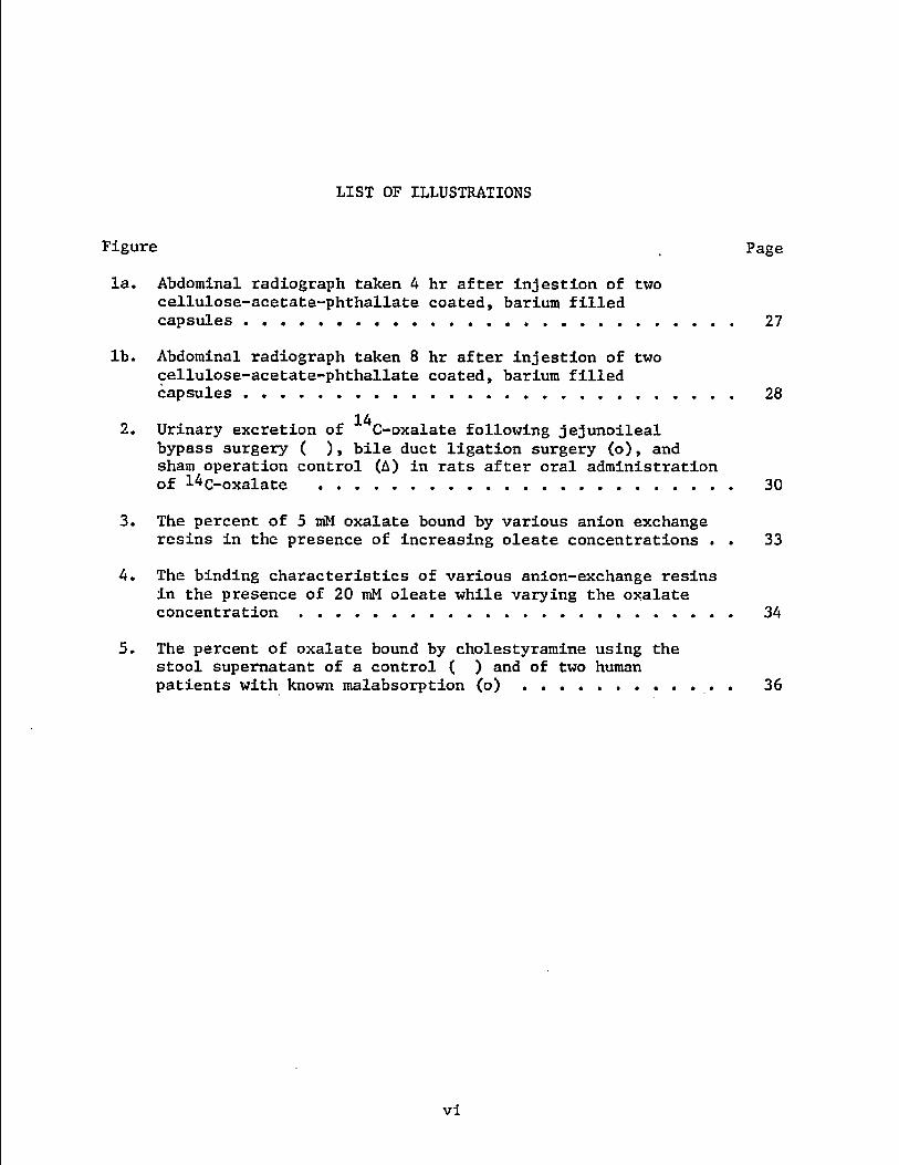
LIST OF ILLUSTRATIONS
Figure Page
la. Abdominal radiograph taken 4 hr after injestion of two cellulose-acetate-phthallate coated, barium filled capsules 27
lb. Abdominal radiograph taken 8 hr after injestion of two cellulose-acetate-phthallate coated, barium filled capsules 28
14 2. Urinary excretion of C-oxalate following jejunoileal
bypass surgery ( ), bile duct ligation surgery (o), and sham operation control (A) in rats after oral administration of -^C-oxalate 30
3. The percent of 5 mM oxalate bound by various anion exchange resins in the presence of increasing oleate concentrations . . 33
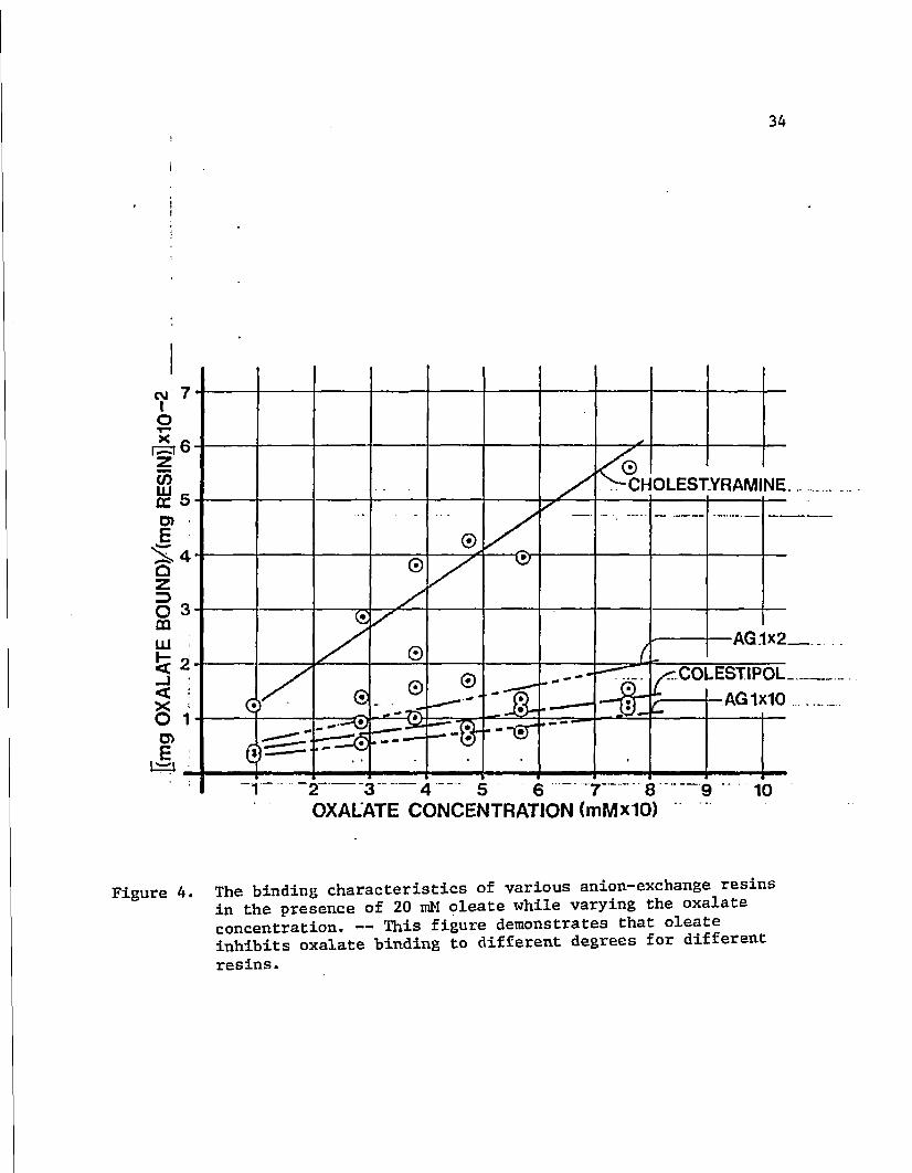
4. The binding characteristics of various anion-exchange resins in the presence of 20 mM oleate while varying the oxalate concentration 34
5. The percent of oxalate bound by cholestyramine using the stool supernatant of a control ( ) and of two human patients with known malabsorption (o) . 36
vi

LIST OP TABLES
Table Page
1. Oxalic acid content of some foods 3
2. Anion-exchange resins 19
3. Cation-exchange resins 20
4. Cation-exchange resins converted to calcium form 22
vii

ABSTRACT
Enteric hyperoxaluria is defined as an elevated urinary oxalate
excretion secondary to increased intestinal absorption. Patients with
enteric hyperoxaluria have an increased propensity for developing
calcium oxalate kidney stones. Clinically, a number of efforts have
been undertaken to decrease the intestinal absorption of oxalate,
including the use of the anion-exchange resin cholestyramine. Thera
peutic efforts have met with inconsistent success. The oxalate
binding affinities and capacities for cholestyramine and a number of
other ion-exchange resins were determined in vitro. Comparison of these
parameters suggest that cholestyramine is the most effective resin for
the binding of oxalate. Even in the presence of fat cholestyramine
bound more oxalate than did the other resins tested. It is suggested
that severe steatorrhea seen in patients with enteric hyperoxaluria may
account for the inability of cholestyramine to reduce urinary oxalate
excretion in previous studies.
viii

INTRODUCTION
In 1978 it was estimated that 88.6% of the kidney stones
occurring in the United States contained oxalate as a major component
(1). Oxalic acid is a naturally occurring dicarboxylic acid which forms
-9 an extremely insoluble calcium salt (Ksp 2.3x10 ). In man, the major
source of oxalate is the metabolism of ascorbate and glyoxylate (2).
Additionally, a small amount of oxalate is absorbed from the diet.
Since oxalate is a metabolic end product, essentially all absorbed or
synthesized oxalate is excreted in the urine (2). In addition to
enhancing renal stone formation, calcium oxalate crystals can deposit in
the renal tubules and parenchyma, and may produce pain, hematuria, upper
urinary tract infection and, in severe cases, progressive renal failure
(3,4). Any condition which results in enhanced oxalate synthesis or
intestinal oxalate absorbtion will cause increased urinary oxalate
excretion (hyperoxaluria) and thereby increase the risk of these
problems.
For many years it was thought that hyperoxaluria was an
uncommon phenomenon and associated mainly with rare genetic disorders
of oxalate metabolism or with accidental ingestion of large quantities
of oxalic acid or its precursors (i.e. ethylene glycol). For example,
in primary hyperoxaluria Type I, large quantities of oxalate and
glyoxylate are produced secondary to an enzymatic defect the conversion
of glyoxylate to a-ketoadipate (5). This condition is characterized
clinically by hyperoxaluria, recurrent calcium oxalate nephrolithiases,
1
2
chronic renal failure and death usually before age 20 due to renal
failure and uremia (5).
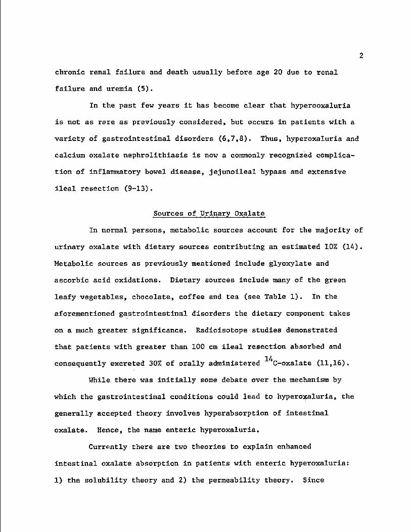
In the past few years it has become clear that hyperooxaluria
is not as rare as previously considered, but occurs in patients with a
variety of gastrointestinal disorders (6,7,8). Thus, hyperoxaluria and
calcium oxalate nephrolithiasis is now a commonly recognized complica
tion of Inflammatory bowel disease, jejunoileal bypass and extensive
ileal resection (9-13).
Sources of Urinary Oxalate
In normal persons, metabolic sources account for the majority of
urinary oxalate with dietary sources contributing an estimated 10% (14).
Metabolic sources as previously mentioned include glyoxylate and
ascorbic acid oxidations. Dietary sources include many of the green
leafy vegetables, chocolate, coffee and tea (see Table 1). In the
aforementioned gastrointestinal disorders the dietary component takes
on a much greater significance. Radioisotope studies demonstrated
that patients with greater than 100 cm ileal resection absorbed and
consequently excreted 30% of orally administered "^C-oxalate (11,16).
While there was initially some debate over the mechanism by
which the gastrointestinal conditions could lead to hyperoxaluria, the
generally accepted theory involves hyperabsorptlon of intestinal
oxalate. Hence, the name enteric hyperoxaluria.
Currently there are two theories to explain enhanced
intestinal oxalate absorption in patients with enteric hyperoxaluria:
1) the solubility theory and 2) the permeability theory. Since
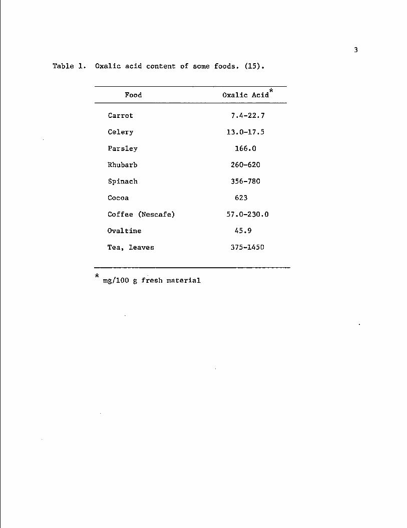
Table 1. Oxalic acid content of some foods. (15).
3
* Food Oxalic Acid
Carrot 7.4-22.7
Celery 13.0-17.5
Parsley 166.0
Rhubarb 260-620
Spinach 356-780
Cocoa 623
Coffee (Nescafe) 57.0-230.0
Ovaltine 45.9
Tea, leaves 375-1450
A mg/100 g fresh material
k
in vitro and in vivo evidence support both, it is likely that both
contribute to enhanced intestinal oxalate absorption (36).
Permeability Theory
It has been established that patients with extreme ilial
resection have malabsorption of both bile and fatty acids (17). It has
also been established that certain of the bile acids, especially 3a,
7a and 3al2a-ones, alter colonic membrane permeability (18). Large
molecular weight compounds such as vitamin readily move
through the colonic mucosal membranes and therefore into the portal
circulation as a result of exposure to these dihydroxy bile salts.
0 Soluble oxalate salts (<5A) are also capable of diffusing through the
colonic mucosa under these conditions. Thus, enhanced absorption of
oxalate would increase urinary excretion producing hyperoxaluria.
Whether the concentration of bile acids required in "vitro to produce
these permeability changes are similar to concentrations present
in vivo is not yet established.
Solubility Theory
Both oxalate and fatty acids form insoluble and consequently
non-absorbable calcium salts. However, in vitro studies have clearly
shown that at intestinal pH, the reaction favors formation of the
calcium salt of the fatty acid over the oxalate salt (19). Therefore,
as the amount of fat malabsorption increases, calcium precipitates
forming fatty acid soaps and the calcium activity is reduced, thus
decreasing the calcium available to precipitate oxalate. As a

5
consequence more soluble oxalate salts are formed. These remain in
solution and available for absorption by the intestinal mucosa.
Ileal resection and jejunoileal bypass cause fat malabsorption and can
thereby produce enteric hyperoxaluria. In added support of this theory
is the fact that under metabolic ward conditions intestinal oxalate
absorption and subsequent urinary oxalate excretion can be varied
simply by changing the degree of fatty acid malabsorption during an
otherwise constant diet (11).
Therapeutic Efforts
Clinically a number of methods have been used to reduce urinary
oxalate excretion in patients with enteric hyperoxaluria and thereby
decrease the likelihood that they will develop calcium oxalate kidney
stones. One method has been a diet low in oxalate. This has not been
successful especially for outpatient management. The oxalate content
of many foods has yet to be determined. Additionally, patients seldom
comply with strict dietary restrictions.
Pharmacologic efforts in the past have included oral administra
tion of supplemental calcium as well as the anion exchange resin
cholestyramine, both given to remove oxalate from solution and thus
prevent its intestinal absorption. While calcium supplementation
effectively reduces urinary oxalate excretion in some patients under a
controlled metabolic ward setting, this treatment has the theoretical
disadvantage of increasing urinary calcium excretion (20,21). If
urinary calcium increased more than oxalate was reduced, the activity
product of these ions could exceed the formation product of calcium

6
oxalate (2). Thus calcium supplementation treatment could theoretically
enhance renal stone formation (21,22). Some investigators have found
that the anion-exchange resin cholestyramine effectively controls
enteric hyperoxaluria, others have not (11,23-27). Thus, since there
is no predictably successful method to bind oxalate in the intestinal
lumen and thus prevent its absorption, the principle goal of this thesis
project was to find a more effective method of complexing oxalate in a
non-absorbable form in the intestinal solution. Specifically, I chose
to evaluate the properties of various ion-exchange resins in order to
1) determine if a better ion exchange resin than cholestyramine
existed for binding oxalate, 2) to determine if resin pore size could
modify oxalate trapping, and 3) to determine whether cationic functional
groups could successfully remove oxalate from solution based on
relative affinities for oxalate.
Ion-Exchange Resins (28)
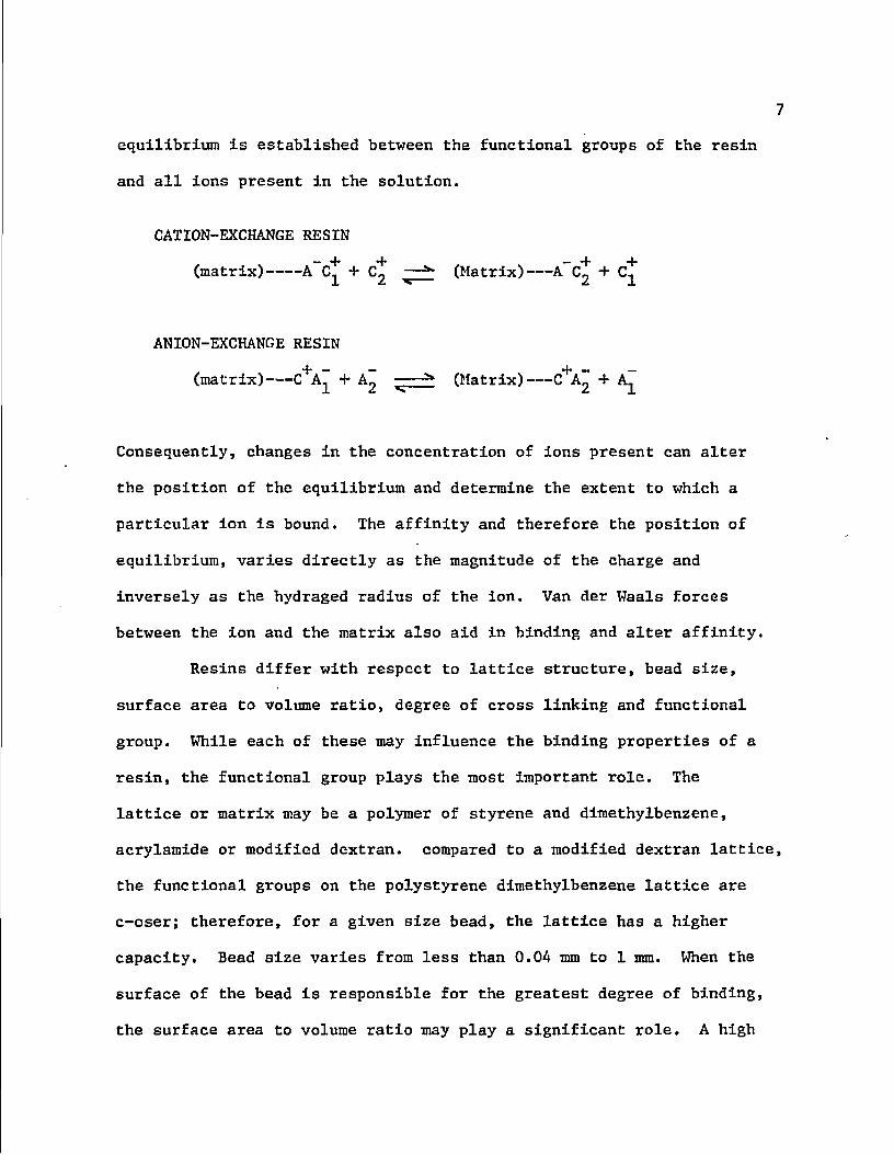
Ion-exchange resins consist of an elastic, three-dimensional
hydrocarbon lattice or matrix to which ionizable groups are covalently
bonded. The hydrocarbon lattice is usually Insoluble in most laboratory
solvents and chemically inert. The ionizable groups are usually acids
or bases and are responsible for binding ions. In normal ion-exchange
procedures, the counter-ion is displaced from the resin functional
group and replaced by an ion of the same charge sign. How well an
ion-exchange resin binds a desired ion depends upon the concentration
of all ions present in the solution, properties of the ions and
properties of the resin. Just as in all chemical reactions, an
7
equilibrium is established between the functional groups of the resin
and all ions present in the solution.
CATION-EXCHANGE RESIN
(matrix) A C* + C* (Matrix) A C^ + C^
ANION-EXCHANGE RESIN
(matrix) C+A^ + ̂2 „ N (Matrix) C+A2 + A^
Consequently, changes in the concentration of ions present can alter
the position of the equilibrium and determine the extent to which a
particular ion is bound. The affinity and therefore the position of
equilibrium, varies directly as the magnitude of the charge and
inversely as the hydraged radius of the ion. Van der Waals forces
between the ion and the matrix also aid in binding and alter affinity.
Resins differ with respect to lattice structure, bead size,
surface area to volume ratio, degree of cross linking and functional
group. While each of these may influence the binding properties of a
resin, the functional group plays the most important role. The
lattice or matrix may be a polymer of styrene and dimethylbenzene,
acrylamide or modified dextran. compared to a modified dextran lattice,
the functional groups on the polystyrene dimethylbenzene lattice are
c-oser; therefore, for a given size bead, the lattice has a higher
capacity. Bead size varies from less than 0.04 mm to 1 mm. When the
surface of the bead is responsible for the greatest degree of binding,
the surface area to volume ratio may play a significant role. A high
8
surface area to volume ratio would be most desirable where the volume
of resin used is limited. Cross-linking determines the pore size, the
amount of swelling that will occur in solution as well as the speed of
the exchange process. Where exclusion of large molecular weight
compounds is desirable, the higher degrees of cross linking may be
desirable. Functional groups for cation-exchange resins are usually
weak or strong acids such as carboxylic acids or sulfuric acids,
respectively. For anion-exchange resins, weak and strong bases such as
tertiary and quaternary amines are examples.
Characterization of Anion-Exchange Resins
Ion-exchange resins may vary with respect to binding affinity
and binding capacity for a given ion. Using the concepts of enzyme
kinetics, resins can be characterized and quantitatively compared. A
plot of mg oxalate bound per mg resin at varying oxalate concentrations
can be used to determine two parameters: 1) the binding affinity of the
resin for oxalate, or K ; and 2) the maximum binding capacity of the m
resin for oxalate, or (not unlike maximum velocity in enzyme
kinetics). Binding affinity of the resin is defined as the oxalate
concentration at which half the available binding sites are filled.
This is derived from Michealis-Menton kinetics as the Michealis
Constant. Beyond a certain oxalate concentration the amount of oxalate
bound remains constant. The amount of oxalate bound per mg resin at
this oxalate concentration si the Maximum Binding Capacity.
9
Statement of Problem
The first objective of this research project was to determine
if there is an ion-exchange resin available that will bind oxalate
better than cholestyramine. Both anion and cation-exchange resins were
evaluated. The resins evaluated varied in functional group,
counter-ion, pore size and lattice structure. After initial screening
to identify resins with the ability to bind oxalate, the resins were
characterized in vitro. Comparison of the binding affinities and
binding capacities allowed objective evaluation.
The next objective was to develop an animal model for enteric
hyperoxaluria in which the best resin could be evaluated. Using rats,
a jejunoileal bypass and a bile duct ligation was performed to create
fat malabsorption. The urinary oxalate excretion of orally administered
^C-oxalate was determined and compared to that of control rats of
similar weight.
The third, objective was to explain why cholestyramine effectively
reduced enteric hyperoxaluria in some patients and not in others. It
was hypothesized that malabsorbed fatty acids reduced oxalate binding
by the resin. Evaluation of resins in calcium-free Ringers solution,
in the presence of fat, and in stool from control and hyperoxaluric
patients was performed.
The aim of this research project was to develop a rational
basis from which to suggest an ion-exchange resin as a method for
reducing intestinal oxalate absorption. The overall goal was to
develop a better and more effective way of reducing the likelihood of
kidney stone formation in a moderately large segment of our population.
MATERIALS AND METHODS
AG 1X2, AG 1X8, AG 1X10, AGMP, AGMP50 and AG 50WX12 were gifts
of Bio-Rad. The amberlite resins IRA93 and IRC 50 were purchased from
Rohm and Haas. Cholestryamine was a gift of Mead-Johns Laboratories.
Colestipol was purchased from the Upjohn Company. Pharmacia Fine
Chemicals generously supplied DEAE 25, DEAE 50, QAE 25, QAE 50, SP 50,
CM 25 and CM 50. Radiolabeled oxalic acid was purchased from Amersham
Corporation (74 mCi/inmol) .
Calcium-free Ringers solution contained 140 mM sodium,
123.2 mM chloride, 5.4 niM potassium, 2.4 mM magnesium, 21 niM bicar
bonate, 0.6 niM dihydrogen phosphate, 3.4 mM biphosphate and 10 mM
glucose. The pH was adjusted to 7.4 with 95% 02, 5% C0£ gas.
Initial Screening of Resins
Each resin was tested for its iji vitro ability to bind oxalate.
Two hundred mg of resin were placed in a 10 ml glass test tube with
9.5 ml of calcium-free Ringers solution. Made in distilled water,
14 0.5 ml of an oxalate solution containing C-oxalate was added. The
final oxalate concentration was 5 mM. The control was Ringers solution
and 5 mM oxalate without resin. Each tube was gently shaken for 30
mintues in a Coulter Test Tube Shaker, then centrifuged at 3000 rpm for
10 mintues in a Beckman Model T0-6 table top centrifuge. An aliquot of
the supernatant was then added to 5 ml of Beta Phase Scintillation
Cocktail and radioactivity measured by counting for 10 minutes in a
10

11
Tracor Analytic Mark III Liquid Scintillation System. All radio
isotope calculations were done using DPM as supplied by the Mark III.
The percentage of oxalate bound by each resin was determined. Only
resins with the ability to bind oxalate were further characterized.
Swelling of Resins
According to manufacturers specifications DEAE25, DEAE50,
QAE25 and QAE50 required swelling prior to use. This was achieved by
washing 200 mg of resin with calcium-free Ringers solution in the tube
used for the test. Each resin was allowed to swell for 24 hours prior
to use, then the washing solution was removed. While 24 hours was
necessary where the tubes were left standing, it was found that
agitation could greatly reduce the time necessary for maximum swelling.
Conversion of Cation-Exchange Resins to Calcium Form
Amberlite 252 was converted from the sodium to hydrogen form by
sequential washings with concentrated hydrochloric acid. Sequential
washings with a calcium citrate solution were then used to convert the
resin to the calcium form. IRC50 was converted from the hydrogen to
calcium form by sequential washings with a calcium citrate solution.
Each conversion was performed by the batch method and the reaction
followed by determining the pH of the eluent. When the eluent pH was
the same as that of the washing solution, it was assumed that the
conversion was complete.
12
Cation-Exchange Resin Binding of Oxalate
In Ringers solution or distilled water, 200 mg resin were
exposed to 5 thM oxalate containing tracer. Samples were shaken,
centrifuged and counted as previously described. The percentage of
oxalate bound by each resin was determined.
Characterization of Anion-Exchange Resins
Resins were characterized by determining mg oxalate bound per mg
resin at varying oxalate concentrations. A 100 mM oxalate stock
14 solution was made in calcium-free Ringers solution; C-oxalate was
added to obtain a specific activity of 12,500 DPM per mg oxalate.
Oxalate solutions of 80, 60, 50, 40, 30, 20, 10, 5, 2.5 and 0.3125 mM
were made by serial dilutions of the stock oxalate solution; Ringers
solution was used to bring dilutions up to volume. In a 10 ml test
tube were placed 200 mg of resin and 10 ml of the desired oxalate solu
tion. Samples were shaken, centrifuged and radioactivity in the
supernatant determined as previously described. At each oxalate
concentration, resin binding was evaluated in duplicate or triplicate
samples. Mg oxalate bound per mg resin was plotted against oxalate
concentration. From these data k., and B were determined. H max
Fatty Acid Inhibition of Oxalate Binding
Oleate was used to evaluate the effects of fatty acid on oxalate
binding. Oleate concentrations of 24, 16 and 8 mM were made by serial
dilutions. In Ringers solution, 100 mg resin were exposed to oleate
14 in 5 mM oxalate with C-oxalate tracer at a final volume of 10 ml.
13
Samples were shaken, centrifuged and radioactivity in the supernatant
determined as described above. At each oleate concentration, each
resin was evaluated in triplicate. Percent oxalate bound per 100 mg
resin was plotted against oleate concentration.
Competition between Oleate and Oxalate for Resin Binding Sites
In 20 mM oleate, 200 mg resin were exposed to varying oxalate
concentrations in Ringers solution. Oxalate concentrations of 76, 57,
47, 38, 28 and 10 uiM were used. Samples were shaken, centrifuged then
radioactivity determined as previously described. Mg oxalate bound per
mg resin was plotted against oxalate concentration.
Jejunoileal Bypass Surgery
Surgical technique was developed with assistance of Don DeYoung,
D.V.M., Ph.D., of the Arizona Health Sciences Center Department of
Animal Resources. Male Sprague-Dawley rats, 300-400 g were fasted for
24 hours prior to surgery. Anesthesia was achieved with Innovar ,
0.33 cc/kg IM. After shaving, the abdomen was prepped with providine-
iodine solution. Midline incisions, one through the cutaneous tissue
and one through the abdominal muscles and peritoneum (using the linea
alba as a guide), were made. The duodenum and ileum were located. The
duodenum was transected less than 0.5 cm distal to the Ligament of
Tritz. The distal duodenum was then closed using mattress pattern
sutures of 6-0 silk material. Approximately 4 cm proximal to the
cecum, a lateral incision was made in the ileum. This incision was of
sufficient length to form an anastomosis with the open end of the
14
jejunum. Using interrupted sutures of 6-0 silk material, the ileum and
duodenum were joined. The peritoneal contents were then placed in
anatomically correct positions, taking care not to strangle any portion
of the intestine. Using 4-0 chromic catgut suture, the peritoneum and
abdominal muscles were closed, then the cutaneous tissue, each by an
interrupted suture pattern. A prophylactic dose of 30,000 units of
penicillin G was administered IM.
The post-operative rats were returned to wire-bottomed cages
and allowed water ad libitum for 24 hours. After 24 hours the rats
were given Purina rat chow and water acl libitum.
Bile Duct Ligation Surgery
Female Sprague Dawley rats 150-200 g, were anesthetized with
Innovar 0.33 cc/kg IM. The abdomen was shaved, then prepped with
providine-lodine, A midline incision was made through the cutaneous
tissue. Then, following the linea alba, an incision was made through
the abdominal muscle and peritoneum. The duodenum was located and
held up to light to locate the common bile duct. Being careful not to
disrupt the mesenteric vasculature, the bile duct was llgated in two
places using single sutures of 6-0 silk material. The bile duct was
then sectioned. The abdominal contents were then placed in anatomically
correct position and the peritoneum and abdomen closed with 4-0 chromic
catgut sutures. Using the same interrupted suture technique, the
cutaneous tissue was closed.
Rats were returned to wire-bottomed cages and fed Purina rat
chow and water ad libitum.
Evaluation of Animal Model
Jejunoileal bypass, bile duct ligation and sham operated control
rats were allowed to adapt to wire bottomed metabolism cages and then
fasted for 24 hours. Each rat then received by gavage 0.3 cc of a
14 5 mM oxalate solution containing 0.1 pCi of C-oxalate, Rats were
subsequently offered Purina rat chow and water ac[ libitum. Urine was
collected for seven days. An aliquot of each sample was placed in
5 ml of scintillation cocktail, radioactivity determined and counted.
The percent urinary oxalate excretion of the oral oxalate dose was
plotted against time.
Fecal Fat Analysis
The method of Van de Kamer (47) was used to determine the total
fatty acid content of stool. Stool was homogenized with 1-3 volumes by
weight of calcium free Ringers in a blender. Approximately 10 g of the
homogenate were saponified with 10 ml of 6.2 M potassium hydroxide.
40 ml of ethanol containing 4% isoamyl alcohol were then added, and the
resulting mixture shaken, then refluxed for 30 mintues. After allowing
the mixture to cool, the fatty acids were liberated with 10 ml of 8.2 M
hydrochloric acid. The solution was then allowed to cool to room
temperature. Free fatty acids were extracted with 50 ml of petroleum
ether, boiling range 47° to 56° C. A 25 ml aliquot of the ether
extract was evaporated to dryness in a 50 ml Erlenmeyer flask containing
a small piece of filter paper. The fatty acids were resuspended in
10 ml of 99% ethanol, and the solution boiled for less than one minute
to remove carbon dioxide. 250 yl of a 0.2 g/100 ml solution of

16
thymol blue in 50% ethanol were then added, and the solution titrated
with 0.1 N sodium hydroxide until the yellow color turned blue. A
blank of 10 g of 99% ethanol is carried through the same procedure. By
use of the following equation, the fatty acid content of the sample was
calculated.
™ . , . . . , (Vu-Vb) • C • 1.04 • 2 • 265 Total fat in sample (g) =
Vu = volume of titrant added (ml)
Vb = volume of titrant for blank (ml)
C = concentration of titrant (M)
1.04 = conversion factor to account for volume change of ether
phase when ethanol added
2 = to account for other 25 ml of ether not titrated
265 = average molecular weight of fat in stool
1000 = convert: ml to liter of titrant
Analysis of Oxalate Binding by Cholestyramine in Stool
Stool samples from patients wity varying degrees of malabsorp
tion were homogenized in an equal weight of calcium-free Ringers
solution. Each sample was then divided and poured into 50 ml poly
styrene tubes, then centrifuged in a Beckman TJ-6 table top centrifuge
at 3000 rpm for 20 minut.es. The supernatants were then frozen until
use.
A 10 g aliquot of each sample was analyzed for total fat content
using the method of Van de Kamer. To each of four more 10 mg samples
14 0.5 pCi of C-oxalate were added. To each of two of these samples
17
was added 200 mg cholestyramine. All tubes were shaken for one hour,
then centrifuged at 3000 rpm for 10 minutes. An aliquot of each sample
was added to 5 ml scintillation cocktail and counted. From these data
the binding of oxalate could be quantified for each of the stool
supernatant and cholestyramine. Binding to cholestyramine was then
plotted against total fat in the supernatant.
Evaluation of Enteric Coated Capsule
Barium filled gelatin capsules were coated with cellulose-
acetate-phthallate by the College of Pharmacy. Two human volunteers
took two capsules each with a meal. Plain abdominal X-rays were taken
four and eight hours after ingestion to determine the integrity and
position of the capsule in the gastrointestinal tracts. X-Rays were
evaluated by the Radiology Department, A. H. S. C.

RESULTS AND DISCUSSION
Qualitative Screening of Resins
Fecal water is extremely variable in composition and difficult
to accurately simulate. Ringers solution was used as the medium in
these studies since Ringers qualitatively simulates the ionic environ
ment of the intestinal solution without the variability encountered
using stool samples. Calcium was eliminated from the Ringers in order
to prevent loss of oxalate due to precipitation. Oxalate binding and
solubility could thus be accurately attributed to the experimental
variables. Both anion and cation-exchange resins were screened to
determine oxalate binding in vitro. Table 2 shows the anion-exchange
resins screened and the percent of 5 iriM oxalate bound by each. The
results show that oxalate was bound to some extent by most of the
resins without specific advantage to lattice structure, functional
group or pore size.
Table 3 shows the percent of 5 mM oxalate bound by the cation-
exchange resins as supplied by the manufacturers. No modifications of
counter ions were made. As would be expected cation-exchange resins
with counter ions that do not produce an insoluble complex with oxalate
showed no oxalate removed from solution.
Cation-Exchange Resins
In normal ion-exchange procedures the desired ion binds directly
to the functional group after displacing the counter-ion. As supplied
18

19
Table 2. AnIon-exchange resins. — The percent of 5 mM oxalate bound to 200 mg resin in Ringers solution. Functional groups: (a) tertiary amine; (b) quaternary amine.
Resin Functional Group % Oxalate Bound
AG 1X2 b 31
AG 1X8 b 47
AG 1X10 b 38
AG MP1 b 33
Cholestyramine b 48
Colestipol b 42
DEAE25 a 53
DEAE50 a 25
IRA93 a 0
QAE25 b 33
QAE50 b 43

20
Cation-exchange resins. — The percent of 5 mM oxalate bound by 200 mg resin in Ringers solution. Resins were tested as supplied by manufacturer, not in calcium form.
Resin % Oxalate Bound
AGMP50 0
AG50WX12 0
CM25 0
CM50 0
IRC 50 0
IRC84 0
SP50 0
252 0

by the manufacturers, the counter-ion of most cation-exchange resins is
sodium. Since calcium oxalate is an extremely insoluble salt, it was
postulated that a cation-exchange resin using calcium as a counter-ion
would decrease oxalate solubility, possibly by oxalate binding to the
resin-calcium comples. Two cation-exchange resins having different
functional groups were converted to the calcium form. Amberlite IRC50
has a weak carboxylic acid functional group and Amberlite 252 has the
strong acid functional group sulfuric acid. In this unique use of a
cation-exchanger, it was hoped that the calcium would not displace from
the resin, but would remain bound to the resin and the oxalate at the
same time. The two different resins were used to determine the effect
of functional groups on the resins' ability to bind both oxalate and
calcium. Table 4 shows the results of these experiments. While the
oxalate solubility in the presence of the resin was extremely low, it
was not bound to the resin. The calcium was displaced from the resin
and precipitated as the calcium oxalate salt. That the precipitate
was indeed calcium oxalate was shown when precipitation occurred only
after addition of oxalate to the supernatant; the resin-Ringers mixture
had been centrifuged. Addition of oxalate to Ringers solution alone
resulted in no precipitate. Thus, calcium was displaced such that the
cation-exchange resins served only as a source of calcium and would
offer no advantage over ionic calcium administration.
Characterization of Anion-Exchange Resins
A series of anion-exchange resins differing in terms of
lattice structure, pore size and functional group were characterized in

22
Table 4. Cation-exchange resins converted to calcium form. — The percent of 5 mM oxalate bound or precipitated from distilled water or Ringers. IRC 50 has carboxylic acid functional group; 252 has sulfuric acid functional group.
Resin Distilled Water Ringers
IRC50 44 N.D.
252 86 96
N.D. - not determined
PLEASE NOTE:
These pages not included with original material. Filmed as received.
University Microfilms International
25
between binding sites in the dextran lattice seems to cancel this
benefit (37).
The functional group on cholestyramine and the AG resins is a
strong base, the quaternary amine benzyl-trimethylamine. The QAE and
colestipol resins also have a quaternary amine functional group, but
the substituents on the amine function are larger, being diethyl-
isopropyl alcohol for QAE and diethylene-trlamine for colestipol.
Perhaps one explanation for the higher B found with cholestyramine
involves steric considerations at the binding site. If the hydrated
oxalate molecule is sterically inhibited from making contact with the
binding site, the binding capacity would be decreased; flow through the
resin would tend to carry the tenuously bound molecule away from the
binding site while replacing it with a smaller anion that would be
inhibited less by the larger substituent molecules.
Cholestyramine and AGlxlO differ only with respect to pore size,
i.e. those of AGlxlO are smaller. While pore sizes vary from a few A O
to 50 A depending on cross linkage, oxalates should still be able to
pass through the pores to the center of the resin C37). On this basis,
AGlxlO and cholestyramine should have equal binding capacities.
However, the data in Table 1 do not support this. One possible
explanation is the resistance encountered by the hydrated oxalate
molecule at the resin surface. A certain pressure is needed to cause
adequate fluid movement into and through the resin. The pressure created
The pressure created at the resin surface in this test system or
in vivo are by no means comparable to those obtained in column
26
chromatography. Consequently, cholestyramine which has less resistance
to solvent flow because of its large pore size, has a higher capacity
in this system. The oxalate thus appears to be able to freely enter the
resin and occupy internal binding sites.
Enteric Coated Capsules
In its currently approved use, cholestyramine is supplied in
powder form. The powder is mixed with juice, then consiimed with meals.
Consequently the resin is exposed along the entire length of the
gastrointestinal tract to various anions other than oxalate that could
compete for resin binding sites. Since the highest concentrations of
competing molecules is present in the stomach and small bowel, the
capacity of the resin for oxalate is probably greatly reduced. The
majority of oxalate absorption in patients with enteric hyperoxaluria
occurs in the colon (40,42). Although the fatty acid content of the
colon is increased in these patients, it is still less than that of the
small bowel. Theoretically, the degree of competition between fatty
acids and oxalate could be reduced by protecting the resin from the
contents of the stomach and small bowel. An enteric coated sample
capable of releasing its contents in the colon could increase resin
binding of oxalate by avoiding the high concentrations of fatty acids
present in the stomach and small bowel. That such a capsule is
capable of selective release in the colon was shown by X-rays taken
after ingestion of cellulose-acetate-phthalate coated, barium filled
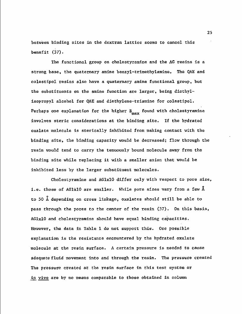
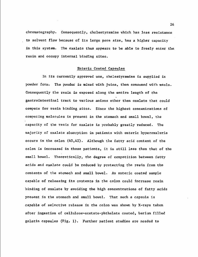
gelatin capsules (Fig. 1). Further patient studies are needed to
Figure la. Abdominal radiograph taken 4 hr after injestion of two cellulose-acetate-phthallate coated, barium filled capsule. — Capsules are intact in distal ileum.
28
Figure lb. Abdominal radiograph taken 8 hr after injestion of two cellulose-acetate-phthallate coated, barium filled capsules. — The capsules were digested exposing the barium contents to the ascending colon.

29
confirm the release pattern of the capsules as only two subjects were
studied here.
Animal Model
Upon completion of the characterization of resins it was
necessary to have an animal model for enteric hyperoxaluria in which to
test the resins since all are not currently approved for human use.
Two types of surgery were performed on Sprague-Dawley rats, bile duct
ligation and jejunoileal bypass. Both forms of surgery create fat
malabsorption and thus potentially lead to enteric hyperoxaluria.
Malabsorption of fatty acids leads to enteric-hyperoxaluria by
increasing the competition between fatty acids and oxalate for intra
luminal calcium (38), As the soluble fraction of oxalate increases, so
does the amount absorbed and then excreted.
Bile acids aid in the lipolysis of triglycerides and micellar
solubilization of fatty acids, both necessary for normal absorption of
dietary fat. Theoretically, ligation of bile duct with subsequently
reduced intestinal bile acid concentration would cause malabsorption
of fatty acids. The resulting formation of calcium soaps of the fatty
acids decreases availability of intraluminal calcium to precipitate
oxalate. Thus, this model should produce hyperoxaluria by increasing
oxalate solubility. While fecal fat measurement in post-operative rats
was not done, consistency of fecal pellets remained normal suggesting
14 no gross malabsorption. Additionally, the amount of C-oxalate
excreted in the urine afte roral challenge was not significantly
different from sham operated controls (Figure 2). These data suggest
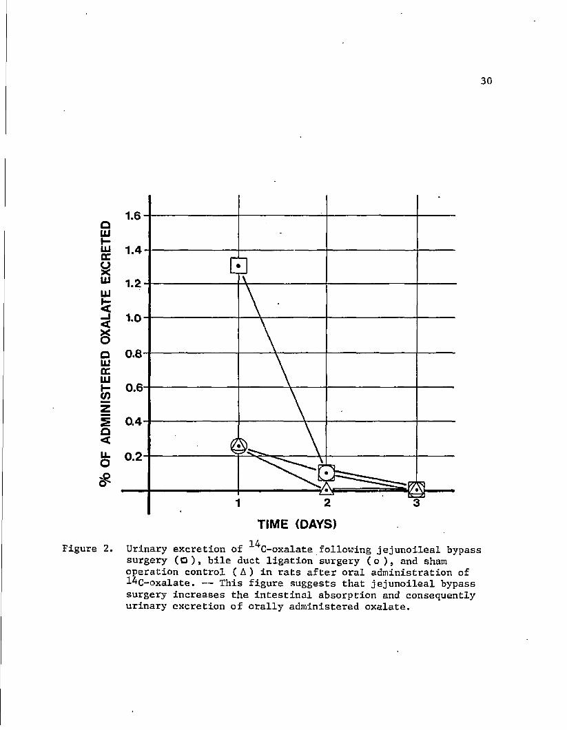
30
o ID h LU CC g UJ
LU
< 8 Q UJ DC UJ H (/)
O <
o #
1.6
1.4
1.2
1.0
0.8
0.6
0.4
0.2
Figure 2.
TIME (DAYS)
14 Urinary excretion of C-oxalate following jejunoileal bypass surgery (O), bile duct ligation surgery (o), and sham operation control (A) in rats after oral administration of l^C-oxalate. — This figure suggests that jejunoileal bypass surgery increases the intestinal absorption and consequently urinary excretion of orally administered oxalate.
31
either bile duct ligation was not complete or that steatorrhea was not
severe enough to cause depletion of intraluminal calcium.
In the jenuno-ileal bypass model, the proximal jejunum was
anastomosed to the distal ileum. In bypassing most of the jejunum and
ileum approximately 86% of the absorptive surface of the small
intestine was avoided. Gross malabsorption was evident by severe
diarrhea for the first seven to ten days post-operatively. Beyond this
time fecal pellets had a more normal appearance. Of five rats which
14 received the oral C-oxalate challenge approximately 30 days post
operatively, four had high normal or slightly increased urinary
"^C-oxalate excretion. However, these rats had variable and small
amounts of small bowel bypassed. The fifth rat, which had the greatest
14 bypass, had an extremely elevated urinary C-oxalate excretion. These
results are similar to those seen in the clinical situation, i.e., the
greater the amount of small bowel bypassed and the greater the degree of
steatorrhea, the greater the fractional absorption of oxalate (.23).
These data suggest that jejuno-ileal bypass can increase intestinal
oxalate absorption but only if a large percentage of the small bowel is
bypassed.
While these animal model experiments were in progress, it was
concluded from the in vitro resin binding studies that cholestyramine
was the best resin to bind oxalate. Because cholestyramine was
already approved for human use, it was no longer necessary to test
resins in an animal model. Consequently, no further data were obtained
regarding the jejuno-ileal bypass animal model.
32
Inhibition of Oxalate Binding
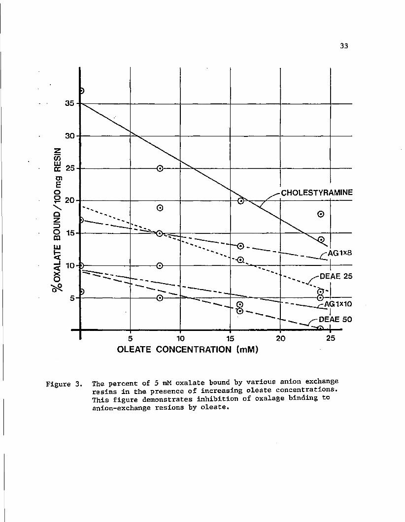
The common denominator of all conditions leading to enteric
hyperoxaluria is the malabsorption of fat (23,24,28,38-40). In the
neutral to basic conditions of the small bowel, the -majority of free
fatty acids in solution exist in theionized form. Consequently, fatty
acids may compete with oxalate for resin anionic binding sites. To
determine if fatty acids could, in fact, inhibit resin oxalate binding,
the fatty acid oleate was incubated with the various anion-exchange
resins and "^C-oxalate (5 mM) in Ringers solution (Fig. 3). As the
concentration of oleate increased, the percent of 5 mM oxalate bound by
resin decreased. Of all resins tested, cholestyramine continued to
bind the greatest amount of oxalate in the presence of oleate. In
another experiment, oxalate concentrations were varied in the presence
of 20 uiM oleate (Fig. 4). As oxalate concentrations increased, so die
the amount of oxalate bound by each resin. Again, cholestyramine
showed the greatest binding at any given oxalate concentration, with the
greatest difference between resins occurring at the higher oxalate
concentration. The anion-exchange resin colestipol, another resin
approved for human use, bound considerably less oxalate than did
cholestyramine. Between 20 and 50 mM oleate, cholestyramine binding of
oxalate decreased by a factor of two, while binding of oxalate by all
other resins tested decreased by a factor of approximately five.
The molecular radius of oxalate is smaller than that of many of
the other organic ions present in the intesting such as fatty acids and
amino acids. It was postulated that by using resins of different
33
35
30
CHOLESTYRAMINE 20
Q 15
AG 1X8
10 DEAE 25
G1X10
DEAE 50
25 20 5 10 15
OLEATE CONCENTRATION (mM)
Figure 3. The percent of 5 mM oxalate bound by various anion exchange resins in the presence of increasing oleate concentrations. This figure demonstrates inhibition of oxalage binding to anion-exchange resions by oleate.
34
i i
OLESTYRAMINE
AG .1x2
COLESTIPOL
AG 1x10
OXALATE CONCENTRATION (mMx10)
Figure 4. The binding characteristics of various anion-exchange resins in the presence of 20 mM oleate while varying the oxalate concentration. — This figure demonstrates that oleate inhibits oxalate binding to different degrees for different
resins.

35
cross linking molecular exclusion of these compounds from the resin
core could increase the amount of oxalate bound by the resin. Pore
size, which determines molecular sieve properties of the resin, varies
inversely as the degree of cross linking. According to manufacturers,
cholestyramine and AGlxlO differ only in the degree of cross linking,
being 1% and 10%, respectively. If molecular exclusion is operating,
theoretically AGlxlO might bind more oxalate than cholestyramine in the
presence of oleate. This was not the case. AGlxlO bound considerably
less oxalate than did cholestyramine. One possible explanation for this
involves the relative sizes of competing molecules. Oleate is
approximately three times larger than oxalate. The molecular sieve
action may become evident only when competing molecules are more
different in size. Consequently oleate and oxalate were actively
competing for internal binding sites. The pressure forcing solvent
through the resin is very low in this test system. A higher pressure
may be required to expose the solvent to the resin core and thus
exhibit the sieve effect.
Analysis of Oxalate Binding by Cholestyramine in Stool
It was postulated that excessive free fatty acid in stool of
patients with malabsorption is responsible for the variable and
frequently inadequate effect of cholestyramine in reducing enteric
hyperoxaluria. In order to evaluate this possibility, fecal samples
were obtained from patients with varying degrees of malabsorption.
Preliminary data suggest there is an inverse correlation between fecal
fat and resin oxalate binding (Fig. 5). However, only three fecal
36
•
f • )
% )
0.5 1.0 1.5 2.0 2.5 3.0
CONCENTRATION OF FAT IN SUPERNATANT (mg FAT/g SUPERNATANT)
Figure 5. The percent of oxalate bound by cholestyramine using the stool supernatant of a control ( ) and of two human patients with known malabsorption (o ). — This figure suggests that the degree of malabsorption in patients may be inversely correlated with the ability of cholestyramine to bind oxalate.

37
samples were analyzed and further studies should be conducted to confirm
these data.
Protocol for the Patient Study
To evaluate and compare the efficacy of the cellulose-acetate-
phthalate-coated capsule and the conventional cholestyramine dosage
forms, the following patient study protocol is suggested. Volunteers
will consist of patients with a history of malabsorption and enteric
hyperoxaluria. Three 24 hour urine samples and three 24 hour fecal
samples will be required of each volunteer prior to the study. The
urine and feces will be evaluated for oxalate and total fat,
respectively. Total fat will be determined by the method of Van de
Van de Kamer (47). Urine oxalate will be determined by the method of
Hatch (45). Volunteers will then be asked to modify their diets to
include 100 g spinach per day as an oxalate load. During the four
pre-resin treatment days, patients will collect urine for oxalate
determination. For the next five days volunteers will continue the
same spinach containing diet plus take the conventional cholestyramine
preparation in a 4 gm dose with meals three times a day. The last
three days of this period, patients will collect urine for oxalate
determination and stool for fat analysis. The patients will take a
week rest without the oxalate load. At the end of this rest period,
24 hour urine and fecal samples will again be collected as previously
for baseline data. The same oxalate load will be added to the diet for
four days, and urine and fecal samples collected. For the next four
days patients will continue on the high oxalate diet and, in addition,

38
will take cholestyramine contained in cellulose-acetate-phthalate
coated gelatin capsules. Urinary oxalate and fecal fat excretion will
be determined at the end of this period. Baseline urine oxalate and
fecal fat levels will be compared with levels during the use of each
dosage form of cholestyramine.
Conclusions
In vitro characterization of oxalate binding of a number of
commercially available anion-exchange resins was completed. Resins
differing in respect to counter ion, functional group, lattice
structure and pore size (or degree of cross linking) were analyzed
allowing objective comparison of each in terms of oxalate binding. The
resin with the greatest oxalate binding capacity was cholestyramine.
Further experiments showed that resin binding of oxalate is indeed
inhibited by free fatty acids, which could be present in the intestinal
water of individuals with conditions leading to enteric hyperoxaluria.
Preliminary data using stool as a medium suggest an inverse correlation
between the degree of steatorrhea and the binding of oxalate by
cholestyramine. These data suggest that a high degree of fat -mal
absorption may adversely influence the effectiveness of cholestyramine
treatment for enteric hyperoxaluria. Though further experiments are
needed to confirm this, perhaps severe steatorrhea should serve as a
marker to identify patients with a greater propensity for developing
calcium oxalate kidney stones even with cholestyramine administration.
As a potential way of limiting the extent of fatty acid
inhibition of resin oxalate binding, it is suggested that a capsule
39
capable of delivering unreacted resin directly into the colon may
increase the efficacy of cholestyramine treatment. A capsule capable
of such selective colonic release was developed. A protocol for a
patient study to confirm the efficacy of encapsulated resin is
suggested.

REFERENCES
1. Pak, C. Y. C. Disorders of stone formation. In The Kidney, Vol. II. Edited by B. M. Brenner and F. C. Rector. Philadelphia: W. B. Saunders Co., 1976, pp. 1326-1354.
2. Coe, F. L, Nephrolithiasis: Pahtogenesis and Treatment. Chicago: Year Book Medical Publishers, 1978, pp. 1-26.
3. Jordan, W. R., Finlayson, B. and Luxenberg, M. Kinetics of early time calcium oxalate nephrolithiasis. Invest. Urol. 15:465-468, 1978.
4. Coe, F. L. Nephrolithiasis: Pathogenesis and Treatment, Chicago: Year Book Medical Publishers, 1978. pp. 144-145.
5. Williams, H. E., and Smith, L. H. Disorders of oxalate metabolism Am. J. Med. 45:715-735, 1968.
6. Suki, W. N. Renal stones. In Pathophysiology of the Kidney. Edited by N. A. Kurtzman and M. Martinez-Maldonado. Springfield: Charles C. Thomas, Publisher, 1977, pp. 1052-1056.
7. Williams, H. E. Oxalic acid: absorption, excretion, and metabolism. In Urolithiasis Research. Edited by H. Fleisch, W. G. Robertson, L. H. Smith, and W. Vahlensiech. London: Plenum Press, 1976, pp. 181-188.
8. Doremus, R. H., Teich, S,, and Silvis, P. K. Crystallization of calcium oxalate from synthetic urine. Invest. Urol. 15: 469-472, 1978.
9. Gardner, G. L. and Doremus, R. H. Crystal growth inhibitors in human urine: effect on calcium oxalate kinetics. Invest, Urol. 15:478-485, 1978.
10. Maher, J. F. Toxic nephropathy. In The Kidney, Vol, II, Edited by B. M. Brenner, and F. C. Rector, Philadelphia: W. B. Saunders Co., 1976, pp. 1367-1368.
11. Coe, F. L. 'Nephrolithiasis: Pathogenesis and Treatment. Chicago Year Book Medical Publishers, 1978, pp. 72-94,
12. Thomas, Jr., W. C. Renal Calculi. Springfield: Charles C. Thomas, Publisher, 1976, p. 118,
40

41
13. Watts, R. E, Oxalate biosynthesis and the primary hyperoxaluria syndromes. In Urolithiasis Research. Edited by H. Fleisch, W. G. Robertson, L. H. Smith, and W. Vahlensiech. London: Plenum Press, 1976, pp. 189-i96.
14. Halpren, B. A., Kempson, R. L.» and Coplon, N. S. Interstitial fibrosis and chronic renal failure following methoxyflurane anesthesia. J.A.M.A, 223:1239-1242, 1973,
15. Mayze, R. I., Trudell, J. R., and Cousins, M. J. Methoxyflurane metabolism and renal dysfunction: clinical correlations in man. Anesthesiology 35:247-252, 1971.
16. Hodgkinson, A. Oxalic Acid in Biology and Medicine. London: Academic Press, 1977, pp. 196-199.
17. Hodgkinson, A, Oxalic Acid in Biology and Medicine. London: Academic Press, 1977, p. 238.
18. Takasaki, E, Urinary magnesium and oxalic acid excretion in patients with recurrent oxalate urolithiasis. Invest. Urol. 12:251-254, 1975.
19. Smith, L. H., Meyer. J, L., McCall, J. T. Chemical nature of crystal inhibitors Isolated from human urine. In Urinary Calculi: Recent Advances in Aetiology, Stone Structure and Treatment. Edited by L. C. Delatte, A. Rapado, and Hodgkinson A. Basel: S. Karger. 1973, pp. 318-327,
20. Robertson, W. G., Peacock, M., and Nordin, B. E. C. Activity products in stone-forming and non-stone-forming urine. Clin. Sci. 34:579-594, 1968.
21. Gelzayed, E. A., Breuer, R. I., and Kirsner, J. B. Nephrolithiasis in inflammmatory bowel disease. Am. J. Dig. Dis, 13:1027-1034, 1968.
22. Deren, J. J., Porush, J. G., Levitt, M. F., and Khilnani, M. T, Nephrolithiasis as a complication of ulcerative colitis and regional enteritis. Ann. Int. Med. 56:843-853, 1962.
23. Earnest, D. L., Johnson, G., Williams, H. E. and Admlrand, N. H, Hyperoxaluria in patients with ileal resection: an abnormality in dietary oxalate absorption. Gastro. 66:1114-1122, 1974.
24. Stauffer, J. Q., Humphreys, M. H., and Weir, G. J. Acquired hyperoxaluria with regional enteritis after ileal resection: role of dietary oxalate, Ann. Int. Med, 79:383-391, 1973,

42
25. Gregory, J. G., Park, K. Y., and Schoenberg, H. W, Oxalate stone disease after intestinal resection. J. Urol. 117:631-634, 1977.
26. Hylander, E., Jarnura, S., Jensen, H. J., and Thale, M. Enteric hyperoxaluria: dependence on small intestinal resection, colectomy, and steatorrhea in chronic inflammatory bowel disease. Scand. J. Gastro. 13:577-588, 1978,
27. Das, S., Joseph, B., and Dick, A. L. Renal failure owing to oxalate nephrosis after jejunoileal bypass. J. Urol. 121: 506-509, 1979.
28. Chadwlck, V. S., Modha, K., and Dowling, R. H. Mechanism for hyperoxaluria in patients with ileal dysfunction. New Eng. J. Med. 289:172-176, 1973.
29. Stauffer, J. Q., Stewart, R. J., and Bertrand, S, G, Acquired hyperoxaluria: relationship to dietary calcium content and severity of steatorrhea. Gastro. 66:783(abstract), 1974.
30. Marshall, R. W., Cochran, M., and Hodgkinson, A. Relationships between calcium and oxalic acid intake in the diet and their excretion in the urine of normal and renal-stone-forming subjects. Clin. Sci. 43:91-99, 1972.
31. Coe, F. L. Nephrolithiasis: Pathogenesis and Treatment. Chicago: Year Book Medical Publishers, 1978, pp. 84-86.
32. Epstein, F. H. Calcium and the kidney. Am. J. Med. 45:700-714, 1968.
33. Smith, L. H., Hofmann, A. F., Tacher, M. M., Fromm, H., and Thomas, P. J. Acquired hyperoxaluria, nephrolithiasis, and intestinal disease. In Urinary Calculi: Recent Advances in Aetiology, Stone Structure and Treatment. Edited by L. C. Delatte, A. Rapado, and A. Hodgkinson. Basel: S. Karger, 1973, pp. 31-40.
34. Smith, L. H., Fromm, H. and Hofraann, A. F. Acquired hyperoxaluria, nephrolithiasis, and intestinal disease: description of a syndrome. New Eng. J. Med, 286:1371-1375, 1972.
35. Caspary, W. F., Tonissen, J., Cankisch, P. G, Enteral hyperoxaluria: effects of cholestyramine, calcium, neomycin and bile acids on oxalate absorption in man. Acta Hepato-Gastro. 24:193-200, 1977.
43
36. Earnest, D. L., Associate Professor, Dept. of Medicine, "University of Arizona, personal communication, 1980,
37. Khyn, J. X. Analytical Ion-Exchange Procedures in Chemistry and Biology: Theory, Equipment, Technique. Nc* Jersey: Prentice-Hall, Inc., 1974.
38. Earnest, D. L., Williams, H, E., and Admirand, W. H. A physicochemical basis for treatment of enteric hyperoxaluria. Trans, Assoc. Am. Phys. 88:224-234, 1975,
39. Admirand, W. H., Carnest, D. L., Williams, H. E. Hyperoxaluria and bowel disease. Trans. Assoc. Am. Phys. 84:307-312, 1971,
40. Modigliani, R., Labayle, L,, Aymes, C., and Denvil, R. Evidence for excessive absorption of oxalate by the colon in enteric hyperoxaluria. Scand. J. Gastro. 13:187-192, 1978.
41. Earnest, D. L. Associate Professor, Dept. of Medicine, University of Arizona, personal communication, November, 1979,
42. Saunders, D. R., Sillerg, J., McDonald, G. B. Regional differences in oxalate absorption by rat intestine: evidence for excessive abosrption by the rat colon in steatorrhea. Gut. 16:543-554, 1975.
43. Scheline, R. R. Metabolism of foreign compounds by gastrointestinal microorganisms. Pharm. Rev, 24:451-523, 1973,
44. Hodgkinson, A. Oxalic Acid in Biology and Medicine, London: Academic Press, 1978, pp. 164-170.
45. Hatch, M., Bourhe, E., Costello, J. New enzymic method for serum oxalate determination. Clin. Chem, 23:76-78, 1977,
46. Costello, J., Hatch, M., Bourke, E. An enzymic method for the spectrophotometry determination of oxalic acid. J, Lab. Clin. Med. 87:903, 1976.
47. Van de Karaer, J. H. Total fatty acids in stool. Standard Methods of Clinical Chemistry. 2:35-44, 1958,