Unfavorable iliac artery anatomy causing access ... · Unfavorable iliac artery anatomy causing...
7
THERAPEUTIC CHALLENGE 318 J Vasc Bras. 2014 Oct.-Dec.; 13(4):318-324 http://dx.doi.org/10.1590/1677-5449.0021 Unfavorable iliac artery anatomy causing access limitations during endovascular abdominal aortic aneurysm repair: application of the endoconduit technique Tratamento endovascular do aneurisma de aorta abdominal associado à dificuldade no acesso aórtico devido a artérias ilíacas de pequeno calibre: aplicação técnica do Endoconduíte Rodrigo Gibin Jaldin 1 , Marcone Lima Sobreira 1 , Regina Moura 1 , Matheus Bertanha 1 , Jamil Víctor de Oliveira Mariaúba 1 , Rafael Elias Farres Pimenta 1 , Ricardo de Alvarenga Yoshida 1 , Winston Bonetti Yoshida 1 Abstract Endovascular aneurysm repair (EVAR) is already considered the first choice treatment for abdominal aortic aneurysms (AAA). Several different strategies have been used to address limitations to arterial access caused by unfavorable iliac artery anatomy. e aim of this report is to illustrate the advantages and limitations of each option and present the results of using the internal endoconduit technique and the difficulties involved. Keywords: aortic aneurysm; endovascular procedures; surgical procedures, minimally invasive; iliac artery; vascular access devices. Resumo O tratamento endovascular para aneurisma de aorta abdominal (AAA) já está bastante difundido, sendo considerado como primeira escolha na maioria dos casos. Limitações no acesso pelas artérias ilíacas tortuosas, com estenoses, calibre pequeno ou doença oclusiva já foram contornadas com o uso de condutos, dissecção direta aortoilíaca, angioplastias, entre outros procedimentos. O objetivo deste desafio é mostrar as vantagens e limitações de cada alternativa, além de apresentar o resultado e as dificuldades com o endoconduíte. Palavras-chave: aneurisma aórtico; procedimentos endovasculares; procedimentos cirúrgicos minimamente invasivos; artéria ilíaca; dispositivos de acesso vascular. 1 Universidade Estadual Paulista – UNESP, Botucatu, SP, Brazil. Financial support: None. Conflicts of interest: No conflicts of interest declared concerning the publication of this article. Submitted: 02.10.14. Accepted: 06.17.14. e study was carried out at the Department of Surgery and Orthopedics, Faculdade de Medicina de Botucatu, Universidade Estadual Paulista (UNESP), Botucatu-SP, Brazil.
Transcript of Unfavorable iliac artery anatomy causing access ... · Unfavorable iliac artery anatomy causing...
THER APEUTIC CHALLENGE
318 J Vasc Bras. 2014 Oct.-Dec.; 13(4):318-324 http://dx.doi.org/10.1590/1677-5449.0021
Unfavorable iliac artery anatomy causing access limitations during endovascular abdominal aortic aneurysm repair:
application of the endoconduit technique
Tratamento endovascular do aneurisma de aorta abdominal associado à dificuldade no acesso aórtico devido a artérias ilíacas de pequeno calibre:
aplicação técnica do Endoconduíte
Rodrigo Gibin Jaldin1, Marcone Lima Sobreira1, Regina Moura1, Matheus Bertanha1, Jamil Víctor de Oliveira Mariaúba1, Rafael Elias Farres Pimenta1,
Ricardo de Alvarenga Yoshida1, Winston Bonetti Yoshida1
AbstractEndovascular aneurysm repair (EVAR) is already considered the first choice treatment for abdominal aortic aneurysms (AAA). Several different strategies have been used to address limitations to arterial access caused by unfavorable iliac artery anatomy. The aim of this report is to illustrate the advantages and limitations of each option and present the results of using the internal endoconduit technique and the difficulties involved.
Keywords: aortic aneurysm; endovascular procedures; surgical procedures, minimally invasive; iliac artery; vascular access devices.
ResumoO tratamento endovascular para aneurisma de aorta abdominal (AAA) já está bastante difundido, sendo considerado como primeira escolha na maioria dos casos. Limitações no acesso pelas artérias ilíacas tortuosas, com estenoses, calibre pequeno ou doença oclusiva já foram contornadas com o uso de condutos, dissecção direta aortoilíaca, angioplastias, entre outros procedimentos. O objetivo deste desafio é mostrar as vantagens e limitações de cada alternativa, além de apresentar o resultado e as dificuldades com o endoconduíte.
Palavras-chave: aneurisma aórtico; procedimentos endovasculares; procedimentos cirúrgicos minimamente invasivos; artéria ilíaca; dispositivos de acesso vascular.
1Universidade Estadual Paulista – UNESP, Botucatu, SP, Brazil.Financial support: None.Conflicts of interest: No conflicts of interest declared concerning the publication of this article.Submitted: 02.10.14. Accepted: 06.17.14.
The study was carried out at the Department of Surgery and Orthopedics, Faculdade de Medicina de Botucatu, Universidade Estadual Paulista (UNESP), Botucatu-SP, Brazil.
319J Vasc Bras. 2014 Oct.-Dec.; 13(4):318-324
Rodrigo Gibin Jaldin, Marcone Lima Sobreira et al.
INTRODUCTION
Endovascular aneurysm repair (EVAR) is now a widely accepted method for treating abdominal aortic aneurysms and is considered the first choice treatment in the majority of cases, especially so when patients have comorbidities and are at high risk of cardiovascular events. However, endoprosthesis delivery systems generally have large calibers (varying from 18 to 24 F) which in some cases can cause difficulties when advancing in the direction of the aorta, navigating the iliac arteries. Attempts to overcome these situations with no prior preparation are associated with complications such as rupture of the iliac arteries, which occurs in around 15% of cases.1 Access limitations caused by tortuous or small caliber iliac arteries or by stenotic or calcifed atherosclerotic disease have previously been surmounted using endarterectomies, angioplasties with and without stents, direct aortoiliac dissection or retroperitoneal conduits.2 We describe the case of the patient treated in an innovative manner about which little has been written in the literature, particularly when used as an adjuvant to abdominal aneurysm treatment.3
PART I – CLINICAL SITUATION
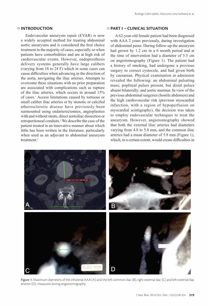
A 62-year-old female patient had been diagnosed with AAA 2 years previously, during investigation of abdominal pains. During follow-up the aneurysm had grown by 1.2 cm in a 6 month period and at the time of intervention had a diameter of 5.5 cm on angiotomography (Figure 1). The patient had a history of smoking, had undergone a previous surgery to correct cystocele, and had given birth by caesarean. Physical examination at admission revealed the following: an abdominal pulsating mass; popliteal pulses present, but distal pulses absent bilaterally; and aortic murmur. In view of the previous abdominal surgeries (hostile abdomen) and the high cardiovascular risk (previous myocardial infarction, with a region of hypoperfusion on myocardial scintigraphy), the decision was taken to employ endovascular techniques to treat the aneurysm. However, angiotomography showed that both the external iliac arteries had diameters varying from 4.8 to 5.0 mm, and the common iliac arteries had a mean diameter of 5.8 mm (Figure 1), which, to a certain extent, would create difficulties in
Figure 1. Maximum diameters of the infrarenal AAA (A) and the left common iliac (B), right external iliac (C) and left external iliac arteries (D), measured during angiotomography.
320 J Vasc Bras. 2014 Oct.-Dec.; 13(4):318-324
Technical application of the endoconduit
achieving femoral access to the aorta for placement of endoprostheses, since their delivery devices had a larger profile caliber than the external iliac arteries of the patient. In such a situation, the available options would be:
1- attempt conventional open surgery on the previously operated abdomen (hostile abdomen);
2- create a retroperitoneal surgical access, implanting a 10 mm Dacron conduit with end-to-side anastomo-ses of the prosthesis to the common iliac arteries;
3- conduct angioplasty of the external iliac arteries with selective stent and balloon;
4- create an Endoconduit for access to the aorta.
PART II – WHAT WAS DONE?
The decision was taken to construct an endoconduit and conduct endovascular treatment of the AAA.
The procedure was conducted under general anesthesia. After antisepsis and placement of sterile fields, the femoral arteries were dissected bilaterally via a transverse inguinotomy and then the left was punctured under direct view and a 5F introducer sheath was fitted. The endoprosthesis chosen for this case is suitable for percutaneous applications via contralateral femoral access, but we preferred bilateral dissection because we did not have a sealing device available and because it afforded
greater safety, considering the small diameters and advanced calcification of the bilateral iliofemoral axes. Abdominal aortography and iliac arteriography were conducted using a cm graded 5F Pigtail catheter (Figure 2).
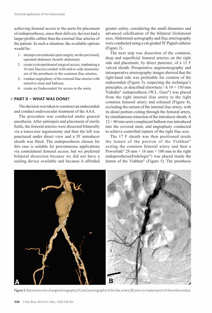
The next step was dissection of the common, deep and superficial femoral arteries on the right side and placement, by direct puncture, of a 12 F valved sheath. Preoperative angiotomography and intraoperative arteriography images showed that the right-hand side was preferable for creation of the endoconduit (Figure 3), respecting the technique’s principles, as described elsewhere.4 A 10 × 150 mm Viabahn endoprosthesis (W.L. Gore) was placed from the right internal iliac artery to the right common femoral artery and released (Figure 4), occluding the ostium of the internal iliac artery, with its distal portion exiting through the femoral artery, by simultaneous retraction of the introducer sheath. A 12 × 80 mm semi-complacent balloon was introduced into the covered stent, and angioplasty conducted to achieve controlled rupture of the right iliac axis.
The 17 F sheath was then positioned inside the lumen of the portion of the Viabhan exiting the common femoral artery and then a Powerlink 28 mm × 16 mm × 100 mm to the right endoprosthesis(Endologix) was placed inside the lumen of the Viabhan (Figure 5). The prosthesis
Figure 2. Reconstruction of angiotomography (A) and arteriography of the iliac artery (B) prior to implantation of the endoconduit.
321J Vasc Bras. 2014 Oct.-Dec.; 13(4):318-324
Rodrigo Gibin Jaldin, Marcone Lima Sobreira et al.
was released according to its instructions for use (Figure 6) and the decision was taken not to use a proximal extension to avoid excessive oversizing at the juxtarenal aorta, since the aneurysm was completely excluded by the bifurcated model.5 Once EVAR was complete, the excess length of the Viabahn was sectioned and it was fitted to the right common femoral artery, to which it was sutured only at the anterior wall during primary arteriorrhaphy of the right femoral artery. However, there was extensive damage to the wall of the common femoral artery at the suture line, complicated by occlusion of the right superficial femoral artery, and embolectomy with a Fogarty catheter was required. There was therefore a need to reconstruct the right common femoral artery, which was accomplished by end-to-end interposition of a Dacron prosthesis, which was anastomosed proximally to the Viabahn and distally to the common femoral artery, just before its bifurcation. The patient developed discrete dehiscence of the right incision and was treated with antibiotic therapy for 14 days. Later (one year after the intervention), the patient
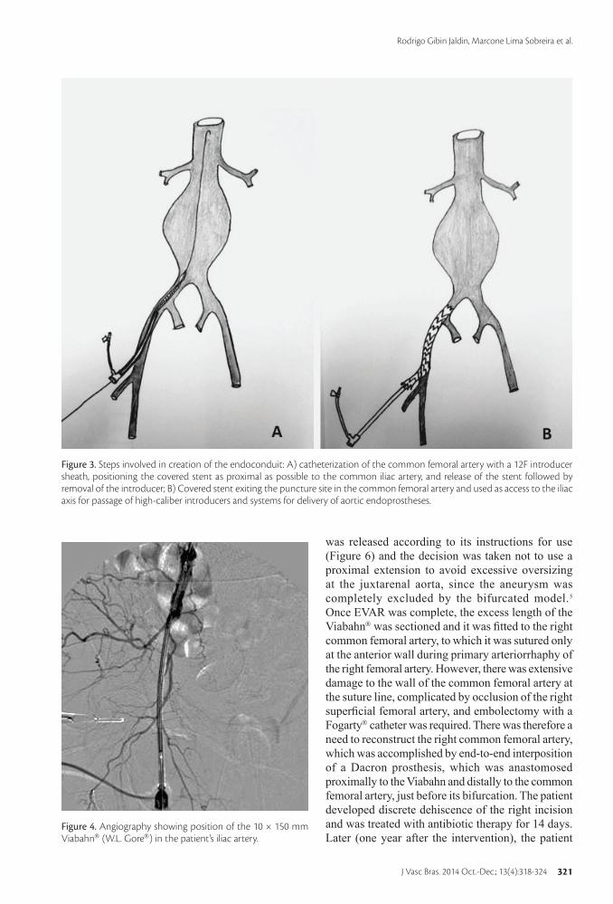
Figure 3. Steps involved in creation of the endoconduit: A) catheterization of the common femoral artery with a 12F introducer sheath, positioning the covered stent as proximal as possible to the common iliac artery, and release of the stent followed by removal of the introducer; B) Covered stent exiting the puncture site in the common femoral artery and used as access to the iliac axis for passage of high-caliber introducers and systems for delivery of aortic endoprostheses.
Figure 4. Angiography showing position of the 10 × 150 mm Viabahn (W.L. Gore) in the patient’s iliac artery.
322 J Vasc Bras. 2014 Oct.-Dec.; 13(4):318-324
Technical application of the endoconduit
suffered occlusion of the right superficial femoral artery, paucisymptomatic, with ankle-brachial indices of 0.78 in anterior and posterior tibial arteries and 0.67 in the fibular (Figures 7 and 8).
DISCUSSION
For endovascular treatment of AAA to be successful, a favorable anatomy is necessary, including the anatomic characteristics of the arteries that will be used to access the aorta. Normally, access is achieved via the iliac arteries, which must have a caliber greater than 8 mm and must not be overly tortuous. Common causes of aortic access difficulties include iliac arteries with naturally small calibers or that have narrowed because of extensive atherosclerosis or calcifications. Certain options have been developed over the years in attempts to surmount these difficulties, including intra-abdominal tunnels or conduits;6 retroperitoneal conduits;7 conduits constructed with the aid of video vascular surgery;8 iliac reconstruction surgeries or bypasses,9 and angioplasty of the iliac arteries with or without stents.10 Iliofemoral endarterectomy,1 dissection and direct puncture of the aortoiliac
segment,9,11 and utilization of internal conduits2,3,12 have also been described.
Dissection of the femoral artery above the inguinal fold offers the advantage of accessing the artery at a point with larger caliber and with few lymph vessels in the vicinity. However, when the iliac arteries do not offer a large enough caliber, this advantage is lost. An earlier solution was to dissect and puncture the common iliac arteries at a point with sufficient caliber to accept the endoprosthesis delivery device. This procedure suffers from the inconvenience of being open and more invasive surgery, canceling out part of the advantage of endovascular procedures. Construction of intraperitoneal or retroperitoneal conduits or bypasses during open surgery did not improve this aspect greatly. Employing videolaparoscopy during construction of the conduit offers the advantage of being much less invasive, but demands a long learning curve and a team trained to conduct the procedure.8 Angioplasties of the iliac arteries, with or without stents, have also been proposed in attempts
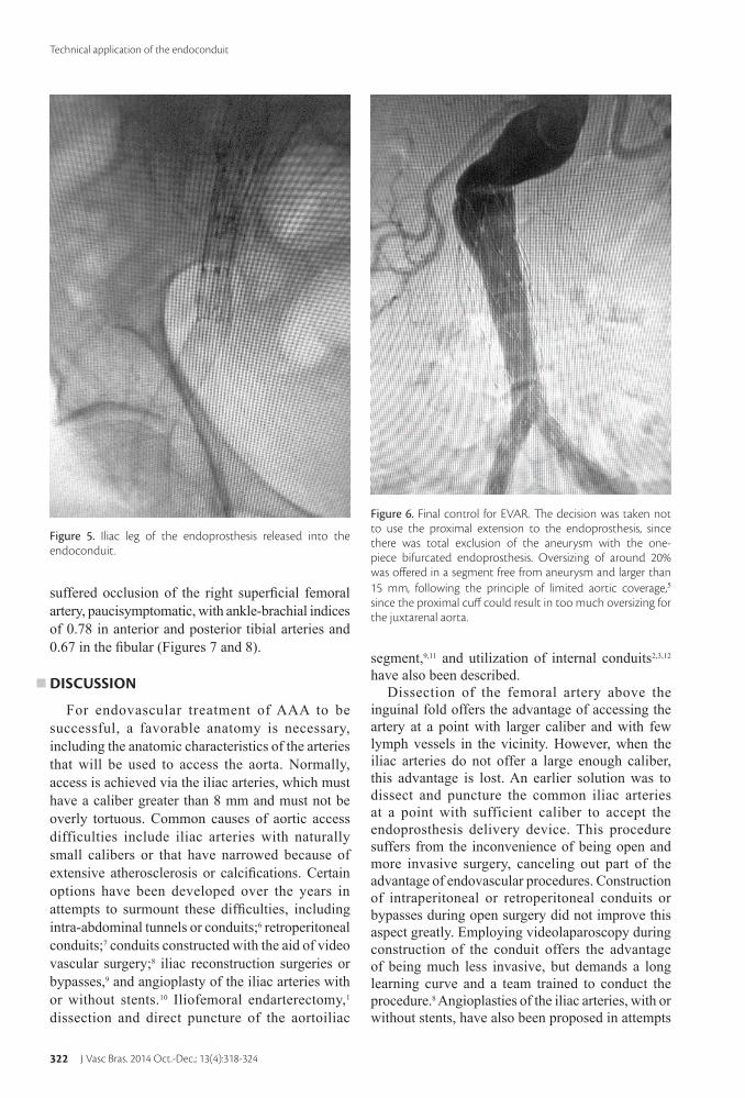
Figure 5. Iliac leg of the endoprosthesis released into the endoconduit.
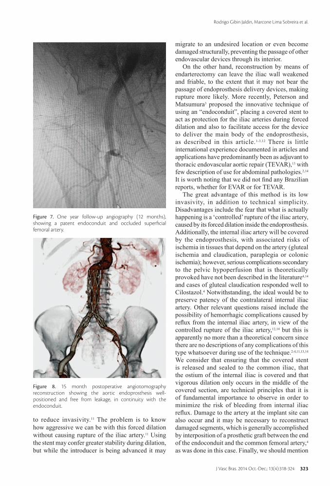
Figure 6. Final control for EVAR. The decision was taken not to use the proximal extension to the endoprosthesis, since there was total exclusion of the aneurysm with the one-piece bifurcated endoprosthesis. Oversizing of around 20% was offered in a segment free from aneurysm and larger than 15 mm, following the principle of limited aortic coverage,5 since the proximal cuff could result in too much oversizing for the juxtarenal aorta.
323J Vasc Bras. 2014 Oct.-Dec.; 13(4):318-324
Rodrigo Gibin Jaldin, Marcone Lima Sobreira et al.
migrate to an undesired location or even become damaged structurally, preventing the passage of other endovascular devices through its interior.
On the other hand, reconstruction by means of endarterectomy can leave the iliac wall weakened and friable, to the extent that it may not bear the passage of endoprosthesis delivery devices, making rupture more likely. More recently, Peterson and Matsumura3 proposed the innovative technique of using an “endoconduit”, placing a covered stent to act as protection for the iliac arteries during forced dilation and also to facilitate access for the device to deliver the main body of the endoprosthesis, as described in this article.1-3,12 There is little international experience documented in articles and applications have predominantly been as adjuvant to thoracic endovascular aortic repair (TEVAR),13 with few description of use for abdominal pathologies.2,14 It is worth noting that we did not find any Brazilian reports, whether for EVAR or for TEVAR.
The great advantage of this method is its low invasivity, in addition to technical simplicity. Disadvantages include the fear that what is actually happening is a ‘controlled’ rupture of the iliac artery, caused by its forced dilation inside the endoprosthesis. Additionally, the internal iliac artery will be covered by the endoprosthesis, with associated risks of ischemia in tissues that depend on the artery (gluteal ischemia and claudication, paraplegia or colonic ischemia); however, serious complications secondary to the pelvic hypoperfusion that is theoretically provoked have not been described in the literature4,14 and cases of gluteal claudication responded well to Cilostazol.4 Notwithstanding, the ideal would be to preserve patency of the contralateral internal iliac artery. Other relevant questions raised include the possibility of hemorrhagic complications caused by reflux from the internal iliac artery, in view of the controlled rupture of the iliac artery,12,14 but this is apparently no more than a theoretical concern since there are no descriptions of any complications of this type whatsoever during use of the technique.2-4,11,13,14 We consider that ensuring that the covered stent is released and sealed to the common iliac, that the ostium of the internal iliac is covered and that vigorous dilation only occurs in the middle of the covered section, are technical principles that it is of fundamental importance to observe in order to minimize the risk of bleeding from internal iliac reflux. Damage to the artery at the implant site can also occur and it may be necessary to reconstruct damaged segments, which is generally accomplished by interposition of a prosthetic graft between the end of the endoconduit and the common femoral artery,4 as was done in this case. Finally, we should mention
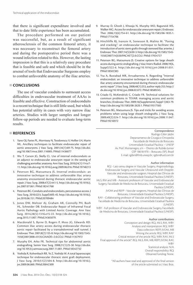
Figure 7. One year follow-up angiography (12 months), showing a patent endoconduit and occluded superficial femoral artery.
Figure 8. 15 month postoperative angiotomography reconstruction showing the aortic endoprosthesis well-positioned and free from leakage, in continuity with the endoconduit.
to reduce invasivity.11 The problem is to know how aggressive we can be with this forced dilation without causing rupture of the iliac artery.11 Using the stent may confer greater stability during dilation, but while the introducer is being advanced it may
324 J Vasc Bras. 2014 Oct.-Dec.; 13(4):318-324
Technical application of the endoconduit
that there is significant expenditure involved and that to date little experience has been accumulated.
The procedure performed on our patient was successful, but, as a result of significant atherosclerosis of the common femoral artery, it was necessary to reconstruct the femoral artery and during the postoperative period there was a wound infection related to this. However, the lasting impression is that this is a relatively easy procedure that is feasible and safe and which should be in the arsenal of tools that Endovascular Surgeons employ to combat unfavorable anatomy of the iliac arteries.
CONCLUSIONS
The use of vascular conduits to surmount access difficulties in endovascular treatment of AAAs is feasible and effective. Construction of endoconduits is a recent technique that is still little used, but which has potential utility in cases with very narrow iliac arteries. Studies with larger samples and longer follow-up periods are needed to evaluate long-term results.
REFERENCES
1. Yano OJ, Faries PL, Morrissey N, Teodorescu V, Hollier LH, Marin ML. Ancillary techniques to facilitate endovascular repair of aortic aneurysms. J Vasc Surg. 2001;34(1):69-75. http://dx.doi.org/10.1067/mva.2001.116005. PMid:11436077
2. Wu T, Carson JG, Skelly CL. Use of internal endoconduits as an adjunct to endovascular aneurysm repair in the setting of challenging aortoiliac anatomy. Ann Vasc Surg. 2010;24(1):114.e7-11. http://dx.doi.org/10.1016/j.avsg.2009.06.016. PMid:19748213
3. Peterson BG, Matsumura JS. Internal endoconduit : an innovative technique to address unfavorable iliac artery anatomy encountered during thoracic endovascular aortic repair. J Vasc Surg. 2008;47(2):441-5. http://dx.doi.org/10.1016/j.jvs.2007.07.061. PMid:18241768
4. Peterson BG. Conduits and endoconduits, percutaneous access. J Vasc Surg. 2010;52(4, Suppl):60S-4S. http://dx.doi.org/10.1016/j.jvs.2010.06.151. PMid:20709484
5. Jones DW, Meltzer AJ, Graham AR, Connolly PH, Bush HL, Schneider DB. Endovascular Repair of Infrarenal Focal Aortic Pathology with Limited Aortic Coverage. Ann Vasc Surg. 2014;28(5):1316.e15-22. http://dx.doi.org/10.1016/j.avsg.2013.11.007. PMid:24365079.
6. Macdonald S, Byrne D, Rogers P, Moss JG, Edwards RD. Common iliac artery access during endovascular thoracic aortic repair facilitated by a transabdominal wall tunnel. J Endovasc Ther. 2001;8(2):135-8. http://dx.doi.org/10.1583/1545-1550(2001)008<0135:CIAADE>2.0.CO;2. PMid:11357972
7. Murphy EH, Arko FR. Technical tips for abdominal aortic endografting. Semin Vasc Surg. 2008;21(1):25-30. http://dx.doi.org/10.1053/j.semvascsurg.2007.11.001. PMid:18342732
8. Yoshida R, Kolvenbach RR, Ye Z, Yoshida W. A total laparoscopic technique for endovascular thoracic stent graft deployment. J Vasc Surg. 2010;51(2):504-8. http://dx.doi.org/10.1016/j.jvs.2009.06.060. PMid:20042309
9. Murray D, Ghosh J, Khwaja N, Murphy MO, Baguneid MS, Walker MG. Access for endovascular aneurysm repair. J Endovasc Ther. 2006;13(6):754-61. http://dx.doi.org/10.1583/06-1835.1. PMid:17154706
10. Hinchliffe RJ, Ivancev K, Sonesson B, Malina M. “Paving and cracking”: an endovascular technique to facilitate the introduction of aortic stent-grafts through stenosed iliac arteries. J Endovasc Ther. 2007;14(5):630-3. http://dx.doi.org/10.1583/1545-1550(2007)14[630:PACAET]2.0.CO;2. PMid:17924727
11. Peterson BG, Matsumura JS. Creative options for large sheath access during aortic endografting. J Vasc Interv Radiol. 2008;19(6, Suppl):S22-6. http://dx.doi.org/10.1016/j.jvir.2008.01.031. PMid:18502383
12. Ysa A, Bustabad MR, Arruabarrena A. Regarding “Internal endoconduit: an innovative technique to address unfavorable iliac artery anatomy encountered during thoracic endovascular aortic repair”. J Vasc Surg. 2008;48(1):253, author reply 253. http://dx.doi.org/10.1016/j.jvs.2008.02.072. PMid:18589245
13. Criado FJ, McKendrick C, Criado FR. Technical solutions for common problems in TEVAR: managing access and aortic branches. J Endovasc Ther. 2009;16(SupplementI, Suppl 1):I63-79. http://dx.doi.org/10.1583/08-2620.1. PMid:19317583
14. Peterson BG, Matsumura JS. Tips and tricks for avoiding access problems when using large sheath endografts. J Vasc Surg. 2009;49(2):524-7. http://dx.doi.org/10.1016/j.jvs.2008.11.047. PMid:19216972
Correspondence Rodrigo Gibin Jaldin
Departamento de Cirurgia e Ortopedia Faculdade de Medicina de Botucatu
Universidade Estadual Paulista – UNESP Av. Prof. Montenegro, s/n – Distrito de Rubião Junior
CEP 18618-970 – Botucatu (SP), Brazil E-mail: [email protected]; [email protected]
Author information RGJ - Lato sensu degree in Vascular Surgery, Angioradiology,
Endovascular Surgery, and Vascular Ultrasound with Doppler. Vascular and endovascular surgeon, Hospital das Clínicas de
Botucatu, Universidade Estadual Paulista (UNESP). MLS, RM and MB - Assistant professors of Vascular and Endovascular
Surgery, Faculdade de Medicina de Botucatu, Universidade Estadual Paulista (UNESP).
JVOM and REFP - Vascular surgeons, Hospital das Clínicas de Botucatu, Universidade Estadual Paulista (UNESP).
RAY - Collaborating professor of Vascular and Endovascular Surgery, Faculdade de Medicina de Botucatu, Universidade Estadual Paulista
(UNESP). WBY - Full professor of Vascular and Endovascular Surgery, Faculdade
de Medicina de Botucatu, Universidade Estadual Paulista (UNESP).
Author contributions Conception and design: RGJ, MLS, RM, WBY
Analysis and interpretation: RGJ, WBY Data collection: REFP, JVOM, MB
Writing the article: RGJ, WBY, RAY Critical revision of the article: RGJ, WBY, RAY, MLS
Final approval of the article*: RGJ, MLS, RM, MB, REFP, JVOM, RAY, WBY
Statistical analysis: N/A Overall responsibility: RGJ Obtained funding: None.
*All authors have read and approved of the final version
of the article submitted to J Vasc Bras.