Understanding the role of MasR in vascular pathology (EDIT)vuir.vu.edu.au/35026/1/RAI...
80
1 Understanding the role of MasR in vascular pathology By Sudarshan Rai For College of Health and Biomedicine Supervisors: A/Prof. Alan Hayes (VU); Dr. Anthony Zulli (VU) Victoria University Submitted in fulfillment of the requirements of the degree of Master of Science (Research) October 2016
Transcript of Understanding the role of MasR in vascular pathology (EDIT)vuir.vu.edu.au/35026/1/RAI...

1
UnderstandingtheroleofMasRinvascularpathology
By
Sudarshan Rai
For
College of Health and Biomedicine
Supervisors: A/Prof. Alan Hayes (VU); Dr. Anthony Zulli (VU)
Victoria University Submitted in fulfillment of the requirements of the degree of Master of Science (Research)
October 2016

2
Abstract
Background: Homocysteine was first suggested as a risk to CVD in 1969. Since then, elevated
plasma homocysteine (derived from methionine) has remained a cardiovascular risk factor with no
current treatment. Homocysteine is known to stimulate NADPH oxidase, and NADPH oxidase can
be inhibited by stimulation of the Mas receptor (MasR) of the renin angiotensin system. Stimulation
of MasR is known to reduce organ fibrosis through the production of NO. However, the role that
MasR plays in homocysteine-induced vascular pathology, including endothelial dysfunction and
organ fibrosis, is not known.
Aims: To determine if high methionine diet in MasR-/- mice will worsen cardiovascular pathology.
Methods: MasR-/- and C57BL/6 (control) mice were fed a 1% methionine or a control diet for 8
weeks (n=16 per group). Aortic endothelial function in response to acetylcholine was performed,
fibrosis was semi-quantified in myocardium and kidney and aorta using fast green/sirius red
staining. Immunohistochemical staining for Collagen I and III were performed.
Results: Aortic acetylcholine-induced relaxation was reduced by >50% in MasR-/- mice vs control
(32.3±6.8% vs 66.1±7.7%, p<0.001), this dysfunction was worsened by methionine in MasR-/- but
not in control (6.6±6.7% vs 74±7%, p<0.0001) mice. Methionine increased glomerular fibrosis by
22% in control (p=0.02), and 2.1 fold (p<0.0001) in MasR-/- mice whereas kidney interstitial/tubular
fibrosis increased 5-fold (p<0.0001) only in MasR-/- mice. Methionine increased myocardial
interstitial fibrosis by >3-fold (p<0.0001) only in MasR-/- mice and perivascular fibrosis by 31% in
control (p<0.001) and 2.6-fold (p<0.001) in MasR-/- mice. However, aortic medial and adventitial
fibrosis was not present in methionine fed MasR-/- mice. By contrast, aortae of methionine fed
control mice (p<0.01) displayed a significant increase in fibrosis in both medial and adventitial
tissue. Positive staining for collagen I and III was detected in heart, kidney and aorta.
Conclusion: Excess dietary methionine for 8 weeks worsens cardiovascular pathology in MasR-/-
mice, suggesting that the MasR is protective against homocysteine-induced cardiovascular
pathologies.

3
Student Declaration Masters by Research Student Declaration
Master by Research Declaration
I, Sudarshan Rai, declare that the Master by Research thesis entitled
“Understanding the role of MasR in vascular pathology” is no more than
60,000 words in length including quotes and exclusive of tables, figures,
appendices, bibliography, references and footnotes. This thesis contains no
material that has been submitted previously, in whole or in part, for the
award of any other academic degree or diploma. Except where otherwise
indicated, this thesis is my own work.
Tuesday, October 11, 16

4
Table of Contents
ABSTRACT....................................................................................................................................................................2
STUDENTDECLARATION........................................................................................................................................3
LISTOFFIGURES........................................................................................................................................................6
ACKNOWLEDGEMENTS............................................................................................................................................8
INTRODUCTION..........................................................................................................................................................9
ENDOTHELIALDYSFUNCTIONANDNITRICOXIDE.....................................................................................................................9
RISKFACTORSFORCVDANDORGANPATHOLOGY................................................................................................................11
MethionineandHomocysteine:..........................................................................................................................................11
ROS:................................................................................................................................................................................................15
RAS:................................................................................................................................................................................................16
Ang(1-7)andMasR:...............................................................................................................................................................19
Aorticfibrosisandaorticcompliance.............................................................................................................................21
MyocardialfibrosisinCVD...................................................................................................................................................22
RenaldiseaseinCVD:.............................................................................................................................................................24
HYPOTHESIS.............................................................................................................................................................26
AIMS...............................................................................................................................................................................................26
MATERIALSANDMETHOD:.................................................................................................................................27
Animals:........................................................................................................................................................................................27
Organbath–endothelialfunction:..................................................................................................................................27
Fibrosis..........................................................................................................................................................................................28
Immunohistochemistry:........................................................................................................................................................29
FibrosisandImmunohistochemistrydatagatheringandanalysis....................................................................31
RESULTS.....................................................................................................................................................................33
DISCUSSION:.............................................................................................................................................................60
EndothelialFunction..............................................................................................................................................................60

5
Fibrosis..........................................................................................................................................................................................63
Conclusion/FutureDirections............................................................................................................................................70
REFERENCES.............................................................................................................................................................71

6
List of Figures
Figure 1. Progression of Atherosclerosis………...…………………………………………………..9
Figure 2. Schematic of homocysteine metabolism………………………………………………….12
Figure 3. Ang II and Ang (1-7) and the respective pathways.……………………………………...18
Figure 4. Overview of proposed rationale scheme.......………………………………………….….21
Figure 5. Myocardial fibrosis imaging process………………………………………………….….31
Figure 6. Glomerular and aorta imaging process………………………………………………...…33
Figure 7. Abdominal aorta relaxation to acetylcholine……………………………..………………36
Figure 8. Aortic medial fibrosis, fast green and direct red staining………..………….……………37
Figure 9. Aortic adventitial fibrosis, fast green and direct red staining…………………..………...38
Figure 10. Myocardial Interstitial fibrosis, fast green and direct red staining…………….………..39
Figure 11. Myocardial perivascular fibrosis, fast green and direct red staining…..………..………40
Figure 12. Renal glomerular fibrosis, fast green and direct red staining…….……………………..41
Figure 13. Renal interstitial/tubular fibrosis, fast green and direct red staining.………….………..42
Figure 14. Aortic medial fibrosis, collagen I…………………………………….……….…………43
Figure 15. Aortic adventitial fibrosis, collagen I…………………………………….…….………..44
Figure 16. Myocardial interstitial fibrosis, collagen I……………………………………..………..45
Figure 17. Myocardial perivascular fibrosis, collagen I…………………………………….………46
Figure 18. Renal glomerular fibrosis, collagen I…………………………………….……………...47
Figure 19. Renal interstitial/tubular fibrosis, collagen I…………………………………….………48
Figure 20. Aortic medial fibrosis, collagen III…………………………………….………………..49
Figure 21. Aortic adventitial fibrosis, collagen III…………………………………….……………50
Figure 22. Myocardial interstitial fibrosis, collagen III…………………………………….………51
Figure 23. Myocardial perivascular fibrosis, collagen III…………………………………….…….52
Figure 24. Renal glomerular fibrosis, collagen III…………………………………….……………53
Figure 25. Renal interstitial/tubular fibrosis, collagen III…………………………………….…….54

7
Figure 26. Aortic medial fibrosis, Rabbit IgG, polyclonal - Isotype Control …….………………..55
Figure 27. Aortic adventitial fibrosis, Rabbit IgG, polyclonal - Isotype Control ..………….……..56
Figure 28. Myocardial interstitial fibrosis, Rabbit IgG, polyclonal - Isotype Control ………....…..57
Figure 29. Myocardial perivascular fibrosis, Rabbit IgG, polyclonal - Isotype Control......….…….58
Figure 30. Renal glomerular fibrosis, Rabbit IgG, polyclonal - Isotype Control….…………..……59
Figure 31. Renal interstitial/tubular fibrosis, Rabbit IgG, polyclonal - Isotype Control .………….60

8
Acknowledgements
I would like to express my sincere gratitude to my supervisor Dr. Anthony Zulli for the continuous
support of my MSc study and related research, for his knowledge, motivation and most importantly
patience. His guidance helped me in all the time of research and writing of this thesis. I could not
have imagined having a better supervisor and mentor for my MSc study. I would also like to take
this opportunity to thank my principal supervisor A/ProfessorAlan Hayes for his guidance and
support through this research process.
Besides my supervisors, a special thanks goes to Dr. Chandana Hearth for his kind donation without
which this research would not be possible. I would also like to thank Professor Robert Widdop who
has offered his expertise throughout my research.
I would also like to thank my fellow lab-mates for the stimulating discussions, and for all the fun
we have had in the last 2 years. Also, I would like to thank Anne Luxford in the animal facility for
all her help and guidance.
Last but not the least, I would like to thank my family: my parents for their support and to my ever-
patient girlfriend Sandra for supporting me throughout this thesis and my life in general.

9
Introduction
Cardiovascular disease (CVD) is well documented as the number one cause of death worldwide.
In 2005 alone, people who died from CVD accounted for 30% of all global deaths (World Health
Organization 2008). It occurs almost equally in men as in women, and is predicted to be the most
common cause of death worldwide by 2020 (1). CVD encompasses ischemic heart disease, stroke
and vascular diseases. CVD usually stems from vascular dysfunction, which leads to
atherosclerosis. Atherosclerosis is an inflammatory disease involving the arterial endothelium (2).
The disease disrupts blood flow through the lumen due to the formation of plaques that may
eventually lead to plaque rupture (2), which can lead to other complications such as myocardial and
cerebral infarction (3).
There are several risk factors associated with CVD, the most common of which are dyslipidemia,
hypertension, smoking, diabetes mellitus (4-6), and high plasma homocysteine, also known as
hyperhomocysteinemia (>15µM) (7). Current treatment for CVD can involve a combination of drugs
such as, low-density lipoproteins (LDL) lowering statins, high blood pressure lowering aspirins or
surgery to repair damaged blood vessels. Current methods of reducing cardiac and atherosclerotic
events mainly include statin therapy, as this also reduces the occurrence of strokes and transient
ischemic attacks (8). However lipid-lowering therapy such as statins itself also carries a risk of
muscular symptoms or myopathy with 7% of outpatients on statins reporting mild muscular pain (9-
11).
Endothelial dysfunction and nitric oxide
It has been well established that endothelial dysfunction is in direct response to cardiovascular risk
factors, and precedes the development of atherosclerosis. Endothelial dysfunction is characterized
by a reduced bioavailability of vasodilators, in particular nitric oxide (NO), whilst endothelium-

10
derived vasoconstrictors are increased (12, 13). The formation of lesions promoted by both early and
late mechanisms of atherosclerosis, including increased chemokine secretion and leukocyte
adherence, enhanced low-density lipoprotein oxidation, increased cell permeability, platelet
activation, cytokine elaboration, up regulation of adhesion molecules, and vascular smooth muscle
cell proliferation and migration, is a result of endothelial dysfunction (12, 14, 15). A major vasodilator
released by the endothelium is NO, as well as bradykinin and prostacyclin (14-16). NO was indicated
to be a vasodilator able to inhibit growth and inflammation(17, 18). During diseased states,
endothelial dysfunction can be characterized by the imbalance between vasodilators and
vasoconstrictors, resulting in an impairment of endothelium-dependent vasodilation (12, 19).
Moreover, endothelial dysfunction has a chronic inflammatory, proliferative, and procoagulatory
state that favors atherogenesis (19). NO is produced when L-arginine is converted to L-citrulline by
endothelial nitric oxide synthase (eNOS). It has been suggested that phosphorylation of eNOS at
either and/or both serine1177 (ser1177) and serine633 (ser633) leads to eNOS activation in
response to stimuli, with ser633 found to have higher efficacy in increasing the eNOS derived NO
bioavailability (20-22).
Figure 1. Evolustion of arterial wall changes in response to injury artery wall thickens as a result
of invasion and accumulation of white blood cells (macrophages and foam cells) and proliferation
of intimal-smooth-muscle cell progressing to atherosclerosis.
11
Previous studies have demonstrated that both NO production and endothelial function are impaired
in diseased vascular tissue(23, 24), however recent results indicate that this may not be as a result of
expression level of mRNA and the protein eNOS, as findings show their expression as either
increased or not changed (25). These findings suggest a reduced generation or bioavailability of NO,
which may be regulated independently of eNOS activity (25, 26). In support of this idea, a recent
study suggested that an uncoupling of eNOS and decreased phosphorylation resulting in reduced
NO synthesis was a major underlying cause of endothelial dysfunction (25, 27).
Risk factors for CVD and organ pathology
Methionine and Homocysteine:
Homocysteine, a metabolite of methionine found in meat, was proposed to be an independent
atherogenic agent by Kilmer McCully more than 30 years ago (28). McCully showed that an increase
in plasma homocysteine affected all artery sizes, with the lumen of many large to medium arteries
exhibiting narrowing due to focal intimal and medial fibrosis, which is often associated with fraying
and discontinuity of internal elastic membrane(28). Moderate thickening of the media and internal
elastic membrane was also noted (28). Additionally, focal proliferation of perivascular connective
tissue of many small arteries, and an increase of collagen bundles, fibroblasts, and small elastic
fibers was observed (28).

12
Figure 2. Schematic of homocysteine metabolism. Methionine is converted to homocysteine which
forms cystathionine (in an irreversible reaction). Cystathionine is hydrolyzed to form cysteine, and
excess cysteine is oxidized to inorganic sulfates and is excreted in urine.
Homocysteine is an endogenous amino acid, that once formed is catabolized to cysteine by
transsulfuration or remethylated to methionine (29, 30). Elevated homocysteine known as
Hyperhomocysteinemia is widely considered to be an independent risk factor for various CVD (31,
32). However, the precise mechanism in which homocysteine interferes with the cardiovascular
system has not yet been elucidated.
Mild elevations in plasma homocysteine concentrations (10-15 µM) are frequently found in
Western populations and are associated with an increase risk of CVD (33). Humans synthesize
between 15-20 mmol of homocysteine per day however the majority of this is converted to cysteine
or to methionine (29). Regulation of homocysteine levels sets the basal concentration between 5 to
15 µM (29, 34, 35). Studies have suggested that elevations in plasma homocysteine are associated with
an increased risk of occlusive artery disease, particularly in the brain, heart and kidney (36).
Hyperhomocysteinemia is described as moderate at 15-30 µM, intermediate between 30-100 µM or

13
severe measured at >100 µM (29, 34, 35), with 300µM being the highest recorded pathological level
detected (12, 37, 38). Additionally, impaired handling and metabolism of homocysteine has been
exhibited after a methionine challenge in individuals with premature atherosclerosis. Essentially,
hyperhomocysteinemia is a state where there is an increased risk of vascular constrictive affects,
atherosclerotic, and atherothrombotic (39).
Reduced endothelial-dependent relaxation has also been observed in humans acutely affected by
hyperhomocysteinemia(40, 41). These observations have some to reason that hyperhomocysteinemia
may not in fact be the cause of atherosclerosis intrinsically (41), but may have a role in altering
vascular function so as to increase risk for complications of atherosclerosis (41). Furthermore,
endothelial-dependent flow-meditated dilation in brachial arteries was lower in
hyperhomocysteinemia patients than in patients with normal homocysteine levels (42). A study
using healthy volunteers with oral methionine loading resulted in increased homocysteine levels
and a reduced NO bioavailability by producing oxidative stress, causing decreased endothelial
function (42-45).
Therefore, several large studies evaluating lowering total homocysteine have been performed to
elucidate its affect on CVD (46). Normalization of total homocysteine is typically done by
supplementation with folic acid and vitamin B12 (33). Consequently, clinical trials using dietary
folate supplementation to efficiently lower total homocysteine in patients produce an array of
effects. While treatment with folate did lower total homocysteine, this did not yield a protective
effect on secondary adverse vascular events, leading investigators to conclude that
hyperhomocysteinemia may be an early biomarker of cardiovascular risk rather than an independent
risk factor (47, 48). However, Weiss et al. 2002, demonstrated that normalizing homocysteine levels
had in fact improved endothelial dysfunction, although it was unclear if this translated to a reduced
risk of CVD (33, 45).

14
A Norwegian group reported in a prospective study that hyperhomocysteinemia was a strong
predictor of mortality, although correlation between hyperhomocysteinemia and coronary artery
atherosclerosis was not clear (41, 49). Alternatively, studies investigating a moderate rise in plasma
homocysteine found that it accelerated the formation of atherosclerotic lesions in ApoE knockout
(ApoE-/-) mice. ApoE-/- mice fed a hyperhomocysteinemic diet for up to 8 weeks developed
atherosclerotic lesions in the aorta that were larger then mice fed on normal chow (41, 50). The ApoE -
/- mice were created to develop severe hypercholesterolemia in addition to lesions of atherosclerosis
with similar characteristics to that those observed in humans (51). The mice fed on the
hyperhomocysteinemic diet had significantly larger mean aortic lesion when compared to control
(52).
Studies such as the Heart Outcomes Prevention Evaluation (HOPE) 2 Investigators trial and the
Norwegian Vitamin (NORVIT) trial have performed extensive investigation into the homocysteine
lowering effects of folic acid and B vitamin treatments. While the trials concluded that treatment
with folic acid and B vitamins did not reduce the risk major cardiovascular events in patients with
vascular disease (53), or lower the risk of recurrent CVD after myocardial infraction (54), they did
however acknowledge plasma total homocysteine levels were a predictor (or marker) of
cardiovascular events (53, 54).
Studies show that oxidation and modification of low density lipoprotein (LDL) plays in important
part in the initiation of atherosclerosis (55), where oxidized LDL cholesterol was able to impair
endothelial-mediated relaxation (55). Rabbits fed on a 0.5% cholesterol diet showed significantly less
blood vessel relaxation then normal chow fed rabbits (56). Our lab has also demonstrated that in
addition to 0.5% cholesterol, when 1% methionine is added to the diet it intensifies the progression
of atherosclerosis (57) and interestingly shows proliferation of mesenchymal stem cells and

15
endothelial hematopoietic stem cells which were identified using cell surface markers specific to the
respective cell types (unpublished data).
Investigation into atherosclerosis, and endothelial integrity prior and during diseased states, shows
the crucial role the endothelial plays; it acts as executive and sensory function, it can generate
effector molecules that are able to regulate thrombosis, vascular tone, inflammation and vascular
remodeling (58). Injury or removal of the endothelial results in rapid migration and proliferation of
smooth muscle cells which then subsides after the endothelium regenerates (58, 59). Therefore,
endothelial-dependent regulation of vascular tone and consequently endothelial dysfunction is
indicative of a reduced bioavailability of endothelial-derived signaling molecule NO (60, 61). Studies
suggest that chronic hyperhomocysteinemic subjects that are free of any overt CVD exhibit reduced
NO bioavailability (62-64). This is supported in studies examining oral methionine administration to
healthy subjects that also show reduced NO (43, 65-67). Thus, a consensus can be drawn that lowering
plasma homocysteine levels by supplementation with B vitamins, improves endothelial dysfunction
by reducing the biochemical markers and improving overall endothelial-dependent vasodilatory
function (45).
ROS:
Reactive oxygen species (ROS) when present at low concentrations at sites of injury and
inflammation function as signaling molecules regulating cell activity and growth. Conversely,
increased levels of ROS is believed to lead to cellular injury and death, often associated with
microvascular pathology in age-related CVD (17, 68, 69). Prolonged exposure to ROS and other
cardiovascular risk factors can eventually fatigue the protective effect of endogenous anti-
inflammatory systems within endothelial cells. As a consequence the endothelial becomes
increasingly permeable and promotes leukocyte adhesion and alters endothelial signaling(12).

16
LDL modified via oxidation also plays a central role in the development of atherosclerosis, and
maybe important in abnormal endothelial vasorelaxation(19, 70). The nicotinamide adenine
dinucleotide phosphate family of oxidases (NOX) is a crucial component of ROS production. NOX
proteins are able to produce super oxide anions (O2-), which further react to form hydrogen
peroxide (H2O2) (71, 72). However, in the presence of NO, O2- combines to NO and forms
peroxynitrite (ONOO-) (71, 72). The NOX family of NADPH oxidases has only one role, to generate
ROS (71, 73). There are seven members of the NADPH oxidase family, NOX1 through to NOX5 and
Duox1 and Duox2. NOX1 is expressed in smooth muscle cells however in endothelial cells a lower
level of expression was detected (71, 74, 75). NOX2 is expressed in endothelial cells and adventitial
fibroblasts (71, 76, 77). NOX4 was found to be ubiquitously expressed, and thus was found in all the
cell types: smooth muscle cells, endothelial cells and adventitial fibroblasts (71, 76, 78-85). Our lab has
previously studied the deleterious affects of homocysteine on endothelial response to acetylcholine
with Nox inhibitor, however, only ML090 a Nox1 inhibitor was found to ameliorate homocysteine
and its actions on the endothelial, which suggested a strong correlation between Nox1 and
homocysteine-induced vascular damage (80). In addition to our previous results, a study
investigating salidroside (an antioxidant) demonstrated it protected against homocysteine-induced
impairment of endothelial relaxation by inhibiting Nox2-dependent ROS overproduction and
increasing NO bioavailability (84). Other researchers have also shown that homocysteine inhibited
endothelial-dependent relaxation in a concentration and time dependent manner (86). These studies
together indicate homocysteine causes endothelial dysfunction by way of Nox-dependent pathways.
RAS:
The renin-angiotensin system (RAS) plays a significant role in the regulation of blood pressure
homeostasis, which includes body fluid control and electrolyte homeostasis (87, 88). Consequently it
has been a principal target for therapeutics for the treatment of hypertension and other such related

17
diseases. Extended stimulation of angiotensin type 1 receptor (AT1R) by angiotensin II (AngII)
signaling leads to fibrosis (89), inflammation, and cellular hypertrophy and cellular proliferation,
which all contribute to disease pathophysiology (90, 91). Studies have indicated that AngII is the
primary vasoactive substance of the RAS and causes renal, arterial and cardiac lesions (91).
A recent discovery of an alternate pathway has expanded the classical view of RAS, where
angiotensin peptide metabolism and signaling forms Ang (1-7) by degradation of AngII, by
angiotensin converting enzyme 2 (ACE2) (90, 92, 93). ACE2, a homologue of angiotensin converting
enzyme (ACE) is highly expressed in the kidney, lungs, testis and heart (94, 95).
ACE2 catalyzes the conversion of Ang I to Ang (1-9) and Ang II to Ang (1-7) (96). ACE2 facilitates
Ang (1-7) production via two pathways, when Ang I is converted to Ang (1-9) allowing ACE to
subsequently convert Ang (1-9) to Ang(1-7), and the direct AngII to Ang(1-7) hydrolysis (97).
Moreover it was discovered that ACE2 is able to catalyze Ang II substrate with a 400-fold
efficiency when compared to the catalytic rate of Ang I, further suggesting that the ACE2/Ang(1-7)
may counter regulatory to Ang I.
Ang (1-7), a heptapeptide that binds to and activates a unique receptor known as Mas (98), opposes
many of the actions of the Ang I and AT1R (87, 90, 92). Therefore, MasR, activated by Ang (1-7) is
observed as the protective arm of the RAS (99, 100) see Figure 2.

18
Figure 3. Ang II and Ang (1-7) and the respective pathways.
Both AT1R and AT2R have been identified in the vessel wall, although it is thought that AT1R
mediates most of the atherogenic actions of Ang II. Though both AT1R and AT2R belong to the
same receptor family they differ greatly in their signaling cascades and biological activities (101).
AT1R were most densely distributed in the vascular smooth muscle cells and endothelial cells (102,
103). Activation of the AT1R was shown to contribute independently to chronic CVD (101, 103). The
effects of Ang II through its AT1R is thought to enhance oxidized LDL infiltration in the vessel wall
(103).
Less is know about the AT2R receptor signaling pathways. However recent evidence suggests that
it may be involved in the control of cell proliferation, cell differentiation and development,
angiogenesis, wound healing, tissue regeneration and generally counteracts the affects of AT1R (101).
A well-established AT2R signaling mechanism is via NO/cGMP pathway, which is thought to be
one of the vasoprotective actions of the AT2R. Expression of AT2R is also observed to be up
regulated during certain circumstances such as in heart failure, post-infarct repair, and skin and
nervous system lesions (101).

19
Ang (1-7) and MasR:
The identification of the Mas receptor (MasR) in 1986 by Young et al. marked an important step in
the understanding of the RAS (104). The Mas proto-oncogene was originally detected due to its
ability to render NIH 3T3 cells tumorigenic and was initially identified as a transforming protein
when overexpressed in the NIH 3T3 cells (104, 105). It is a G-protein coupled receptor, which
includes a large family of cell surface receptors that mediate the actions of a diverse array of
extracellular ligands (87, 104, 105), and is highly expressed within the cardiovascular system (106).
Transfection studies had proposed that the Mas gene encoded an AngII receptor (98), however
several other studies concluded that AngII induced activation was only in cells endogenously
expressing the AngII AT1R (87, 107). Furthermore, a radio-ligand-binding study with AngII in the
kidney of wild type and MasR knockout (-/-) (108) mice found that there is a clear interaction between
MasR and Ang (1-7) (87). Additionally the vasodilatory effects of Ang (1-7) were absent in the
MasR-/- model (92, 109). The anti-fibrotic effects of ACE2 were predominantly achieved through the
MasR, as the MasR-/- mice displayed a predisposition to pro-fibrosis and mesenchymal stem cell
proliferation in the cardiovascular system (110). Therefore, based on these findings it was concluded
that Ang (1-7) binding with MasR exerts a definite biological action, including prevention of organ
fibrosis (87).
Long term infusion of Ang (1-7) was shown to have a vasoprotective and atheroprotective effect in
ApoE knockout (ApoE -/-) mice with increased eNOS expression and an overall improvement of
endothelial function, along with reduced atherosclerosis development in both ApoE -/- and low-
density lipoprotein receptor deficient mice fed on a high fat diet (106, 110-112). Ligation studies where
the left coronary artery is ligated and perfused with either Ang (1-7) or saline revealed that Ang (1-
7) prevented the deterioration of cardiac function (113). Furthermore AT1R blockers and ACE
inhibitors were administered to partially ligated abdominal aorta of rats to examine the affects on
the expression levels of ACE2 and Mas receptor (MasR) (91). AT1R blocker caused an increase in

20
the expression of cardiac ACE2 along with an increased MasR level indicating an activation of the
ACE2/Ang (1-7)/MasR axis (91). However, the ACE inhibitor effects were shown elevate the ratio
of cardiac ACE2/ACE since it was able to considerably reduce the level of ACE (91). This suggests
that an increased level of ACE2 and MasR may protect the heart against hypertension (91).
The potential for the ACE2/Ang (1-7)/MasR axis to be manipulated as a therapy for CVD, vascular
occlusions that induce cardiac hypertrophy, hypertension and fibrosis has also been examined (90, 91),
as it has anti-inflammatory, anti-proliferative and anti-oxidative stress properties (106). The
ACE2/Ang (1-7)/MasR axis has been demonstrated to counter the adverse effects of AngII and the
AT1R in a variety of inflammatory disease models, including hypertensive kidney disease, arthritis,
asthma, acute respiratory disease and atherosclerosis (90, 111, 114, 115). Treatment with Ang (1-7) on
bleomycin lung fibrosis model, a model dependent on inflammation and oxidative stress was
protected by the inhibition of ROS-meditated pathways (99, 116).
In recent studies the action of the ACE2/Ang (1-7)/MasR axis was shown to be regulated by
changing the MasR expression in response to different pathophysiological conditions (92). However,
no significant changes were observed in MasR expression in normotensive rats undergoing
physiological training, therefore supporting the view that physical training changes MasR
expression primarily in diseased states (92). Our lab has previously shown that MasR-/- mice model
significantly increased intimal thickening by 4.5 fold when compared against a control group (117).
Furthermore, recent unpublished data has suggested that the cells that proliferate in an in vitro
model in MasR-/- are mesenchymal stem cells. Our preliminary findings from unpublished data
supports the idea that cellular migration and proliferation observed in diseased arteries could be
caused by a lack of MasR regulatory anti-athero-protective properties (117).
21
Ang (1-7) increases NO production through the MasR and counters the effects of Ang II (e.g.
stimulation of endothelial cells, cell proliferation, stimulation of NADPH in vascular smooth
muscle cells), thus retaining endothelial function (111). It has also been shown that stimulation of the
MasR is able to inhibit the deleterious effects of Nox (118), as well as mediating fibrosis (116), cellular
proliferation, and vascular remodeling (110, 117, 118). Thus MasR-/- mice should exhibit a worsened
disease stated when compared with the wild type mice; where Nox activity is elevated leading to
increased fibrosis, cellular proliferation and vascular remodeling see Figure 3. Evidence from our
lab had initially suggested that MasR (-/-) did not effect cellular proliferation(117) however recent
data in our laboratory (unpublished data) was able to show proliferation of mesenchymal stem cells,
although we suggest that a different number of markers may be required to identify a possible
second population of cells.
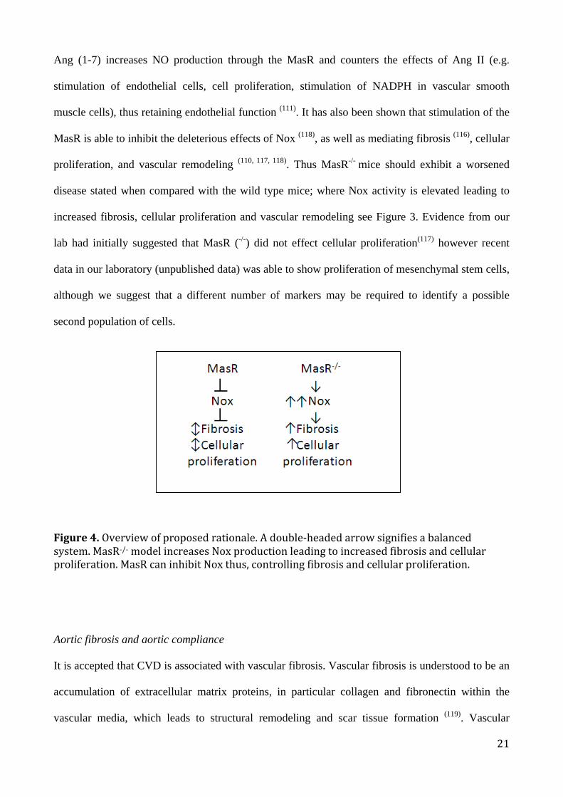
Figure4.Overviewofproposedrationale.Adouble-headedarrowsignifiesabalancedsystem.MasR-/-modelincreasesNoxproductionleadingtoincreasedfibrosisandcellularproliferation.MasRcaninhibitNoxthus,controllingfibrosisandcellularproliferation.
Aortic fibrosis and aortic compliance
It is accepted that CVD is associated with vascular fibrosis. Vascular fibrosis is understood to be an
accumulation of extracellular matrix proteins, in particular collagen and fibronectin within the
vascular media, which leads to structural remodeling and scar tissue formation (119). Vascular

22
fibrosis is recognized as an important contributor to arterial stiffening and is also known to promote
the development of atherosclerosis (120, 121). Fibrosis or increased stiffening of the vascular walls can
be attributed to a lack of elastin or excessive collagen (122).
Hyperhomocysteinemia has been reported in previous studies to increase collagen accumulation in
the carotid artery in mice and rats (123). Remodeling of vascular tissue contributes to an increased
peripheral resistance, and is known to impact both development and complications of hypertension
(124). Biopsies of abdominal aortic aneurysms from patients revealed vascular remodeling and
degeneration as well as a dilation of two to six times the normal aortic diameter (125). Contrary to the
belief that an enlarged aorta with the risk of rupture increasing, that the walls of the aorta may
become thinner, it was discovered that the opposite was true, where the larger the aneurysm the
thicker the walls and the smaller the aneurysm the thinner the walls (125). Although the larger
aneurysms present with thicker walls, they were thought to be weaker, thus leading to the much
higher risk of rupture than smaller aneurysms (125).
Myocardial fibrosis in CVD
Long-term experimental models have established that hyperhomocysteinemia is a risk factor for
systolic heart failure (126), and may also accelerate hyperhomocysteinemia-induced cardiac
remodeling (127). These hyperhomocysteinemic models have also suggested the presence of
ventricular hypertrophy and heart fibrosis (126, 128, 129). It was presented that homocysteine was able
to induce an increase of collagen content and ventricular hypertrophy (130), which is in line with
previous studies exhibiting a marked interstitial myocardial fibrosis in hypertensive and normal rats,
as well as rabbits subjected to a diet supplemented with methionine and cholesterol enriched diet
(126, 128, 129, 131). Hyperhomocysteinemia also affects several other tissues, for example, along with
myocardial fibrosis, liver fibrosis, lung fibrosis, and glomerulosclerosis; it also increases the
collagen content of aortic atherosclerotic plaque in atherosclerosis mouse models (132).
23
Preclinical studies have further added to the understanding that hyperhomocysteinemia promotes
myocardial fibrosis and dysfunction (126, 133, 134). Remodeling of the cardiac tissue was also observed
in rat models of mild hyperhomocysteinemia, which was associated with dysregulation of
extracellular matrix degradation (130). Although these collagen deposits can be attributed to an
increase of collagen synthesis and/or decrease in collagen degradation, homocysteine was revealed
to stimulate and elevate collagen, which was accompanied by cell proliferation(135-137).
All organs including cardiac tissue are composed of specialized parenchymal cells enclosed in
stroma consisting of extracellular matrix, tissue fluid and undifferentiated, pluripotent mesenchymal
cells (138). Matrix integrity relies on synthesis and degradation of all its major components, including
collagens. Extracellular matrix homeostasis is integral for cardiac function, as alterations can result
in development of myocardial fibrosis leading to systolic and diastolic dysfunction (139).
Accumulation of collagen in response to injury further exacerbates the structural loss of myocyte
due to ischemic necrosis, and progresses scar tissue formation (139). This continued accumulation of
collagen compromises and impairs contractile behavior leading to heart failure (138). Myocardial
fibrosis has also been linked with various other myocardial diseases, all of which fibrosis is
observed to be increased, thus leading to a correspondingly increased susceptibility to arrhythmia
(140). Patchy fibrosis throughout the right ventricle was shown to increase susceptibility to
arrhythmias and is thought to act as an anatomical substrate favoring arrhythmias (141).
The myocardial collagen networks consists of collagen I and III (85% and 10%, respectively), as
observed in rodent and non-human heart tissue, with scar tissue consisting of 90% type I collagen
(139, 142, 143). Type I collagen provides rigidity and myocardial stiffness due to its tensile strength (144),
while type III collagen contributes to elasticity as it consists of a fine reticular network (144, 145).
Alterations to the concentration of collagen and several other factors determine the development of

24
fibrosis (139, 143). Cells capable of synthesizing collagen include endothelial cells, osteoblasts, and
smooth muscle cells, with myofibroblasts being identified as the predominant cells responsible for
the development of cardiac fibrosis (138, 139). Studies have identified a correlation between cardiac
function and myocardial remodeling with reduced ejection fraction related to worsening fibrosis
and myocyte degeneration and elevated left ventricular end-diastolic pressure (146).
Renal disease in CVD:
Endothelial dysfunction is a well-accepted characteristic of atherosclerosis and is present within
both large and small arteries in renal disease (147). Incubation of rat small renal arteries with
homocysteine reduces NO in renal artery endothelium. This decrease in NO not only results in
endothelial dysfunction, but also in an increase in superoxide levels in the arterial endothelium (36,
39). In normal kidney physiology, NO regulates vascular homeostasis, glomerular, and tubular
function maintaining normal renal function, GFR and renal vascular resistance, vascular resistance
(148, 149).
Biopsies taken from patients with severe kidney disease exhibit prominent fibrosis in the renal
vasculature, glomeruli and tubuolinterstituim (95). Studies have demonstrated that chronic activation
of RAS can result in fibrosis and inflammation (150, 151) and production of Ang II (a potent
vasoconstrictor) is a significant mediator of tubulointerstitial fibrosis and progression towards end-
stage renal disease (95). Moreover, evidence suggests hyperhomocysteinemia leads to renal oxidative
stress and renal hemodynamic dysfunction due to altered L-Agr-NO pathway, and this may
aggravate vascular disease as a result of renal failure (152).
Thus, the idea of the ACE2/Ang (1-7)/MasR axis, and its role in countering the effects of ACE/Ang
II/AT1R effects needs to be further investigated. It is known that local RAS is expressed in the
kidneys and is able to function independently of the systemic RAS and is thought to contribute to

25
long-term blood pressure regulation and renal injury (153). In order to elucidate the protective
function of the ACE2/Ang (1-7)/MasR pathway, a study investigating the protective affects of
MasR-/- mice with 1% methionine enriched diet was performed (154). It was discovered that the
diuresis effect produced by AVE 0991, a MasR nonpeptide agonist, was significantly reduced in
MasR-/- mice (154). The researchers concluding that MasR plays a role in kidney fluid homeostasis
(154). As the ACE2/Ang (1-7)/MasR axis are critically involved in kidney function, it remains
unclear if this axis is protective against an unbalanced diet causing kidney disease, such as renal
fibrosis.

26
Hypothesis
It is hypothesized that a high methionine diet will exacerbate vascular pathology in MasR(-/-) mice.
Aims
To determine if a high methionine diet in MasR(-/-) mice will worsen:
a) endothelial function
b) blood vessel collagen accumulation
c) myocardial interstitial and perivascular collagen accumulation
d) renal glomerular and tubule collagen accumulation

27
Materials and Method: Animals:
Male MasR-/- (n=24) mice were be donated by Dr. Chandana Hearth, Austin Health. Wild type
control (C57BL/6J), (n=16) were purchased from the Animal Research Centre, WA. The mice
were divided into groups and fed their diet for 8 weeks:
1. MasR-/- - control diet (MasR-Control), n=12;
2. MasR-/- + 1% methionine (MasR+Meth) diet, n=12;
3. C57BL/6J – control diet (C57-Control), n=8
4. and C57BL/6J + 1% methionine diet (C57+Meth), n=8 (131, 155).
Mice were housed maximum 5 per cage and maintained on 12-h dark/12-h light cycle in an air-
conditioned room (22.5±0.5°C, 50±5% humidity) with access to diet and water ad libitum. The
mice were divided into 2 study groups, an acute organ bath and a fibrosis study group. Mice were
culled and organs extracted (aorta, heart, and kidney). The experiments were approved by Victoria
University, Animal Ethics Committee (approval # 14/005).
Organ bath – endothelial function:
The mice were anesthetized using oxygen and 4% isoflurane. A foot pinch test was performed to
ensuring the mouse has been completely anesthetized before the mouse were then culled by cervical
dislocation, and ensuring the mouse is unresponsive and not breathing. The aorta were taken from
mice and cut into ~3mm rings. The rings were then mounted between two metal hooks in 5ml organ
baths attached to force displacement transducers, which was in turn connected to a PC using
MediDAQ software that allows the tension to be measured (OB8, Zultek, Engineering, VIC,
Australia). The ring were placed in a normal physiological salt solution filled with Krebs solution
[mmol/L: (NaCl 118mM, KCl 4.7mM, NaHCO3 25mM, MgSO47H2O 1.2mM, CaCl2 2.5mM,
KH2PO4 1.2mM, glucose 11.7mM)] and kept at a constant temperature of 37oC and continuously

28
bubbled with 95% O2 and 5% CO2. The organ bath system is a traditional method allowing
isometric or isotonic measurement (155, 156). The rings were held to a basal tension of 0.5g and
allowed to equilibrate for 30 minutes (155). The rings were then washed once with fresh Krebs
solution. The rings were then pre-constricted with U46619 (Sigma, #D8174-5MG) a thromboxane
A2 mimetic to approximately 30% - 50% of maximal contraction (where maximal range is between
0.9-1.2g total tension ); after which a cumulative response curve to acetylcholine-induced relaxation
was performed (155).
Fibrosis
These diets have previously been used in our lab to induce an atherosclerotic state in rabbits (57, 131,
155) and it was predicted to also occur in the current mice. The heart, kidney and aorta were removed
and immediately placed in 4% PFA (Sigma, #158127-500G) for 24hrs, after which the 4% PFA was
removed and replaced with 1 x PBS and refrigerated at 4oC ready for processing. The tissue was
processed by initially removing water from the tissue using a series of ethanol solutions of increase
concentration until 100% water-free ethanol. Then a clearing process that involves using 3 different
xylene immersions, which gradually replaces the ethanol with xylene. Finally the xylene is replaced
by molten paraffin wax (SkinPath Pathology, Hawthorn East, VIC). Tissue was then embedded in
paraffin and refrigerated at 4oC until required. Sections were cut at 5 microns and dried over night
at 37oC on 26mm x 76mm glass slides.
Organ fibrosis was determined using a combination of Fast Green FCF (Sigma, #F7252-5G), Picric
acid (Sigma, #197378) and Direct Red (sirius red) (Sigma,#365548-5G), which binds to collagen
fibrils (131, 157). Initial tests using fast green-picro-sirius red were performed to optimize the visual
staining, as each tissue varied in colour with the same concentration. The following concentrations
of fast green and Sirius red were dissolved in picric acid; 0.1% of both fast green and Sirius red,
0.01% fast green and 0.1% Sirius red, and 0.01% of both fast green and sirius red. The de-waxing

29
and dehydrating method is as follows; The picro-sirius red/fast green stain solution was prepared
with the 3 different concentrations, a) 0.1% fast green and 0.1% sirius red in 100ml (saturated
aqueous picric acid solution) in Coplin jar, b) 0.01% fast green and 0.1% sirius red in 100ml
(saturated aqueous picric acid solution) in Coplin jar, c) 0.01% fast green and 0.01% sirius red in
100ml (saturated aqueous picric acid solution) in Coplin jar. Then the slides were run through 100%
xylenes 2X 10min, 100% ethanol 2X 20dips, 90% ethanol 1X 20dips, ddH2O 2X 2min, and placed
in appropriate Coplin jars for 60 minutes. The slides were rinsed quickly in 10dips of ddH2O and
immediately rinsed in 90% ethanol 1X 20dips, 100% ethanol 2X 20dips, 100% xylenes 2X 20dips.
The slide is then placed in a third 100% xylene, and finally a coverslip was placed on the slide with
DPX mounting medium and left to dry overnight.
After initial analysis, the optimal concentration for heart was c) 0.01% fast green and 0.01% sirius
red in 100ml (saturated aqueous picric acid solution), with kidney being b) 0.01% fast green and
0.1% sirius red in 100ml, and finally aorta with a concentration of a) 0.1% fast green and 0.1%
sirius red in 100ml. These three concentrations were then used in the final analysis process as
shown in Figures 4 - 12.
Immunohistochemistry:
Standard immunohistochemistry process was performed for collagen I (rabbit polyclonal anti-
collagen I antibody, ab34710, abcam) and III (rabbit polyclonal anti-collagen III antibody , ab7778,
abcam) with a dilution of 1:100 as follows; slides were run through 100% xylenes 2X 10min, 100%
ethanol 2X 20dips, 90% ethanol 1X 20dips, ddH2O 2X 2min, and 10mM Tris and left for 5mins.
200µl of blocking solution (ImmPRESS 2.5% normal goat serum blocking solution) was added
onto tissue and placed into humidified sealed incubation camber for 20mins. The blocking solution
was tipped off at 20mins and 200µl of primary antibody was added to the tissue and placed into
humidified sealed incubation chamber overnight. The primary antibody was then tipped off and the

30
slide washed in Tris 10mM for 20dips and left for 5mins. 200µl of Secondary antibody
(ImmPRESS HRP anti-rabbit IgG) was then added to the tissue and incubated for 1 hour. The slide
was washed in Tris 10mM for 20dips and left for 5mins. DAB Chromogen was prepared by adding
1 drop of DAB chromogen per 1ml of substrate buffer. 200µl of DAB solution was added to tissue
until browning was visible and no longer than 5mins to prevent over exposure. The DAB solution
was tipped off and washed in ddH2O 20dips. A 10ml syringe was used to draw up haematoxylin
and a filter attached prior to applying to the slide for 1-2mins to ensure nuclei staining. Slides were
washed once again in ddH2O 20dips, and then placed in Scott’s tap water (bluing solution) for 5-
10mins. Slides were washed in ddH2O 10dips and put through the dehydration process; 90%
ethanol 20dips, 100% ethanol 2X 20dips, 100% xylene 2X 20dips, and finally into a third 100%
xylene before mounting the coverslip with DPX mounting solution and left to dry overnight.
Negative controls were performed using Rabbit IgG, polyclonal - Isotype Control (Abcam,
#ab37415).
a) b)
Figure 5.
Four sections of tissue were imaged, shown with the blue dotted line indicating the image observed
on the Lecia software at 40X magnification. Black arrows indicate myocardial fibrosis detected by

31
fast green/sirus red staining. At 5x magnification a) blue doted squares were imaged at 40X. The b)
tissue was traced using a square template, observed here as a black dashed line. Myocardial vessels
were traced to analyze fibrosis.
Fibrosis and Immunohistochemistry data gathering and analysis
All pictures were taken using the Olympus BX53 upright microscope and the Leica DFC425 digital
microscope camera, and the Leica Application Suite X Core software. For each heart tissue section,
four randomised pictures, representative of the whole muscle tissue as well as any visible blood
vessels were taken at 40X magnification see Figure 4. Approximately a ~75% proportion of the
images were then analyzed using MCID Core 7.1. Heart perivascular tissue was analyzed for
fibrosis see Figure 4, by using a tracing pointer to draw around the perivascular wall and
surrounding fibrosis and subsequently allowing MCID Core 7.1 software to determine differences is
colour changes via predetermined colour parameters. The blue dotted squares areas were chosen
due to fibrosis. The glomeruli and aortic tissue was chosen according to relative size and
tractability, thus ensuring uniform tissue analysis data.

32
Figure 6.
In kidney a) black solid square represents the whole tissue, while the blue dotted square indicates
the image observed on the Lecia software at 40X. The green circles with red fill represent glomeruli
taken at 40X. b) Glomeruli and interstitial/tubule fibrosis were analyzed separately as shown by the
yellow dashed oval. c) Aorta pictures taken are represented by the red dotted square at 40X. The d)
adventitia and media were then traced (yellow dashed line) and analyzed separately.
Ten glomeruli images from each kidney section were taken at 40X magnification Figure 5. The
glomeruli and interstitial/tubule images were separately analyzed, using MCID Core 7.1. Between
two and eight images of the aorta were taken depending on the size of the aorta with larger aortas
requiring greater amounts of images, following the contour of the blood vessel as observed in the
Figure 5. The adventitia and media were traced and analyzed separately on MCID Core 7.1.
a) b)
c) d)

33
Results
To investigate the role of MasR in homocysteine induced vascular pathology, MasR-/- and C57BL/6
mice were fed a 1% methionine enriched or control diet. Endothelial function was determined in
mouse aorta by organ bath methods and the response to acetylcholine was measured via signal
transducers. Relaxation of the abdominal aorta is shown in Figure 6. Both C57BL/6 groups
displayed a normal relaxation and there was no significant difference between the control and
methionine diets. In contrast, the MasR-/- aorta displayed significantly less relaxation compared to
the C57BL/6 mice (32.3±6.8% vs 66.1±7.7%, p<0.001). These results were further exacerbated in
the MasR+Meth on the methionine diet, which displayed a further reduction in relaxation
(6.6±6.7% vs 74±7%, p<0.0001).
In myocardial tissue analysis using immunohistochemistry (IHC), see Figures 9 and 10, 1%
methionine feeding increased myocardial interstitial fibrosis by >3-fold (p<0.0001) in MasR+Meth
mice. However there was no significant increase in fibrosis observed in C57BL/6 mice between on
1% methionine or normal chow diet. In contrast, myocardial perivascular fibrosis was present in
C57BL/6 mice, displaying a 31% (p<0.001) increase while MasR+Meth mice displayed a 2.6-fold
(p<0.001) in crease in fibrosis.
Organ fibrosis was investigated using IHC in the heart, kidney and aorta in order to investigate
whether deletion of the MasR worsened the pathology caused by homocysteine. Renal fibrosis
shown in figure 11 and 12, displayed a significant increase in glomerular and interstitial/tubular
fibrosis. Methionine increased glomerular fibrosis by 22% in control mice (p=0.02), while a 2.1
fold (p<0.0001) increase was observed in MasR+Meth mice when compared to MasR+control
mice. Renal interstitial/tubular tissue displayed the greatest level of fibrosis displaying a 5-fold
(p<0.0001) increase however; this was only in MasR+Meth mice.

34
Interestingly, we found IHC analysis of our aortic medial and adventitial tissue did not present any
significant increase in fibrosis, in MasR+Meth mice. By contrast, 1% methionine fed control mice
(p<0.01) displayed significant aortic increase in fibrosis in both medial and adventitial tissue.
In addition, collagen I (Abcam, #ab34710) and III (Abcam, #ab7778) distribution was analyzed
using IHC, see figures 13 – 18 for collagen I, and figures 19 – 24 for collagen III staining. Aorta,
heart and renal tissues were analyzed, however no significant difference was detected between the
animal groups; MasR-/- and C57BL/6 on either diets (1% methionine and normal chow).
Furthermore, Rabbit IgG, polyclonal - Isotype Control (Abcam, #ab37415) used to analysis
negative control displayed no positive staining.

35
Figure 7.
Four groups were fed an 8-week normal mouse chow diet supplemented with 1% methionine as
well as a control diet. MasR+Meth (n=12) (l), MasR-/- (n=12) and control (q), C57 (n=8) and
control (o), and C57 (n=8) + methionine (p), were subjected to concentration response curves of
endothelial dependent relaxation to acetylcholine in aortic rings. Preconstriction of (U46619,
Sigma) aorta showed both C57 diet groups display relaxation, while a relaxation response was
limited with MasR-/- + methionine group with the least relaxation present. Means ± SEM, **p<0.01
n=12
n=12
n=8
n=8

36
Figure 8.
Aortic medial fibrosis, analyzed using fast green and sirius red, where red is indicative of collagen,
I and III. a) C57+control diet showing the least collagen content of all groups, b) C57+methionine
diet appears to have to most collagen content with a higher distribution of red within the tissue
sample, c) MasR-/- +control diet displays a higher collagen content (than C57 + control), d) MasR-/-
+methionine only showed a slight increase in collagen content. Means ± SEM, ***p<0.001
a)b)c)d)

37
Figure 9.
Aortic adventitial fibrosis, analyzed using fast green and sirius red, where the colour red is
indicative of collagen, I and III. a) C57 + control diet while not appearing to differ between the four
samples displays the least collagen content post analysis, b) C57 + methionine diet was shown to
have to most collagen content, c) MasR-/- + control diet had the second highest collagen content,
with d) MasR -/- + methionine showing a slightly lower collagen content than the control. Means ±
SEM, **p<0.01
a)b)c)d)

38
Figure 10.
Myocardial interstitial fibrosis was analyzed using fast green and sirius red, staining for protein and
collagen respectively; arrows are used to denote collagen. a) C57 + control diet shows a sparse,
though even distribution of collagen, while b) C57 + methionine indicates a similar distribution of
collagen, however there seems to be a higher distribution of collagen in the myocardial tissue. c)
MasR-/- + control shows the least collagen content, with very sparse distribution of collagen, and in
contrast to the previous three groups, d) MasR-/- + methionine diet yielded the highest level (a 2
fold increase) of collagen content within myocardial tissue, with even and more visible distribution.
Means ± SEM, **p<0.01
a)b)c)d)

39
Figure 11.
Myocardial perivascular fibrosis is analyzed using fast green/sirus red staining for protein and
collagen respectively. a) C57-Control diet shows a thin layer of perivascular collagen and has the
second lowest content of collagen, however b) C57 + methionine had a more pronounced increase
in perivascular collagen content which is also quite evident visually, while the c) MasR-/- + control
has the lowest detected level of perivascular collagen distribution, and finally d) MasR-/- + Meth
had a high level of perivascular collagen detected. Means ± SEM, *p<0.05
a)b)c)d)

40
Figure 12.
Glomerular fibrosis is analyzed using fast green and sirius red staining for protein and collagen
respectively. a) C57 + control, b) C57 + methionine, and c) MasR-/- + control all show similar
levels of glomerular collagen content, with MasR-/- + methionine indicating the lowest collagen
content, however d) MasR-/- + methionine has a significant (almost a 2 fold) increase in glomerular
collagen content, which can visualized. Means ± SEM, ***p<0.001
a)b)c)d)

41
Figure 13.
Interstitial/tubular fibrosis is analyzed using fast green and sirius red staining for protein and
collagen respectively. a) C57 + control, b) C57 + methionine, and c) MasR-/- + control all show
similar levels of glomerular collagen content, interestingly the MasR-/- + methionine shows the
lowest collagen content, however d) MasR-/- + methionine has a significant (more than a 2 fold)
increase in glomerular collagen content, which can easily visualized by the deep red. Means ±
SEM, ***p<0.0001
a)b)c)d)

42
Figure 14.
Aortic medial fibrosis collagen I. Positive generalized non-specific brown staining is observed
throughout the aortic media tissue. Positive adventitial staining is also displayed. No significant
increase was observed between the groups.
a)b)c)d)
43
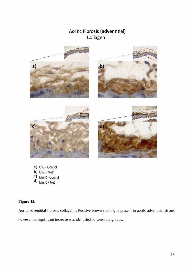
Figure 15.
Aortic adventitial fibrosis collagen I. Positive brown staining is present in aortic adventitial tissue,
however no significant increase was identified between the groups.
a)b)c)d)
44
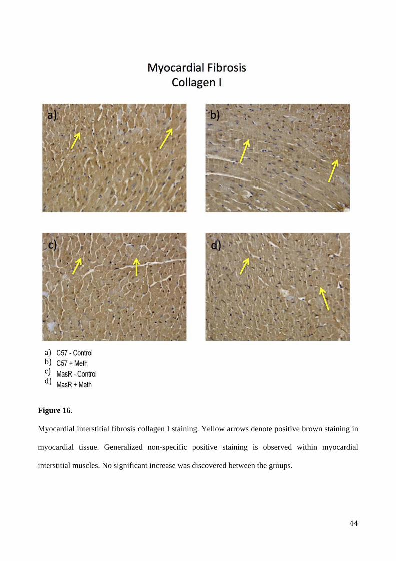
Figure 16.
Myocardial interstitial fibrosis collagen I staining. Yellow arrows denote positive brown staining in
myocardial tissue. Generalized non-specific positive staining is observed within myocardial
interstitial muscles. No significant increase was discovered between the groups.
a)b)c)d)

45
Figure 17.
Myocardial perivascular fibrosis collagen I. Yellow arrows denote positive brown staining in
myocardial tissue. Generalized non-specific staining is positive for collagen I within vascular
muscles as well as surrounding interstitial cardiomyocytes. No significant increase was detected
between the groups.
46
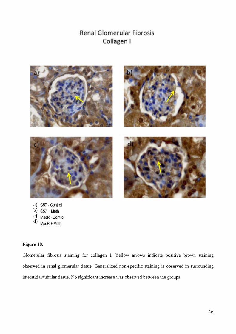
Figure 18.
Glomerular fibrosis staining for collagen I. Yellow arrows indicate positive brown staining
observed in renal glomerular tissue. Generalized non-specific staining is observed in surrounding
interstitial/tubular tissue. No significant increase was observed between the groups.
a)b)c)d)

47
Figure 19.
Renal interstitial/tubular fibrosis staining for collagen I. Yellow arrows indicate positive brown
staining detected throughout the renal interstitial/tubular tissue. Positive staining is detected in
glomerular tissue. No significant increase was seen between the groups.
a) b)
c) d)
a)b)c)d)

48
Figure 20.
Aortic medial fibrosis collagen III staining. Positive brown staining is observed in aortic medial
tissue. This generalized non-specific staining is observed in the media in addition to positive
staining observed in the adventitia. No significant increase was detected between the groups.
a)b)c)d)

49
Figure 21.
Aortic adventitial fibrosis collagen III staining. Positive brown staining is observed in the aortic
adventitial tissue. No significant increase was seen between the groups.
a)b)c)d)
50
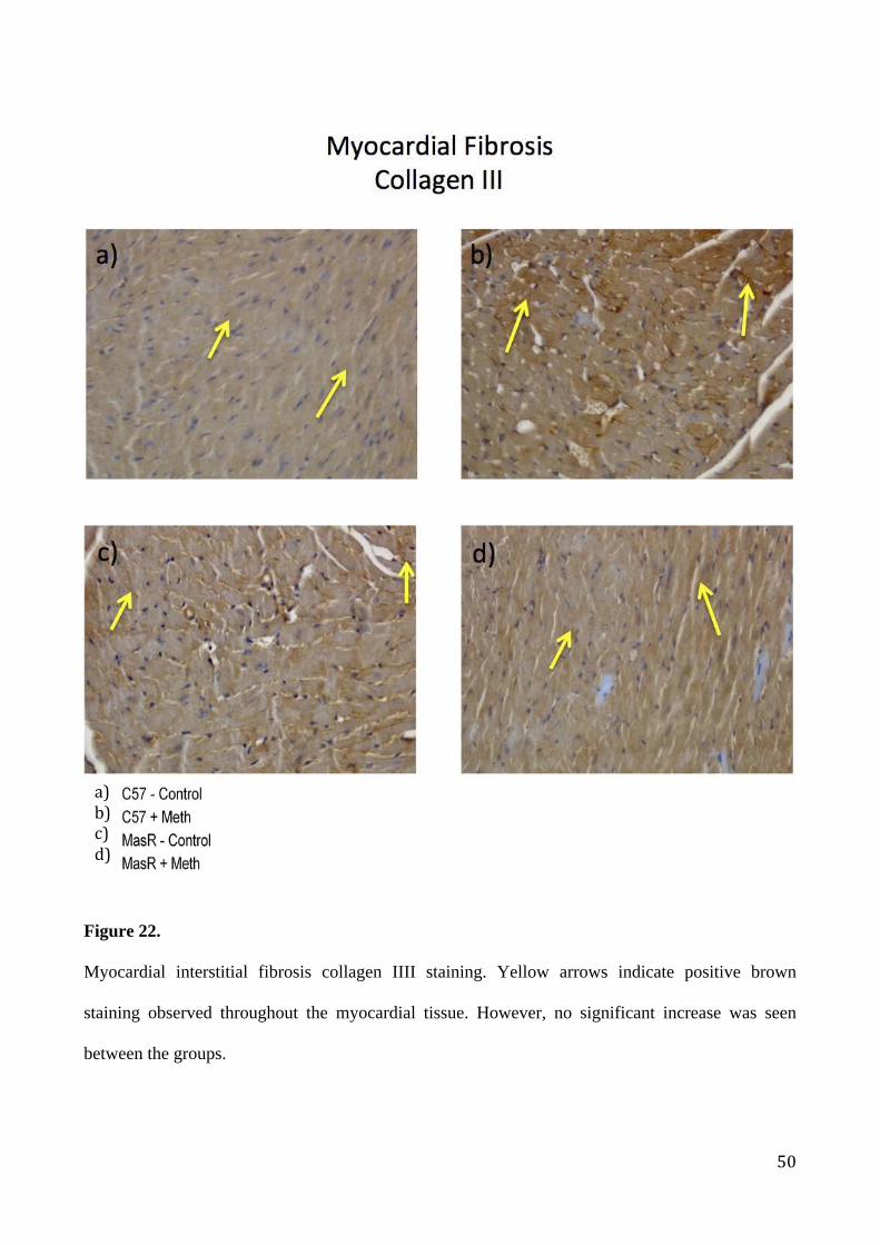
Figure 22.
Myocardial interstitial fibrosis collagen IIII staining. Yellow arrows indicate positive brown
staining observed throughout the myocardial tissue. However, no significant increase was seen
between the groups.
a)b)c)d)

51
Figure 23.
Myocardial perivascular fibrosis collagen III staining. Yellow arrows denote positive brown
staining observed throughout the myocardial tissue. This generalized non-specific staining is
observed throughout vascular muscles, however post-analysis did not detect any significant increase
in collagen III between the groups.
a)b)c)d)

52
Figure 24.
Glomerular fibrosis staining for collagen III. Yellow arrows indicate positive brown staining
observed throughout the renal glomerular tissue. This generalized non-specific staining is observed
within the glomerular and surrounding interstitial/tubular tissue. No significant increase was
observed between the groups.
a)b)c)d)

53
Figure 25.
Renal interstitial/tubular fibrosis staining for collagen III. Yellow arrows denote positive brown
staining. Generalized brown non-specific staining is observed throughout renal interstitial/tubular
tissue.
a)b)c)d)

54
Figure 26. IHC analysis of aortic medial fibrosis using rabbit IgG, polyclonal - isotype control did not display positive staining.
a)b)c)d)
RabbitIgG,polyclonal-IsotypeControl

55
Figure 27. IHC analysis of aortic adventitial fibrosis using rabbit IgG, polyclonal - isotype control did not display positive staining.
a)b)c)d)
RabbitIgG,polyclonal-IsotypeControl
56
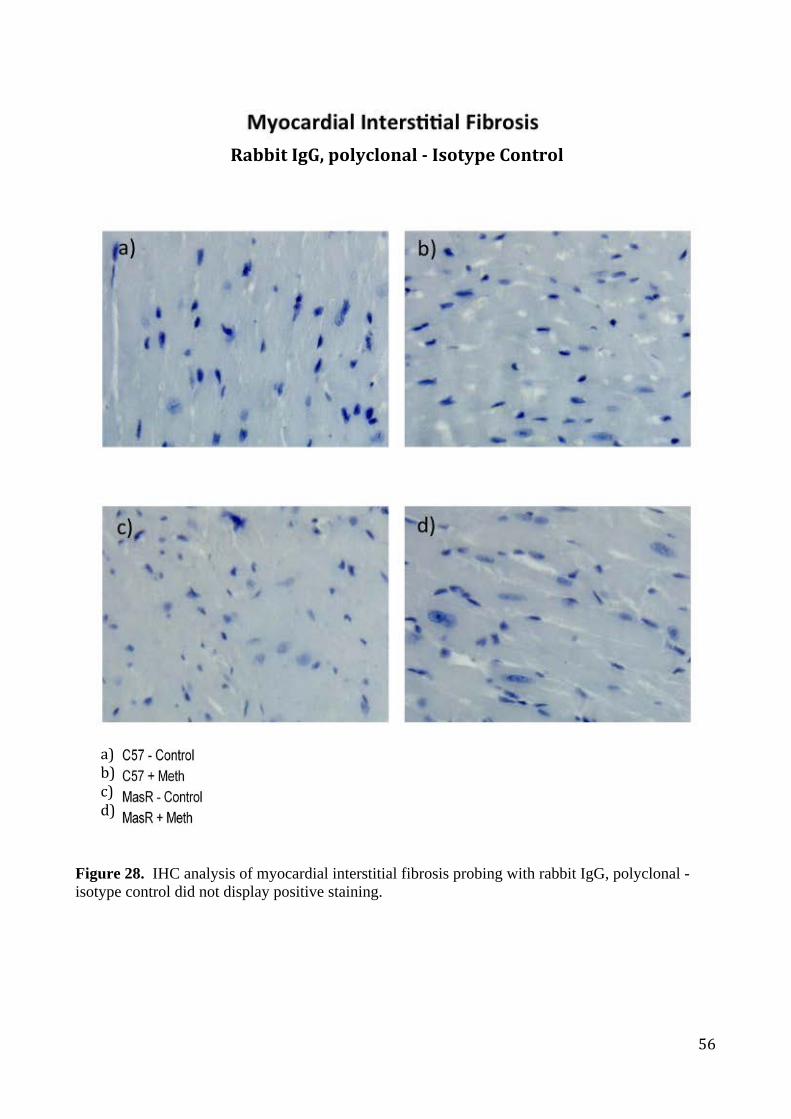
Figure 28. IHC analysis of myocardial interstitial fibrosis probing with rabbit IgG, polyclonal - isotype control did not display positive staining.
a)b)c)d)
RabbitIgG,polyclonal-IsotypeControl

57
Figure 29. IHC analysis of myocardial perivascular fibrosis using rabbit IgG, polyclonal - isotype control did not display positive staining.
a)b)c)d)
RabbitIgG,polyclonal-IsotypeControl

58
Figure 30. IHC analysis of renal interstitial/tubular fibrosis using rabbit IgG, polyclonal - isotype control did not display positive staining.
a)b)c)d)
RabbitIgG,polyclonal-IsotypeControl

59
Figure 31. IHC analysis of renal glomerular fibrosis using rabbit IgG, polyclonal - isotype control did not display positive staining.
a)b)c)d)
RabbitIgG,polyclonal-IsotypeControl

60
Discussion: In the present investigation, the major findings are that in both C57BL/6 and MasR-/- mice, a high
methionine diet (1% methionine) caused an increase in collagen accumulation as detected by sirus
red in the vasculature. This was worsened in MasR-/- mice, which also showed worsened
endothelial function.
These studies suggest that the MasR may play a protective role against methionine-induced fibrosis
within the myocardium (interstitially and perivascular), renal (glomeruli and interstitial/tubules),
however, interestingly, this was not the case within aortic tissue, which showed an increase in
fibrosis in C57BL/6 fed on 1% methionine but not in the MasR-/- mice. Aortic endothelial function
was also negatively affected by 1% methionine diet in the MasR-/-, however in the C57BL/6 mice
methionine did not display any significant affects.
Endothelial Function
We investigated the affects of homocysteine on MasR-/- and C57BL/6 mice with regards to
abdominal aorta relaxation to acetylcholine. Our study suggests that there is a significant reduction
in endothelial relaxation in MasR-/- mice when compared to C57BL/6, and this is further
exacerbated by the introduction of a methionine diet.
In this study we hypothesized to clarify the link between the role of the MasR, homocysteine and
CVD. More recently the MasR and in particular the ACE2/Ang (1-7)/MasR axis has been identified
as a possible treatment pathway in preventing the deleterious affects of the ACE/Ang II/AT1R (158).
This current research determined that our endothelial function study exhibited a clear difference in
the ability of the abdominal aorta to maximally relax in response to acetylcholine between the
MasR-/- and the C57BL/6 mice. Thus, there was an indication that endothelial dysfunction had
occurred and that this may directly relate to the lack of MasR. Our findings support the theory that

61
MasR plays a protective role in the normal homeostasis of blood vessels. MasR-/- mouse abdominal
aorta had a reduced relaxation in response to acetylcholine compared to the control. Previous studies
investigating the role of Ang (1-7) and its protective role on vascular beds was first demonstrated in
MasR-/- mice, where MasR-/- mice exhibited impaired in vivo endothelial-dependent vasorelaxant
response to acetylcholine (159, 160).
Studies have demonstrated the cardioprotective effects of the Ang (1-7)/MasR axis by the increased
synthesis and phosphorylation of eNOS (161). There is also evidence to suggest that defects that lead
to a reduced production of NO may lead to endothelial dysfunction, which can also be predictive of
future cardiovascular events (16). It is well understood that the endothelium is vital in cardiovascular
homeostasis while also providing protection against vascular disease. These cardioprotective effects
are achieved through the production of NO, a free radical responsible for inhibition of vascular
smooth muscle cells (VSMC) hypertrophy and relaxation of VSMC, platelet activation and
adhesion, inflammatory molecule expression. It has been previously been reported that Ang (1-7) is
an endogenous ligand for the MasR, thus the antidiuretic effects of Ang (1-7) on water-loaded mice
and the vasodilator effects on aortic rings of wild type mice were eliminated in MasR-/- mice (87).
Other studies investigating Ang (1-7) agonists such as AVE0991 and CGEN-856S found that they
were able to increase NO production, while the antagonist A-779 was able to inhibit the production
of NO (109, 162). Therefore the difference in relaxation in response to acetylcholine observed may be
due to a reduced NO bioavailability. Our investigation is in line with results demonstrated in other
studies suggesting that a reduced response to acetylcholine is indicative of a reduced
production/bioavailability of NO in MasR-/- model.
While there is still no well-defined understanding of the direct or indirect affects of homocysteine
on the vascular system, some have suggested that there is a risk associated with even a mild
increase of plasma homocysteine levels, with studies suggesting that homocysteine is elevated in

62
cases of sudden death as a result of severe coronary artery disease (163). Investigators have also
argued that their data identified elevated homocysteine level as being casually associated with
sudden death or the marker of a process related to sudden death (163). However, other studies have
revealed that lowering elevated homocysteine levels did not directly translate towards a healthy
cardiovascular system, and in fact even after homocysteine levels were reduced, the risk of major
cardiovascular events in patients with vascular disease did not significantly change (53, 54, 164). We
found that after an 8-week diet of 1% methionine, the response to acetylcholine was significantly
reduced only in the MasR-/- mouse. Homocysteine is known to instigate the production of ROS via
increased expression of NADPH oxidase leading to an alteration in tissue morphology. This
increase in ROS production is also linked to increased generation of ONOO- and decreased NO
bioavailability(165). Additionally MasR-/- mice were shown to upregulate gene and protein
expression of the NADPH oxidase catalytic subunit Nox2, while also reducing aortic SOD activity
(159, 160), taken together, these findings suggest that the reduction in endothelial response to
acetylcholine in MasR-/- could be due to upregulation of free radicals. The reduction of endothelial
response to acetylcholine was further exacerbated by the introduction of a methionine-enriched diet.
Other studies have also demonstrated that increased homocysteine levels induced higher O2
production (152). Xiao et al. demonstrated that MasR protects human brain microvascular endothelial
cells against Ang II-induced oxidative stress and dysfunction and it has also been speculated that
the underlying mechanism may rely on the ROS and NO signaling pathways(166). It was also
demonstrated that homocysteine reduced the resting cerebrovascular blood flow and additional, was
able to attenuate the increase in blood flow produced by acetylcholine and S-nitroso-N-
acetylpenicillamine (SNAP) (167). Yet, our data suggests that there was no significant difference
detected between the 1% methionine and control diet on endothelial function in C57BL/6 mice as
observed in other studies, which is contrasting as others have previously determined an increment
of 2 to 3mol/L in plasma homocysteine via oral methionine induced endothelial dysfunction in
healthy volunteers (n=18) (168). However, a 1% methionine diet was able to worsen endothelial

63
dysfunction in MasR-/- but no affect was observed in the C57BL/6 mice. 1% methionine diet and
MasR-/- mouse and consequently higher levels of homocysteine, suggest that without the protective
affects of the MasR, elevated plasma homocysteine levels are in part if not directly affecting
vascular tissue function leading to endothelial dysfunction.
Fibrosis
Our investigation suggests that hyperhomocysteinemia, by the introduction of a 1% methionine
diet, significantly increased fibrosis within myocardial and renal tissue in MasR-/- mice. However,
there was no significant increase of fibrosis identified in C57BL/6 mice despite the 1% methionine
diet. Interestingly however, with the same diet combination, the aortic tissue showed a significant
increase in fibrosis, the C57BL/6 mouse and not in the MasR-/- mouse.
Our investigation of renal fibrosis within glomerular tissue showed a 2.1 fold significant increase of
fibrosis in the MasR-/- mouse fed on 1% methionine diet over the control diet. However, there was
only a 22% in control significant increase of fibrosis observed in the 1% methionine diet in the
C57BL/6 mouse. Observations of fibrosis were even greater in interstitial/tubular tissue, with a
greater than 5 fold increase in the MasR-/- on 1% methionine diet over the control diet. C57BL/6
showed no significant increase in fibrosis between the two diets in interstitial/tubular tissue. The
Ang (1-7)/MasR axis is known to oppose the vasoconstrictor, profibrotic, and proliferative actions
of Ang II (158), and is thought to play an important role in renal function. Furthermore, enzymes
involved in the configuration of Ang (1-7) are abundantly found in the kidney (158) however, in
MasR-/- mice the configuration and production of Ang (1-7) may have little to no affect due to its
receptor binding site being absent. There are several other physiological ligands that also bind to
and activate MasR such as the neuropeptide FF (NPFF) (87), Ang III and Ang IV (169) however, most
effects of MasR were found to be mediated by Ang (1-7) (170). In addition, a study investigating the
distribution of enzymes involved in the metabolism of Ang II within the mouse renal tissue found

64
that Ang (1-7) was predominantly formed in the renal cortex (171), which suggested that ACE2 was
mainly localized in the renal cortex(158). Shi, Y. et al. 2015, demonstrated that Ang (1-7) treatment
in Akita mice was able to effectively attenuate oxidative, normalise ACE2 and MasR expression, as
well as suppressing expression of pro-fibrotic, pro-hypertensive and pro-apoptotic proteins, in renal
proximal tubular cells (172). These findings support our investigation as we demonstrated a clear
protective role of MasR against the deleterious effects of the methionine diet.
Homocysteine is understood to cause oxidative stress via way of ROS production. It was reported
that total homocysteine levels in patients with end stage renal failure was 3-5 times greater than
normal, and the incidence of hyperhomocysteinemia in the patient group was at 85-100% (173).
Currently, elevated homocysteine is observed as having a significantly positive correlation with
renal disease and is linked to kidney function however, it is debated as to whether elevated
homocysteine is an independent risk factor for kidney disease. Interestingly, previous reports have
suggested endothelial cells can detoxify homocysteine by stimulating the release of NO (174). While
there is limited literature on homocysteine and the role MasR plays in renal fibrosis, studies have
demonstrated human endothelial cells treated with homocysteine for a 24h period at 100µM was
able to reduce NO release, increase ROS and eventually lead to eNOS uncoupling by reducing
intracellular BH4 availability (175). This is reflective in part to our findings, where C57BL/6 mice on
1% methionine displayed an increase in glomerular but not interstitial/tubular fibrosis. However,
our MasR-/- presented with the greatest increase in glomerular and interstitial/tubular fibrosis
indicating that homocysteine in the absence of MasR produces an even greater increase of fibrotic
tissue which, may of course be due to the reduced NO production leading to a reduced ability to
detoxify ROS. In a 2004 study Ninomiya et al. reported that a 5-year investigation into total
homocysteine levels in local Japanese community suggested that moderately elevated serum total
homocysteine levels were a significant risk factor in the development of chronic kidney disease in
the general population (176). Interstitial fibrosis, vascular and glomerular sclerosis have been

65
identified as pathological abnormalities in chronic kidney disease (177). Studies demonstrating that
administration of diets that induced hyperhomocysteinemia for 12 weeks spontaneously provoked
arterial and arteriolar wall thickening and renal tubulointerstitial fibrosis are supportive of our
findings, in particular where we showed significant increase in fibrosis in our MasR-/- mice.
Furthermore, the worsening of renal function and renal tubulointerstitial remodeling were closely
correlated with the plasma total homocysteine levels (178). Our MasR-/- mouse model, without the
ability of protect against adverse affects of hyperhomocysteinemia and Ang II/AT1R axis, presented
with a significantly greater fibrosis level than that of the C57BL/6 mice. Interestingly, a study
revealed that ACE2 was 20-fold more active in the mouse renal cortex than in cardiac tissue (179).
However, no obvious difference in terms of fold increase was detected.
Hyperhomocysteinemia has been linked with the burden of CVD causing adverse cardiac
remodeling. Several experimental models of hyperhomocysteinemia have suggested heart fibrosis
and ventricular hypertrophy as a result of methionine diet (126, 128, 129). Investigators have also
suggested that a methionine enriched diet increased plasma total homocysteine levels resulting in
mild to moderate hyperhomocysteinemia (50, 126, 131). However, currently there is little information in
terms of literature as to whether the MasR functions to protect against hyperhomocysteinemia,
therefore our aim was to elucidate to role of MasR in myocardial tissue. In our study we observed a
more than 3-fold increase in myocardial interstitial fibrosis with the MasR-/- mice on the 1%
methionine diet over the control diet. C57BL/6 mice did not present with any significant increase in
collagen within the interstitial tissue. Previous reports have shown that hyperhomocysteinemia
induces an increase of collagen content (130), with preclinical studies suggesting that
hyperhomocysteinemia promotes myocardial fibrosis and tissue dysfunction (126, 133). We showed
that with our 8 weeks diet our study was in support of these previous findings although, the increase
was only observed in MasR-/- mice. In addition, others have described a marked interstitial
myocardial fibrosis in both hypertensive and normal rat subjects to homocysteine-supplemented

66
diet, as well as in rabbits on a methionine and cholesterol enriched diet (126, 131, 133). The deposition
of collagen may be a result of an increase of collagen synthesis and/or a decrease of its degradation
(130). Interestingly, while others have demonstrated an increase in fibrosis in control animals with
methionine enriched diets (126, 131), we only had a marked increase in our MasR-/- mice which,
suggests the within the myocardial interstitial tissue the MasR protects against homocysteine-
induced remodeling and fibrosis. However, within the myocardium we observed an increase in
perivascular fibrosis in both MasR-/- and C57BL/6 mice on the 1% methionine enriched diet. The
locational variation observed in fibrosis accumulation between interstitial and perivascular may to
NO availability. It is understood that homocysteine initiates matrix metalloproteinases (MMP)
deterioration of elastic structures causing stiff collagen deposition resulting in vascular remodeling
(180). Furthermore, a reduced NO availability will also lead to increased MMPs activity responsible
for extra cellular matrix degradation (181), with NO being shown to regulate MMP activity, vascular
function and remodeling within the endothelium (165).
Interestingly, while the fold increase was not as pronounced as in the interstitial tissue, we
identified a significant 2.6 fold increase in perivascular fibrosis in MasR-/-, as well as a 31%
increase in C57BL/6 mice fed on 1% methionine diet. Consideration must also be made that
homocysteine may have a tissue specific response, hence the difference in fibrosis observed
interstitially versus perivascularly. A 2002 study suggested that hyperhomocysteinemia was
associated with a significant increase in perivascular collagen in spontaneously hypertensive mice
hearts, additionally it was also suggested that while interstitial collagen level also increased in
hyperhomocysteinemia groups relative to control, no statistical significance was evident (133). The
interstitial increase in collagen may be due to replacement fibrosis following myocyte loss (182).
Total collagen content was showing to increase proportionally to increasing levels of total plasma
homocysteine (133).

67
Homocysteine is thought to promote oxidative stress via ROS(27), and this homocysteine-induced
oxidative stress is thought to be dependent mainly on NOS activity (175). Joseph, J. et al. in 2008
demonstrated that hyperhomocysteinemia acted through oxidative stress in order to promote
myocardial fibrosis (129). Additionally, oxidative stress is understood to be the most injurious
stressor in the progression and pathogenesis of most diseases, with homocysteine itself having the
ability to produce ROS when oxidized due to its reactive sulfhydryl group (181). Homocysteine is
also understood to indirectly decrease NO bioavailability via generation of superoxide to rapidly
consume NO leading to generation of ONOO- (183). In additional to the deleterious affects of
hyperhomocysteinemia, our MasR-/- mice would also have a significantly reduced ability to produce
NO via eNOS pathway, which has also been linked to myocardial fibrosis (184-186). Studies have
suggested that the increase in fibrosis due to hyperhomocysteinemia may be a result of excess
accumulation of extracellular matrix factors such as collagen I, where, changes of histone
modifications contributed to homocysteine-regulated gene expression of the collagen I gene (187).
Similar observations of tissue remodeling were demonstrated with long term administration of L-
NAME resulting in decreased NO synthesis (186). Our data suggests that 1% methionine diet was
causing adverse remodeling by way of perivascular fibrosis. However we were unable to
demonstrate a similar affect on interstitial tissue where only the MasR-/- mouse displayed an
increase in fibrosis. This may be a result of actions by various chemokines understood to affect
myocardial remodeling such as AngII and aldosterone, whose activities result in myocardial
collagen deposition in the perivascular area trailed by interstitial distribution (182, 188).
Our aortic tissue fibrosis results are however different from the results obtained from myocardial
and renal tissue. The significant increase was observed in the C57BL/6 mouse on the 1%
methionine diet in both medial and adventitial tissue. Hyperhomocysteinemia has been recognized
as an independent cardiovascular risk factor that may be involved in altering vascular structure (189),
and has been linked in the development of several aortic diseases (190-192). Furthermore, previous

68
studies involving vascular pathology associated with hyperhomocysteinemia have been
characterized by excess extracellular matrix turnover, initiating increased deposition of collagen
leading to endothelial dysfunction (193, 194). We observed a significant increase in aortic fibrosis
(medial and adventitial) in C57BL/6 mice fed on the 1% methionine diet. These results are in line
with previous studies reporting an accumulation of collagen as well as injured vessels being
susceptible to homocysteine-mediated vascular wall thickening (28, 120). Homocysteine was also
reported to increase collagen expression in smooth muscle cells in atherosclerotic plaques in ApoE
knock out mice (195). Interestingly, while our study is suggesting that homocysteine is causing aortic
endothelial dysfunction in MasR-/- mice as measured by response to acetylcholine, we were not able
discover any significant endothelial remodeling in either the adventitia or media of the aortic tissue.
MasR, activated by Ang (1-7) is understood to limit NADPH oxidase expression and activation. In
addition MasR-/- mice exhibit an increased expression of Nox2 and elevated vascular ROS levels in
comparison to control mice (160). Furthermore, Ang (1-7)/MasR axis was found to protect human
brain microvascular endothelial cells (HbmECs) against Ang II-induced dysfunction and oxidation,
suggesting that the underlying mechanism may rely on modulation of ROS and NO signaling
pathway (166). Additionally, Ang (1-7) and the nonpeptide agonist AVE 0991 was observed to
produce NO in wild-type mice however, this action was completed suppressed by blocking the
Ang (1-7) receptor and also by deletion of the Mas gene (109). We ascertained that
hyperhomocysteinemia was responsible for endothelial dysfunction in our study, which is in line
with previous studies. Therefore it was interesting to discover that our C57BL/6 mice presented
with a higher level of fibrosis, when previous studies have suggested that MasR-/- should be more
susceptible to fibrosis injury. With both adventitial and medial aortic tissue exhibiting no significant
increase in fibrosis, our results suggest that a different pathway may be involved in the reduced
fibrosis observed in MasR-/- mice fed on the 1% methionine and control diet. Apart form the MasR,
it is known that other G-coupled protein receptor such as the AT2R (196) and MrgD (197) also counter

69
the deleterious affects of AT1R. The AT2R receptor has shown to be upregulated in certain
pathological conditions, such as heart failure, aging, vascular injury, hypertension, and
atherosclerosis (131, 198-200), which may suggest a possible pathway in protection against fibrosis
however, the role of the AT2R is largely undefined. Additionally data suggests that Ang (1-7) may
also be acting through the AT2R(87, 201, 202) and not only the MasR. Furthermore, studies have
demonstrated the heptapeptide alamandine (that only differs from Ang (1-7) by an N-terminal Ala1
[Ala1-Ang (1-7)]) functions in a similar manner to Ang (1-7), such as endothelial-induced
vasodilation of aortic rings which, can be attenuated by NO synthase inhibitor N-nitro-l-arginine-
methyl ester (203). However, in our MasR-/- the functions of alamandine would becomes redundant
without MasR, thus it is interesting to note that alamandine also functions through the MrgD
receptor. A study demonstrated that using the MasR antagonist A-779 did not abolish alamandine-
induced vasodilation and thus maintaining the vasodilatory affects of alamandine in MasR-/- mice
(203). However, the alamandine-induced vasodilation was blocked using the Ang (1-7) antagonist D-
Pro7-Ang(1-7) which suggests that alamandine may be exerting its actions via a different G-coupled
protein receptor related to the MasR, the MrgD (169). The low levels of fibrosis detected within the
MasR-/- mouse suggests other protective, antifibrotic pathways are compensating for MasR, with the
AT2R and MrgD two likely receptors playing a protective role in the absence of MasR. It will
interesting to investigate whether the use of a combination of N-nitro-l-arginine- methyl ester, A-
779 and D-Pro7-Ang(1-7) on MasR-/- mice will be able to produce a fibrotic mouse however more
studies need to be performed to elucidated MasR role in aortic fibrosis.
As well, we immunohistochemically examined collagen I and III distribution in the heart, kidney
and aorta between MasR-/- and C57BL/6 mice. Positive staining of collagen I and III was detected
in the heart, kidney and aorta in both MasR-/- and C57BL/6 diet groups. We detected presence of
collagen I and III in myocardial interstitial and perivascular tissue which supports our fast
green/Sirius red results found in MasR+Meth and other groups. A study determined that

70
homocysteine is associated with significant increases in perivascular collagen in hypertensive mice
heart, with interstitial collagen also increasing (133), hence our results show positive generalized
binding of collagen I and III. However, a study determined that treatment with AVE 0991 did
significantly reduce collagen volume fraction as verified by reduced mRNA expression of collagen
I and III (204). This supports our results as the MasR+Meth mouse displayed a positive and possibly
generalized non-specific binding to collagen I and III. Previously, collagen release measurements
on human arterial smooth muscle cells by ELISA method had shown an increase of collagen I and
III in response to homocysteine (195). Collagen I was found to accumulate at a greater amount than
collagen III even though numbers of cells (SMC) had not increased (195). Collagen synthesis by
SMC in response to homocysteine dramatically increased in a dose-dependent manner, with even
the lowest concentration (50µM) significantly increasing collagen synthesis (136). Our aorta media
results indicated positive general non-specific Collagen I and III staining.
Conclusion/Future Directions
In conclusion, this study is the first to show a relationship between homocysteine and MasR, in that
the MasR is protective against homocysteine-induced vascular pathologies. We have shown the
presence of MasR is critical for protecting the cardiovascular system during CVD by maintaining
endothelial function and preventing tissue fibrosis. Future studies should focus on western blot
analysis of collagen I and III in heart, kidney, and aortic tissue to perform quantification of collagen
types.

71
References 1. Crackower MA, Sarao R, Oudit GY, Yagil C, Kozieradzki I, Scanga SE, et al. Angiotensin-converting enzyme 2 is an essential regulator of heart function. Nature. 2002;417(6891):822-8. 2. Paoletti R, Gotto AM, Jr., Hajjar DP. Inflammation in atherosclerosis and implications for therapy. Circulation. 2004;109(23 Suppl 1):Iii20-6. 3. Ross R. The pathogenesis of atherosclerosis: a perspective for the 1990s. Nature. 1993;362(6423):801-9. 4. Organization WH. Global Burden of Disease: 2004 Update. Geneva, Switzerland: World Health Organization, 2008. 5. Porth CM. Pathophysiology: Concepts of Altered Health States (6th edtion). 6th Edition ed: Lippincott Williams & Wilkins.; 2002. 1552 p. 6. Solberg LA, Strong JP. Risk factors and atherosclerotic lesions. A review of autopsy studies. Arteriosclerosis (Dallas, Tex). 1983;3(3):187-98. 7. Brattstrom L, Wilcken DE. Homocysteine and cardiovascular disease: cause or effect? The American journal of clinical nutrition. 2000;72(2):315-23. 8. Wierzbicki AS. Lipid-altering therapies and the progression of atherosclerotic disease. Cardiovascular and interventional radiology. 2007;30(2):155-60. 9. Bruckert E, Hayem G, Dejager S, Yau C, Begaud B. Mild to moderate muscular symptoms with high-dosage statin therapy in hyperlipidemic patients--the PRIMO study. Cardiovascular drugs and therapy / sponsored by the International Society of Cardiovascular Pharmacotherapy. 2005;19(6):403-14. 10. Bruckert E, Simonetta C, Giral P. Compliance with fluvastatin treatment characterization of the noncompliant population within a population of 3845 patients with hyperlipidemia. CREOLE Study Team. Journal of clinical epidemiology. 1999;52(6):589-94. 11. Lambrecht LJ, Malini PL. Efficacy and tolerability of simvastatin 20 mg vs pravastatin 20 mg in patients with primary hypercholesterolemia. European Study Group. Acta cardiologica. 1993;48(6):541-54. 12. Hadi HA, Carr CS, Al Suwaidi J. Endothelial dysfunction: cardiovascular risk factors, therapy, and outcome. Vascular health and risk management. 2005;1(3):183-98. 13. Bonetti PO, Lerman LO, Lerman A. Endothelial dysfunction: a marker of atherosclerotic risk. Arteriosclerosis, thrombosis, and vascular biology. 2003;23(2):168-75. 14. Anderson TJ, Gerhard MD, Meredith IT, Charbonneau F, Delagrange D, Creager MA, et al. Systemic nature of endothelial dysfunction in atherosclerosis. The American journal of cardiology. 1995;75(6):71b-4b. 15. Kinlay S, Ganz P. Role of endothelial dysfunction in coronary artery disease and implications for therapy. The American journal of cardiology. 1997;80(9a):11i-6i. 16. Davignon J, Ganz P. Role of endothelial dysfunction in atherosclerosis. Circulation. 2004;109(23 Suppl 1):Iii27-32. 17. Deanfield JE, Halcox JP, Rabelink TJ. Endothelial function and dysfunction: testing and clinical relevance. Circulation. 2007;115(10):1285-95. 18. Furchgott RF, Zawadzki JV. The obligatory role of endothelial cells in the relaxation of arterial smooth muscle by acetylcholine. Nature. 1980;288(5789):373-6. 19. Anderson TJ. Assessment and treatment of endothelial dysfunction in humans. Journal of the American College of Cardiology. 1999;34(3):631-8. 20. Chen Z, Peng IC, Sun W, Su MI, Hsu PH, Fu Y, et al. AMP-activated protein kinase functionally phosphorylates endothelial nitric oxide synthase Ser633. Circulation research. 2009;104(4):496-505. 21. Heller R, Hecker M, Stahmann N, Thiele JJ, Werner-Felmayer G, Werner ER. Alpha-tocopherol amplifies phosphorylation of endothelial nitric oxide synthase at serine 1177 and its short-chain derivative trolox stabilizes tetrahydrobiopterin. Free radical biology & medicine. 2004;37(5):620-31. 22. Bauer PM, Fulton D, Boo YC, Sorescu GP, Kemp BE, Jo H, et al. Compensatory phosphorylation and protein-protein interactions revealed by loss of function and gain of function mutants of multiple serine phosphorylation sites in endothelial nitric-oxide synthase. The Journal of biological chemistry. 2003;278(17):14841-9. 23. Forstermann U, Li H. Therapeutic effect of enhancing endothelial nitric oxide synthase (eNOS) expression and preventing eNOS uncoupling. British journal of pharmacology. 2011;164(2):213-23. 24. Mount PF, Kemp BE, Power DA. Regulation of endothelial and myocardial NO synthesis by multi-site eNOS phosphorylation. Journal of molecular and cellular cardiology. 2007;42(2):271-9.

72
25. Kobayashi T, Nemoto S, Ishida K, Taguchi K, Matsumoto T, Kamata K. Involvement of CaM kinase II in the impairment of endothelial function and eNOS activity in aortas of Type 2 diabetic rats. Clinical science (London, England : 1979). 2012;123(6):375-86. 26. Kobayashi T, Taguchi K, Yasuhiro T, Matsumoto T, Kamata K. Impairment of PI3-K/Akt pathway underlies attenuated endothelial function in aorta of type 2 diabetic mouse model. Hypertension. 2004;44(6):956-62. 27. Kietadisorn R, Juni RP, Moens AL. Tackling endothelial dysfunction by modulating NOS uncoupling: new insights into its pathogenesis and therapeutic possibilities. American journal of physiology Endocrinology and metabolism. 2012;302(5):E481-95. 28. McCully KS. Vascular pathology of homocysteinemia: implications for the pathogenesis of arteriosclerosis. The American journal of pathology. 1969;56(1):111-28. 29. Ducros V, Demuth K, Sauvant MP, Quillard M, Causse E, Candito M, et al. Methods for homocysteine analysis and biological relevance of the results. Journal of chromatography B, Analytical technologies in the biomedical and life sciences. 2002;781(1-2):207-26. 30. Fonseca V, Guba SC, Fink LM. Hyperhomocysteinemia and the endocrine system: implications for atherosclerosis and thrombosis. Endocrine reviews. 1999;20(5):738-59. 31. Mahalle N, Kulkarni MV, Garg MK, Naik SS. Vitamin B12 deficiency and hyperhomocysteinemia as correlates of cardiovascular risk factors in Indian subjects with coronary artery disease. Journal of cardiology. 2013;61(4):289-94. 32. Wang Y, Liu J, Jiang Y, Zhang H, Leng S, Wang G. Hyperhomocysteinemia is associated with decreased apolipoprotein AI levels in normal healthy people. BMC cardiovascular disorders. 2016;16:10. 33. Weiss N, Ide N, Abahji T, Nill L, Keller C, Hoffmann U. Aged garlic extract improves homocysteine-induced endothelial dysfunction in macro- and microcirculation. The Journal of nutrition. 2006;136(3 Suppl):750s-4s. 34. Brouwer DA, Welten HT, van Doormaal JJ, Reijngoud DJ, Muskiet FA. [Recommended dietary allowance of folic acid is insufficient for optimal homocysteine levels]. Nederlands tijdschrift voor geneeskunde. 1998;142(14):782-6. 35. Refsum H, Ueland PM, Nygard O, Vollset SE. Homocysteine and cardiovascular disease. Annual review of medicine. 1998;49:31-62. 36. Li N, Yi FX, Rute E, Zhang DX, Slocum GR, Zou AP. Effects of homocysteine on intracellular nitric oxide and superoxide levels in the renal arterial endothelium. American journal of physiology Heart and circulatory physiology. 2002;283(3):H1237-43. 37. Lipton SA, Kim WK, Choi YB, Kumar S, D'Emilia DM, Rayudu PV, et al. Neurotoxicity associated with dual actions of homocysteine at the N-methyl-D-aspartate receptor. Proceedings of the National Academy of Sciences of the United States of America. 1997;94(11):5923-8. 38. Handy DE, Zhang Y, Loscalzo J. Homocysteine down-regulates cellular glutathione peroxidase (GPx1) by decreasing translation. The Journal of biological chemistry. 2005;280(16):15518-25. 39. Toda N, Okamura T. Hyperhomocysteinemia impairs regional blood flow: involvements of endothelial and neuronal nitric oxide. Pflugers Archiv : European journal of physiology. 2016. 40. Bellamy MF, McDowell IF, Ramsey MW, Brownlee M, Bones C, Newcombe RG, et al. Hyperhomocysteinemia after an oral methionine load acutely impairs endothelial function in healthy adults. Circulation. 1998;98(18):1848-52. 41. Lentz SR. Does homocysteine promote atherosclerosis? Arteriosclerosis, thrombosis, and vascular biology. 2001;21(9):1385-6. 42. Holmdahl R, Malissen B. The need for littermate controls. European journal of immunology. 2012;42(1):45-7. 43. Abahji TN, Nill L, Ide N, Keller C, Hoffmann U, Weiss N. Acute hyperhomocysteinemia induces microvascular and macrovascular endothelial dysfunction. Archives of medical research. 2007;38(4):411-6. 44. Taddei S, Virdis A, Ghiadoni L, Sudano I, Salvetti A. Endothelial dysfunction in hypertension. Journal of cardiovascular pharmacology. 2001;38 Suppl 2:S11-4. 45. Weiss N, Keller C, Hoffmann U, Loscalzo J. Endothelial dysfunction and atherothrombosis in mild hyperhomocysteinemia. Vascular medicine (London, England). 2002;7(3):227-39. 46. Loscalzo J. Homocysteine trials--clear outcomes for complex reasons. The New England journal of medicine. 2006;354(15):1629-32. 47. den Heijer M, Willems HP, Blom HJ, Gerrits WB, Cattaneo M, Eichinger S, et al. Homocysteine lowering by B vitamins and the secondary prevention of deep vein thrombosis and pulmonary embolism: A randomized, placebo-controlled, double-blind trial. Blood. 2007;109(1):139-44.

73
48. Jamison RL, Hartigan P, Kaufman JS, Goldfarb DS, Warren SR, Guarino PD, et al. Effect of homocysteine lowering on mortality and vascular disease in advanced chronic kidney disease and end-stage renal disease: a randomized controlled trial. Jama. 2007;298(10):1163-70. 49. Nygard O, Nordrehaug JE, Refsum H, Ueland PM, Farstad M, Vollset SE. Plasma homocysteine levels and mortality in patients with coronary artery disease. The New England journal of medicine. 1997;337(4):230-6. 50. Hofmann MA, Lalla E, Lu Y, Gleason MR, Wolf BM, Tanji N, et al. Hyperhomocysteinemia enhances vascular inflammation and accelerates atherosclerosis in a murine model. The Journal of clinical investigation. 2001;107(6):675-83. 51. Plump AS, Smith JD, Hayek T, Aalto-Setala K, Walsh A, Verstuyft JG, et al. Severe hypercholesterolemia and atherosclerosis in apolipoprotein E-deficient mice created by homologous recombination in ES cells. Cell. 1992;71(2):343-53. 52. Zhou J, Moller J, Danielsen CC, Bentzon J, Ravn HB, Austin RC, et al. Dietary supplementation with methionine and homocysteine promotes early atherosclerosis but not plaque rupture in ApoE-deficient mice. Arteriosclerosis, thrombosis, and vascular biology. 2001;21(9):1470-6. 53. Lonn E, Yusuf S, Arnold MJ, Sheridan P, Pogue J, Micks M, et al. Homocysteine lowering with folic acid and B vitamins in vascular disease. The New England journal of medicine. 2006;354(15):1567-77. 54. Bonaa KH, Njolstad I, Ueland PM, Schirmer H, Tverdal A, Steigen T, et al. Homocysteine lowering and cardiovascular events after acute myocardial infarction. The New England journal of medicine. 2006;354(15):1578-88. 55. Kamata K, Nakajima M. Ca2+ mobilization in the aortic endothelium in streptozotocin-induced diabetic and cholesterol-fed mice. British journal of pharmacology. 1998;123(8):1509-16. 56. Cayatte AJ, Palacino JJ, Horten K, Cohen RA. Chronic inhibition of nitric oxide production accelerates neointima formation and impairs endothelial function in hypercholesterolemic rabbits. Arteriosclerosis and thrombosis : a journal of vascular biology / American Heart Association. 1994;14(5):753-9. 57. Zulli A, Hare DL. High dietary methionine plus cholesterol stimulates early atherosclerosis and late fibrous cap development which is associated with a decrease in GRP78 positive plaque cells. International journal of experimental pathology. 2009;90(3):311-20. 58. Lusis AJ. Atherosclerosis. Nature. 2000;407(6801):233-41. 59. Gimbrone MA, Jr. Vascular endothelium, hemodynamic forces, and atherogenesis. The American journal of pathology. 1999;155(1):1-5. 60. Guerra R, Jr., Brotherton AF, Goodwin PJ, Clark CR, Armstrong ML, Harrison DG. Mechanisms of abnormal endothelium-dependent vascular relaxation in atherosclerosis: implications for altered autocrine and paracrine functions of EDRF. Blood vessels. 1989;26(5):300-14. 61. Luscher TF, Richard V, Tschudi M, Yang ZH, Boulanger C. Endothelial control of vascular tone in large and small coronary arteries. Journal of the American College of Cardiology. 1990;15(3):519-27. 62. Holven KB, Holm T, Aukrust P, Christensen B, Kjekshus J, Andreassen AK, et al. Effect of folic acid treatment on endothelium-dependent vasodilation and nitric oxide-derived end products in hyperhomocysteinemic subjects. The American journal of medicine. 2001;110(7):536-42. 63. Tawakol A, Omland T, Gerhard M, Wu JT, Creager MA. Hyperhomocyst(e)inemia is associated with impaired endothelium-dependent vasodilation in humans. Circulation. 1997;95(5):1119-21. 64. Woo KS, Chook P, Lolin YI, Cheung AS, Chan LT, Sun YY, et al. Hyperhomocyst(e)inemia is a risk factor for arterial endothelial dysfunction in humans. Circulation. 1997;96(8):2542-4. 65. Chambers JC, McGregor A, Jean-Marie J, Obeid OA, Kooner JS. Demonstration of rapid onset vascular endothelial dysfunction after hyperhomocysteinemia: an effect reversible with vitamin C therapy. Circulation. 1999;99(9):1156-60. 66. Chao CL, Kuo TL, Lee YT. Effects of methionine-induced hyperhomocysteinemia on endothelium-dependent vasodilation and oxidative status in healthy adults. Circulation. 2000;101(5):485-90. 67. Kanani PM, Sinkey CA, Browning RL, Allaman M, Knapp HR, Haynes WG. Role of oxidant stress in endothelial dysfunction produced by experimental hyperhomocyst(e)inemia in humans. Circulation. 1999;100(11):1161-8. 68. Rhee SG. Cell signaling. H2O2, a necessary evil for cell signaling. Science (New York, NY). 2006;312(5782):1882-3. 69. Shafique E, Choy WC, Liu Y, Feng J, Cordeiro B, Lyra A, et al. Oxidative stress improves coronary endothelial function through activation of the pro-survival kinase AMPK. Aging (Albany NY). 2013;5(7):515-30.

74
70. Steinberg D. Antioxidants and atherosclerosis. A current assessment. Circulation. 1991;84(3):1420-5. 71. Takac I, Schroder K, Brandes RP. The Nox family of NADPH oxidases: friend or foe of the vascular system? Current hypertension reports. 2012;14(1):70-8. 72. Takac I, Schroder K, Zhang L, Lardy B, Anilkumar N, Lambeth JD, et al. The E-loop is involved in hydrogen peroxide formation by the NADPH oxidase Nox4. The Journal of biological chemistry. 2011;286(15):13304-13. 73. Sela M, Tirza G, Ravid O, Volovitz I, Solodeev I, Friedman O, et al. NOX1-induced accumulation of reactive oxygen species in abdominal fat-derived mesenchymal stromal cells impinges on long-term proliferation. Cell death & disease. 2015;6:e1728. 74. Lassegue B, Sorescu D, Szocs K, Yin Q, Akers M, Zhang Y, et al. Novel gp91(phox) homologues in vascular smooth muscle cells : nox1 mediates angiotensin II-induced superoxide formation and redox-sensitive signaling pathways. Circulation research. 2001;88(9):888-94. 75. Sorescu GP, Song H, Tressel SL, Hwang J, Dikalov S, Smith DA, et al. Bone morphogenic protein 4 produced in endothelial cells by oscillatory shear stress induces monocyte adhesion by stimulating reactive oxygen species production from a nox1-based NADPH oxidase. Circulation research. 2004;95(8):773-9. 76. Chamseddine AH, Miller FJ, Jr. Gp91phox contributes to NADPH oxidase activity in aortic fibroblasts but not smooth muscle cells. American journal of physiology Heart and circulatory physiology. 2003;285(6):H2284-9. 77. Gorlach A, Brandes RP, Nguyen K, Amidi M, Dehghani F, Busse R. A gp91phox containing NADPH oxidase selectively expressed in endothelial cells is a major source of oxygen radical generation in the arterial wall. Circulation research. 2000;87(1):26-32. 78. Ellmark SH, Dusting GJ, Fui MN, Guzzo-Pernell N, Drummond GR. The contribution of Nox4 to NADPH oxidase activity in mouse vascular smooth muscle. Cardiovascular research. 2005;65(2):495-504. 79. Van Buul JD, Fernandez-Borja M, Anthony EC, Hordijk PL. Expression and localization of NOX2 and NOX4 in primary human endothelial cells. Antioxidants & redox signaling. 2005;7(3-4):308-17. 80. Smith RM, Kruzliak P, Adamcikova Z, Zulli A. Role of Nox inhibitors plumbagin, ML090 and gp91ds-tat peptide on homocysteine thiolactone induced blood vessel dysfunction. Clinical and experimental pharmacology & physiology. 2015;42(8):860-4. 81. Takeno A, Kanazawa I, Tanaka K, Notsu M, Yokomoto M, Yamaguchi T, et al. Activation of AMP-activated protein kinase protects against homocysteine-induced apoptosis of osteocytic MLO-Y4 cells by regulating the expressions of NADPH oxidase 1 (Nox1) and Nox2. Bone. 2015;77:135-41. 82. Edirimanne VE, Woo CW, Siow YL, Pierce GN, Xie JY, O K. Homocysteine stimulates NADPH oxidase-mediated superoxide production leading to endothelial dysfunction in rats. Canadian journal of physiology and pharmacology. 2007;85(12):1236-47. 83. Sipkens JA, Hahn N, van den Brand CS, Meischl C, Cillessen SA, Smith DE, et al. Homocysteine-induced apoptosis in endothelial cells coincides with nuclear NOX2 and peri-nuclear NOX4 activity. Cell biochemistry and biophysics. 2013;67(2):341-52. 84. Leung SB, Zhang H, Lau CW, Huang Y, Lin Z. Salidroside improves homocysteine-induced endothelial dysfunction by reducing oxidative stress. Evidence-based complementary and alternative medicine : eCAM. 2013;2013:679635. 85. Sipkens JA, Krijnen PA, Meischl C, Cillessen SA, Smulders YM, Smith DE, et al. Homocysteine affects cardiomyocyte viability: concentration-dependent effects on reversible flip-flop, apoptosis and necrosis. Apoptosis : an international journal on programmed cell death. 2007;12(8):1407-18. 86. Lang D, Kredan MB, Moat SJ, Hussain SA, Powell CA, Bellamy MF, et al. Homocysteine-induced inhibition of endothelium-dependent relaxation in rabbit aorta: role for superoxide anions. Arteriosclerosis, thrombosis, and vascular biology. 2000;20(2):422-7. 87. Santos RA, Simoes e Silva AC, Maric C, Silva DM, Machado RP, de Buhr I, et al. Angiotensin-(1-7) is an endogenous ligand for the G protein-coupled receptor Mas. Proceedings of the National Academy of Sciences of the United States of America. 2003;100(14):8258-63. 88. Kim S, Iwao H. Molecular and cellular mechanisms of angiotensin II-mediated cardiovascular and renal diseases. Pharmacological reviews. 2000;52(1):11-34. 89. Sadoshima J, Izumo S. Molecular characterization of angiotensin II--induced hypertrophy of cardiac myocytes and hyperplasia of cardiac fibroblasts. Critical role of the AT1 receptor subtype. Circulation research. 1993;73(3):413-23.

75
90. Bennion DM, Haltigan E, Regenhardt RW, Steckelings UM, Sumners C. Neuroprotective Mechanisms of the ACE2-Angiotensin-(1-7)-Mas Axis in Stroke. Current hypertension reports. 2015;17(2):512. 91. Zhang Y, Li B, Wang B, Zhang J, Wu J, Morgan T. Alteration of Cardiac ACE2/Mas Expression and Cardiac Remodelling in Rats with Aortic Constriction. The Chinese journal of physiology. 2014;57(6):335-42. 92. Dias-Peixoto MF, Ferreira AJ, Almeida PW, Braga VB, Coutinho DC, Melo DS, et al. The cardiac expression of Mas receptor is responsive to different physiological and pathological stimuli. Peptides. 2012;35(2):196-201. 93. Wang J, Liu R, Qi H, Wang Y, Cui L, Wen Y, et al. The ACE2-Angiotensin-(1-7)-Mas Axis Protects Against Pancreatic Cell Damage in Cell Culture. Pancreas. 2015;44(2):266-72. 94. Gembardt F, Sterner-Kock A, Imboden H, Spalteholz M, Reibitz F, Schultheiss HP, et al. Organ-specific distribution of ACE2 mRNA and correlating peptidase activity in rodents. Peptides. 2005;26(7):1270-7. 95. Zhong J, Guo D, Chen CB, Wang W, Schuster M, Loibner H, et al. Prevention of angiotensin II-mediated renal oxidative stress, inflammation, and fibrosis by angiotensin-converting enzyme 2. Hypertension. 2011;57(2):314-22. 96. Ferrario CM, Chappell MC, Tallant EA, Brosnihan KB, Diz DI. Counterregulatory actions of angiotensin-(1-7). Hypertension. 1997;30(3 Pt 2):535-41. 97. Iwai M, Horiuchi M. Devil and angel in the renin-angiotensin system: ACE-angiotensin II-AT1 receptor axis vs. ACE2-angiotensin-(1-7)-Mas receptor axis. Hypertension research : official journal of the Japanese Society of Hypertension. 2009;32(7):533-6. 98. Jackson TR, Blair LA, Marshall J, Goedert M, Hanley MR. The mas oncogene encodes an angiotensin receptor. Nature. 1988;335(6189):437-40. 99. Papinska AM, Soto M, Meeks CJ, Rodgers KE. Long-term administration of angiotensin (1-7) prevents heart and lung dysfunction in a mouse model of type 2 diabetes (db/db) by reducing oxidative stress, inflammation and pathological remodeling. Pharmacological research. 2016;107:372-80. 100. Bader M, Santos RA, Unger T, Steckelings UM. New therapeutic pathways in the RAS. Journal of the renin-angiotensin-aldosterone system : JRAAS. 2012;13(4):505-8. 101. Unger T. The role of the renin-angiotensin system in the development of cardiovascular disease. The American journal of cardiology. 2002;89(2a):3A-9A; discussion 10A. 102. Fukuda D, Sata M. Role of bone marrow renin-angiotensin system in the pathogenesis of atherosclerosis. Pharmacology & therapeutics. 2008;118(2):268-76. 103. Grote K, Drexler H, Schieffer B. Renin-angiotensin system and atherosclerosis. Nephrology, dialysis, transplantation : official publication of the European Dialysis and Transplant Association - European Renal Association. 2004;19(4):770-3. 104. Young D, O'Neill K, Jessell T, Wigler M. Characterization of the rat mas oncogene and its high-level expression in the hippocampus and cerebral cortex of rat brain. Proceedings of the National Academy of Sciences of the United States of America. 1988;85(14):5339-42. 105. Zohn IE, Symons M, Chrzanowska-Wodnicka M, Westwick JK, Der CJ. Mas oncogene signaling and transformation require the small GTP-binding protein Rac. Molecular and cellular biology. 1998;18(3):1225-35. 106. Wang Y, Tikellis C, Thomas MC, Golledge J. Angiotensin converting enzyme 2 and atherosclerosis. Atherosclerosis. 2013;226(1):3-8. 107. Ambroz C, Clark AJ, Catt KJ. The mas oncogene enhances angiotensin-induced [Ca2+]i responses in cells with pre-existing angiotensin II receptors. Biochimica et biophysica acta. 1991;1133(1):107-11. 108. Walther T, Balschun D, Voigt JP, Fink H, Zuschratter W, Birchmeier C, et al. Sustained long term potentiation and anxiety in mice lacking the Mas protooncogene. The Journal of biological chemistry. 1998;273(19):11867-73. 109. Lemos VS, Silva DM, Walther T, Alenina N, Bader M, Santos RA. The endothelium-dependent vasodilator effect of the nonpeptide Ang(1-7) mimic AVE 0991 is abolished in the aorta of mas-knockout mice. Journal of cardiovascular pharmacology. 2005;46(3):274-9. 110. Zhang Z, Chen L, Zhong J, Gao P, Oudit GY. ACE2/Ang-(1-7) signaling and vascular remodeling. Science China Life sciences. 2014;57(8):802-8. 111. Tesanovic S, Vinh A, Gaspari TA, Casley D, Widdop RE. Vasoprotective and atheroprotective effects of angiotensin (1-7) in apolipoprotein E-deficient mice. Arteriosclerosis, thrombosis, and vascular biology. 2010;30(8):1606-13.

76
112. Thatcher SE, Zhang X, Howatt DA, Lu H, Gurley SB, Daugherty A, et al. Angiotensin-converting enzyme 2 deficiency in whole body or bone marrow-derived cells increases atherosclerosis in low-density lipoprotein receptor-/- mice. Arteriosclerosis, thrombosis, and vascular biology. 2011;31(4):758-65. 113. Loot AE, Roks AJ, Henning RH, Tio RA, Suurmeijer AJ, Boomsma F, et al. Angiotensin-(1-7) attenuates the development of heart failure after myocardial infarction in rats. Circulation. 2002;105(13):1548-50. 114. da Silveira KD, Coelho FM, Vieira AT, Sachs D, Barroso LC, Costa VV, et al. Anti-inflammatory effects of the activation of the angiotensin-(1-7) receptor, MAS, in experimental models of arthritis. Journal of immunology (Baltimore, Md : 1950). 2010;185(9):5569-76. 115. Giani JF, Gironacci MM, Munoz MC, Pena C, Turyn D, Dominici FP. Angiotensin-(1 7) stimulates the phosphorylation of JAK2, IRS-1 and Akt in rat heart in vivo: role of the AT1 and Mas receptors. American journal of physiology Heart and circulatory physiology. 2007;293(2):H1154-63. 116. Meng Y, Li T, Zhou GS, Chen Y, Yu CH, Pang MX, et al. The angiotensin-converting enzyme 2/angiotensin (1-7)/Mas axis protects against lung fibroblast migration and lung fibrosis by inhibiting the NOX4-derived ROS-mediated RhoA/Rho kinase pathway. Antioxidants & redox signaling. 2015;22(3):241-58. 117. Alsaadon H, Kruzliak P, Smardencas A, Hayes A, Bader M, Angus P, et al. Increased aortic intimal proliferation due to MasR deletion in vitro. International journal of experimental pathology. 2015. 118. Zheng J, Li G, Chen S, Bihl J, Buck J, Zhu Y, et al. Activation of the ACE2/Ang-(1-7)/Mas pathway reduces oxygen-glucose deprivation-induced tissue swelling, ROS production, and cell death in mouse brain with angiotensin II overproduction. Neuroscience. 2014;273:39-51. 119. Touyz RM. Intracellular mechanisms involved in vascular remodelling of resistance arteries in hypertension: role of angiotensin II. Experimental physiology. 2005;90(4):449-55. 120. Lan TH, Huang XQ, Tan HM. Vascular fibrosis in atherosclerosis. Cardiovascular pathology : the official journal of the Society for Cardiovascular Pathology. 2013;22(5):401-7. 121. Shirwany NA, Zou MH. Arterial stiffness: a brief review. Acta pharmacologica Sinica. 2010;31(10):1267-76. 122. Arribas SM, Hinek A, Gonzalez MC. Elastic fibres and vascular structure in hypertension. Pharmacology & therapeutics. 2006;111(3):771-91. 123. Guo YH, Chen FY, Wang GS, Chen L, Gao W. Diet-induced hyperhomocysteinemia exacerbates vascular reverse remodeling of balloon-injured arteries in rat. Chinese medical journal. 2008;121(22):2265-71. 124. Intengan HD, Schiffrin EL. Vascular remodeling in hypertension: roles of apoptosis, inflammation, and fibrosis. Hypertension. 2001;38(3 Pt 2):581-7. 125. Freestone T, Turner RJ, Coady A, Higman DJ, Greenhalgh RM, Powell JT. Inflammation and matrix metalloproteinases in the enlarging abdominal aortic aneurysm. Arteriosclerosis, thrombosis, and vascular biology. 1995;15(8):1145-51. 126. Devi S, Kennedy RH, Joseph L, Shekhawat NS, Melchert RB, Joseph J. Effect of long-term hyperhomocysteinemia on myocardial structure and function in hypertensive rats. Cardiovascular pathology : the official journal of the Society for Cardiovascular Pathology. 2006;15(2):75-82. 127. Zhang JS, Hou YL, Lu WW, Ni XQ, Lin F, Yu YR, et al. Intermedin1-53 Protects Against Myocardial Fibrosis by Inhibiting Endoplasmic Reticulum Stress and Inflammation Induced by Homocysteine in Apolipoprotein E-Deficient Mice. Journal of atherosclerosis and thrombosis. 2016. 128. Kundu S, Kumar M, Sen U, Mishra PK, Tyagi N, Metreveli N, et al. Nitrotyrosinylation, remodeling and endothelial-myocyte uncoupling in iNOS, cystathionine beta synthase (CBS) knockouts and iNOS/CBS double knockout mice. Journal of cellular biochemistry. 2009;106(1):119-26. 129. Joseph J, Joseph L, Devi S, Kennedy RH. Effect of anti-oxidant treatment on hyperhomocysteinemia-induced myocardial fibrosis and diastolic dysfunction. The Journal of heart and lung transplantation : the official publication of the International Society for Heart Transplantation. 2008;27(11):1237-41. 130. Raaf L, Noll C, Cherifi Mel H, Samuel JL, Delcayre C, Delabar JM, et al. Myocardial fibrosis and TGFB expression in hyperhomocysteinemic rats. Molecular and cellular biochemistry. 2011;347(1-2):63-70. 131. Zulli A, Hare DL, Buxton BF, Black MJ. The combination of high dietary methionine plus cholesterol induces myocardial fibrosis in rabbits. Atherosclerosis. 2006;185(2):278-81. 132. Dai J, Li W, Chang L, Zhang Z, Tang C, Wang N, et al. Role of redox factor-1 in hyperhomocysteinemia-accelerated atherosclerosis. Free radical biology & medicine. 2006;41(10):1566-77.
77
133. Joseph J, Washington A, Joseph L, Koehler L, Fink LM, Hauer-Jensen M, et al. Hyperhomocysteinemia leads to adverse cardiac remodeling in hypertensive rats. American journal of physiology Heart and circulatory physiology. 2002;283(6):H2567-74. 134. Miller A, Mujumdar V, Palmer L, Bower JD, Tyagi SC. Reversal of endocardial endothelial dysfunction by folic acid in homocysteinemic hypertensive rats. American journal of hypertension. 2002;15(2 Pt 1):157-63. 135. Garcia-Tevijano ER, Berasain C, Rodriguez JA, Corrales FJ, Arias R, Martin-Duce A, et al. Hyperhomocysteinemia in liver cirrhosis: mechanisms and role in vascular and hepatic fibrosis. Hypertension. 2001;38(5):1217-21. 136. Majors A, Ehrhart LA, Pezacka EH. Homocysteine as a risk factor for vascular disease. Enhanced collagen production and accumulation by smooth muscle cells. Arteriosclerosis, thrombosis, and vascular biology. 1997;17(10):2074-81. 137. Zhi H, Luptak I, Alreja G, Shi J, Guan J, Metes-Kosik N, et al. Effects of direct Renin inhibition on myocardial fibrosis and cardiac fibroblast function. PloS one. 2013;8(12):e81612. 138. Gonzalez A, Lopez B, Querejeta R, Diez J. Regulation of myocardial fibrillar collagen by angiotensin II. A role in hypertensive heart disease? Journal of molecular and cellular cardiology. 2002;34(12):1585-93. 139. Segura AM, Frazier OH, Buja LM. Fibrosis and heart failure. Heart failure reviews. 2014;19(2):173-85. 140. Akar FG, Spragg DD, Tunin RS, Kass DA, Tomaselli GF. Mechanisms underlying conduction slowing and arrhythmogenesis in nonischemic dilated cardiomyopathy. Circulation research. 2004;95(7):717-25. 141. Stein M, Boulaksil M, Jansen JA, Herold E, Noorman M, Joles JA, et al. Reduction of fibrosis-related arrhythmias by chronic renin-angiotensin-aldosterone system inhibitors in an aged mouse model. American journal of physiology Heart and circulatory physiology. 2010;299(2):H310-21. 142. Medugorac I, Jacob R. Characterisation of left ventricular collagen in the rat. Cardiovascular research. 1983;17(1):15-21. 143. Weber KT. Cardiac interstitium in health and disease: the fibrillar collagen network. Journal of the American College of Cardiology. 1989;13(7):1637-52. 144. Brilla CG, Zhou G, Rupp H, Maisch B, Weber KT. Role of angiotensin II and prostaglandin E2 in regulating cardiac fibroblast collagen turnover. The American journal of cardiology. 1995;76(13):8d-13d. 145. Weber KT. Extracellular matrix remodeling in heart failure: a role for de novo angiotensin II generation. Circulation. 1997;96(11):4065-82. 146. Villarreal FJ, Kim NN, Ungab GD, Printz MP, Dillmann WH. Identification of functional angiotensin II receptors on rat cardiac fibroblasts. Circulation. 1993;88(6):2849-61. 147. Schiffrin EL, Lipman ML, Mann JF. Chronic kidney disease: effects on the cardiovascular system. Circulation. 2007;116(1):85-97. 148. Kone BC, Baylis C. Biosynthesis and homeostatic roles of nitric oxide in the normal kidney. The American journal of physiology. 1997;272(5 Pt 2):F561-78. 149. Raij L, Baylis C. Glomerular actions of nitric oxide. Kidney international. 1995;48(1):20-32. 150. Bache RJ, Chen Y. NOX2-induced myocardial fibrosis and diastolic dysfunction: role of the endothelium. Journal of the American College of Cardiology. 2014;63(24):2742-4. 151. Lassegue B, San Martin A, Griendling KK. Biochemistry, physiology, and pathophysiology of NADPH oxidases in the cardiovascular system. Circulation research. 2012;110(10):1364-90. 152. Fischer PA, Dominguez GN, Cuniberti LA, Martinez V, Werba JP, Ramirez AJ, et al. Hyperhomocysteinemia induces renal hemodynamic dysfunction: is nitric oxide involved? Journal of the American Society of Nephrology : JASN. 2003;14(3):653-60. 153. Samuel P, Ali Q, Sabuhi R, Wu Y, Hussain T. High Na intake increases renal angiotensin II levels and reduces expression of the ACE2-AT(2)R-MasR axis in obese Zucker rats. American journal of physiology Renal physiology. 2012;303(3):F412-9. 154. Pinheiro SV, Simoes e Silva AC, Sampaio WO, de Paula RD, Mendes EP, Bontempo ED, et al. Nonpeptide AVE 0991 is an angiotensin-(1-7) receptor Mas agonist in the mouse kidney. Hypertension. 2004;44(4):490-6. 155. Zulli A, Widdop RE, Hare DL, Buxton BF, Black MJ. High methionine and cholesterol diet abolishes endothelial relaxation. Arteriosclerosis, thrombosis, and vascular biology. 2003;23(8):1358-63. 156. Ko EA, Song MY, Donthamsetty R, Makino A, Yuan JX. Tension Measurement in Isolated Rat and Mouse Pulmonary Artery. Drug Discov Today Dis Models. 2010;7(3-4):123-30.

78
157. Kruzliak PH, D.L.; Zulli, A. Simvastatin Impairs The Induction Of Pulmonary Fibrosis Caused By A Western Style Diet: A Preliminary Study. Journal of cellular and molecular medicine. 2015. 158. Santos RA, Ferreira AJ, Verano-Braga T, Bader M. Angiotensin-converting enzyme 2, angiotensin-(1-7) and Mas: new players of the renin-angiotensin system. The Journal of endocrinology. 2013;216(2):R1-r17. 159. Rabelo LA, Xu P, Todiras M, Sampaio WO, Buttgereit J, Bader M, et al. Ablation of angiotensin (1-7) receptor Mas in C57Bl/6 mice causes endothelial dysfunction. Journal of the American Society of Hypertension : JASH. 2008;2(6):418-24. 160. Xu P, Costa-Goncalves AC, Todiras M, Rabelo LA, Sampaio WO, Moura MM, et al. Endothelial dysfunction and elevated blood pressure in MAS gene-deleted mice. Hypertension. 2008;51(2):574-80. 161. Abwainy A, Babiker F, Akhtar S, Benter IF. Endogenous angiotensin-(1-7)/Mas receptor/NO pathway mediates the cardioprotective effects of pacing postconditioning. American journal of physiology Heart and circulatory physiology. 2016;310(1):H104-12. 162. Savergnini SQ, Beiman M, Lautner RQ, de Paula-Carvalho V, Allahdadi K, Pessoa DC, et al. Vascular relaxation, antihypertensive effect, and cardioprotection of a novel peptide agonist of the MAS receptor. Hypertension. 2010;56(1):112-20. 163. Burke AP, Fonseca V, Kolodgie F, Zieske A, Fink L, Virmani R. Increased serum homocysteine and sudden death resulting from coronary atherosclerosis with fibrous plaques. Arteriosclerosis, thrombosis, and vascular biology. 2002;22(11):1936-41. 164. Toole JF, Malinow MR, Chambless LE, Spence JD, Pettigrew LC, Howard VJ, et al. Lowering homocysteine in patients with ischemic stroke to prevent recurrent stroke, myocardial infarction, and death: the Vitamin Intervention for Stroke Prevention (VISP) randomized controlled trial. Jama. 2004;291(5):565-75. 165. Steed MM, Tyagi N, Sen U, Schuschke DA, Joshua IG, Tyagi SC. Functional consequences of the collagen/elastin switch in vascular remodeling in hyperhomocysteinemic wild-type, eNOS-/-, and iNOS-/- mice. American journal of physiology Lung cellular and molecular physiology. 2010;299(3):L301-11. 166. Xiao X, Zhang C, Ma X, Miao H, Wang J, Liu L, et al. Angiotensin-(1-7) counteracts angiotensin II-induced dysfunction in cerebral endothelial cells via modulating Nox2/ROS and PI3K/NO pathways. Experimental cell research. 2015;336(1):58-65. 167. Zhang F, Slungaard A, Vercellotti GM, Iadecola C. Superoxide-dependent cerebrovascular effects of homocysteine. The American journal of physiology. 1998;274(6 Pt 2):R1704-11. 168. Chambers JC, Obeid OA, Kooner JS. Physiological increments in plasma homocysteine induce vascular endothelial dysfunction in normal human subjects. Arteriosclerosis, thrombosis, and vascular biology. 1999;19(12):2922-7. 169. Gembardt F, Grajewski S, Vahl M, Schultheiss HP, Walther T. Angiotensin metabolites can stimulate receptors of the Mas-related genes family. Molecular and cellular biochemistry. 2008;319(1-2):115-23. 170. Santos RA, Castro CH, Gava E, Pinheiro SV, Almeida AP, Paula RD, et al. Impairment of in vitro and in vivo heart function in angiotensin-(1-7) receptor MAS knockout mice. Hypertension. 2006;47(5):996-1002. 171. Grobe N, Elased KM, Cool DR, Morris M. Mass spectrometry for the molecular imaging of angiotensin metabolism in kidney. American journal of physiology Endocrinology and metabolism. 2012;302(8):E1016-24. 172. Shi Y, Lo CS, Padda R, Abdo S, Chenier I, Filep JG, et al. Angiotensin-(1-7) prevents systemic hypertension, attenuates oxidative stress and tubulointerstitial fibrosis, and normalizes renal angiotensin-converting enzyme 2 and Mas receptor expression in diabetic mice. Clinical science (London, England : 1979). 2015;128(10):649-63. 173. Long Y, Nie J. Homocysteine in Renal Injury. Kidney diseases (Basel, Switzerland). 2016;2(2):80-7. 174. Stamler JS, Osborne JA, Jaraki O, Rabbani LE, Mullins M, Singel D, et al. Adverse vascular effects of homocysteine are modulated by endothelium-derived relaxing factor and related oxides of nitrogen. The Journal of clinical investigation. 1993;91(1):308-18. 175. Topal G, Brunet A, Millanvoye E, Boucher JL, Rendu F, Devynck MA, et al. Homocysteine induces oxidative stress by uncoupling of NO synthase activity through reduction of tetrahydrobiopterin. Free radical biology & medicine. 2004;36(12):1532-41. 176. Ninomiya T, Kiyohara Y, Kubo M, Tanizaki Y, Tanaka K, Okubo K, et al. Hyperhomocysteinemia and the development of chronic kidney disease in a general population: the Hisayama study. American journal of kidney diseases : the official journal of the National Kidney Foundation. 2004;44(3):437-45.

79
177. Levey AS, Coresh J. Chronic kidney disease. Lancet (London, England). 2012;379(9811):165-80. 178. Kumagai H, Katoh S, Hirosawa K, Kimura M, Hishida A, Ikegaya N. Renal tubulointerstitial injury in weanling rats with hyperhomocysteinemia. Kidney international. 2002;62(4):1219-28. 179. Wysocki J, Ye M, Soler MJ, Gurley SB, Xiao HD, Bernstein KE, et al. ACE and ACE2 activity in diabetic mice. Diabetes. 2006;55(7):2132-9. 180. Hayden MR, Tyagi SC. Intimal redox stress: accelerated atherosclerosis in metabolic syndrome and type 2 diabetes mellitus. Atheroscleropathy. Cardiovascular diabetology. 2002;1:3. 181. Steed MM, Tyagi SC. Mechanisms of cardiovascular remodeling in hyperhomocysteinemia. Antioxidants & redox signaling. 2011;15(7):1927-43. 182. Weber KT, Brilla CG. Pathological hypertrophy and cardiac interstitium. Fibrosis and renin-angiotensin-aldosterone system. Circulation. 1991;83(6):1849-65. 183. Faraci FM. Hyperhomocysteinemia: a million ways to lose control. Arteriosclerosis, thrombosis, and vascular biology. 2003;23(3):371-3. 184. Kobayashi N, Hara K, Watanabe S, Higashi T, Matsuoka H. Effect of imidapril on myocardial remodeling in L-NAME-induced hypertensive rats is associated with gene expression of NOS and ACE mRNA. American journal of hypertension. 2000;13(2):199-207. 185. Takemoto M, Egashira K, Tomita H, Usui M, Okamoto H, Kitabatake A, et al. Chronic angiotensin-converting enzyme inhibition and angiotensin II type 1 receptor blockade: effects on cardiovascular remodeling in rats induced by the long-term blockade of nitric oxide synthesis. Hypertension. 1997;30(6):1621-7. 186. Takemoto M, Egashira K, Usui M, Numaguchi K, Tomita H, Tsutsui H, et al. Important role of tissue angiotensin-converting enzyme activity in the pathogenesis of coronary vascular and myocardial structural changes induced by long-term blockade of nitric oxide synthesis in rats. The Journal of clinical investigation. 1997;99(2):278-87. 187. Lei W, Long Y, Li S, Liu Z, Zhu F, Hou FF, et al. Homocysteine Induces Collagen I Expression by Downregulating Histone Methyltransferase G9a. PloS one. 2015;10(7):e0130421. 188. Foglia MJ, Poss KD. Building and re-building the heart by cardiomyocyte proliferation. Development (Cambridge, England). 2016;143(5):729-40. 189. Kumar M, Tyagi N, Moshal KS, Sen U, Pushpakumar SB, Vacek T, et al. GABAA receptor agonist mitigates homocysteine-induced cerebrovascular remodeling in knockout mice. Brain research. 2008;1221:147-53. 190. Giusti B, Marcucci R, Lapini I, Sestini I, Lenti M, Yacoub M, et al. Role of hyperhomocysteinemia in aortic disease. Cellular and molecular biology (Noisy-le-Grand, France). 2004;50(8):945-52. 191. Novaro GM, Aronow HD, Mayer-Sabik E, Griffin BP. Plasma homocysteine and calcific aortic valve disease. Heart (British Cardiac Society). 2004;90(7):802-3. 192. Tyagi N, Sedoris KC, Steed M, Ovechkin AV, Moshal KS, Tyagi SC. Mechanisms of homocysteine-induced oxidative stress. American journal of physiology Heart and circulatory physiology. 2005;289(6):H2649-56. 193. Ovechkin AV, Tyagi N, Sen U, Lominadze D, Steed MM, Moshal KS, et al. 3-Deazaadenosine mitigates arterial remodeling and hypertension in hyperhomocysteinemic mice. American journal of physiology Lung cellular and molecular physiology. 2006;291(5):L905-11. 194. Sundstrom J, Sullivan L, D'Agostino RB, Jacques PF, Selhub J, Rosenberg IH, et al. Plasma homocysteine, hypertension incidence, and blood pressure tracking: the Framingham Heart Study. Hypertension. 2003;42(6):1100-5. 195. Rasmussen LM, Hansen PR, Ledet T. Homocysteine and the production of collagens, proliferation and apoptosis in human arterial smooth muscle cells. APMIS : acta pathologica, microbiologica, et immunologica Scandinavica. 2004;112(9):598-604. 196. Carey RM. Newly discovered components and actions of the renin-angiotensin system. Hypertension. 2013;62(5):818-22. 197. Hrenak J, Paulis L, Simko F. Angiotensin A/Alamandine/MrgD Axis: Another Clue to Understanding Cardiovascular Pathophysiology. International journal of molecular sciences. 2016;17(7). 198. Pinaud F, Bocquet A, Dumont O, Retailleau K, Baufreton C, Andriantsitohaina R, et al. Paradoxical role of angiotensin II type 2 receptors in resistance arteries of old rats. Hypertension. 2007;50(1):96-102. 199. Vinh A, Widdop RE, Drummond GR, Gaspari TA. Chronic angiotensin IV treatment reverses endothelial dysfunction in ApoE-deficient mice. Cardiovascular research. 2008;77(1):178-87. 200. Widdop RE, Vinh A, Henrion D, Jones ES. Vascular angiotensin AT2 receptors in hypertension and ageing. Clinical and experimental pharmacology & physiology. 2008;35(4):386-90.

80
201. Faria-Silva R, Duarte FV, Santos RA. Short-term angiotensin(1-7) receptor MAS stimulation improves endothelial function in normotensive rats. Hypertension. 2005;46(4):948-52. 202. Sampaio WO, Souza dos Santos RA, Faria-Silva R, da Mata Machado LT, Schiffrin EL, Touyz RM. Angiotensin-(1-7) through receptor Mas mediates endothelial nitric oxide synthase activation via Akt-dependent pathways. Hypertension. 2007;49(1):185-92. 203. Lautner RQ, Villela DC, Fraga-Silva RA, Silva N, Verano-Braga T, Costa-Fraga F, et al. Discovery and characterization of alamandine: a novel component of the renin-angiotensin system. Circulation research. 2013;112(8):1104-11. 204. Zeng WT, Chen WY, Leng XY, Tang LL, Sun XT, Li CL, et al. Impairment of cardiac function and remodeling induced by myocardial infarction in rats are attenuated by the nonpeptide angiotensin-(1-7) analog AVE 0991. Cardiovasc Ther. 2012;30(3):152-61.