Understanding Clostridium difficile ColonizationUnderstanding Clostridium difficile Colonization...
29
Understanding Clostridium difficile Colonization Monique J. T. Crobach, a Jonathan J. Vernon, b Vivian G. Loo, c,d Ling Yuan Kong, c,d Séverine Péchiné, e Mark H. Wilcox, b Ed J. Kuijper a a Department of Medical Microbiology, Centre for Infectious Diseases, Leiden University Medical Centre, Leiden, the Netherlands b Department of Microbiology, Leeds Teaching Hospitals NHS Trust & University of Leeds, Leeds, United Kingdom c Division of Infectious Diseases, Department of Medicine, McGill University Health Centre, McGill University, Montréal, Québec, Canada d Department of Medical Microbiology, McGill University Health Centre, McGill University, Montréal, Québec, Canada e Unités Bactéries Pathogènes et Santé (UBaPS), Université Paris-Sud, Université Paris-Saclay, Châtenay-Malabry, France SUMMARY ........................................................................................ 1 INTRODUCTION .................................................................................. 2 DEFINITIONS ...................................................................................... 2 Definition of C. difficile Colonization ......................................................... 2 Assessing Asymptomatic Colonization ....................................................... 3 MECHANISMS OF C. DIFFICILE COLONIZATION ............................................. 5 Disruptions in the Microbiota ................................................................. 5 Roles of the Microbiota ........................................................................ 7 Bile acid metabolism ........................................................................ 7 Other mechanisms ........................................................................... 7 Roles of the Immune System .................................................................. 8 Innate immunity ............................................................................. 8 Adaptive immunity .......................................................................... 8 HUMAN SOURCES OF C. DIFFICILE ............................................................ 9 ANIMAL AND ENVIRONMENTAL SOURCES OF C. DIFFICILE............................... 10 Animals ........................................................................................ 10 Food ........................................................................................... 11 Environment .................................................................................. 11 EPIDEMIOLOGY OF ASYMPTOMATIC COLONIZATION .................................... 12 Infants (0 to 24 Months) ..................................................................... 12 Children (2 to 16 Years) ..................................................................... 13 Healthy Adults ................................................................................ 13 Patients at Admission to Hospital ........................................................... 14 Hospitalized Patients ......................................................................... 15 Patients in LTCF .............................................................................. 15 HCWs .......................................................................................... 16 Duration of Carriage ......................................................................... 16 Ribotype-Specific Differences ................................................................ 17 RISK FACTORS FOR C. DIFFICILE COLONIZATION ......................................... 18 Colonization in a Community Setting ...................................................... 18 Colonization at Hospital Admission ......................................................... 18 Acquiring C. difficile during Hospital Admission ........................................... 18 Colonization by Toxigenic versus Nontoxigenic Strains ................................... 19 C. DIFFICILE COLONIZATION AND SUBSEQUENT CDI ..................................... 19 INFECTION CONTROL AND ANTIMICROBIAL STEWARDSHIP IMPLICATIONS FOR ASYMPTOMATIC CARRIERS ............................................................. 20 CONCLUDING REMARKS AND FUTURE DIRECTIONS ...................................... 22 ACKNOWLEDGMENTS ......................................................................... 23 REFERENCES ..................................................................................... 23 AUTHOR BIOS ................................................................................... 28 SUMMARY Clostridium difficile is the main causative agent of antibiotic-associated and health care-associated infective diarrhea. Recently, there has been growing in- terest in alternative sources of C. difficile other than patients with Clostridium difficile Published 14 March 2018 Citation Crobach MJT, Vernon JJ, Loo VG, Kong LY, Péchiné S, Wilcox MH, Kuijper EJ. 2018. Understanding Clostridium difficile colonization. Clin Microbiol Rev 31:e00021-17. https://doi .org/10.1128/CMR.00021-17. Copyright © 2018 American Society for Microbiology. All Rights Reserved. Address correspondence to Monique J. T. Crobach, [email protected]. REVIEW crossm April 2018 Volume 31 Issue 2 e00021-17 cmr.asm.org 1 Clinical Microbiology Reviews on May 27, 2020 by guest http://cmr.asm.org/ Downloaded from
Transcript of Understanding Clostridium difficile ColonizationUnderstanding Clostridium difficile Colonization...

Understanding Clostridium difficile Colonization
Monique J. T. Crobach,a Jonathan J. Vernon,b Vivian G. Loo,c,d Ling Yuan Kong,c,d Séverine Péchiné,e Mark H. Wilcox,b
Ed J. Kuijpera
aDepartment of Medical Microbiology, Centre for Infectious Diseases, Leiden University Medical Centre,Leiden, the Netherlands
bDepartment of Microbiology, Leeds Teaching Hospitals NHS Trust & University of Leeds, Leeds, UnitedKingdom
cDivision of Infectious Diseases, Department of Medicine, McGill University Health Centre, McGill University,Montréal, Québec, Canada
dDepartment of Medical Microbiology, McGill University Health Centre, McGill University, Montréal, Québec, CanadaeUnités Bactéries Pathogènes et Santé (UBaPS), Université Paris-Sud, Université Paris-Saclay, Châtenay-Malabry,France
SUMMARY . . . . . . . . . . . . . . . . . . . . . . . . . . . . . . . . . . . . . . . . . . . . . . . . . . . . . . . . . . . . . . . . . . . . . . . . . . . . . . . . . . . . . . . . 1INTRODUCTION . . . . . . . . . . . . . . . . . . . . . . . . . . . . . . . . . . . . . . . . . . . . . . . . . . . . . . . . . . . . . . . . . . . . . . . . . . . . . . . . . . 2DEFINITIONS . . . . . . . . . . . . . . . . . . . . . . . . . . . . . . . . . . . . . . . . . . . . . . . . . . . . . . . . . . . . . . . . . . . . . . . . . . . . . . . . . . . . . . 2
Definition of C. difficile Colonization . . . . . . . . . . . . . . . . . . . . . . . . . . . . . . . . . . . . . . . . . . . . . . . . . . . . . . . . . 2Assessing Asymptomatic Colonization . . . . . . . . . . . . . . . . . . . . . . . . . . . . . . . . . . . . . . . . . . . . . . . . . . . . . . . 3
MECHANISMS OF C. DIFFICILE COLONIZATION . . . . . . . . . . . . . . . . . . . . . . . . . . . . . . . . . . . . . . . . . . . . . 5Disruptions in the Microbiota . . . . . . . . . . . . . . . . . . . . . . . . . . . . . . . . . . . . . . . . . . . . . . . . . . . . . . . . . . . . . . . . . 5Roles of the Microbiota . . . . . . . . . . . . . . . . . . . . . . . . . . . . . . . . . . . . . . . . . . . . . . . . . . . . . . . . . . . . . . . . . . . . . . . . 7
Bile acid metabolism . . . . . . . . . . . . . . . . . . . . . . . . . . . . . . . . . . . . . . . . . . . . . . . . . . . . . . . . . . . . . . . . . . . . . . . . 7Other mechanisms . . . . . . . . . . . . . . . . . . . . . . . . . . . . . . . . . . . . . . . . . . . . . . . . . . . . . . . . . . . . . . . . . . . . . . . . . . . 7
Roles of the Immune System . . . . . . . . . . . . . . . . . . . . . . . . . . . . . . . . . . . . . . . . . . . . . . . . . . . . . . . . . . . . . . . . . . 8Innate immunity . . . . . . . . . . . . . . . . . . . . . . . . . . . . . . . . . . . . . . . . . . . . . . . . . . . . . . . . . . . . . . . . . . . . . . . . . . . . . 8Adaptive immunity . . . . . . . . . . . . . . . . . . . . . . . . . . . . . . . . . . . . . . . . . . . . . . . . . . . . . . . . . . . . . . . . . . . . . . . . . . 8
HUMAN SOURCES OF C. DIFFICILE . . . . . . . . . . . . . . . . . . . . . . . . . . . . . . . . . . . . . . . . . . . . . . . . . . . . . . . . . . . . 9ANIMAL AND ENVIRONMENTAL SOURCES OF C. DIFFICILE. . . . . . . . . . . . . . . . . . . . . . . . . . . . . . . 10
Animals . . . . . . . . . . . . . . . . . . . . . . . . . . . . . . . . . . . . . . . . . . . . . . . . . . . . . . . . . . . . . . . . . . . . . . . . . . . . . . . . . . . . . . . . 10Food . . . . . . . . . . . . . . . . . . . . . . . . . . . . . . . . . . . . . . . . . . . . . . . . . . . . . . . . . . . . . . . . . . . . . . . . . . . . . . . . . . . . . . . . . . . 11Environment . . . . . . . . . . . . . . . . . . . . . . . . . . . . . . . . . . . . . . . . . . . . . . . . . . . . . . . . . . . . . . . . . . . . . . . . . . . . . . . . . . 11
EPIDEMIOLOGY OF ASYMPTOMATIC COLONIZATION . . . . . . . . . . . . . . . . . . . . . . . . . . . . . . . . . . . . 12Infants (0 to 24 Months) . . . . . . . . . . . . . . . . . . . . . . . . . . . . . . . . . . . . . . . . . . . . . . . . . . . . . . . . . . . . . . . . . . . . . 12Children (2 to 16 Years) . . . . . . . . . . . . . . . . . . . . . . . . . . . . . . . . . . . . . . . . . . . . . . . . . . . . . . . . . . . . . . . . . . . . . 13Healthy Adults . . . . . . . . . . . . . . . . . . . . . . . . . . . . . . . . . . . . . . . . . . . . . . . . . . . . . . . . . . . . . . . . . . . . . . . . . . . . . . . . 13Patients at Admission to Hospital . . . . . . . . . . . . . . . . . . . . . . . . . . . . . . . . . . . . . . . . . . . . . . . . . . . . . . . . . . . 14Hospitalized Patients . . . . . . . . . . . . . . . . . . . . . . . . . . . . . . . . . . . . . . . . . . . . . . . . . . . . . . . . . . . . . . . . . . . . . . . . . 15Patients in LTCF . . . . . . . . . . . . . . . . . . . . . . . . . . . . . . . . . . . . . . . . . . . . . . . . . . . . . . . . . . . . . . . . . . . . . . . . . . . . . . 15HCWs . . . . . . . . . . . . . . . . . . . . . . . . . . . . . . . . . . . . . . . . . . . . . . . . . . . . . . . . . . . . . . . . . . . . . . . . . . . . . . . . . . . . . . . . . . 16Duration of Carriage . . . . . . . . . . . . . . . . . . . . . . . . . . . . . . . . . . . . . . . . . . . . . . . . . . . . . . . . . . . . . . . . . . . . . . . . . 16Ribotype-Specific Differences . . . . . . . . . . . . . . . . . . . . . . . . . . . . . . . . . . . . . . . . . . . . . . . . . . . . . . . . . . . . . . . . 17
RISK FACTORS FOR C. DIFFICILE COLONIZATION . . . . . . . . . . . . . . . . . . . . . . . . . . . . . . . . . . . . . . . . . 18Colonization in a Community Setting . . . . . . . . . . . . . . . . . . . . . . . . . . . . . . . . . . . . . . . . . . . . . . . . . . . . . . 18Colonization at Hospital Admission . . . . . . . . . . . . . . . . . . . . . . . . . . . . . . . . . . . . . . . . . . . . . . . . . . . . . . . . . 18Acquiring C. difficile during Hospital Admission . . . . . . . . . . . . . . . . . . . . . . . . . . . . . . . . . . . . . . . . . . . 18Colonization by Toxigenic versus Nontoxigenic Strains . . . . . . . . . . . . . . . . . . . . . . . . . . . . . . . . . . . 19
C. DIFFICILE COLONIZATION AND SUBSEQUENT CDI . . . . . . . . . . . . . . . . . . . . . . . . . . . . . . . . . . . . . 19INFECTION CONTROL AND ANTIMICROBIAL STEWARDSHIP IMPLICATIONS FOR
ASYMPTOMATIC CARRIERS . . . . . . . . . . . . . . . . . . . . . . . . . . . . . . . . . . . . . . . . . . . . . . . . . . . . . . . . . . . . . 20CONCLUDING REMARKS AND FUTURE DIRECTIONS . . . . . . . . . . . . . . . . . . . . . . . . . . . . . . . . . . . . . . 22ACKNOWLEDGMENTS . . . . . . . . . . . . . . . . . . . . . . . . . . . . . . . . . . . . . . . . . . . . . . . . . . . . . . . . . . . . . . . . . . . . . . . . . 23REFERENCES . . . . . . . . . . . . . . . . . . . . . . . . . . . . . . . . . . . . . . . . . . . . . . . . . . . . . . . . . . . . . . . . . . . . . . . . . . . . . . . . . . . . . 23AUTHOR BIOS . . . . . . . . . . . . . . . . . . . . . . . . . . . . . . . . . . . . . . . . . . . . . . . . . . . . . . . . . . . . . . . . . . . . . . . . . . . . . . . . . . . 28
SUMMARY Clostridium difficile is the main causative agent of antibiotic-associatedand health care-associated infective diarrhea. Recently, there has been growing in-terest in alternative sources of C. difficile other than patients with Clostridium difficile
Published 14 March 2018
Citation Crobach MJT, Vernon JJ, Loo VG, KongLY, Péchiné S, Wilcox MH, Kuijper EJ. 2018.Understanding Clostridium difficile colonization.Clin Microbiol Rev 31:e00021-17. https://doi.org/10.1128/CMR.00021-17.
Copyright © 2018 American Society forMicrobiology. All Rights Reserved.
Address correspondence to Monique J. T.Crobach, [email protected].
REVIEW
crossm
April 2018 Volume 31 Issue 2 e00021-17 cmr.asm.org 1Clinical Microbiology Reviews
on May 27, 2020 by guest
http://cmr.asm
.org/D
ownloaded from

infection (CDI) and the hospital environment. Notably, the role of C. difficile-colonized patients as a possible source of transmission has received attention. In thisreview, we present a comprehensive overview of the current understanding of C. dif-ficile colonization. Findings from gut microbiota studies yield more insights into de-terminants that are important for acquiring or resisting colonization and progressionto CDI. In discussions on the prevalence of C. difficile colonization among popula-tions and its associated risk factors, colonized patients at hospital admission meritmore attention, as findings from the literature have pointed to their role in bothhealth care-associated transmission of C. difficile and a higher risk of progression toCDI once admitted. C. difficile colonization among patients at admission may haveclinical implications, although further research is needed to identify if interventionsare beneficial for preventing transmission or overcoming progression to CDI.
KEYWORDS Clostridium difficile, health care-associated infections, intestinalcolonization
INTRODUCTION
Clostridium difficile is a spore-forming, Gram-positive rod that causes Clostridiumdifficile infection (CDI), whose symptoms range from mild diarrhea to life-
threatening pseudomembranous colitis. Clostridium difficile infection has been consid-ered a health care-associated infection transmitted primarily from other symptomaticCDI patients. Recent studies, notably based on highly discriminatory techniques, suchas whole-genome sequencing, have emphasized that assumptions about the sourcesand transmission of C. difficile may not be correct (1–3). The realization that a largeproportion of CDI cases are not due to transmission from other CDI cases has under-lined the need to reexamine the many diverse potential sources of C. difficile and todetermine their contributions to the epidemiology of this disease. Paramount to ourunderstanding is the issue of colonization of C. difficile, which is the subject of thisreview.
DEFINITIONSDefinition of C. difficile Colonization
We define “C. difficile colonization” as the detection of the organism in the absenceof CDI symptoms and “C. difficile infection” as the presence of C. difficile toxin (ideally)or a toxigenic strain type and clinical manifestations of CDI (Fig. 1). Clinical presenta-tions compatible with CDI include diarrhea (defined as Bristol stool chart types 5 to 7plus a stool frequency of three stools in 24 or fewer consecutive hours, or morefrequently than is normal for the individual), ileus (defined as signs of severely dis-turbed bowel function, such as vomiting and absence of stool with radiological signsof bowel distention), and toxic megacolon (defined as radiological signs of distentionof the colon, usually to �10 cm in diameter, and signs of a severe systemic inflamma-tory response) (4).
However, as a previous review highlighted, the definitions for CDI used in theInfectious Diseases Society of America (IDSA) and European Society of Clinical Micro-biology and Infectious Diseases (ESCMID) guidelines differ (5–7). IDSA guidelines accepta CDI diagnosis if C. difficile symptoms are identified in combination with either thepresence of a toxigenic strain, free toxin in the stool, or histopathological evidence ofpseudomembranous colitis, whereas recent ESCMID guidelines require the additionalexclusion of alternative etiologies for diarrhea. Differences in definitions for CDI mayaffect the proportion of patients regarded as asymptomatically or symptomaticallycolonized instead of having symptomatic CDI.
Moreover, the criteria used to define asymptomatic carriage/colonization vary con-siderably among studies. Strict definitions of colonization have been described (8, 9)and include classifying asymptomatic carriers as those testing positive for C. difficiletoxins but having no signs of CDI for 12 weeks pre- or post-specimen collection, basedon a retrospective record review (2). Highly restrictive definitions are difficult to applyin practice, and therefore use of a simplified definition of multiple positive stools from
Crobach et al. Clinical Microbiology Reviews
April 2018 Volume 31 Issue 2 e00021-17 cmr.asm.org 2
on May 27, 2020 by guest
http://cmr.asm
.org/D
ownloaded from
multiple time points to determine colonization has been recommended (10). In con-trast, other studies utilized the less strict definition of colonization as a single C.difficile-positive stool and the absence of diarrhea (11–13). Clearly, this has implicationsfor who is classified as colonized by C. difficile and how asymptomatic cohorts areperceived as potential transmission sources. Donskey and colleagues demonstratedthat a single C. difficile-positive fecal sample may imply either colonization, transientcarriage, or even “passing through” (10). We thus indicate the importance of furtherdelineation of asymptomatic carriage into transient and persistent colonization, asoutlined in a transmission study by Curry et al. (2). Differentiating repeat, persistentdetection (carriage) and point detection (colonization) would enable a greater under-standing of transmission events and the infection control practices necessary toprevent CDI spread. However, at the moment, longitudinal studies on this topic arelacking.
Assessing Asymptomatic Colonization
The rates of asymptomatic colonization vary considerably due to different defini-tions of diarrhea and laboratory methodological differences.
Standardization of the definition of diarrhea is essential, since McFarland et al.defined diarrhea as �3 unformed stools for at least two consecutive days (14), whileothers accepted the same number of loose stools, but over a single 24-h period (12, 15).Therefore, the absence of diarrhea is not synonymous with a lack of loose stools,potentially resulting in inconsistent designations of asymptomatic patients.
Besides the disparate definitions of diarrhea, assays or methodologies to test for CDIor C. difficile colonization also vary and affect the incidence rates of both conditions (13)(Table 1). Methods used for CDI diagnosis can sometimes also be used to diagnose C.difficile colonization, but on the other hand, some methods used for routine diagnosisof CDI may falsely classify colonized patients with diarrhea (due to a non-C. difficilecause) as CDI patients.
Despite its labor-intensive and time-consuming characteristics and susceptibility totoxin degradation in stool samples with incorrect storage, the cell cytotoxicity neutral-ization assay (CCNA) is frequently considered the gold standard for CDI diagnosis dueto its high specificity and direct detection of the main virulence factor (toxin) (16, 17).However, as CCNA detects C. difficile toxins, not the presence of the organism itself, itsutility is limited in detecting C. difficile colonization. Nonetheless, in infants, a positiveCCNA result without clinical symptoms has been used to consider these infants
FIG 1 C. difficile colonization versus C. difficile infection. CDI, Clostridium difficile infection.
Clostridium difficile Colonization Clinical Microbiology Reviews
April 2018 Volume 31 Issue 2 e00021-17 cmr.asm.org 3
on May 27, 2020 by guest
http://cmr.asm
.org/D
ownloaded from

colonized by C. difficile (18), indicating the aberrant association between toxin presenceand clinical symptoms in this age group.
An alternative gold standard for CDI is toxigenic culture, which includes culture ofthe organism followed by detection of its in vitro toxin-producing capacity by a toxinenzyme immunoassay (Tox A/B EIA), CCNA, or a nucleic acid amplification test (NAAT)for detection of the toxin genes. A major study of �12,000 fecal specimens by Plancheet al. highlighted no increase in mortality in patients harboring a toxigenic C. difficilestrain without the presence of detectable toxin (19), suggesting that free toxin posi-tivity reflects CDI, while toxigenic culture positivity encompasses some patients withcolonization. Therefore, the use of toxigenic culture to diagnose CDI may lead tooverdiagnosis of CDI, and hence an underestimation of C. difficile colonization. How-ever, if the goal is detection of toxigenic C. difficile colonization in asymptomaticpatients, toxigenic culture is a suitable option.
As both gold standard methods for diagnosing CDI are time-consuming and labo-rious, rapid assays are more appealing for CDI testing in daily practice. If rapid assaysare used to test for CDI, it is recommended that they be used in an algorithm in orderto optimize positive and negative predictive values. Concerning the relationship be-tween free toxins and true disease as described above, the algorithm should include aTox A/B EIA to test for free toxins in stool. However, in clinical practice, rapid assays(especially NAATs) are often used as stand-alone tests instead of as part of an algorithm,and this may again lead to C. difficile colonization erroneously being classified as CDI.A study by Polage et al. demonstrated that 39.9% of NAAT-positive specimens testednegative for toxin by cell cytotoxicity assay (20), showing that reliance on a stand-aloneNAAT may lead to overdiagnosis of CDI, and consequently an underestimation ofasymptomatic colonization, similar to the situation described above for toxigenicculture.
There are some specific limitations that have to be taken into account in assessingC. difficile colonization. With C. difficile colonization, bacterial loads can be lower thanthose for CDI. Direct culture of the organism is quite sensitive, although detection rateswill differ as the sensitivities of the culture media vary. Nonetheless, culture-independent detection techniques, such as enzyme immunoassays, have lower sensi-tivity and specificity than those of culture methods. As stools with lower counts of C.difficile may be deemed falsely negative, these assays may lead to underestimation ofthe asymptomatic colonization rate, making them less suitable for detection of colo-nization. For example, glutamate dehydrogenase (GDH) screening is regarded as highlyspecific for detection of C. difficile in clinical specimens (7, 21); however, potential issueshave been highlighted with the use of this methodology for reporting asymptomatic
TABLE 1 Diagnostic methodologies for detecting C. difficile or its toxinsa
Diagnostic test Detection targetAbility to detectcolonization Remarks
Direct culture C. difficile Yes Does not differentiate between colonization and infection by C. difficile,does not differentiate between toxigenic and nontoxigenic C. difficile
Enrichment culture C. difficile Yes Does not differentiate between colonization and infection by C. difficile,does not differentiate between toxigenic and nontoxigenic C. difficile,thought to be more sensitive than direct culture when smallnumbers of vegetative cells or spores are present
GDH EIA GDH Yes Does not differentiate between colonization and infection by C. difficile,does not differentiate between toxigenic and nontoxigenic C. difficile
Toxigenic culture ToxigenicC. difficile
Yes Does not differentiate between infection and colonization by toxigenicC. difficile
PCR assay of toxin genes tcdA, tcdB, binarytoxin genes
Yes Does not differentiate between infection and colonization by toxigenicC. difficile
Toxin A/B EIA Toxins A and B No Detects toxins A and B, not the presence of the organism, andtherefore cannot be utilized to identify asymptomatic colonization
CCNA Toxin B No Detects toxin B, not the presence of the organism, and thereforecannot be utilized to identify asymptomatic colonization
aGDH, glutamate dehydrogenase; EIA, enzyme immunoassay; CCNA, cell cytotoxicity neutralization assay.
Crobach et al. Clinical Microbiology Reviews
April 2018 Volume 31 Issue 2 e00021-17 cmr.asm.org 4
on May 27, 2020 by guest
http://cmr.asm
.org/D
ownloaded from

colonization (22). In a study by Miyajima et al., only one of five positive cases deter-mined by an enrichment culture method was positive by GDH assay (22), probably dueto low levels of GDH antigen (below the lower limit of detection for this assay) innondiarrheal stools.
As the above observations illustrate, the diagnosis of CDI should not be based onlaboratory results alone but should always be supported by clinical signs and symp-toms suggestive of CDI (7, 23). This is especially important in cases where methodol-ogies which cannot discern CDI from colonization (stand-alone NAATs and toxigenicculture) are applied for routine CDI testing.
Likewise, we suggest that an optimal diagnostic method for the determination ofasymptomatic colonization should include a confirmation of the absence of clinicalsymptoms (i.e., absence of diarrhea, ileus, and toxic megacolon per the criteria de-scribed above) or the presence of an alternative explanation for these clinical symp-toms. The laboratory methods should include (enrichment) stool culture and eithertoxigenic culture or PCR confirmation. This combination of sensitive techniques, al-though expensive, will yield more reliable data and support interstudy comparisons.
MECHANISMS OF C. DIFFICILE COLONIZATION
After the definition of C. difficile colonization, a closer look at mechanisms thatunderlie C. difficile colonization is needed. Key factors in acquiring or resisting coloni-zation (and subsequent infection) are the gut microbiota and the host immuneresponse against C. difficile.
Disruptions in the Microbiota
The gut microbiota has a prominent role in the whole life cycle of C. difficile, fromgermination and colonization to establishing symptomatic disease. Results from studieson the differences in microbial composition in patients with CDI, asymptomatic carriers,and noninfected patients can elucidate which alterations determine either susceptibil-ity to colonization and/or disease development or colonization resistance (defined asthe resistance to colonization by ingested bacteria or inhibition of overgrowth ofresident bacteria normally present at low levels within the intestinal tract) (24, 25). Theoptimal method for studying the impact of the microbiota in spore germination,colonization, and toxin production by C. difficile would be to take luminal samples andbiopsy specimens to study both the microbiota attached to the intestinal wall and thatpresent in the lumen, as C. difficile was actually found in biofilm-like structures in themucus layer of the murine gut and in a human CDI gut model (26, 27). Also, ideally,samples from different locations along the intestine should be examined, because itwas demonstrated that in mice C. difficile spores did germinate and grow in ilealcontents, while this was not possible in cecal contents unless the mice had beentreated with specific antibiotics (28). Obtaining these samples from human subjects isnot feasible, though ingestible, remotely controlled capsules that are capable of takingsamples from the small intestinal tract are in development. However, most humanstudies use easy-to-obtain fecal samples to analyze the intestinal microbiota, althoughthese may not actually optimally reflect the microbial composition in the more prox-imal intestine, where bile acid-induced germination of ingested spores occurs (seebelow).
Alterations in gut microbial composition that have been described for CDI patientsinclude a lower species richness and lower microbial diversity than those in healthycontrols (29–31). Greater heterogeneity was observed between samples from CDIpatients than between individual samples from healthy controls (31). At the phylumlevel, Bacteroidetes was less prevalent in CDI patients than in healthy controls, whilethere was an increase in the Proteobacteria. Within the Firmicutes phylum, a decrease inthe Clostridia, especially from the Ruminococcaceae and Lachnospiraceae families andbutyrate-producing anaerobic bacteria from Clostridium clusters IV and XIVa, was notedin CDI patients (31). In addition to these depletions, increases in bacteria of the ordersEnterobacteriales, Pseudomonadales (Proteobacteria), and Lactobacillales (Firmicutes)
Clostridium difficile Colonization Clinical Microbiology Reviews
April 2018 Volume 31 Issue 2 e00021-17 cmr.asm.org 5
on May 27, 2020 by guest
http://cmr.asm
.org/D
ownloaded from

were observed (30, 31). Also, in human fecal samples collected prior to onset of a firstCDI episode, decreased diversity, a decrease in the phylum Bacteroidetes, and changeswithin the phylum Firmicutes (a decrease in Clostridiales family XI incertae sedis and anincrease in Enterococcaceae from the order Lactobacillales) were observed in compar-ison to those for samples from hospitalized patients who did not develop CDI (32). Areduction in Clostridiales family XI incertae sedis in these samples was demonstrated tobe independently associated with CDI development. Moreover, changes in microbialcomposition comparable to those found in CDI patients have been described forpatients with nosocomial diarrhea who tested negative for C. difficile or its toxins. Thesechanges included comparable decreases in species richness and microbial diversity and,again, a decrease in butyrate-producing bacteria from the Ruminococcaceae and Lach-nospiraceae families in comparison to those in healthy controls (30, 31, 33). This mayindicate that patients with nosocomial diarrhea not due to CDI are also susceptible todevelopment of CDI once they are exposed to C. difficile spores. It also suggests thatCDI itself did not much alter the gut microbial composition (31). For mice that weregiven clindamycin to render them susceptible to CDI development, luminal samplesand biopsy specimens generally confirmed the findings for humans and demonstrateda decreased species richness (34). Mice without antibiotic preexposure, and thereforewith an undisturbed microbiota, do not develop CDI symptoms after administration ofC. difficile spores (34). Also, a microbiota dominated by Proteobacteria was demon-strated for mice with CDI, instead of a Firmicutes- and Bacteroidetes-dominated micro-biota like that found in healthy mice (34, 35).
Alterations in gut microbial composition in C. difficile carriers are less well describedbut may give more insight into the mechanisms that allow for colonization whileprotecting against the development of overt disease. One of the few available studiesreports a decreased species richness and decreased microbial diversity not only insamples from 8 CDI patients but also in samples from 8 asymptomatic carriers com-pared to those in samples from 9 healthy subjects (29). However, the structures of themicrobial communities were significantly different among CDI patients and carriers,and therefore it is suggested that the absence or presence of certain bacterial taxa ismore important in determining the development of CDI or C. difficile colonization thanthe diversity or species richness alone. Fewer Proteobacteria and a larger proportion ofFirmicutes and Bacteroidetes were found for carriers than for CDI patients, so thisdistribution more closely resembled that of healthy individuals (29). Another study of98 hospitalized patients (including 4 CDI patients and 4 C. difficile-colonized patients)showed that compared to that in CDI patients, a higher level of Clostridiales family XIincertae sedis, Clostridium, or Eubacterium was found just before C. difficile colonizationwas detected, also supporting the notion that the presence of certain bacterial taxa isimportant for preventing overgrowth or progression from colonization to overt infec-tion (36). Evidence from murine studies also indicates that colonization with certainbacterial taxa may prevent the progression from colonization to CDI; mice precolonizedwith a murine Lachnospiraceae isolate showed significantly reduced C. difficile coloni-zation (37). Similarly, administration of Clostridium scindens to antibiotic-treated micewas associated with resistance to CDI (38). Moreover, in antibiotic-exposed micechallenged with C. difficile spores, different patterns of microbiota composition wereseen for those that developed severe CDI symptoms versus animals that became onlycolonized by C. difficile (35). In the first group, a shift toward Proteobacteria was noted,while the latter group had a microbiota that was dominated by Firmicutes (includingLachnospiraceae), resembling that of mice who had not been exposed to antibiotics.The presence of a Firmicutes-dominated microbiota seemed to be protective againstthe development of clinical symptoms in this experiment (35).
Interestingly, a recent longitudinal study of a C. difficile-colonized infant showedimportant changes in microbiota composition during weaning. An increase in therelative abundance of Bacteroides, Blautia, Parabacteroides, Coprococcus, Ruminococcus,and Oscillospira was noted, suggesting that these bacterial genera likely account for theexpulsion of C. difficile (39).
Crobach et al. Clinical Microbiology Reviews
April 2018 Volume 31 Issue 2 e00021-17 cmr.asm.org 6
on May 27, 2020 by guest
http://cmr.asm
.org/D
ownloaded from

In conclusion, there are only a few studies on the intestinal microbiota in patientswith asymptomatic C. difficile colonization, which also have very limited sample sizes.However, these studies and findings from mouse studies support the idea that de-creased species richness and decreased microbial diversity appear to allow for coloni-zation, although the presence of certain bacterial taxa seems to protect from progres-sion to CDI. Mechanisms by which the microbiome, and in particular the presence ofcertain bacterial taxa, may offer colonization resistance and protection against infectionare described below.
Roles of the MicrobiotaBile acid metabolism. The first step in establishing C. difficile colonization is the
germination of spores. Primary bile acids are known to stimulate this germinationprocess (40). The physiological function of primary bile acids is to assist in digesting fat.To be able to do so, after being produced in the liver, primary bile acids are releasedinto and reabsorbed from the small intestine. However, a small amount of the primarybile acids is not reabsorbed and is passed into the colon. In the colon, these primary bileacids are metabolized into secondary bile acids by certain members of the normal gutmicrobiota. Secondary bile acids inhibit C. difficile growth (40). The capacity to metab-olize primary bile acids into secondary bile acids by the production of bile acid7�-dehydroxylating enzymes has been shown for members of the Lachnospiraceae,Ruminococcaceae, and Blautia families, all of which belong to the phylum Firmicutes (28,41). A disruption in the intestinal microbiota and depletion of Firmicutes may thereforecause an increase in primary bile acids and a decrease in secondary bile acids. This wasshown in antibiotic-treated mice, in which loss of members of the Lachnospiraceae andRuminococcaceae families was found to be correlated with a significant loss of second-ary bile acids (28). More specifically, this was also shown for one of the members of theLachnospiraceae family, C. scindens; the administration of this bacterium was shown torestore physiological levels of secondary bile acid synthesis (38). Loss of secondary bileacids and an increase in primary bile acids create a favorable environment for C. difficile.Support for the role of bile acid metabolism in susceptibility to C. difficile colonizationis obtained from both in vitro and in vivo studies. In vitro, spores were able to germinatein the presence of bile acid concentrations found in feces of CDI patients; however,spore germination and vegetative growth were inhibited in the presence of bile acidsat concentrations found in patients after fecal microbiota transplant (FMT) or in miceresistant to C. difficile (28, 42). In vivo, significantly higher levels of primary bile acids andlower levels of secondary bile acids were found in feces from CDI patients than in thosefrom controls, especially for patients with a recurrent CDI episode (43). Notably, theamount of germination in response to bile acids seems to vary between strains, whichmay be related to mutations in the CspC germinant receptor that recognizes theprimary bile acids (42). A C. difficile mutant completely deficient in the CspC receptorgene was demonstrated to cause less severe clinical symptoms in a hamster model (40).
Other mechanisms. Apart from the altered bile acid composition, other mechanismsalso induced by disruptions of the microbiota are suggested to play a role in conferringsusceptibility to C. difficile.
First, disruptions in the microbiota that lead to diminished production of short-chainfatty acids (SCFAs) may be important. SCFAs are produced from dietary and host-derived carbohydrates, mainly by Lachnospiraceae and Ruminococcaceae, the familiesthat were less abundant in CDI patients and carriers. They may have an effect oncolonization resistance through reducing the luminal pH (and thereby creating anunfavorable environment for C. difficile) (44) and stimulating the defensive barrier, asone of the SCFAs (butyrate) is the main energy source of the gut epithelium (45, 46).Amino acids may also play a role in susceptibility to C. difficile colonization, as they canenhance germination in the presence of secondary bile acids and may influence theimmune system. Moreover, the digestion of carbohydrates in the gut may have animpact on susceptibility to CDI development. The Bacteroidetes are mainly responsiblefor this carbohydrate digestion, which results in production of substrates essential for
Clostridium difficile Colonization Clinical Microbiology Reviews
April 2018 Volume 31 Issue 2 e00021-17 cmr.asm.org 7
on May 27, 2020 by guest
http://cmr.asm
.org/D
ownloaded from

homeostasis of colonocytes (47). A reduction in the level of Bacteroidetes may thereforehave a negative impact on colonic health.
Besides the indirect mechanisms described above, the microbiota may also havedirect resistance mechanisms against C. difficile. These include competition for nichesand nutrients and the production of antimicrobials (48, 49).
Roles of the Immune SystemInnate immunity. The precise protective factors of innate immunity that prevent
colonization and progression to CDI are unknown but are probably less important thanthe role of the microbiota and bile acid metabolism. Virulence factors of C. difficileinduce a rapid innate immune response that results in an inflammatory response whichis necessary to induce adaptive immunity.
CDI is characterized by a severe intestinal inflammatory response in which neutro-phils infiltrate the mucosa. TcdA and TcdB play an important role in eliciting thisinflammatory response (50). After epithelial barrier disruption, TcdA and TcdB triggerinflammatory signaling cascades through activation of NF-�B, AP-1, and inflam-masomes, and they stimulate production of proinflammatory cytokines and chemo-kines in epithelial cells. This promotes the recruitment of immune cells, includingneutrophils, and induces the production of defensins. Surface proteins also trigger aninnate immune response. Challenge of macrophages with C. difficile surface proteins(surface layer proteins [SLPs]) leads to production of proinflammatory cytokines, suchas tumor necrosis factor alpha (TNF-�), interleukin-1� (IL-1�), and IL-8 (51).
Additionally, C. difficile SLPs interact in vitro with Toll-like receptor 4 (TLR4), leadingto dendritic cell (DC) maturation, robust Th1 and Th17 responses with production ofgamma interferon (IFN-�) and IL-17, and a weak Th2 response leading to antibodyproduction (52). Ryan et al. showed that TLR4- and myeloid differentiation primaryresponse protein 88 (MyD88)-deficient mice were more prone to C. difficile infection(53). The C. difficile flagellin FliC also activates an innate immune response via itsinteraction with TLR5, inducing activation predominantly of p38 mitogen-activatedprotein kinase (MAPK) and, to a lesser extent, NF-�B, resulting in upregulation of theexpression of proinflammatory cytokine genes and the production of proinflammatoryfactors (54, 55). In vivo, Batah et al. showed a synergic effect of C. difficile flagellin andtoxins in inducing mucosal inflammation (56).
In summary, the innate immune response induces an inflammatory response whichpromotes an adaptive immune response with memory and long-lasting immunity (seebelow), but its effects on C. difficile colonization are unknown.
Adaptive immunity. Adaptive immunity against C. difficile colonization or CDI hasbeen studied mainly for its antibody-mediated response, whereas the role of thecell-mediated immune response remains unknown.
Serum antibodies against somatic antigens and surface components have beenfound in asymptomatic carriers and patients who recovered from CDI (57, 58), whichsuggests that surface proteins induce an immune response and modulate diseaseoutcome. Vaccination assays with these proteins have been performed in animalmodels. Parenteral or mucosal vaccination with the S-layer proteins led to specificantibody production but only partial protection in the hamster model (59, 60). Immu-nization studies with Cwp84 and the flagellar proteins FliC and FliD administered toanimals by the mucosal route resulted in a significant decrease in intestinal C. difficilecolonization in the mouse model and partial protection in the hamster model (61, 62).Likewise, Ghose et al. immunized mice and hamsters intraperitoneally with FliC adju-vanted with alum, inducing a high circulating anti-FliC IgG response in animal sera andfull protection in mice against the clinical 072/NAP1 strain but only partial protectionin hamsters against the 630Δerm strain (63). All these results suggest that antibodiesagainst C. difficile surface proteins have a protective role against colonization. At themoment, studies with surface protein-based vaccines to prevent colonization in hu-mans are lacking.
Antibodies to TcdA and TcdB do not protect from colonization, but they influence
Crobach et al. Clinical Microbiology Reviews
April 2018 Volume 31 Issue 2 e00021-17 cmr.asm.org 8
on May 27, 2020 by guest
http://cmr.asm
.org/D
ownloaded from
disease susceptibility and, subsequently, the progression from colonization to CDI. Kyneet al. studied anti-TcdA IgG antibody levels in patients who became colonized after C.difficile exposure. They found that patients who remained asymptomatically colonizedhad greater increases in anti-TcdA IgG antibodies than patients who progressed fromcolonization to CDI (64).
Monoclonal antibody (MAb)-based passive immunotherapy directed to toxins wasable to protect hamsters from CDI. In humans, two MAbs, one targeting TcdA (actox-umab) and another targeting TcdB (bezlotoxumab), were tested in human clinical trialsaimed at the prevention of recurrent disease (65). Bezlotoxumab prevented approxi-mately 40% of recurrences. A recently published hypothesis suggested that this reduc-tion in recurrences is presumably due to limiting epithelial damage and facilitatingrapid microbiome recovery (66), suggesting that reduced (re)colonization may be animportant factor, although this should be explored further. Currently, two pharmaceu-tical firms (Pfizer and Valneva) have vaccine clinical trial development programs, withthe two toxins (toxoids or toxin fragments) but no colonization factors as antigens (67);Sanofi Pasteur recently announced the cessation of its vaccine development program,which was also based on toxin antigens alone. Therefore, these vaccines protect againstthe toxic effects of C. difficile on the intestinal mucosa and can thereby hinder theprogression from colonization to CDI.
In conclusion, a rapid innate immune response induces adaptive immunity to CDI,for which the antibody-mediated response is best understood. Antibodies against C.difficile surface proteins are thought to protect against colonization, while antibodiesagainst C. difficile toxins protect against disease, directly by a toxin neutralizing effectand possibly also indirectly by limiting epithelial damage and restoring colonizationresistance.
HUMAN SOURCES OF C. DIFFICILE
Patients with CDI can shed C. difficile not only during the diarrheal episode but alsoafter completion of therapy. In a study of 52 patients receiving treatment for CDI,samples from stool, skin, and environmental sites were cultured for C. difficile beforetreatment, every 2 to 3 days during treatment, and weekly after therapy was completed(68). Prior to treatment, 100% of stool samples and approximately 90% of skin andenvironmental samples were culture positive for C. difficile. Stool cultures became C.difficile negative in most patients by the time diarrhea resolved at a mean of 4.2 days.However, at the same time, skin and environmental contamination levels with C. difficileremained high, at 60% and 37%, respectively. In addition, stool detection of C. difficilewas 56% at 1 to 4 weeks posttreatment among asymptomatic patients recovering fromCDI. Moreover, 58% had skin contamination with C. difficile 1 to 4 weeks after comple-tion of treatment, and 50% had sustained environmental shedding. Persistent skin andenvironmental contamination was associated with receipt of additional antibiotictherapy. Prior to treatment, the mean density of C. difficile in stool samples wassignificantly higher than that at the time that the diarrhea resolved, at the end oftreatment, and at 1 to 6 weeks posttreatment. This study highlights that patients withCDI can be a source of C. difficile spores and that they can potentially transmit C. difficileto other patients even after diarrhea has resolved. In addition, similar to the case inanimal models, continued antibiotic treatment can trigger a “supershedder” state inpatients, in which there is C. difficile overgrowth and excretion of high concentrationsof spores (69).
CDI was historically regarded as a health care-associated infection transmittedprimarily (directly or indirectly) by symptomatic patients, but a growing body ofevidence demonstrates that asymptomatic carriers can also transmit the disease.
One study using multilocus sequence typing (MLST) could link only 25% of patientswith symptomatic CDI to a previously identified CDI patient (1). A follow-up study of thesame large patient cohort (�1,200 cases) used whole-genome sequencing and wasable to link, at most, only 55% (and more likely only 35%) of new cases to previouspatients with CDI (3). A much smaller study (�50 cases) using multilocus variable-
Clostridium difficile Colonization Clinical Microbiology Reviews
April 2018 Volume 31 Issue 2 e00021-17 cmr.asm.org 9
on May 27, 2020 by guest
http://cmr.asm
.org/D
ownloaded from

number tandem-repeat analysis (MLVA) found that only 30% of new cases could belinked to previously identified cases (2). One could argue that the inability to link newcases to previous ones might be caused by patients with CDI who are clinicallyundetected. However, strict criteria were used to determine which samples should betested for CDI in the large UK study (1, 3); although the study used a toxin EIA, whichis not as sensitive as a reference test, repeat sampling was carried out according toclinical suspicion of CDI. Depending on the reference test used, the sensitivity of toxinEIA is approximately 60 to 85%, which means that 15 to 40% of patients with CDI maygo undetected. Nonetheless, this does not account completely for the 45 to 75% ofcases that were not closely linked to symptomatic patients (1, 3). This raises thequestion of what source(s) accounts for approximately half of new CDI cases. Curry etal. examined patients for C. difficile carriage who were selected to undergo screeningfor vancomycin-resistant enterococci. They found that 29% of CDIs could be linked toasymptomatic C. difficile carriers (2).
As asymptomatic carriers and the associated shedding of spores usually go unde-tected because of a lack of routine screening, they can play a role in spread of C. difficileto the environment and other patients. Although transmission events from one indi-vidual asymptomatic carrier may be rare, as shown in a relatively small study (15),asymptomatic carriers may still importantly contribute to the transmission of thedisease, as they likely outnumber symptomatic CDI patients. A recent study showedthat 2.6% of patients who were not exposed to C. difficile-colonized patients developedCDI, while this percentage increased to 4.6% for patients who were exposed (70).Unfortunately, however, the case definition of CDI in this study was based on detectionof a toxin gene rather than toxin, so overdiagnosis of true cases likely occurred.Asymptomatic carriers who are colonized at admission appear to contribute to sus-taining transmission in the ward. Already in 1992, it was recognized that C. difficilestrains introduced to the ward by asymptomatic carriers were important sources ofonwards health care-associated transmission (71), although definitive proof of linkagewas hampered by use of a nonspecific typing technique. More recently, using anepidemiological model of C. difficile transmission in health care settings, Lanzas et al.confirmed that patients colonized on admission likely play a significant role in sustain-ing ward-based transmission (72).
ANIMAL AND ENVIRONMENTAL SOURCES OF C. DIFFICILEAnimals
Similar to that in humans, CDI or asymptomatic carriage can occur among domestic,farm, and wild animals (73–80). Carriage rates in these studies range from 0 to 100%.These varied observed rates may be related to different culture methodologies anddifferent study settings. Much of this subject has been reviewed in this journal, but newinformation has emerged on possible transmission from domestic and farm animals(81, 82).
C. difficile can cause diarrhea in domestic companion animals, such as dogs and cats,but asymptomatic transient carriage of C. difficile by household pets is common (11 to40%) (73, 78, 83, 84). However, many of these studies did not analyze isolates fromhumans and pets within the same household. A recent study examined the potentialfor transmission to pets from 8 patients with recurrent CDI (85), but in that study C.difficile was not found in any of the pets. In contrast, Loo et al. studied 51 families with15 domestic pets that included 9 cats, 5 dogs, and 1 bird (86). During follow-up visits,toxigenic C. difficile was found in cultures of 2 cats and 2 dogs. Probable transmissionoccurred in 3 of the 15 domestic pet contacts. None of the domestic pets had diarrhea.Typing by pulsed-field gel electrophoresis (PFGE) showed that the profiles of all 4domestic pet isolates were indistinguishable or closely related to those of their respec-tive index patients. It is conceivable that household pets can serve as a potential sourceof C. difficile for humans.
Transmission from farm animals to humans has been examined by whole-genomesequencing of 40 Australian ribotype 014/NAP4 isolates of human or porcine origin
Crobach et al. Clinical Microbiology Reviews
April 2018 Volume 31 Issue 2 e00021-17 cmr.asm.org 10
on May 27, 2020 by guest
http://cmr.asm
.org/D
ownloaded from

(87). A clonal relationship with one or more porcine strains was demonstrated among42% of human strains, underscoring the potential for interspecies transmission. Similarfindings were obtained in a study of 65 C. difficile 078/NAP7 isolates collected between2002 and 2011 that included 12 pairs of human and pig isolates from 12 different pigfarms (88). Five (41.7%) of the 12 farmer-pig pairs were colonized with identical andnearly identical C. difficile clones (88); the remaining 7 (58.3%) farmer-pig isolate pairswere not clonal, suggesting exposure to different sources, such as the environment.
Food
With reports that C. difficile can be detected among farm animals, studies of C.difficile detection in retail food products appeared.
Studies from Canada and the United States report that C. difficile can be recoveredfrom retail meat, including ground beef, ready-to-eat beef, ground pork, ground turkey,pork sausage, summer sausage, pork chorizo, and pork braunschweiger, with preva-lences ranging from 20 to 63% (89–92).
However, the prevalences of C. difficile in retail meat products were lower inEuropean countries, ranging from 0 to 6.3% (93–95). The observed differences inprevalence of C. difficile culture positivity in retail meats in North America and Europeare striking. These may be related to seasonal and temporal changes or may be trueobserved geographical differences.
Using both quantitative and enrichment culture methods, Weese et al. sought toprovide a measure of the degree of contamination of 230 samples of retail ground beefand pork (96). C. difficile was isolated from 28 (12%) samples, and notably, approxi-mately 70% of these samples were positive by enrichment culture only. Among thesamples that were positive on direct culture, the concentrations of spores ranged from20 to 240 spores/g. Although the infectious dose of C. difficile is not known, thesefindings suggest that while C. difficile can readily be recovered from retail meatproducts, the concentration of C. difficile spores is low.
Stabler et al. investigated the MLST profiles of 385 C. difficile isolates from human,animal, and food sources and from geographically diverse regions (97). Animal andfood strains were associated with the ST-1 and ST-11 profiles, and these strains havebeen associated with CDI outbreaks in humans. Although the majority of C. difficileisolates recovered from retail food products are toxigenic and are of the same ribotypesor MLST types as those of human isolates, there have not been any human CDI casesthat have been confirmed to be foodborne in origin.
Environment
C. difficile spores can survive in the environment for months or years due to theirresistance to heat, drying, and certain disinfectants. Within hospitals, the surfaceenvironment is frequently contaminated with C. difficile. C. difficile has been culturedfrom many surfaces, including floors, commodes, toilets, bedpans, and high-touchsurfaces, such as call bells and overbed tables (14, 98). The frequency of environmentalcontamination depends on the C. difficile status of the patient: fewer than 8% of roomsof culture-negative patients, 8 to 30% of rooms of patients with asymptomatic colo-nization, and 9 to 50% of rooms of CDI patients were found to be contaminated withC. difficile (14, 99, 100).
To examine environmental sources outside the health care milieu, al Saif and Brazierundertook a large study in Cardiff, South Wales, of 2,580 samples from various sources,including water, domestic and farm animals, soil, raw vegetables, surface samples fromhealth care facilities, veterinary clinics, and private residences (101). One hundredeighty-four (7.1%) samples were positive. Water samples gave the highest yield ofculture positivity (36%), followed by soil (21%) and health care environments (20%). C.difficile was found in 59% of lawn samples collected in public spaces in Perth, Australia,and toxigenic ribotypes 014/NAP4 and 020/NAP4 were predominant (102). A Canadianstudy demonstrated that C. difficile was found in 39% of sediments sampled from rivers
Clostridium difficile Colonization Clinical Microbiology Reviews
April 2018 Volume 31 Issue 2 e00021-17 cmr.asm.org 11
on May 27, 2020 by guest
http://cmr.asm
.org/D
ownloaded from

connected to the discharge effluent pipes of wastewater treatment plants (103). Themost common PCR ribotype was 078/NAP7.
In summary, C. difficile has been isolated from animals, retail food, and the environ-ment. Based on results obtained by ribotyping and whole-genome sequencing tech-niques, there appears to be interspecies and environmental transmission, but thedirectionality of the transmission remains to be elucidated.
EPIDEMIOLOGY OF ASYMPTOMATIC COLONIZATION
After the discussion of possible sources of C. difficile and underlying mechanisms ofcolonization, a description of the epidemiology of colonization, including the preva-lence of colonization rates among different populations, is essential.
Infants (0 to 24 Months)
Asymptomatic colonization rates in neonates and infants (�2 years) are widelyreported as high but range from 4 to 71% (18, 104–108). Although the clinical relevanceof C. difficile colonization in infants is considered less significant due to low rates ofdisease in this population (109), its potential as a transmission reservoir for adultpopulations remains.
An early study researching the prevalence of C. difficile in the neonate populationfound that approximately 30% of all newborns were asymptomatically colonized withintheir first month of life (18). However, these data included four specimens deemedpositive but with no identifiable organism, only toxin. Nonetheless, the transient natureof colonization at this early stage was highlighted, with only 4 of 10 babies who wereculture positive in the first week of life remaining positive at 14 and 28 days. A morerecent review corroborated these early figures, pooling data from 5,887 subjects todetermine a colonization rate of approximately 35% of infants under 1 year of age(105). This large-scale analysis suggests that colonization peaks at 6 to 12 monthsbefore substantially decreasing toward adult rates. Although that major review pro-vides a valuable assemblage of data, the variability across methodologies used by theincluded studies should be taken into consideration.
Geographical differences in infant colonization rates have been identified, with onestudy indicating variances of 4 to 35% across Estonian and Swedish infant populations(108). The colonization rate was inversely associated with an elevated presence ofinhibitory lactobacilli in Estonian subjects, which may be determined by variations indiet and environmental exposure. A U.S. study of hospitalized infants demonstrated a20% colonization rate (110), whereas Furuichi et al. found no evidence of C. difficilecolonization among Japanese newborns (111). However, the Japanese data were basedon culture only, with no attempt to utilize an EIA or NAAT to detect low levels oforganism. These studies emphasize the variable epidemiology among diverse geo-graphical populations.
The source of infant colonization is uncertain, with suggestions that the presence ofC. difficile in the urogenital tract implicated vaginal delivery as a potential route oftransmission to neonates (112). However, later work contradicted this suggestion,failing to detect any C. difficile-positive vaginal swabs from postpartum mothers (18,104). Molecular analysis of both infant and environmental isolates demonstrated likelyacquisition from environmental sources and patient-to-patient transmission (113).
Infants are rarely diagnosed with CDI. Bolton and colleagues found that almost 50%of colonized infants carried toxin-positive strains but showed no sign of diarrhea,suggesting that although the relevant toxin genes may be present, they may beminimally (or not) expressed and so fail to cause disease; alternatively, absent orimmature toxin receptors may explain the infrequency of CDI despite high colonizationrates (18). However, understanding toxigenic strain colonization rates may providegreater insight into the relevance of this population as a reservoir for transmission toadults. Isolates from infants have shown a predominance of ribotypes associated withCDI (106). Adlerberth et al. found that 71% of colonized infants had toxigenic strains,with more than half identified as ribotypes 001/NAP2 and 014/NAP4, which can cause
Crobach et al. Clinical Microbiology Reviews
April 2018 Volume 31 Issue 2 e00021-17 cmr.asm.org 12
on May 27, 2020 by guest
http://cmr.asm
.org/D
ownloaded from
endemic CDI (114). A comparison of C. difficile strains in children (�30 months) andthose circulating in the adult (�18 years) CDI population within the same institutiondetermined nine shared sequence types among the 20% asymptomatic pediatricsubjects (115). This may further implicate infants as a potential reservoir for C. difficiledissemination; nonetheless, no direct transmission events were documented in thatlimited pilot study. Potential community-based transmission from infant carriers to theadult population was alluded to in a longitudinal study demonstrating colonization inall 10 infants at some point in the first year of life, with 3 infants colonized for 4 to 9months (116).
Children (2 to 16 Years)
A meta-analysis of studies examining pediatric C. difficile epidemiology reported anasymptomatic colonization rate of 15% for children older than 1 year of age, with theprevalence reduced to 5% in those older than 2 years of age (117). One explanation forthe reduction in colonization rates after infancy is that, by 12 months, the distributionof gut flora begins to closely resemble that of a healthy adult, providing a colonizationresistance effect. Nonetheless, contemporaneous studies have reported higher rates(up to 30%) of asymptomatic colonization among noninfant pediatric populations (111,118, 119). Similarly, Merino and colleagues found that around a quarter of U.S. childrenaged 1 to 5 years were colonized asymptomatically by C. difficile (120). By using amolecular identification method to classify groups by the presence of the toxin A gene(tcdA), the toxin B gene (tcdB), and the binary toxin genes (cdtA/B), they found thatalthough 3/37 asymptomatically colonized children harbored a strain with the toxigenicgenes (tcdA and tcdB), none carried the binary toxin genes (cdtA/cdtB). Ferreira et al.(121) found low levels of toxigenic C. difficile in Brazilian children, arguing that themajority of cases of acute diarrhea in this cohort are likely to be associated with entirelydifferent enteropathogens. These epidemiological variations should be considered inthe context of widely differing enteric pathogen populations between developing anddeveloped countries.
Healthy Adults
Previous studies indicated that the asymptomatic colonization rates among healthyindividuals range from 4 to 15% (Fig. 2). However, these studies were often based onpoint prevalence detection of C. difficile, making the true carriage rate difficult toascertain. Nevertheless, such a prevalence of even transient colonization by C. difficilesuggests significant potential for exposure to the bacterium in the community settingamong healthy populations.
It is important to note the proportions of toxigenic strains because of their impor-tance for transmission and potential for causing CDI. Work carried out among healthyJapanese adults reported a high colonization rate (15.4%), with around 70% of colo-nized individuals harboring toxigenic strains (122). However, a more recent U.S. studydiscovered that all strains contributing to a 6.6% asymptomatic colonization rate weretoxigenic (13). This rate is higher than those seen in large patient transmission studies(2, 12, 71), suggesting that the healthy adult data may be skewed by relatively smallstudy cohorts (n � 149 [122] and n � 139 [123]).
Ozaki identified matching PCR ribotypes among a cohort of healthy companyemployees as a potential indication of a shared workplace as a common source orrepresenting human cross-transmission within this cohort (123). In addition, theyhighlighted the transient nature of colonization, with only 37.5% of individualsdemonstrating carriage with the same strain within a follow-up period of 1 year.Galdys et al. also found that approximately 33% of participants remained positivewith the same strain in samples submitted 1 month apart (13). Another study usedcluster analysis to highlight that although colonization among healthy groups actsas a reservoir for community-acquired CDI, it may only occur infrequently betweenfamilies (124). Although a previous study implicated the family environment as asource of transmission of C. difficile (125), Kato et al. (124) found only one instance
Clostridium difficile Colonization Clinical Microbiology Reviews
April 2018 Volume 31 Issue 2 e00021-17 cmr.asm.org 13
on May 27, 2020 by guest
http://cmr.asm
.org/D
ownloaded from

of a shared strain type among family members across 22 families with 1 C.difficile-colonized index patient.
Patients at Admission to Hospital
Patients at admission to a hospital are a considerable reservoir for C. difficile and,importantly, a potential source of nosocomial transmission. Asymptomatic colonizationrates among patients at admission to a hospital range from 3 to 21% (11, 12, 14, 98,126–131) (Fig. 2). A large study by Clabots and colleagues reported that 9.6% ofpatients admitted to the study ward were colonized; admissions from home had thelowest colonization rate (6%) but nonetheless accounted for the second most prevalentmethod of C. difficile introduction due to their larger numbers (71). A major Canadianstudy of over 5,000 admissions demonstrated a lower C. difficile prevalence rate, with4.05% of patients colonized asymptomatically (132); this rate was very similar in a morerecent large-scale study (4.8%) (133). Kong et al. suggested that these low rates may bedue to regional distribution, as the majority of C. difficile-colonized patients in thismulti-institution study were based in hospitals with larger proportions of NAP1-associated CDI (132).
A recent meta-analysis of studies reporting toxigenic C. difficile colonization ratesupon hospital admission reported a rate of 8.1% among almost 9,000 patients (134).Although this overall rate provides a strong insight into the prevalence of toxigenic C.difficile colonization, the meta-analysis excluded certain large studies due to method-ology differences in order to attain maximum compatibility of the data sets. Suchexclusions may well have had an impact on the reported colonization rates.
Two considerably smaller studies reported higher C. difficile colonization rates,highlighting the potential for sampling bias. Hung et al. found that 20% of 441 patientsadmitted to a Taiwanese hospital were C. difficile positive, with two-thirds of these
FIG 2 Prevalence of colonization among community-dwelling adults, patients at hospital admission, andLTCF residents. Hollow circles represent C. difficile colonization (including nontoxigenic and toxigenicstrains) prevalences, and solid circles represent toxigenic C. difficile colonization prevalences. Sizes of thecircles represent sample sizes. The different colors represent the different studies (see the legend). Seereferences 11 to 14, 22, 70, 98, 122 to 124, 127 to 131, 133, 138, 139, and 141.
Crobach et al. Clinical Microbiology Reviews
April 2018 Volume 31 Issue 2 e00021-17 cmr.asm.org 14
on May 27, 2020 by guest
http://cmr.asm
.org/D
ownloaded from

carrying toxigenic C. difficile (11), while Alasmari and colleagues reported a rate of21.2% (n � 259), with almost 75% of carriers harboring toxigenic strains (127). Priorhealth care exposure was very common and not statistically different between patientscolonized with a toxigenic strain and noncolonized patients (prevalences of prior healthcare exposure were 90% and 85%, respectively). However, Leekha and colleaguesdemonstrated recent health care exposure as a significant risk factor, reporting a 9.7%toxigenic C. difficile colonization rate on admission (129).
Hospitalized Patients
Determination of hospital C. difficile colonization rates is helpful for understandingthe potential for nosocomial transmission. Rates of asymptomatic acquisition duringhospital admission have generally been demonstrated to range from 3 to 21% (11, 12,14, 71, 98, 130, 135, 136). McFarland et al. were able to separate their study cohort intogroups with early (�2 weeks) and late (�2 weeks) acquisition relative to hospitaladmission (14). The majority of patients had early colonization, with a significantincrease in disease severity associated with subjects progressing to CDI after lateacquisition. However, this understandably correlates with significant increases in otherrecognized CDI risk factors, including exposure to antibiotics and multiple comorbidi-ties.
Nevertheless, a study that involved mainly HIV-positive (and young) participantsdemonstrated that all 44 C. difficile-negative patients remained noncolonized through-out the period of hospitalization (137). This study population was largely accommo-dated in single rooms, which may have diminished the impact of positive carriers ontransmission. In addition, Guerrero et al. demonstrated that rectal and skin swabs fromhospitalized, colonized patients yielded much lower counts than those from subjectswith diarrhea, suggesting a reduced transmission potential associated with colonizedindividuals (8). Furthermore, Longtin and colleagues were able to show a significantdecreasing trend in health care-associated CDI cases after the implementation ofcontact isolation precautions for colonized patients identified upon admission (133).
Length of hospital stay, not surprisingly, is related to the risk of C. difficile coloni-zation: a large study reported a 50% acquisition rate for patients with a length of staygreater than 4 weeks. For patients who screened negative on admission, averagedurations of hospital stay before a positive C. difficile culture ranged from 12 to 71 days(11, 14, 136).
Patients in LTCF
Previous reports of C. difficile colonization rates among residents of long-term healthcare facilities (LTCF) have ranged widely (4 to 51%) (138–141). A major caveat in thestudy reporting the highest colonization rate was that it was conducted during a CDIoutbreak (142). Furthermore, two studies that found high rates examined relativelysmall cohorts (n � 68 [142] and n � 32 [140]). Interestingly, data from the work of Riggsand colleagues showed that 37% of colonized residents harbored the outbreak strain(RT027/NAP1) asymptomatically (142), while Rea et al. also isolated a range of outbreak-associated strains, including RT027/NAP1, 078/NAP7, 018, 014/NAP4, and 026, from anasymptomatic group (141). These rates must be considered with caution, as thepresence of an epidemic strain in a given community is likely to inflate asymptomaticcolonization rates. For example, the asymptomatic colonization rates before and aftera CDI outbreak were reported to be 6.5% and 30.1%, respectively (P � 0.01) (143).
Arvand et al. identified colonization rates that ranged from 0 to 10% across 11nursing homes in Germany and concluded that additional factors influenced theasymptomatic colonization prevalence, including antibiotic exposure rates, comorbidi-ties of residents, and individual facilities’ infection control procedures (139). Ryan et al.found similar distributions, likely reflecting differing resident morbidities and regionalstrain prevalences (138). Arvand and colleagues found that nursing home residentswere 10 times more likely to be colonized with toxigenic strains than with nontoxigenictypes (139), similar to the results of other reports (122, 138) demonstrating the presence
Clostridium difficile Colonization Clinical Microbiology Reviews
April 2018 Volume 31 Issue 2 e00021-17 cmr.asm.org 15
on May 27, 2020 by guest
http://cmr.asm
.org/D
ownloaded from

of the toxin genes tcdA and tcdB in 70% of strains from the asymptomatic cohorts.Conversely, Rogers et al. found only toxigenic C. difficile in those with asymptomaticcolonization (140). In one study where follow-up samples from colonized residents (1 to3 months after initial screening) were tested, 10/12 individuals displayed persistentcarriage of the same C. difficile PFGE type, possibly indicating a less transient natureamong individuals in LTCFs (142). These data demonstrate the variability across studies,which likely reflects multiple confounders, including the stringency of infection controlprocedures, strain type, antibiotic use, comorbidities, and issues such as single-roomversus shared accommodation.
HCWs
Asymptomatic gut colonization of health care workers (HCWs) is a potential butunproven source of C. difficile transmission. HCWs may well have a role in transmissiondue to their frequent patient contact, but this may simply be due to transient handcontamination.
Kato et al. carried out a large-scale study of Japanese groups, including two cohortsof HCWs, and identified 4.2% of hospital employees as colonized by C. difficile (124). VanNood et al. attempted to clarify whether intestinal colonization was related to thepresence of spores on HCW’s hands. Of 50 Dutch hospital workers, 0% and 13% wereC. difficile culture positive based on handprint agar plates and fecal samples, respec-tively (144). Also, in demonstrating that colonization rates were similar across staffworking on wards with and without CDI patients, they highlighted the potential foracquisition and/or transmission by means other than HCW’s hands. Unfortunately, nostrain typing was carried out in this study, and definitive transmission relationshipstherefore could not be determined.
Several studies demonstrated low to nonexistent intestinal colonization levels, with0 to 1% of health care workers being C. difficile positive (145–148). Friedman et al. did,however, point out the voluntary nature of study recruitment, in which case HCWs withpoorer hand hygiene may have opted out, leading to a nonrepresentative cohort (146).Furthermore, these studies sampled subjects only once.
Landelle et al. detected C. difficile spores on the hands of 24% of HCWs who weredirectly caring for CDI patients (149). Other studies have also shown that after caring forpatients with CDI, the proportions of health care workers with hand contaminationwhen gloves are not worn range from 8 to 59% (14, 150). This highlights the challengein determining the relative importance of patients’ fecal C. difficile burden versus HCWhand or environmental contamination as a potential source of transmission.
Duration of Carriage
There is a paucity of research reporting the duration of asymptomatic C. difficilecarriage. Large-scale longitudinal studies are required to investigate length of carriageand the associated determinants. Nonetheless, some research does provide follow-updata on asymptomatic hosts.
Several studies have assessed the duration of short-term carriage (98, 151, 152).During weekly follow-up of 32 asymptomatic subjects, Samore et al. found that 84%remained positive until discharge, although the mean duration of sampling was only8.5 days (range, 7 to 29 days) (98). Johnson et al. continued surveillance on 51asymptomatic patients with long-term hospital stays for up to 9 weeks, with nodevelopment of CDI during this time (151). Later, on investigating treatment efficaciesfor asymptomatic carriage, the same investigators found that 60, 80, and 100% ofcarriers had lost C. difficile colonization after 40, 70, and �90 days, respectively (in theabsence of a targeted intervention) (152). Contemporaneous research demonstratedthat only two of six healthy, colonized volunteers retained the same strain 1 monthlater (13). Although the data are limited, they indicate the short-term, transient natureof symptomless C. difficile colonization, at least in the absence of repeated exposure toC. difficile risk factors, such as antibiotics. Nonetheless, variation among patient cohortsand environments must be considered.
Crobach et al. Clinical Microbiology Reviews
April 2018 Volume 31 Issue 2 e00021-17 cmr.asm.org 16
on May 27, 2020 by guest
http://cmr.asm
.org/D
ownloaded from

Longitudinal studies of healthy Japanese populations have monitored asymptom-atic carriers among students, employees, and hospital workers. Kato et al. performed alongitudinal surveillance study of 38 asymptomatic carriers for 5 to 7 months anddetermined that 12 (31.6%) remained C. difficile positive during this time (124). Half ofthese maintained the same PFGE type, while 5 had acquired a new strain. Theremaining participant retained the original strain and acquired a new type. Therefore,only 18.4% of participants retained the same strain after 6 months, again implying ahigh rate of transient colonization. Nonetheless, analysis of a single, 6-month follow-upsample does not permit in-depth analysis of the dynamics of carriage, and it remainsunclear if carriage was lost after a few days, weeks, or months. Testing of 18 asymp-tomatic subjects in 3-month intervals over a 1-year period found that 10 participants(55.6%) tested positive for C. difficile on only a single sampling occasion, indicating lossof carriage within 3 months; only 3 participants (16.7%) were persistently colonizedthroughout the study (123). This further supports the suggestion that intestinal colo-nization in healthy adults is largely a transient phenomenon. Of those testing positivein three or four instances, 5 individuals harbored the same strain on consecutivesampling occasions (3 students and 2 employees), potentially indicating an element ofcross-transmission within cohorts sharing common physical areas, and even the pos-sibility of a subject contaminating his or her own environment and reacquiring thestrain later.
A recent study of healthy subjects from Pittsburgh, PA, provided an analysis ofparticipant demographics and dietary data in relation to the duration of C. difficilecarriage (13). No correlations were found between previous CDI, prior antibiotic use,health care exposure, race, ethnicity, consumption of uncooked meat or seafood, andduration of carriage.
Ribotype-Specific Differences
Determining the prevalences of ribotypes among asymptomatically colonized indi-viduals may help to improve the understanding of potential sources of C. difficile, andspecifically which toxigenic and common strain types originate from such individuals.Studies of colonizing strains have shown a broad distribution of PCR ribotypes, withreports of 37 ribotypes among 94 isolates (124) and 29 diverse sequence types from112 carriers (115). While it might be expected that there is a diverse strain distributionamong asymptomatically colonized individuals, as with CDI patients the prevalences ofindividual strain types are likely to vary depending on the virulence potential of specificribotypes. Nonetheless, the relationship between ribotype prevalence in CDI patientsand strain distribution among asymptomatic carriers remains unclear.
In the context of outbreaks, colonization rates by hypervirulent strains appear to bemarkedly increased. Loo et al. and Riggs et al. found very similar (asymptomatic)colonization rates for PCR ribotype 027/NAP1 strains (36.1% and 37%, respectively) (12,142). Contemporaneous research highlighted the persistence of PCR ribotype 027/NAP1 in a New York long-term health care facility, where half of the asymptomaticpopulation (19.3% of all residents) carried this strain (153). This is likely to be due toincreased prevalence in the patient populations and consequent spore shedding intothe environment (154). Interestingly, three of the five asymptomatically colonizedpatients that developed subsequent CDI harbored the epidemic 027/NAP1 strain,hinting at its potential superiority in progression from colonization to symptomaticdisease.
Other ribotypes have also been implicated as dominant colonizing strains. Earlierwork reported that 51.7% of asymptomatically colonized elderly patients were positivefor ribotype 001/NAP2 on admission, with the remaining 48.3% of colonizers consistingof 12 other ribotypes (155). As ribotype 001/NAP2 was deemed to predominate inWelsh hospitals at the time, this may be as expected. Other prevalent Europeanribotypes (156), including 012/NAPcr1, 014/NAP4, and 020/NAP4, have also been re-ported as predominant strains among asymptomatic populations (127, 139).
Clostridium difficile Colonization Clinical Microbiology Reviews
April 2018 Volume 31 Issue 2 e00021-17 cmr.asm.org 17
on May 27, 2020 by guest
http://cmr.asm
.org/D
ownloaded from
Conversely, in recent studies covering a period of marked reduction in PCR ribotype027/NAP1-associated CDI (156), asymptomatic colonization rates of this strain wereconsiderably lower (139, 141). These data were supported by a large-scale UK trans-mission study (15) which also found no evidence of PCR ribotype 027/NAP1 coloniza-tion in hospitalized UK patients; no single strain predominated in this study.
RISK FACTORS FOR C. DIFFICILE COLONIZATION
Clinical and epidemiological risk factors for CDI are well known, but risk factorsfor colonization with C. difficile have only recently come to attention. An importantdistinction has to be made between risk factors for being colonized in the communityor at admission to a hospital and risk factors for acquiring colonization during hospitaladmission.
Colonization in a Community Setting
Risk factors for being or becoming colonized in the community are not extensivelystudied. Clusters of colonized patients with identical C. difficile types have beenidentified within community settings (e.g., employees or students) and families, indi-cating cross-transmission from colonized individuals or acquisition from a commonsource (124). A study of 106 healthy adults in Pennsylvania found no statisticallysignificant differences in patient characteristics or exposures between 7 colonized and99 noncolonized subjects, but this may have been due to the small sample size (13).Living in the proximity of livestock farms was not found to be a risk factor in a recentstudy of 2,494 adults in the Netherlands (157). Antibiotic exposure in the 3 precedingmonths was, however, associated with a 3.7-fold increased risk of C. difficile colonizationin the same study (157). A recent study of 338 predominantly healthy infants (�2 yearsof age) showed that the risk of C. difficile colonization was increased in those with petdogs (158).
Colonization at Hospital Admission
Recognition of risk factors for being colonized at admission is important, as patientswith these risk factors may introduce and spread C. difficile in the hospital. Epidemio-logical and clinical risk factors for (overall or toxigenic) colonization at the time ofadmission include recent hospitalization (15, 129, 132), chronic dialysis (129), cortico-steroid/immunosuppressant use (15, 129, 132), gastric acid suppressant medication(15), and antibodies against toxin B (132) (Table 2). The consistent association betweenprevious health care contact and colonization by C. difficile likely means that hospitalsremain important sources of C. difficile, with relationships to host factors at the time ofadmission (e.g., altered microbiota composition due to antibiotic use) and increasedexposure to strains. However, patients colonized at admission may have acquired C.difficile from diverse sources. Notably, the health care-associated C. difficile ribotype027/NAP1 is found less frequently in carriers at admission than in those who becomecolonized during admission (128, 132).
Acquiring C. difficile during Hospital Admission
Previous hospitalization in the last 2 months, use of proton pump inhibitors, H2
blockers, or chemotherapy (within the 8 weeks preceding the hospitalization or duringhospitalization but before colonization was acquired), and cephalosporin use duringadmission were significant risk factors for becoming colonized (with toxigenic ornontoxigenic strains) during admission (12, 128) (Table 2). In one study, cefepime useand a Toll-like receptor 4 polymorphism were risk factors for acquiring toxigenic C.difficile colonization during admission (11). The presence of toxin B antibodies wasassociated with asymptomatic colonization during admission (12). Interestingly, anti-bodies against toxin B may have a protective effect against the development of CDI.Likewise, compared to patients who acquired C. difficile and subsequently developedCDI, patients who acquired C. difficile colonization but remained asymptomatic hadhigher levels of IgG antibody against toxin A at the time of colonization (64). Theseobservations may indicate that antibodies and/or acquired immunity (e.g., due to
Crobach et al. Clinical Microbiology Reviews
April 2018 Volume 31 Issue 2 e00021-17 cmr.asm.org 18
on May 27, 2020 by guest
http://cmr.asm
.org/D
ownloaded from
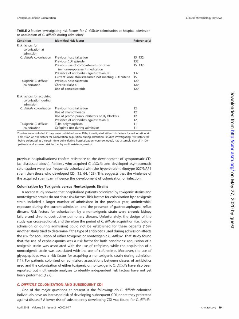
previous hospitalizations) confers resistance to the development of symptomatic CDI(as discussed above). Patients who acquired C. difficile and developed asymptomaticcolonization were less frequently colonized with the hypervirulent ribotype 027/NAP1strain than those who developed CDI (12, 64, 128). This suggests that the virulence ofthe acquired strain can influence the development of colonization or infection.
Colonization by Toxigenic versus Nontoxigenic Strains
A recent study showed that hospitalized patients colonized by toxigenic strains andnontoxigenic strains do not share risk factors. Risk factors for colonization by a toxigenicstrain included a larger number of admissions in the previous year, antimicrobialexposure during the current admission, and the presence of gastroesophageal refluxdisease. Risk factors for colonization by a nontoxigenic strain were chronic kidneyfailure and chronic obstructive pulmonary disease. Unfortunately, the design of thestudy was cross-sectional, and therefore the period of C. difficile acquisition (i.e., beforeadmission or during admission) could not be established for these patients (159).Another study tried to determine if the type of antibiotics used during admission affectsthe risk for acquisition of either toxigenic or nontoxigenic C. difficile. That study foundthat the use of cephalosporins was a risk factor for both conditions: acquisition of atoxigenic strain was associated with the use of cefepime, while the acquisition of anontoxigenic strain was associated with the use of cefuroxime. Moreover, the use ofglycopeptides was a risk factor for acquiring a nontoxigenic strain during admission(11). For patients colonized on admission, associations between classes of antibioticsused and the colonization of either toxigenic or nontoxigenic C. difficile have also beenreported, but multivariate analyses to identify independent risk factors have not yetbeen performed (127).
C. DIFFICILE COLONIZATION AND SUBSEQUENT CDI
One of the major questions at present is the following: do C. difficile-colonizedindividuals have an increased risk of developing subsequent CDI, or are they protectedagainst disease? A lower risk of subsequently developing CDI was found for C. difficile-
TABLE 2 Studies investigating risk factors for C. difficile colonization at hospital admissionor acquisition of C. difficile during admissiona
Condition Identified risk factor Reference(s)
Risk factors forcolonization atadmission
C. difficile colonization Previous hospitalization 15, 132Previous CDI episode 132Previous use of corticosteroids or other
immunosuppressant medication15, 132
Presence of antibodies against toxin B 132Current loose stools/diarrhea not meeting CDI criteria 15
Toxigenic C. difficilecolonization
Previous hospitalization 129Chronic dialysis 129Use of corticosteroids 129
Risk factors for acquiringcolonization duringadmission
C. difficile colonization Previous hospitalization 12Use of chemotherapy 12Use of proton pump inhibitors or H2 blockers 12Presence of antibodies against toxin B 12
Toxigenic C. difficilecolonization
TLR4 polymorphism 11Cefepime use during admission 11
aStudies were included if they were published since 1994, investigated either risk factors for colonization atadmission or risk factors for colonization acquisition during admission (studies investigating risk factors forbeing colonized at a certain time point during hospitalization were excluded), had a sample size of �100patients, and assessed risk factors by multivariate regression.
Clostridium difficile Colonization Clinical Microbiology Reviews
April 2018 Volume 31 Issue 2 e00021-17 cmr.asm.org 19
on May 27, 2020 by guest
http://cmr.asm
.org/D
ownloaded from

colonized patients in a frequently cited but older meta-analysis of four studies (160).The major drawback of that review, however, is that patients colonized by toxigenic ornontoxigenic strains were not analyzed separately; this difference may be important, as44% of colonized patients in the meta-analysis harbored a nontoxigenic strain. Also, allfour studies were performed pre-1994, before the emergence of hypervirulent strainsand recognition of community-associated CDI. Furthermore, colonization was deter-mined at different time points: at admission (71, 98); at the start of tube feeding, withpatients colonized at admission excluded (161); and after a hospital stay of at least 7days (151). Colonized patients therefore included some patients that acquired coloni-zation during admission. The risk of the latter patients going on to develop CDI duringthe hospital stay may be different from that for individuals already colonized atadmission. A recent meta-analysis aimed to include studies in which patients werecolonized at admission with toxigenic strains only (11, 15, 98, 127, 130, 134, 162–164).However, not all included studies succeeded in obtaining samples within 48 or 72 h ofadmission (15, 98). Also, a study that included patients at admission to a rehabilitationunit (after an average stay of 30 days in an acute care facility) was included (164). In onestudy, the distinction between colonization of a toxigenic strain and CDI was difficultto establish, as all patients received a hematopoietic stem cell transplantation anddonor lymphocyte infusion; almost all such patients subsequently develop diarrhea. Forpatients known to carry a toxigenic C. difficile strain, diarrhea may have been falselyattributed to CDI (162). Notwithstanding these limitations, all studies pointed to anincreased risk for patients colonized with toxigenic C. difficile at admission to progressto CDI: overall, the relative risk was 5.86 (95% confidence interval [CI], 4.21 to 8.16)(Table 3). Some recent studies were not included in the meta-analysis. A recent largestudy which screened 3,605 of 4,508 hospital admissions found that patients carryingtoxigenic strains on admission were at a much higher risk of developing CDI (CDI rateof 9.4% versus 2.3% for nontoxigenic C. difficile carriers) (70). The risk of CDI innoncolonized patients who were exposed to subjects colonized by a toxigenic strainwas also significantly increased (4.6% versus 2.6% for nonexposed patients) (odds ratiofor CDI if exposed to carrier, 1.79; 95% CI, 1.16 to 2.76). However, the study appearedto diagnose CDI based on the presence of toxigenic C. difficile strains rather than toxin,so the case incidence is likely to have been overestimated. In turn, the associationbetween colonization by or exposure to toxigenic strains and subsequent CDI may havebeen exaggerated (70). A much smaller study did not report any CDI cases among 37patients colonized on admission (128) (Table 3).
Two other recent studies described the risk of developing CDI for colonized inten-sive care unit (ICU) patients. A study by Tschudin-Sutter et al. with a cohort of 542 ICUpatients described a relative risk of developing CDI of 8.6 for patients colonized onadmission and a relative risk of 10.9 for patients who became colonized duringhospitalization (131). Zhang and colleagues, however, identified 6 patients who werecolonized on admission to the ICU, but none of them developed CDI. During the studyperiod, 4 patients developed CDI, but all were not colonized on admission to the ICU(165). These conflicting results are probably caused by small sample sizes, a relativelyrare outcome event (3 versus 0 colonized patients who progressed to CDI), anddifferent predominant strains.
From the above-mentioned observations, we can conclude that patients asymp-tomatically colonized by toxigenic strains may progress to CDI during admission.However, for patients asymptomatically colonized by nontoxigenic strains, there seemsto be no increased risk of progressing to CDI, and these patients may even be protectedfrom developing CDI.
INFECTION CONTROL AND ANTIMICROBIAL STEWARDSHIP IMPLICATIONS FORASYMPTOMATIC CARRIERS
Symptomatic CDI patients are believed to be the main source of nosocomialtransmission, and current guidelines recommend their systematic detection and isola-tion (5). Due to a paucity of data at the time of writing of this review, the isolation of
Crobach et al. Clinical Microbiology Reviews
April 2018 Volume 31 Issue 2 e00021-17 cmr.asm.org 20
on May 27, 2020 by guest
http://cmr.asm
.org/D
ownloaded from

TAB
LE3
Stud
ies
inve
stig
atin
gth
eris
kof
deve
lop
men
tof
CD
Iam
ong
pat
ient
sw
ithto
xige
nic
C.di
ffici
leco
loni
zatio
non
hosp
ital
adm
issi
ona
Stud
yau
thor
s(r
efer
ence
)C
oun
try,
yr(s
)Se
ttin
gan
dp
atie
nts
Follo
w-u
pp
erio
d
No.
ofin
clud
edp
atie
nts
Prev
alen
ce(n
o.of
pos
itiv
ep
atie
nts
/tot
aln
o.of
pat
ien
ts[%
])
RRfo
rC
DI
(95%
CI)
Rem
arks
Toxi
gen
icC
.di
ffici
leco
lon
izat
ion
CD
Iam
ong
pat
ien
tsw
ith
toxi
gen
icC
.di
ffici
leco
lon
izat
ion
CD
Iam
ong
con
trol
s
Sam
ore
etal
.(98
)U
SA,1
991
Patie
nts
with
anan
ticip
ated
LOS
ofat
leas
t5
days
adm
itted
ortr
ansf
erre
dto
gene
ral
med
ical
and
surg
ical
war
dsan
dIC
Us
Unt
ildi
scha
rge
496
24/4
96(4
.8)
1/24
(4.2
)8/
472
(1.7
)2.
46(0
.32–
18.8
7)90
of49
6sa
mp
les
(18.
1%)
wer
eno
tob
tain
edw
ithin
72h
ofad
mis
sion
Soyl
etir
etal
.(13
0)Tu
rkey
,pub
lishe
d19
96Pa
tient
sad
mitt
edto
age
nera
lm
edic
alw
ard
with
anLO
Sof
atle
ast
48h
Unt
ildi
scha
rge
202
0/20
2(0
)0/
0(0
)0/
202
(0)
NA
Non
eof
the
pat
ient
sw
ere
colo
nize
dat
adm
issi
on
Gup
taet
al.(
163)
USA
and
Can
ada,
2009
–201
1Pa
tient
sof
�60
yrad
mitt
edto
gene
ral
med
ical
and
surg
ical
units
and
onan
tibio
tics
Unt
il30
days
afte
rdi
scha
rge
or60
days
inho
spita
l(w
hich
ever
cam
efir
st)
1,09
991
/1,0
99(8
.3)
9/91
(9.9
)11
/1,0
08(1
.1)
9.06
(3.8
6–21
.30)
Asy
mp
tom
atic
carr
iage
was
diag
nose
db
ycu
ltur
ean
dRE
Aty
pin
gb
utco
uld
have
incl
uded
bot
hto
xige
nic
C.di
ffici
leco
loni
zatio
nan
dno
ntox
igen
icC.
diffi
cile
colo
niza
tion
Ala
smar
iet
al.(
127)
USA
,201
0–20
11A
dult
pat
ient
sw
ithan
antic
ipat
edLO
Sof
�48
had
mitt
edto
gene
ral
med
ical
and
surg
ical
war
ds
Unt
il60
days
afte
rdi
scha
rge
259
40/2
59(1
5.4)
1/40
(2.5
)2/
219
(0.9
)2.
74(0
.25–
29.4
8)
Dub
ber
keet
al.(
128)
USA
,201
0–20
12A
dult
pat
ient
sad
mitt
edto
med
ical
orsu
rgic
alw
ards
with
anan
ticip
ated
LOS
of�
48h
Unt
il60
days
afte
rdi
scha
rge
235
37/2
35(1
5.7)
0/37
(0)
2/19
8(1
.0)
NA
Part
lyth
esa
me
pat
ient
coho
rtas
that
inth
ew
ork
ofA
lasm
ari
etal
.
Brum
inhe
ntet
al.
( 162
)U
SA,2
011–
2012
Patie
nts
adm
itted
toa
bon
em
arro
wtr
ansp
lant
unit
for
HSC
T
Unt
il10
0da
ysaf
ter
HSC
T15
016
/150
(10.
7)14
/16
(87.
5)23
/134
(17.
2)5.
10(3
.36–
7.72
)D
istin
ctio
nb
etw
een
CD
Ian
dco
loni
zatio
nb
yto
xige
nic
stra
ins
isdi
fficu
ltto
esta
blis
hb
ecau
seal
mos
tal
lp
atie
nts
deve
lop
diar
rhea
afte
rH
SCT,
and
CD
Ite
stin
gdi
dno
tin
clud
efr
eeto
xin
dete
ctio
nin
all
case
sH
ung
etal
.(11
)Ta
iwan
,20
11–2
012
Adu
ltp
atie
nts
with
anan
ticip
ated
LOS
ofat
leas
t5
days
adm
itted
toa
gene
ral
med
ical
war
d
Unt
ildi
scha
rge
from
last
hosp
italiz
atio
n44
158
/441
(13.
2)8/
58(1
3.8)
6/38
3(1
.6)
8.80
(3.1
7–24
.46)
Blix
tet
al.(
70)
Den
mar
k,20
12–2
013
Patie
nts
adm
itted
tom
edic
alde
par
tmen
tsat
2un
iver
sity
hosp
itals
1m
o(in
and
outs
ide
hosp
itals
)3,
464
213/
3,46
4(6
.1)
20/2
13(9
.4)
76/3
,251
(2.3
)4.
02(2
.50–
6.44
)
Tsch
udin
-Sut
ter
etal
.(13
1)U
SA, 2
013
Patie
nts
adm
itted
toan
ICU
with
in48
hof
hosp
ital
adm
issi
on
Unt
ildi
scha
rge
542
17/5
42(3
.1)
2/17
(11.
8)6/
525
(1.1
)10
.29
(2.2
4–47
.3)
aSt
udie
sw
ere
incl
uded
ifth
eyw
ere
pub
lishe
dsi
nce
1994
,had
asa
mp
lesi
zeof
�10
0p
atie
nts,
com
par
edp
atie
nts
with
toxi
geni
cC.
diffi
cile
colo
niza
tion
onad
mis
sion
toco
ntro
ls(p
atie
nts
with
nont
oxig
enic
C.di
ffici
leco
loni
zatio
nan
dno
ncol
oniz
edp
atie
nts
toge
ther
).Re
lativ
eris
ksw
ere
calc
ulat
edas
the
risk
for
toxi
geni
cC.
diffi
cile
-col
oniz
edp
atie
nts
com
par
edto
the
risk
for
nonc
olon
ized
and
nont
oxig
enic
C.di
ffici
le-c
olon
ized
pat
ient
sto
geth
eran
dw
ere
unad
just
ed.R
R,re
lativ
eris
k;H
SCT,
hem
atop
oiet
icst
emce
lltr
ansp
lant
atio
n;C
DI,
Clos
trid
ium
diffi
cile
infe
ctio
n;LO
S,le
ngth
ofst
ay;I
CU
,int
ensi
veca
reun
it;N
A,n
otav
aila
ble
;REA
,res
tric
tion
endo
nucl
ease
anal
ysis
.
Clostridium difficile Colonization Clinical Microbiology Reviews
April 2018 Volume 31 Issue 2 e00021-17 cmr.asm.org 21
on May 27, 2020 by guest
http://cmr.asm
.org/D
ownloaded from

asymptomatic carriers is not recommended. Whether these carriers should be isolatedremains an important clinical question stemming from the growing body of literatureon the subject. Mathematical modeling of C. difficile transmission and simulation ofscreening and isolation of carriers have shown the intervention to be effective atreducing CDI rates (166, 167). However, a clinically based study to directly answer thisquestion was not conducted until recently (133).
Longtin et al. explored the effect of isolating asymptomatic C. difficile carriers on theincidence of hospital-acquired CDI in an acute care hospital in Quebec, Canada, withhigh baseline rates of CDI (133). A quasi-experimental design was employed, usingchanges in CDI incidence in other Quebec hospitals as controls. The effect of theintervention (isolation of carriers) was evaluated through a time series analysis. Com-pared with that in the preintervention period, the incidence of CDI decreased signifi-cantly after the intervention. In addition, the effect was confirmed using two methodsof analysis, segmented regression analysis and autoregressive integrated moving av-erage (ARIMA) modeling, indicating the robustness of the results. Incidence rates of CDIin the study hospital remained low a year after the study terminated, demonstratingthe sustained effect of this intervention.
This study provides the most convincing evidence, to date, for the significant effectof isolating carriers. The authors assessed confounding elements, such as the intensityof CDI testing, total antimicrobial use, and proton pump inhibitor use, which remainedstable during the study period. Concurrently, a significant decrease in the use ofmetronidazole and oral vancomycin suggested a true clinical impact from the observeddecrease in incidence. Compliance with hand hygiene increased but utilized an alcohol-based solution not effective against C. difficile spores. Some potential confounders thatwere not assessed include compliance with isolation precautions, environmental clean-ing, improvement in appropriate antibiotic use, and knowledge of C. difficile carrierstatus for the management of a patient (168).
Ultimately, these promising findings need to be reproduced in a multicenter clusterrandomized trial prior to being considered for widespread implementation. If theseresults are confirmed in various hospital settings, adoption of screening and isolationof asymptomatic carriers may be an important strategy for decreasing CDI rates.However, this will raise several practical questions, such as whether universal versustargeted screening should be adopted and which screening method is optimal. Giventhe known risk factors for colonization on admission, a reasonable approach may be toselectively target high-risk patients and to isolate them on admission to hospital (132).Other issues that would need to be addressed include the frequency of screeningduring hospitalization, the optimal isolation protocol, the impact on patient perceptionof care, and the additional workload burden on frontline health care workers and themicrobiology laboratory.
Reducing inappropriate antimicrobial use through antimicrobial stewardship pro-grams (ASPs) has been shown to decrease rates of CDI (169–171), but given the lack ofwidespread screening for asymptomatic carriers, ASPs targeted at this population havenot been studied. It does not necessarily follow that targeting colonized patients as awhole group would decrease CDI rates, as some of these patients may be long-term-colonized patients with immunity and a decreased risk of developing symptomatic CDI.These patients are likely different from patients who may still be colonized with C.difficile after an episode of symptomatic CDI (10, 68). One study showed a 3-foldincrease in recurrence of CDI in patients exposed to antimicrobials after resolved CDIcompared to that in patients who were not exposed (172). Therefore, patients withprior CDI, an easily identifiable subset of asymptomatic carriers, probably representcolonized patients at the highest risk of developing infection and may representsuitable targets for focused stewardship efforts.
CONCLUDING REMARKS AND FUTURE DIRECTIONS
The intriguing concept of C. difficile colonization has garnered much attentionduring the last decade. Gut microbiota studies and immunologic studies have provided
Crobach et al. Clinical Microbiology Reviews
April 2018 Volume 31 Issue 2 e00021-17 cmr.asm.org 22
on May 27, 2020 by guest
http://cmr.asm
.org/D
ownloaded from

some insight into the conditions that allow for colonization and protect against diseaseprogression. However, more studies are needed to assess the precise role of changes inthe microbiota and the precise triggers of spore germination and colonization, as wellas changes and initiators that lead to toxin production. Why some individuals transitionto C. difficile carrier status and what interventions can terminate colonization or blockthe progression to CDI also need to be explored.
The realization that C. difficile-colonized patients may be the most importantunexplained reservoir for C. difficile transmission has led to epidemiological studiesinvestigating colonization rates among different populations and risk factors for thiscondition. Colonized patients at hospital admission appear to play an important role inintroducing and maintaining transmission in the ward, and hence, risk factors forcolonization on admission are of specific interest. To further study the acquisition andtransmission of C. difficile, all patients admitted to the hospital should be screened forcolonization by (and preferably sustained carriage of) C. difficile. C. difficile-positiveindividuals should be questioned about risk factors for acquisition and should bemonitored during admission for the development of symptomatic CDI. Epidemiologicalinvestigations and molecular typing methods should be applied to examine possiblelinkage of C. difficile-colonized individuals to CDI cases. In this way, risk factors for C.difficile colonization can be identified, and the role of C. difficile-positive individuals intransmission of the disease can be elucidated. It would be interesting to determine ifthere are host and pathogen factors that affect the transmissibility of C. difficile. Moreevidence from different settings is needed to determine whether specific controlmeasures targeting colonized patients may be justified to prevent spread. In addition,the protective effects of C. difficile vaccines are being examined, but information on theconsequences of colonization and spread to nonvaccinated individuals would berelevant.
ACKNOWLEDGMENTSM.H.W. has received consulting fees from Actelion, Astellas, bioMerieux, MedIm-
mune, Merck, Pfizer, Qiagen, Sanofi-Pasteur, Seres, Summit, Synthetic Biologics, andValneva; lecture fees from Alere, Astellas, Merck, and Pfizer; and grant support fromActelion, Astellas, bioMerieux, Da Volterra, Merck, Sanofi-Pasteur, Seres, and Summit.V.G.L. has received consulting fees from Merck. M.J.T.C., J.J.V., L.Y.K., S.P., and E.J.K. haveno conflicts of interest.
REFERENCES1. Walker AS, Eyre DW, Wyllie DH, Dingle KE, Harding RM, O’Connor L,
Griffiths D, Vaughan A, Finney J, Wilcox MH, Crook DW, Peto TE. 2012.Characterisation of Clostridium difficile hospital ward-based transmis-sion using extensive epidemiological data and molecular typing. PLoSMed 9:e1001172. https://doi.org/10.1371/journal.pmed.1001172.
2. Curry SR, Muto CA, Schlackman JL, Pasculle AW, Shutt KA, Marsh JW,Harrison LH. 2013. Use of multilocus variable number of tandem re-peats analysis genotyping to determine the role of asymptomaticcarriers in Clostridium difficile transmission. Clin Infect Dis 57:1094 –1102. https://doi.org/10.1093/cid/cit475.
3. Eyre DW, Cule ML, Wilson DJ, Griffiths D, Vaughan A, O’Connor L, Ip CL,Golubchik T, Batty EM, Finney JM, Wyllie DH, Didelot X, Piazza P,Bowden R, Dingle KE, Harding RM, Crook DW, Wilcox MH, Peto TE,Walker AS. 2013. Diverse sources of C. difficile infection identified onwhole-genome sequencing. N Engl J Med 369:1195–1205. https://doi.org/10.1056/NEJMoa1216064.
4. Debast SB, Bauer MP, Kuijper EJ, European Society of Clinical Microbi-ology and Infectious Diseases. 2014. European Society of Clinical Mi-crobiology and Infectious Diseases: update of the treatment guidancedocument for Clostridium difficile infection. Clin Microbiol Infect20(Suppl 2):S1–S26. https://doi.org/10.1111/1469-0691.12418.
5. Cohen SH, Gerding DN, Johnson S, Kelly CP, Loo VG, McDonald LC,Pepin J, Wilcox MH, Society for Healthcare Epidemiology of America,Infectious Diseases Society of America. 2010. Clinical practice guide-lines for Clostridium difficile infection in adults: 2010 update by the
Society for Healthcare Epidemiology of America (SHEA) and the Infec-tious Diseases Society of America (IDSA). Infect Control Hosp Epidemiol31:431– 455. https://doi.org/10.1086/651706.
6. Galdys AL, Curry SR, Harrison LH. 2014. Asymptomatic Clostridiumdifficile colonization as a reservoir for Clostridium difficile infection.Expert Rev Anti Infect Ther 12:967–980. https://doi.org/10.1586/14787210.2014.920252.
7. Crobach MJ, Planche T, Eckert C, Barbut F, Terveer EM, Dekkers OM,Wilcox MH, Kuijper EJ. 2016. European Society of Clinical Microbiologyand Infectious Diseases: update of the diagnostic guidance documentfor Clostridium difficile infection. Clin Microbiol Infect 22(Suppl 4):S63–S81. https://doi.org/10.1016/j.cmi.2016.03.010.
8. Guerrero DM, Becker JC, Eckstein EC, Kundrapu S, Deshpande A, SethiAK, Donskey CJ. 2013. Asymptomatic carriage of toxigenic Clostridiumdifficile by hospitalized patients. J Hosp Infect 85:155–158. https://doi.org/10.1016/j.jhin.2013.07.002.
9. Freter R, Brickner H, Fekete J, Vickerman MM, Carey KE. 1983. Survivaland implantation of Escherichia-coli in the intestinal-tract. Infect Im-mun 39:686 –703.
10. Donskey CJ, Kundrapu S, Deshpande A. 2015. Colonization versuscarriage of Clostridium difficile. Infect Dis Clin North Am 29:13–28.https://doi.org/10.1016/j.idc.2014.11.001.
11. Hung YP, Lin HJ, Wu TC, Liu HC, Lee JC, Lee CI, Wu YH, Wan L, Tsai PJ,Ko WC. 2013. Risk factors of fecal toxigenic or non-toxigenic Clostrid-ium difficile colonization: impact of Toll-like receptor polymorphisms
Clostridium difficile Colonization Clinical Microbiology Reviews
April 2018 Volume 31 Issue 2 e00021-17 cmr.asm.org 23
on May 27, 2020 by guest
http://cmr.asm
.org/D
ownloaded from

and prior antibiotic exposure. PLoS One 8:e69577. https://doi.org/10.1371/journal.pone.0069577.
12. Loo VG, Bourgault AM, Poirier L, Lamothe F, Michaud S, Turgeon N,Toye B, Beaudoin A, Frost EH, Gilca R, Brassard P, Dendukuri N, BeliveauC, Oughton M, Brukner I, Dascal A. 2011. Host and pathogen factors forClostridium difficile infection and colonization. N Engl J Med 365:1693–1703. https://doi.org/10.1056/NEJMoa1012413.
13. Galdys AL, Nelson JS, Shutt KA, Schlackman JL, Pakstis DL, Pasculle AW,Marsh JW, Harrison LH, Curry SR. 2014. Prevalence and duration ofasymptomatic Clostridium difficile carriage among healthy subjects inPittsburgh, Pennsylvania. J Clin Microbiol 52:2406 –2409. https://doi.org/10.1128/JCM.00222-14.
14. McFarland LV, Mulligan ME, Kwok RY, Stamm WE. 1989. Nosocomialacquisition of Clostridium difficile infection. N Engl J Med 320:204 –210.https://doi.org/10.1056/NEJM198901263200402.
15. Eyre DW, Griffiths D, Vaughan A, Golubchik T, Acharya M, O’Connor L,Crook DW, Walker AS, Peto TE. 2013. Asymptomatic Clostridium difficilecolonisation and onward transmission. PLoS One 8:e78445. https://doi.org/10.1371/journal.pone.0078445.
16. Planche T, Wilcox M. 2011. Reference assays for Clostridium difficileinfection: one or two gold standards? J Clin Pathol 64:1–5. https://doi.org/10.1136/jcp.2010.080135.
17. Freeman J, Wilcox MH. 2003. The effects of storage conditions onviability of Clostridium difficile vegetative cells and spores and toxinactivity in human faeces. J Clin Pathol 56:126 –128. https://doi.org/10.1136/jcp.56.2.126.
18. Bolton RP, Tait SK, Dear PRF, Losowsky MS. 1984. Asymptomatic neo-natal colonization by Clostridium-difficile. Arch Dis Child 59:466 – 472.https://doi.org/10.1136/adc.59.5.466.
19. Planche TD, Davies KA, Coen PG, Finney JM, Monahan IM, Morris KA,O’Connor L, Oakley SJ, Pope CF, Wren MW, Shetty NP, Crook DW,Wilcox MH. 2013. Differences in outcome according to Clostridiumdifficile testing method: a prospective multicentre diagnostic validationstudy of C difficile infection. Lancet Infect Dis 13:936 –945. https://doi.org/10.1016/S1473-3099(13)70200-7.
20. Polage CR, Gyorke CE, Kennedy MA, Leslie JL, Chin DL, Wang S, NguyenHH, Huang B, Tang YW, Lee LW, Kim K, Taylor S, Romano PS, PanacekEA, Goodell PB, Solnick JV, Cohen SH. 2015. Overdiagnosis of Clostrid-ium difficile infection in the molecular test era. JAMA Intern Med175:1792–1801. https://doi.org/10.1001/jamainternmed.2015.4114.
21. Eastwood K, Else P, Charlett A, Wilcox M. 2009. Comparison of ninecommercially available Clostridium difficile toxin detection assays, areal-time PCR assay for C. difficile tcdB, and a glutamate dehydroge-nase detection assay to cytotoxin testing and cytotoxigenic culturemethods. J Clin Microbiol 47:3211–3217. https://doi.org/10.1128/JCM.01082-09.
22. Miyajima F, Roberts P, Swale A, Price V, Jones M, Horan M, Beeching N,Brazier J, Parry C, Pendleton N, Pirmohamed M. 2011. Characterisationand carriage ratio of Clostridium difficile strains isolated from acommunity-dwelling elderly population in the United Kingdom. PLoSOne 6:e22804. https://doi.org/10.1371/journal.pone.0022804.
23. Burnham C-AD, Carroll KC. 2013. Diagnosis of Clostridium difficileinfection: an ongoing conundrum for clinicians and for clinical labora-tories. Clin Microbiol Rev 26:604 – 630. https://doi.org/10.1128/CMR.00016-13.
24. Lawley TD, Walker AW. 2013. Intestinal colonization resistance. Immu-nology 138:1–11. https://doi.org/10.1111/j.1365-2567.2012.03616.x.
25. Theriot CM, Young VB. 2015. Interactions between the gastrointestinalmicrobiome and Clostridium difficile. Annu Rev Microbiol 69:445– 461.https://doi.org/10.1146/annurev-micro-091014-104115.
26. Semenyuk EG, Poroyko VA, Johnston PF, Jones SE, Knight KL, GerdingDN, Driks A. 2015. Analysis of bacterial communities during Clostridiumdifficile infection in the mouse. Infect Immun 83:4383– 4391. https://doi.org/10.1128/IAI.00145-15.
27. Crowther GS, Chilton CH, Todhunter SL, Nicholson S, Freeman J, BainesSD, Wilcox MH. 2014. Comparison of planktonic and biofilm-associatedcommunities of Clostridium difficile and indigenous gut microbiota ina triple-stage chemostat gut model. J Antimicrob Chemother 69:2137–2147. https://doi.org/10.1093/jac/dku116.
28. Theriot CM, Bowman AA, Young VB. 2016. Antibiotic-induced altera-tions of the gut microbiota alter secondary bile acid production andallow for Clostridium difficile spore germination and outgrowth in thelarge intestine. mSphere 1:e00045-15. https://doi.org/10.1128/mSphere.00045-15.
29. Zhang L, Dong D, Jiang C, Li Z, Wang X, Peng Y. 2015. Insight intoalteration of gut microbiota in Clostridium difficile infection andasymptomatic C. difficile colonization. Anaerobe 34:1–7. https://doi.org/10.1016/j.anaerobe.2015.03.008.
30. Gu S, Chen Y, Zhang X, Lu H, Lv T, Shen P, Lv L, Zheng B, Jiang X, Li L.2016. Identification of key taxa that favor intestinal colonization ofClostridium difficile in an adult Chinese population. Microbes Infect18:30 –38. https://doi.org/10.1016/j.micinf.2015.09.008.
31. Antharam VC, Li EC, Ishmael A, Sharma A, Mai V, Rand KH, Wang GP.2013. Intestinal dysbiosis and depletion of butyrogenic bacteria inClostridium difficile infection and nosocomial diarrhea. J Clin Microbiol51:2884 –2892. https://doi.org/10.1128/JCM.00845-13.
32. Vincent C, Stephens DA, Loo VG, Edens TJ, Behr MA, Dewar K, MangesAR. 2013. Reductions in intestinal Clostridiales precede the develop-ment of nosocomial Clostridium difficile infection. Microbiome 1:18.https://doi.org/10.1186/2049-2618-1-18.
33. Schubert AM, Rogers MA, Ring C, Mogle J, Petrosino JP, Young VB,Aronoff DM, Schloss PD. 2014. Microbiome data distinguish patientswith Clostridium difficile infection and non-C. difficile-associated diar-rhea from healthy controls. mBio 5:e01021-14. https://doi.org/10.1128/mBio.01021-14.
34. Buffie CG, Jarchum I, Equinda M, Lipuma L, Gobourne A, Viale A, UbedaC, Xavier J, Pamer EG. 2012. Profound alterations of intestinal microbi-ota following a single dose of clindamycin results in sustained suscep-tibility to Clostridium difficile-induced colitis. Infect Immun 80:62–73.https://doi.org/10.1128/IAI.05496-11.
35. Reeves AE, Theriot CM, Bergin IL, Huffnagle GB, Schloss PD, Young VB.2011. The interplay between microbiome dynamics and pathogendynamics in a murine model of Clostridium difficile infection. GutMicrobes 2:145–158. https://doi.org/10.4161/gmic.2.3.16333.
36. Vincent C, Miller MA, Edens TJ, Mehrotra S, Dewar K, Manges AR. 2016.Bloom and bust: intestinal microbiota dynamics in response to hospitalexposures and Clostridium difficile colonization or infection. Micro-biome 4:12. https://doi.org/10.1186/s40168-016-0156-3.
37. Reeves AE, Koenigsknecht MJ, Bergin IL, Young VB. 2012. Suppressionof Clostridium difficile in the gastrointestinal tracts of germfree miceinoculated with a murine isolate from the family Lachnospiraceae.Infect Immun 80:3786 –3794. https://doi.org/10.1128/IAI.00647-12.
38. Buffie CG, Bucci V, Stein RR, McKenney PT, Ling L, Gobourne A, No D,Liu H, Kinnebrew M, Viale A, Littmann E, van den Brink MR, Jenq RR,Taur Y, Sander C, Cross JR, Toussaint NC, Xavier JB, Pamer EG. 2015.Precision microbiome reconstitution restores bile acid mediated resis-tance to Clostridium difficile. Nature 517:205–208. https://doi.org/10.1038/nature13828.
39. Davis MY, Zhang H, Brannan LE, Carman RJ, Boone JH. 2016. Rapidchange of fecal microbiome and disappearance of Clostridium difficilein a colonized infant after transition from breast milk to cow milk.Microbiome 4:53. https://doi.org/10.1186/s40168-016-0198-6.
40. Francis MB, Allen CA, Shrestha R, Sorg JA. 2013. Bile acid recognition bythe Clostridium difficile germinant receptor, CspC, is important forestablishing infection. PLoS Pathog 9:e1003356. https://doi.org/10.1371/journal.ppat.1003356.
41. Wells JE, Hylemon PB. 2000. Identification and characterization of a bileacid 7alpha-dehydroxylation operon in Clostridium sp. strain TO-931, ahighly active 7alpha-dehydroxylating strain isolated from human feces.Appl Environ Microbiol 66:1107–1113. https://doi.org/10.1128/AEM.66.3.1107-1113.2000.
42. Weingarden AR, Dosa PI, DeWinter E, Steer CJ, Shaughnessy MK, John-son JR, Khoruts A, Sadowsky MJ. 2016. Changes in colonic bile acidcomposition following fecal microbiota transplantation are sufficient tocontrol Clostridium difficile germination and growth. PLoS One 11:e0147210. https://doi.org/10.1371/journal.pone.0147210.
43. Allegretti JR, Kearney S, Li N, Bogart E, Bullock K, Gerber GK, Bry L, ClishCB, Alm E, Korzenik JR. 2016. Recurrent Clostridium difficile infectionassociates with distinct bile acid and microbiome profiles. AlimentPharmacol Ther 43:1142–1153. https://doi.org/10.1111/apt.13616.
44. Gupta P, Yakubov S, Tin K, Zea D, Garankina O, Ghitan M, Chapnick EK,Homel P, Lin YS, Koegel MM. 2016. Does alkaline colonic pH predisposeto Clostridium difficile infection? South Med J 109:91–96. https://doi.org/10.14423/SMJ.0000000000000414.
45. Ross CL, Spinler JK, Savidge TC. 2016. Structural and functional changeswithin the gut microbiota and susceptibility to Clostridium difficileinfection. Anaerobe 41:37– 43. https://doi.org/10.1016/j.anaerobe.2016.05.006.
Crobach et al. Clinical Microbiology Reviews
April 2018 Volume 31 Issue 2 e00021-17 cmr.asm.org 24
on May 27, 2020 by guest
http://cmr.asm
.org/D
ownloaded from

46. Wong JM, de Souza R, Kendall CW, Emam A, Jenkins DJ. 2006. Colonichealth: fermentation and short chain fatty acids. J Clin Gastroenterol40:235–243. https://doi.org/10.1097/00004836-200603000-00015.
47. Bibbo S, Lopetuso LR, Ianiro G, Di Rienzo T, Gasbarrini A, Cammarota G.2014. Role of microbiota and innate immunity in recurrent Clostridiumdifficile infection. J Immunol Res 2014:462740. https://doi.org/10.1155/2014/462740.
48. Rea MC, Sit CS, Clayton E, O’Connor PM, Whittal RM, Zheng J, VederasJC, Ross RP, Hill C. 2010. Thuricin CD, a posttranslationally modifiedbacteriocin with a narrow spectrum of activity against Clostridiumdifficile. Proc Natl Acad Sci U S A 107:9352–9357. https://doi.org/10.1073/pnas.0913554107.
49. Sambol SP, Merrigan MM, Tang JK, Johnson S, Gerding DN. 2002.Colonization for the prevention of Clostridium difficile disease in ham-sters. J Infect Dis 186:1781–1789. https://doi.org/10.1086/345676.
50. Popoff MR, Geny B. 2011. Rho/Ras-GTPase-dependent and-independent activity of clostridial glucosylating toxins. J Med Micro-biol 60:1057–1069. https://doi.org/10.1099/jmm.0.029314-0.
51. Pechine S, Collignon A. 2016. Immune responses induced by Clostrid-ium difficile. Anaerobe 41:68 –78. https://doi.org/10.1016/j.anaerobe.2016.04.014.
52. Vohra P, Poxton IR. 2012. Induction of cytokines in a macrophage cellline by proteins of Clostridium difficile. FEMS Immunol Med Microbiol65:96 –104. https://doi.org/10.1111/j.1574-695X.2012.00952.x.
53. Ryan A, Lynch M, Smith SM, Amu S, Nel HJ, McCoy CE, Dowling JK,Draper E, O’Reilly V, McCarthy C, O’Brien J, Ni Eidhin D, O’Connell MJ,Keogh B, Morton CO, Rogers TR, Fallon PG, O’Neill LA, Kelleher D,Loscher CE. 2011. A role for TLR4 in Clostridium difficile infection andthe recognition of surface layer proteins. PLoS Pathog 7:e1002076.https://doi.org/10.1371/journal.ppat.1002076.
54. Yoshino Y, Kitazawa T, Ikeda M, Tatsuno K, Yanagimoto S, Okugawa S,Yotsuyanagi H, Ota Y. 2013. Clostridium difficile flagellin stimulatesToll-like receptor 5, and toxin B promotes flagellin-induced chemokineproduction via TLR5. Life Sci 92:211–217. https://doi.org/10.1016/j.lfs.2012.11.017.
55. Batah J, Deneve-Larrazet C, Jolivot PA, Kuehne S, Collignon A, MarvaudJC, Kansau I. 2016. Clostridium difficile flagella predominantly activateTLR5-linked NF-kappaB pathway in epithelial cells. Anaerobe 38:116 –124. https://doi.org/10.1016/j.anaerobe.2016.01.002.
56. Batah J, Kobeissy H, Bui Pham PT, Deneve-Larrazet C, Kuehne S, Colli-gnon A, Janoir-Jouveshomme C, Marvaud JC, Kansau I. 2017. Clostrid-ium difficile flagella induce a pro-inflammatory response in intestinalepithelium of mice in cooperation with toxins. Sci Rep 7:3256. https://doi.org/10.1038/s41598-017-03621-z.
57. Drudy D, Calabi E, Kyne L, Sougioultzis S, Kelly E, Fairweather N, KellyCP. 2004. Human antibody response to surface layer proteins in Clos-tridium difficile infection. FEMS Immunol Med Microbiol 41:237–242.https://doi.org/10.1016/j.femsim.2004.03.007.
58. Wright A, Drudy D, Kyne L, Brown K, Fairweather NF. 2008. Immu-noreactive cell wall proteins of Clostridium difficile identified byhuman sera. J Med Microbiol 57:750 –756. https://doi.org/10.1099/jmm.0.47532-0.
59. Bruxelle JF, Mizrahi A, Hoys S, Collignon A, Janoir C, Pechine S. 2016.Immunogenic properties of the surface layer precursor of Clostridiumdifficile and vaccination assays in animal models. Anaerobe 37:78 – 84.https://doi.org/10.1016/j.anaerobe.2015.10.010.
60. Ni Eidhin DB, O’Brien JB, McCabe MS, Athie-Morales V, Kelleher DP.2008. Active immunization of hamsters against Clostridium difficileinfection using surface-layer protein. FEMS Immunol Med Microbiol52:207–218. https://doi.org/10.1111/j.1574-695X.2007.00363.x.
61. Pechine S, Janoir C, Boureau H, Gleizes A, Tsapis N, Hoys S, Fattal E,Collignon A. 2007. Diminished intestinal colonization by Clostridiumdifficile and immune response in mice after mucosal immunizationwith surface proteins of Clostridium difficile. Vaccine 25:3946 –3954.https://doi.org/10.1016/j.vaccine.2007.02.055.
62. Pechine S, Deneve C, Le Monnier A, Hoys S, Janoir C, Collignon A. 2011.Immunization of hamsters against Clostridium difficile infection usingthe Cwp84 protease as an antigen. FEMS Immunol Med Microbiol63:73– 81. https://doi.org/10.1111/j.1574-695X.2011.00832.x.
63. Ghose C, Eugenis I, Sun X, Edwards AN, McBride SM, Pride DT, Kelly CP,Ho DD. 2016. Immunogenicity and protective efficacy of recombinantClostridium difficile flagellar protein FliC. Emerg Microbes Infect 5:e8.https://doi.org/10.1038/emi.2016.8.
64. Kyne L, Warny M, Qamar A, Kelly CP. 2000. Asymptomatic carriage of
Clostridium difficile and serum levels of IgG antibody against toxin A. NEngl J Med 342:390–397. https://doi.org/10.1056/NEJM200002103420604.
65. Wilcox MH, Gerding DN, Poxton IR, Kelly C, Nathan R, Birch T,Cornely OA, Rahav G, Bouza E, Lee C, Jenkin G, Jensen W, Kim YS,Yoshida J, Gabryelski L, Pedley A, Eves K, Tipping R, Guris D, Kart-sonis N, Dorr MB. 2017. Bezlotoxumab for prevention of recurrentClostridium difficile infection. N Engl J Med 376:305–317. https://doi.org/10.1056/NEJMoa1602615.
66. Dieterle MG, Young VB. 2017. Reducing recurrence of C. difficile infec-tion. Cell 169:375. https://doi.org/10.1016/j.cell.2017.03.039.
67. Pechine S, Janoir C, Collignon A. 2017. Emerging monoclonal antibod-ies against Clostridium difficile infection. Expert Opin Biol Ther 17:415– 427. https://doi.org/10.1080/14712598.2017.1300655.
68. Sethi AK, Al-Nassir WN, Nerandzic MM, Bobulsky GS, Donskey CJ. 2010.Persistence of skin contamination and environmental shedding of Clos-tridium difficile during and after treatment of C. difficile infection. InfectControl Hosp Epidemiol 31:21–27. https://doi.org/10.1086/649016.
69. Lawley TD, Clare S, Walker AW, Goulding D, Stabler RA, Croucher N,Mastroeni P, Scott P, Raisen C, Mottram L, Fairweather NF, Wren BW,Parkhill J, Dougan G. 2009. Antibiotic treatment of Clostridium difficilecarrier mice triggers a supershedder state, spore-mediated transmis-sion, and severe disease in immunocompromised hosts. Infect Immun77:3661–3669. https://doi.org/10.1128/IAI.00558-09.
70. Blixt T, Gradel KO, Homann C, Seidelin JB, Schonning K, Lester A,Houlind J, Stangerup M, Gottlieb M, Knudsen JD. 2017. Asymptomaticcarriers contribute to nosocomial Clostridium difficile infection: a co-hort study of 4508 patients. Gastroenterology 152:1031.e2–1041.e2.https://doi.org/10.1053/j.gastro.2016.12.035.
71. Clabots CR, Johnson S, Olson MM, Peterson LR, Gerding DN. 1992.Acquisition of Clostridium difficile by hospitalized patients: evidencefor colonized new admissions as a source of infection. J Infect Dis166:561–567. https://doi.org/10.1093/infdis/166.3.561.
72. Lanzas C, Dubberke ER, Lu Z, Reske KA, Grohn YT. 2011. Epidemiolog-ical model for Clostridium difficile transmission in healthcare settings.Infect Control Hosp Epidemiol 32:553–561. https://doi.org/10.1086/660013.
73. Stone NE, Sidak-Loftis LC, Sahl JW, Vazquez AJ, Wiggins KB, Gillece JD,Hicks ND, Schupp JM, Busch JD, Keim P, Wagner DM. 2016. More than50% of Clostridium difficile isolates from pet dogs in Flagstaff, USA,carry toxigenic genotypes. PLoS One 11:e0164504. https://doi.org/10.1371/journal.pone.0164504.
74. Pelaez T, Alcala L, Blanco JL, Alvarez-Perez S, Marin M, Martin-Lopez A,Catalan P, Reigadas E, Garcia ME, Bouza E. 2013. Characterization ofswine isolates of Clostridium difficile in Spain: a potential source ofepidemic multidrug resistant strains? Anaerobe 22:45– 49. https://doi.org/10.1016/j.anaerobe.2013.05.009.
75. Schneeberg A, Neubauer H, Schmoock G, Baier S, Harlizius J, NienhoffH, Brase K, Zimmermann S, Seyboldt C. 2013. Clostridium difficilegenotypes in piglet populations in Germany. J Clin Microbiol 51:3796 –3803. https://doi.org/10.1128/JCM.01440-13.
76. Knight DR, Squire MM, Riley TV. 2015. Nationwide surveillance studyof Clostridium difficile in Australian neonatal pigs shows high prev-alence and heterogeneity of PCR ribotypes. Appl Environ Microbiol81:119 –123. https://doi.org/10.1128/AEM.03032-14.
77. Riley TV, Adams JE, O’Neill GL, Bowman RA. 1991. Gastrointestinalcarriage of Clostridium difficile in cats and dogs attending veterinaryclinics. Epidemiol Infect 107:659 – 665. https://doi.org/10.1017/S0950268800049359.
78. Borriello SP, Honour P, Turner T, Barclay F. 1983. Household pets as apotential reservoir for Clostridium difficile infection. J Clin Pathol 36:84 – 87. https://doi.org/10.1136/jcp.36.1.84.
79. Bojesen AM, Olsen KE, Bertelsen MF. 2006. Fatal enterocolitis in Asianelephants (Elephas maximus) caused by Clostridium difficile. Vet Mi-crobiol 116:329 –335. https://doi.org/10.1016/j.vetmic.2006.04.025.
80. Orchard JL, Fekety R, Smith JR. 1983. Antibiotic-associated colitis due toClostridium difficile in a Kodiak bear. Am J Vet Res 44:1547–1548.
81. Freeman J, Bauer MP, Baines SD, Corver J, Fawley WN, Goorhuis B,Kuijper EJ, Wilcox MH. 2010. The changing epidemiology of Clostridiumdifficile infections. Clin Microbiol Rev 23:529 –549. https://doi.org/10.1128/CMR.00082-09.
82. Knight DR, Elliott B, Chang BJ, Perkins TT, Riley TV. 2015. Diversity andevolution in the genome of Clostridium difficile. Clin Microbiol Rev28:721–741. https://doi.org/10.1128/CMR.00127-14.
83. Weese JS, Staempfli HR, Prescott JF, Kruth SA, Greenwood SJ, Weese
Clostridium difficile Colonization Clinical Microbiology Reviews
April 2018 Volume 31 Issue 2 e00021-17 cmr.asm.org 25
on May 27, 2020 by guest
http://cmr.asm
.org/D
ownloaded from
HE. 2001. The roles of Clostridium difficile and enterotoxigenic Clos-tridium perfringens in diarrhea in dogs. J Vet Intern Med 15:374 –378.
84. Weese JS, Weese HE, Bourdeau TL, Staempfli HR. 2001. SuspectedClostridium difficile-associated diarrhea in two cats. J Am Vet MedAssoc 218:1421, 1436 –1439. https://doi.org/10.2460/javma.2001.218.1421.
85. Shaughnessy MK, Bobr A, Kuskowski MA, Johnston BD, SadowskyMJ, Khoruts A, Johnson JR. 2016. Environmental contamination inhouseholds of patients with recurrent Clostridium difficile infection.Appl Environ Microbiol 82:2686 –2692. https://doi.org/10.1128/AEM.03888-15.
86. Loo VG, Brassard P, Miller MA. 2016. Household transmission of Clos-tridium difficile to family members and domestic pets. Infect ControlHosp Epidemiol 37:1342–1348. https://doi.org/10.1017/ice.2016.178.
87. Knight DR, Squire MM, Collins DA, Riley TV. 2017. Genome analysis ofClostridium difficile PCR ribotype 014 lineage in Australian pigs andhumans reveals a diverse genetic repertoire and signatures of long-range interspecies transmission. Front Microbiol 7:2138. https://doi.org/10.3389/fmicb.2016.02138.
88. Knetsch CW, Connor TR, Mutreja A, van Dorp SM, Sanders IM, BrowneHP, Harris D, Lipman L, Keessen EC, Corver J, Kuijper EJ, Lawley TD.2014. Whole genome sequencing reveals potential spread of Clostrid-ium difficile between humans and farm animals in the Netherlands,2002 to 2011. Euro Surveill 19:20954. https://doi.org/10.2807/1560-7917.ES2014.19.45.20954.
89. Rodriguez-Palacios A, Staempfli HR, Duffield T, Weese JS. 2007. Clos-tridium difficile in retail ground meat, Canada. Emerg Infect Dis 13:485– 487. https://doi.org/10.3201/eid1303.060988.
90. Loo VG, Poirier L, Miller MA, Oughton M, Libman MD, Michaud S,Bourgault AM, Nguyen T, Frenette C, Kelly M, Vibien A, Brassard P, FennS, Dewar K, Hudson TJ, Horn R, Rene P, Monczak Y, Dascal A. 2005. Apredominantly clonal multi-institutional outbreak of Clostridiumdifficile-associated diarrhea with high morbidity and mortality. N EnglJ Med 353:2442–2449. https://doi.org/10.1056/NEJMoa051639.
91. Rodriguez-Palacios A, Reid-Smith RJ, Staempfli HR, Daignault D, Jan-ecko N, Avery BP, Martin H, Thomspon AD, McDonald LC, Limbago B,Weese JS. 2009. Possible seasonality of Clostridium difficile in retailmeat, Canada. Emerg Infect Dis 15:802– 805. https://doi.org/10.3201/eid1505.081084.
92. Songer JG, Trinh HT, Killgore GE, Thompson AD, McDonald LC, LimbagoBM. 2009. Clostridium difficile in retail meat products, USA, 2007.Emerg Infect Dis 15:819 – 821. https://doi.org/10.3201/eid1505.081071.
93. de Boer E, Zwartkruis-Nahuis A, Heuvelink AE, Harmanus C, Kuijper EJ.2011. Prevalence of Clostridium difficile in retailed meat in the Neth-erlands. Int J Food Microbiol 144:561–564. https://doi.org/10.1016/j.ijfoodmicro.2010.11.007.
94. Hoffer E, Haechler H, Frei R, Stephan R. 2010. Low occurrence ofClostridium difficile in fecal samples of healthy calves and pigs atslaughter and in minced meat in Switzerland. J Food Prot 73:973–975.https://doi.org/10.4315/0362-028X-73.5.973.
95. Bouttier S, Barc MC, Felix B, Lambert S, Collignon A, Barbut F. 2010.Clostridium difficile in ground meat, France. Emerg Infect Dis 16:733–735. https://doi.org/10.3201/eid1604.091138.
96. Weese JS, Avery BP, Rousseau J, Reid-Smith RJ. 2009. Detection andenumeration of Clostridium difficile spores in retail beef and pork.Appl Environ Microbiol 75:5009 –5011. https://doi.org/10.1128/AEM.00480-09.
97. Stabler RA, Dawson LF, Valiente E, Cairns MD, Martin MJ, Donahue EH,Riley TV, Songer JG, Kuijper EJ, Dingle KE, Wren BW. 2012. Macro andmicro diversity of Clostridium difficile isolates from diverse sources andgeographical locations. PLoS One 7:e31559. https://doi.org/10.1371/journal.pone.0031559.
98. Samore MH, DeGirolami PC, Tlucko A, Lichtenberg DA, Melvin ZA,Karchmer AW. 1994. Clostridium difficile colonization and diarrhea at atertiary care hospital. Clin Infect Dis 18:181–187. https://doi.org/10.1093/clinids/18.2.181.
99. Kim KH, Fekety R, Batts DH, Brown D, Cudmore M, Silva J, Jr, Waters D.1981. Isolation of Clostridium difficile from the environment and con-tacts of patients with antibiotic-associated colitis. J Infect Dis 143:42–50. https://doi.org/10.1093/infdis/143.1.42.
100. Biswas JS, Patel A, Otter JA, van Kleef E, Goldenberg SD. 2015. Con-tamination of the hospital environment from potential Clostridiumdifficile excretors without active infection. Infect Control Hosp Epide-miol 36:975–977. https://doi.org/10.1017/ice.2015.79.
101. al Saif N, Brazier JS. 1996. The distribution of Clostridium difficile in theenvironment of South Wales. J Med Microbiol 45:133–137. https://doi.org/10.1099/00222615-45-2-133.
102. Moono P, Lim SC, Riley TV. 2017. High prevalence of toxigenic Clos-tridium difficile in public space lawns in western Australia. Sci Rep7:41196. https://doi.org/10.1038/srep41196.
103. Xu C, Weese JS, Flemming C, Odumeru J, Warriner K. 2014. Fate ofClostridium difficile during wastewater treatment and incidence inSouthern Ontario watersheds. J Appl Microbiol 117:891–904. https://doi.org/10.1111/jam.12575.
104. Al-Jumaili IJ, Shibley M, Lishman AH, Record CO. 1984. Incidence andorigin of Clostridium-difficile in neonates. J Clin Microbiol 19:77–78.
105. Enoch DA, Butler MJ, Pai S, Aliyu SH, Karas JA. 2011. Clostridium difficilein children: colonisation and disease. J Infect 63:105–113. https://doi.org/10.1016/j.jinf.2011.05.016.
106. Rousseau C, Lemee L, Le Monnier A, Poilane I, Pons JL, Collignon A. 2011.Prevalence and diversity of Clostridium difficile strains in infants. J MedMicrobiol 60:1112–1118. https://doi.org/10.1099/jmm.0.029736-0.
107. Burgner D, Siarakas S, Eagles G, McCarthy A, Bradbury R, Stevens M.1997. A prospective study of Clostridium difficile infection and coloni-zation in pediatric oncology patients. Pediatr Infect Dis J 16:1131–1134.https://doi.org/10.1097/00006454-199712000-00006.
108. Naaber P, Klaus K, Sepp E, Bjorksten B, Mikelsaar M. 1997. Colonizationof infants and hospitalized patients with Clostridium difficile and lac-tobacilli. Clin Infect Dis 25:S189 –S190. https://doi.org/10.1086/516183.
109. Zilberberg MD, Tillotson GS, McDonald C. 2010. Clostridium difficileinfections among hospitalized children, United States, 1997–2006.Emerg Infect Dis 16:604 – 609. https://doi.org/10.3201/eid1604.090680.
110. Guido K, Khattab H, Bay C, Ostovar GA. 2015. Clostridium difficilecolonization in asymptomatic infants 1 to 12 months old, admitted toa community hospital. Clin Pediatr 55:1176 –1179. https://doi.org/10.1177/0009922815614354.
111. Furuichi M, Imajo E, Sato Y, Tanno S, Kawada M, Sato S. 2014. Character-istics of Clostridium difficile colonization in Japanese children. J InfectChemother 20:307–311. https://doi.org/10.1016/j.jiac.2014.01.009.
112. Hafiz S, Morton RS, McEntegart MG, Waitkins SA. 1975. Clostridiumdifficile in urogenital tract of males and females. Lancet i:420 – 421.
113. Delmee M, Verellen G, Avesani V, Francois G. 1988. Clostridium-difficilein neonates—serogrouping and epidemiology. Eur J Pediatr 147:36 – 40. https://doi.org/10.1007/BF00442608.
114. Adlerberth I, Huang HH, Lindberg E, Aberg N, Hesselmar B, Saalman R,Nord CE, Wold AE, Weintraub A. 2014. Toxin-producing Clostridiumdifficile strains as long-term gut colonizers in healthy infants. J ClinMicrobiol 52:173–179. https://doi.org/10.1128/JCM.01701-13.
115. Stoesser N, Crook DW, Fung R, Griffiths D, Harding RM, KachrimanidouM, Keshav S, Peto TE, Vaughan A, Walker AS, Dingle KE. 2011. Molecularepidemiology of Clostridium difficile strains in children compared withthat of strains circulating in adults with Clostridium difficile-associatedinfection. J Clin Microbiol 49:3994 –3996. https://doi.org/10.1128/JCM.05349-11.
116. Rousseau C, Poilane I, De Pontual L, Maherault AC, Le Monnier A,Collignon A. 2012. Clostridium difficile carriage in healthy infants in thecommunity: a potential reservoir for pathogenic strains. Clin Infect Dis55:1209 –1215. https://doi.org/10.1093/cid/cis637.
117. Lees EA, Miyajima F, Pirmohamed M, Carrol ED. 2016. The role ofClostridium difficile in the paediatric and neonatal gut—a narrativereview. Eur J Clin Microbiol Infect Dis 35:1047–1057. https://doi.org/10.1007/s10096-016-2639-3.
118. Leibowitz J, Soma VL, Rosen L, Ginocchio CC, Rubin LG. 2015. Similarproportions of stool specimens from hospitalized children with andwithout diarrhea test positive for Clostridium difficile. Pediatr Infect DisJ 34:261–266. https://doi.org/10.1097/INF.0000000000000556.
119. Matsuki S, Ozaki E, Shozu M, Inoue M, Shimizu S, Yamaguchi N,Karasawa T, Yamagishi T, Nakamura S. 2005. Colonization by Clostrid-ium difficile of neonates in a hospital, and infants and children in threeday-care facilities of Kanazawa, Japan. Int Microbiol 8:43– 48.
120. Merino VR, Nakano V, Finegold SM, Avila-Campos MJ. 2014. Genes encod-ing toxin of Clostridium difficile in children with and without diarrhea.Scientifica 2014:594014. https://doi.org/10.1155/2014/594014.
121. Ferreira CEA, Nakano V, Durigon EL, Avila-Campos MJ. 2003. Prevalenceof Clostridium spp. and Clostridium difficile in children with acutediarrhea in Sao Paulo City, Brazil. Mem Inst Oswaldo Cruz 98:451– 454.https://doi.org/10.1590/S0074-02762003000400003.
122. Nakamura S, Mikawa M, Nakashio S, Takabatake M, Okado I, Yamakawa
Crobach et al. Clinical Microbiology Reviews
April 2018 Volume 31 Issue 2 e00021-17 cmr.asm.org 26
on May 27, 2020 by guest
http://cmr.asm
.org/D
ownloaded from

K, Serikawa T, Okumura S, Nishida S. 1981. Isolation of Clostridium-difficile from the feces and the antibody in sera of young and elderlyadults. Microbiol Immunol 25:345–351. https://doi.org/10.1111/j.1348-0421.1981.tb00036.x.
123. Ozaki E. 2004. Clostridium difficile colonization in healthy adults: tran-sient colonization and correlation with enterococcal colonization. JMed Microbiol 53:167–172. https://doi.org/10.1099/jmm.0.05376-0.
124. Kato H, Kita H, Karasawa T, Maegawa T, Koino Y, Takakuwa H, Saikai T,Kobayashi K, Yamagishi T, Nakamura S. 2001. Colonisation and trans-mission of Clostridium difficile in healthy individuals examined by PCRribotyping and pulsed-field gel electrophoresis. J Med Microbiol 50:720 –727. https://doi.org/10.1099/0022-1317-50-8-720.
125. Sutphen JL, Grand RJ, Flores A, Chang TW, Bartlett JG. 1983. Chronicdiarrhea associated with Clostridium difficile in children. Am J Dis Child137:275–278.
126. Koo HL, Van JN, Zhao M, Ye X, Revell PA, Jiang ZD, Grimes CZ, Koo DC,Lasco T, Kozinetz CA, Garey KW, DuPont HL. 2014. Real-time polymer-ase chain reaction detection of asymptomatic Clostridium difficile col-onization and rising C. difficile-associated disease rates. Infect ControlHosp Epidemiol 35:667– 673. https://doi.org/10.1086/676433.
127. Alasmari F, Seiler SM, Hink T, Burnham CA, Dubberke ER. 2014. Preva-lence and risk factors for asymptomatic Clostridium difficile carriage.Clin Infect Dis 59:216 –222. https://doi.org/10.1093/cid/ciu258.
128. Dubberke ER, Reske KA, Seiler S, Hink T, Kwon JH, Burnham CA. 2015.Risk factors for acquisition and loss of Clostridium difficile colonizationin hospitalized patients. Antimicrob Agents Chemother 59:4533– 4543.https://doi.org/10.1128/AAC.00642-15.
129. Leekha S, Aronhalt KC, Sloan LM, Patel R, Orenstein R. 2013. Asymp-tomatic Clostridium difficile colonization in a tertiary care hospital:admission prevalence and risk factors. Am J Infect Control 41:390 –393.https://doi.org/10.1016/j.ajic.2012.09.023.
130. Soyletir G, Eskiturk A, Kilic G, Korten V, Tozun N. 1996. Clostridiumdifficile acquisition rate and its role in nosocomial diarrhoea at auniversity hospital in Turkey. Eur J Epidemiol 12:391–394. https://doi.org/10.1007/BF00145303.
131. Tschudin-Sutter S, Carroll KC, Tamma PD, Sudekum ML, Frei R, WidmerAF, Ellis BC, Bartlett J, Perl TM. 2015. Impact of toxigenic Clostridiumdifficile colonization on the risk of subsequent C. difficile infection inintensive care unit patients. Infect Control Hosp Epidemiol 36:1324 –1329. https://doi.org/10.1017/ice.2015.177.
132. Kong LY, Dendukuri N, Schiller I, Bourgault AM, Brassard P, Poirier L,Lamothe F, Beliveau C, Michaud S, Turgeon N, Toye B, Frost EH, Gilca R,Dascal A, Loo VG. 2015. Predictors of asymptomatic Clostridium difficilecolonization on hospital admission. Am J Infect Control 43:248 –253.https://doi.org/10.1016/j.ajic.2014.11.024.
133. Longtin Y, Paquet-Bolduc B, Gilca R, Garenc C, Fortin E, Longtin J,Trottier S, Gervais P, Roussy JF, Levesque S, Ben-David D, Cloutier I, LooVG. 2016. Effect of detecting and isolating Clostridium difficile carriersat hospital admission on the incidence of C difficile infections: aquasi-experimental controlled study. JAMA Intern Med 176:796 – 804.https://doi.org/10.1001/jamainternmed.2016.0177.
134. Zacharioudakis IM, Zervou FN, Pliakos EE, Ziakas PD, Mylonakis E. 2015.Colonization with toxinogenic C. difficile upon hospital admission, andrisk of infection: a systematic review and meta-analysis. Am J Gastro-enterol 110:381–390. https://doi.org/10.1038/ajg.2015.22.
135. Rudensky B, Rosner S, Sonnenblick M, Vandijk Y, Shapira E, Isaacsohn M.1993. The prevalence and nosocomial acquisition of Clostridium-difficile in elderly hospitalized-patients. Postgrad Med J 69:45– 47.https://doi.org/10.1136/pgmj.69.807.45.
136. Hung YP, Tsai PJ, Hung KH, Liu HC, Lee CI, Lin HJ, Wu YH, Wu JJ, Ko WC.2012. Impact of toxigenic Clostridium difficile colonization and infec-tion among hospitalized adults at a district hospital in southern Taiwan.PLoS One 7:e42415. https://doi.org/10.1371/journal.pone.0042415.
137. Mainardi JL, Lacassin F, Guilloy Y, Goldstein FW, Leport C, Acar JF, Vilde JL.1998. Low rate of Clostridium difficile colonization in ambulatory andhospitalized HIV-infected patients in a hospital unit: a prospective survey.J Infect 37:108–111. https://doi.org/10.1016/S0163-4453(98)80162-X.
138. Ryan J, Murphy C, Twomey C, Ross RP, Rea MC, MacSharry J, Sheil B,Shanahan F. 2010. Asymptomatic carriage of Clostridium difficile in anIrish continuing care institution for the elderly: prevalence and char-acteristics. Irish J Med Sci 179:245–250. https://doi.org/10.1007/s11845-009-0361-1.
139. Arvand M, Moser V, Schwehn C, Bettge-Weller G, Hensgens MP, KuijperEJ. 2012. High prevalence of Clostridium difficile colonization among
nursing home residents in Hesse, Germany. PLoS One 7:e30183. https://doi.org/10.1371/journal.pone.0030183.
140. Rogers DS, Kundrapu S, Sunkesula VC, Donskey CJ. 2013. Comparisonof perirectal versus rectal swabs for detection of asymptomatic carriersof toxigenic Clostridium difficile. J Clin Microbiol 51:3421–3422. https://doi.org/10.1128/JCM.01418-13.
141. Rea MC, O’Sullivan O, Shanahan F, O’Toole PW, Stanton C, Ross RP, HillC. 2012. Clostridium difficile carriage in elderly subjects and associatedchanges in the intestinal microbiota. J Clin Microbiol 50:867– 875.https://doi.org/10.1128/JCM.05176-11.
142. Riggs MM, Sethi AK, Zabarsky TF, Eckstein EC, Jump RL, Donskey CJ.2007. Asymptomatic carriers are a potential source for transmission ofepidemic and nonepidemic Clostridium difficile strains among long-term care facility residents. Clin Infect Dis 45:992–998. https://doi.org/10.1086/521854.
143. Ziakas PD, Zacharioudakis IM, Zervou FN, Grigoras C, Pliakos EE, My-lonakis E. 2015. Asymptomatic carriers of toxigenic C. difficile in long-term care facilities: a meta-analysis of prevalence and risk factors. PLoSOne 10:e0117195. https://doi.org/10.1371/journal.pone.0117195.
144. van Nood E, van Dijk K, Hegeman Z, Speelman P, Visser CE. 2009.Asymptomatic carriage of Clostridium difficile among HCWs: do wedisregard the doctor? Infect Control Hosp Epidemiol 30:924 –925.https://doi.org/10.1086/605642.
145. Carmeli YVL, DeGirolami PC, Lichtenberg DA, Karchmer AW, SamoreMB. 1998. Stool colonization of healthcare workers with selected resis-tant bacteria. Infect Control Hosp Epidemiol 19:38 – 40.
146. Friedman ND, Pollard J, Stupart D, Knight DR, Khajehnoori M, Davey EK,Parry L, Riley TV. 2013. Prevalence of Clostridium difficile colonizationamong healthcare workers. BMC Infect Dis 13:459. https://doi.org/10.1186/1471-2334-13-459.
147. Hell M, Sickau K, Chmelizek G, Kern JM, Maass M, Huhulescu S, Aller-berger F. 2012. Absence of Clostridium difficile in asymptomatic hos-pital staff. Am J Infect Control 40:1023–1024. https://doi.org/10.1016/j.ajic.2012.01.018.
148. Sall O, Johansson K, Noren T. 2015. Low colonization rates of Clostrid-ium difficile among patients and healthcare workers at Orebro Univer-sity Hospital in Sweden. APMIS 123:240 –244. https://doi.org/10.1111/apm.12353.
149. Landelle C, Verachten M, Legrand P, Girou E, Barbut F, Brun Buisson C.2014. Contamination of healthcare workers’ hands with Clostridiumdifficile spores after caring for patients with C. difficile infection. InfectControl Hosp Epidemiol 35:10 –15. https://doi.org/10.1086/674396.
150. Samore MH, Venkataraman L, DeGirolami PC, Arbeit RD, Karchmer AW.1996. Clinical and molecular epidemiology of sporadic and clusteredcases of nosocomial Clostridium difficile diarrhea. Am J Med 100:32– 40.https://doi.org/10.1016/S0002-9343(96)90008-X.
151. Johnson S, Clabots CR, Linn FV, Olson MM, Peterson LR, Gerding DN.1990. Nosocomial Clostridium-difficile colonization and disease. Lancet336:97–100. https://doi.org/10.1016/0140-6736(90)91605-A.
152. Johnson S, Homann SR, Bettin KM, Quick JN, Clabots CR, Peterson LR,Gerding DN. 1992. Treatment of asymptomatic Clostridium difficilecarriers (fecal excretors) with vancomycin or metronidazole. A random-ized, placebo-controlled trial. Ann Intern Med 117:297–302.
153. Prasad N, Labaze G, Kopacz J, Chwa S, Platis D, Pan CX, Russo D,LaBombardi VJ, Osorio G, Pollack S, Kreiswirth BN, Chen L, Urban C,Segal-Maurer S. 2016. Asymptomatic rectal colonization withcarbapenem-resistant Enterobacteriaceae and Clostridium difficileamong residents of a long-term care facility in New York City. Am JInfect Control 44:525–532. https://doi.org/10.1016/j.ajic.2015.11.021.
154. Akerlund T, Persson I, Unemo M, Noren T, Svenungsson B, Wullt M,Burman LG. 2008. Increased sporulation rate of epidemic Clostridiumdifficile type 027/NAP1. J Clin Microbiol 46:1530 –1533. https://doi.org/10.1128/JCM.01964-07.
155. Brazier JS, Fitzgerald TC, Hosein I, Cefai C, Looker N, Walker M, BushellAC, Rooney P. 1999. Screening for carriage and nosocomial acquisitionof Clostridium difficile by culture: a study of 284 admissions of elderlypatients to six general hospitals in Wales. J Hosp Infect 43:317–319.https://doi.org/10.1016/S0195-6701(99)90431-0.
156. Freeman J, Vernon J, Morris K, Nicholson S, Todhunter S, Longshaw C,Wilcox MH, Pan-European Longitudinal Surveillance of Antibiotic Re-sistance among Prevalent Clostridium difficile Ribotypes’ Study Group.2015. Pan-European longitudinal surveillance of antibiotic resistanceamong prevalent Clostridium difficile ribotypes. Clin Microbiol Infect21:248.e9 –248.e16. https://doi.org/10.1016/j.cmi.2014.09.017.
Clostridium difficile Colonization Clinical Microbiology Reviews
April 2018 Volume 31 Issue 2 e00021-17 cmr.asm.org 27
on May 27, 2020 by guest
http://cmr.asm
.org/D
ownloaded from

157. Zomer TP, Van Duijkeren E, Wielders CCH, Veenman C, Hengeveld P,Van Der Hoek W, De Greeff SC, Smit LAM, Heederik DJ, Yzermans CJ,Kuijper EJ, Maassen CBM. 2017. Prevalence and risk factors for coloni-zation of Clostridium difficile among adults living near livestock farmsin the Netherlands. Epidemiol Infect 145:2745–2749. https://doi.org/10.1017/S0950268817001753.
158. Stoesser N, Eyre DW, Quan TP, Godwin H, Pill G, Mbuvi E, Vaughan A,Griffiths D, Martin J, Fawley W, Dingle KE, Oakley S, Wanelik K, FinneyJM, Kachrimanidou M, Moore CE, Gorbach S, Riley TV, Crook DW, PetoTEA, Wilcox MH, Walker AS. 2017. Epidemiology of Clostridium difficilein infants in Oxfordshire, UK: risk factors for colonization and carriage,and genetic overlap with regional C. difficile infection strains. PLoS One12:e0182307. https://doi.org/10.1371/journal.pone.0182307.
159. Furuya-Kanamori L, Clements AC, Foster NF, Huber CA, Hong S, Harris-Brown T, Yakob L, Paterson DL, Riley TV. 2017. Asymptomatic Clostrid-ium difficile colonisation in two Australian tertiary hospitals,2012–2014: a prospective, repeated cross-sectional study. Clin Micro-biol Infect 23:48.e1– 48.e7. https://doi.org/10.1016/j.cmi.2016.08.030.
160. Shim JK, Johnson S, Samore MH, Bliss DZ, Gerding DN. 1998. Primarysymptomless colonisation by Clostridium difficile and decreased risk ofsubsequent diarrhoea. Lancet 351:633– 636. https://doi.org/10.1016/S0140-6736(97)08062-8.
161. Bliss DZ, Johnson S, Savik K, Clabots CR, Willard K, Gerding DN. 1998.Acquisition of Clostridium difficile and Clostridium difficile-associated diarrhea in hospitalized patients receiving tube feeding.Ann Intern Med 129:1012–1019. https://doi.org/10.7326/0003-4819-129-12-199812150-00004.
162. Bruminhent J, Wang ZX, Hu C, Wagner J, Sunday R, Bobik B, Hegarty S,Keith S, Alpdogan S, Carabasi M, Filicko-O’Hara J, Flomenberg N, KasnerM, Outschoorn UM, Weiss M, Flomenberg P. 2014. Clostridium difficilecolonization and disease in patients undergoing hematopoietic stemcell transplantation. Biol Blood Marrow Transplant 20:1329 –1334.https://doi.org/10.1016/j.bbmt.2014.04.026.
163. Gupta SB, Mehta V, Herring TA, Dubberke E, Gerding DN, Saddier P,Sambol SP, Walter T, Kaslow DC, Miller M. 2012. A large prospectiveNorth American epidemiologic study of hospital-associated Clostrid-
ium difficile colonization & infection, abstr 020. 4th Int Clostridiumdifficile Symp, Bled, Slovenia.
164. Marciniak C, Chen D, Stein AC, Semik PE. 2006. Prevalence of Clostrid-ium difficile colonization at admission to rehabilitation. Arch Phys MedRehab 87:1086 –1090. https://doi.org/10.1016/j.apmr.2006.03.020.
165. Zhang X, Wang X, Yang J, Liu X, Cai L, Zong Z. 2016. Colonization oftoxigenic Clostridium difficile among ICU patients: a prospective study.BMC Infect Dis 16:397. https://doi.org/10.1186/s12879-016-1729-2.
166. Grigoras CA, Zervou FN, Zacharioudakis IM, Siettos CI, Mylonakis E.2016. Isolation of C. difficile carriers alone and as part of a bundleapproach for the prevention of Clostridium difficile infection (CDI): amathematical model based on clinical study data. PLoS One 11:e0156577. https://doi.org/10.1371/journal.pone.0156577.
167. Lanzas C, Dubberke ER. 2014. Effectiveness of screening hospital ad-missions to detect asymptomatic carriers of Clostridium difficile: amodeling evaluation. Infect Control Hosp Epidemiol 35:1043–1050.https://doi.org/10.1086/677162.
168. Crobach MJ, Terveer EM, Kuijper EJ. 2016. Effect of detecting andisolating asymptomatic Clostridium difficile carriers. JAMA Intern Med176:1572–1573. https://doi.org/10.1001/jamainternmed.2016.5339.
169. Feazel LM, Malhotra A, Perencevich EN, Kaboli P, Diekema DJ, Schwei-zer ML. 2014. Effect of antibiotic stewardship programmes on Clostrid-ium difficile incidence: a systematic review and meta-analysis. J Anti-microb Chemother 69:1748 –1754. https://doi.org/10.1093/jac/dku046.
170. Wenzler E, Mulugeta SG, Danziger LH. 2015. The antimicrobial stew-ardship approach to combating Clostridium difficile. Antibiotics (Basel)4:198 –215. https://doi.org/10.3390/antibiotics4020198.
171. Davey P, Brown E, Charani E, Fenelon L, Gould IM, Holmes A, Ramsay CR,Wiffen PJ, Wilcox M. 2013. Interventions to improve antibiotic prescribingpractices for hospital inpatients. Cochrane Database Syst Rev 2013:CD003543. https://doi.org/10.1002/14651858.CD003543.pub3.
172. Drekonja DM, Amundson WH, Decarolis DD, Kuskowski MA, Lederle FA,Johnson JR. 2011. Antimicrobial use and risk for recurrent Clostridiumdifficile infection. Am J Med 124:1081.e1–1081.e7. https://doi.org/10.1016/j.amjmed.2011.05.032.
Monique J. T. Crobach obtained her medi-cal degree from Leiden University, the Neth-erlands. She subsequently started her inter-nal medicine residency in The BronovoHospital, The Hague, and the Leiden Univer-sity Medical Centre in the Netherlands. Cur-rently, she is combining this training withher work as a Ph.D. student in the Depart-ment of Medical Microbiology at the LeidenUniversity Medical Centre. Her research fo-cuses on diagnostics of C. difficile infectionsand asymptomatic C. difficile colonization. She took the lead in thedevelopment of the European Society of Clinical Microbiology andInfectious Diseases (ESCMID) diagnostic guidance document for CDI, isconducting a multicenter study on asymptomatic C. difficile coloniza-tion, and is involved in the Dutch national CDI surveillance.
Jonathan J. Vernon is currently a final-yearPh.D. research student at the Leeds Institute ofBiomedical and Clinical Sciences at the Univer-sity of Leeds. His research interests are in thedevelopment of multidrug resistance in Clos-tridium difficile, particularly by epidemiologicalinvestigations of resistance determinants andphenotypes. He has been involved in theHealthcare-Associated Infection (HCAI) Re-search Group in Leeds for over 5 years, work-ing primarily with C. difficile as part of a large-scale, longitudinal, pan-European surveillance study. His other researchinvolvements include in vitro gut modeling of C. difficile infections forinvestigation of treatment regimens and infection recurrence, efficacytesting of novel compounds for antimicrobial activity, and work as part ofthe Clostridium difficile Ribotyping Network (CDRN) reference laboratory.Before joining the HCAI research group, he acquired his B.Sc. (Honors) atthe University of Lincoln and previously worked as a forensic DNA analystfor the Forensic Science Service, United Kingdom.
Crobach et al. Clinical Microbiology Reviews
April 2018 Volume 31 Issue 2 e00021-17 cmr.asm.org 28
on May 27, 2020 by guest
http://cmr.asm
.org/D
ownloaded from

Vivian G. Loo is currently a Professor ofMedicine at McGill University and Director ofthe Infectious Disease Clinic at the McGillUniversity Health Centre. Dr. Loo receivedher medical degree, M.S. in epidemiology,and internal medicine residency trainingfrom McGill University. She completed herclinical fellowships in infectious diseases andmedical microbiology at the University ofToronto. In 1991, Dr. Loo joined the Facultyof Medicine of McGill University in the Divi-sion of Infectious Diseases. At the McGill University Health Centre, shewas the Director of the Infection Control and Prevention Service for 10years as well as the Chief of the Department of Microbiology for 9 years.Dr. Loo’s research interests include the epidemiology, transmission, andlaboratory diagnosis of Clostridium difficile infections.
Ling Yuan Kong is an infectious diseasesand medical microbiology fellow in her finalyear of training at McGill University, Mon-treal, Canada. She completed medical schooland residency in internal medicine at McGillUniversity. During her residency and fellow-ship, she developed an interest in infectioncontrol and conducted research on C. difficilecolonization risk factors and transmission dy-namics. She plans to pursue further trainingin epidemiology and a career combiningclinical infectious diseases, medical microbiology, and hospital epide-miology research.
Séverine Péchiné, Pharm.D., Ph.D., has beenan Assistant Professor in the Faculty of Phar-macy, Department of Microbiology, Paris-Sud University, since 2006. From 1998 to2001, she worked for chemical and pharma-ceutical companies (Schweizerhall-Franceand Inalco-Italy). During her Ph.D. training,she studied Clostridium difficile, especiallythe surface proteins and their use in thedevelopment of vaccine strategies, at theParis-Sud University. She has developed ex-pertise on Clostridium difficile colonization factors, immune responses ofthe host, and vaccine strategies in animal models. She has published inthe field of Clostridium and acts as a referee for peer-reviewed journals.
Mark H. Wilcox is a Consultant Microbiolo-gist, Head of Research and Development inMicrobiology at the Leeds Teaching Hospi-tals (LTHT), Professor of Medical Microbiol-ogy at the University of Leeds (Leeds Insti-tute of Biomedical and Clinical Sciences),Lead on Clostridium difficile and Head of theUK C. difficile Reference Laboratory for PublicHealth England (PHE), and Medical Advisorto the National Infection Prevention & Con-trol Lead (NHS Improvement), England. Hewas formerly a Director of Infection Prevention (4 years), InfectionControl Doctor (8 years), Clinical Director of Pathology (6 years), andHead of Microbiology (15 years) at LTHT. Professor Wilcox heads ahealth care-associated infection research team at the University ofLeeds; projects include multiple aspects of Clostridium difficile infection,diagnostics, antibiotic resistance and the gut microbiome, staphylococ-cal infection, and the clinical development of new antimicrobial agents.
Ed J. Kuijper is Professor and Head of Exper-imental Bacteriology at the Department ofMedical Microbiology, Leiden University Med-ical Center. He received his education at theUniversity of Amsterdam and obtained a med-ical degree in 1982. His Ph.D. was achieved in1987 with the thesis “Aeromonas-associateddiarrhoea in the Netherlands.” In 1987, he alsocompleted his training as a medical microbiol-ogist, and he worked as a researcher on thetopics of meningococcal infections, fungal in-fections, and mycobacterial infections until 2000. In 2001, he was ap-pointed at Leiden University and started a research group on Clostridiumdifficile infections (CDI), in close collaboration with the National Center forInfectious Diseases at the RIVM. The research group focuses on the patho-genesis, epidemiology, and treatment of CDI.
Clostridium difficile Colonization Clinical Microbiology Reviews
April 2018 Volume 31 Issue 2 e00021-17 cmr.asm.org 29
on May 27, 2020 by guest
http://cmr.asm
.org/D
ownloaded from

![Clostridium difficile-associated Diarrhea in Developing ......ties) known to be associated with nosocomial infection [11, 13, 14, 17, 18]. Clostridium difficile-associated diarrhea](https://static.fdocuments.net/doc/165x107/60f6b6bec20f9f1cf54c1df4/clostridium-difficile-associated-diarrhea-in-developing-ties-known-to-be.jpg)