Uncommon Manifestations of Scrub Typhus …...338 Uncommon Manifestations of Scrub Typhus...
6
Copyrights © 2015 The Korean Society of Radiology 337 Case Report pISSN 1738-2637 / eISSN 2288-2928 J Korean Soc Radiol 2015;73(5):337-342 http://dx.doi.org/10.3348/jksr.2015.73.5.337 INTRODUCTION Scrub typhus causes systemic vasculitis characterized by the destruction of endothelial cells that line small blood vessels and inflammatory lesions caused by infiltrating leukocytes (1). It is a rickettsial disease caused by Orientia tsutsugamushi and is trans- mitted through chigger bites. Its prevalence is high in East Asia, Korea, and the western Pacific region (1). It involves multiple or- gans, and is characterized by a fever, headache, myalgia, rash, lymphadenopathy, eschar formation, pneumonia, and meningo- encephalitis. Scrub typhus with central nervous system (CNS) involvement presents with various features from aseptic menin- gitis to meningoencephalitis (2). Cerebrospinal fluid (CSF) anal- ysis generally shows pleocytosis, a normal level of glucose, and a mild increase in the protein level (1). e early diagnosis of scrub typhus with CNS involvement is important, as it can alter the pa- tients’ prognoses and reduce mortality rates. Scrub typhus with CNS involvement is not rare and has been reported in several cases. However, to the best of our knowledge, scrub typhus en- cephalitis with extensive white matter involvement and subepen- dymal enhancement are rarely described findings in the previous literature. Uncommon Manifestations of Scrub Typhus Encephalitis in Two Cases: Clinical and Magnetic Resonance Imaging Findings 흔하지 않은 소견을 보였던 두 증례의 쯔쯔가무시 뇌염: 임상적, 자기공명영상 소견 Young Jin Heo, MD, Hae Woong Jeong, MD * Department of Radiology, Inje University Busan Paik Hospital, Busan, Korea Scrub typhus is a well-known acute febrile illness caused by Orientia tsutsugamushi. This disease has multiorgan involvement, which includes the lungs, heart, liver, spleen, and the central or peripheral nervous system. Scrub typhus involving the central nervous system (CNS) is not rare. However, meningitis and meningoenceph- alitis can cause changes in mentation and death and are therefore associated with a poor prognosis. We report two consecutive cases of scrub typhus with CNS in- volvement. One patient presented with extensive white matter involvement, similar to that observed in acute disseminated encephalomyelitis, whereas the other patient presented with subependymal enhancement along the lateral ventricles. To the best of our knowledge, scrub typhus encephalitis, with extensive white matter involve- ment and subependymal enhancement, are very rarely described findings in the previous literature. Our patients did not show complete recovery, but the symptoms resolved with treatment. Recognizing these uncommon radiologic findings of scrub typhus may be helpful in the early diagnosis of scrub typhus with CNS involvement, which may alter the prognoses of patients. Index terms Scrub Typhus White Matter Subependymal Magnetic Resonance Imaging Diagnosis Received June 10, 2015 Revised July 13, 2015 Accepted September 10, 2015 *Corresponding author: Hae Woong Jeong, MD Department of Radiology, Inje University Busan Paik Hospital, 75 Bokji-ro, Busanjin-gu, Busan 47392, Korea. Tel. 82-51-890-6549 Fax. 82-51-896-1085 E-mail: [email protected] This is an Open Access article distributed under the terms of the Creative Commons Attribution Non-Commercial License (http://creativecommons.org/licenses/by-nc/3.0) which permits unrestricted non-commercial use, distri- bution, and reproduction in any medium, provided the original work is properly cited.
Transcript of Uncommon Manifestations of Scrub Typhus …...338 Uncommon Manifestations of Scrub Typhus...
Copyrights © 2015 The Korean Society of Radiology 337
Case ReportpISSN 1738-2637 / eISSN 2288-2928J Korean Soc Radiol 2015;73(5):337-342http://dx.doi.org/10.3348/jksr.2015.73.5.337
INTRODUCTION
Scrub typhus causes systemic vasculitis characterized by the destruction of endothelial cells that line small blood vessels and inflammatory lesions caused by infiltrating leukocytes (1). It is a rickettsial disease caused by Orientia tsutsugamushi and is trans-mitted through chigger bites. Its prevalence is high in East Asia, Korea, and the western Pacific region (1). It involves multiple or-gans, and is characterized by a fever, headache, myalgia, rash, lymphadenopathy, eschar formation, pneumonia, and meningo-encephalitis. Scrub typhus with central nervous system (CNS)
involvement presents with various features from aseptic menin-gitis to meningoencephalitis (2). Cerebrospinal fluid (CSF) anal-ysis generally shows pleocytosis, a normal level of glucose, and a mild increase in the protein level (1). The early diagnosis of scrub typhus with CNS involvement is important, as it can alter the pa-tients’ prognoses and reduce mortality rates. Scrub typhus with CNS involvement is not rare and has been reported in several cases. However, to the best of our knowledge, scrub typhus en-cephalitis with extensive white matter involvement and subepen-dymal enhancement are rarely described findings in the previous literature.
Uncommon Manifestations of Scrub Typhus Encephalitis in Two Cases: Clinical and Magnetic Resonance Imaging Findings흔하지 않은 소견을 보였던 두 증례의 쯔쯔가무시 뇌염: 임상적, 자기공명영상 소견
Young Jin Heo, MD, Hae Woong Jeong, MD*Department of Radiology, Inje University Busan Paik Hospital, Busan, Korea
Scrub typhus is a well-known acute febrile illness caused by Orientia tsutsugamushi. This disease has multiorgan involvement, which includes the lungs, heart, liver, spleen, and the central or peripheral nervous system. Scrub typhus involving the central nervous system (CNS) is not rare. However, meningitis and meningoenceph-alitis can cause changes in mentation and death and are therefore associated with a poor prognosis. We report two consecutive cases of scrub typhus with CNS in-volvement. One patient presented with extensive white matter involvement, similar to that observed in acute disseminated encephalomyelitis, whereas the other patient presented with subependymal enhancement along the lateral ventricles. To the best of our knowledge, scrub typhus encephalitis, with extensive white matter involve-ment and subependymal enhancement, are very rarely described findings in the previous literature. Our patients did not show complete recovery, but the symptoms resolved with treatment. Recognizing these uncommon radiologic findings of scrub typhus may be helpful in the early diagnosis of scrub typhus with CNS involvement, which may alter the prognoses of patients.
Index termsScrub TyphusWhite MatterSubependymalMagnetic Resonance ImagingDiagnosis
Received June 10, 2015Revised July 13, 2015 Accepted September 10, 2015*Corresponding author: Hae Woong Jeong, MDDepartment of Radiology, Inje University Busan Paik Hospital, 75 Bokji-ro, Busanjin-gu, Busan 47392, Korea.Tel. 82-51-890-6549 Fax. 82-51-896-1085E-mail: [email protected]
This is an Open Access article distributed under the terms of the Creative Commons Attribution Non-Commercial License (http://creativecommons.org/licenses/by-nc/3.0) which permits unrestricted non-commercial use, distri-bution, and reproduction in any medium, provided the original work is properly cited.
338
Uncommon Manifestations of Scrub Typhus Encephalitis in Two Cases
jksronline.orgJ Korean Soc Radiol 2015;73(5):337-342
CASE REPORT
Case 1
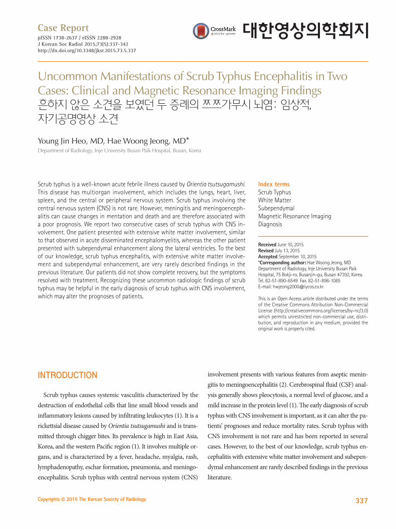
A 57-year-old man was admitted to our hospital for general weakness, gait disturbance, and mental change. He had visited a family member’s grave 1 month previously. For 3 days before ad-mission, the patient experienced a febrile sensation and skin rash. He had no previous history of medical diseases, such as diabetes mellitus, hypertension, or tuberculosis. Physical examination showed a temperature of 37.4°C, blood pressure of 120/70 mm Hg, and pulse rate of 70/min. Eschars were noted over the upper portion of his left arm and back. A neurologic examination indi-cated that his consciousness ranged from a stupor to a semico-matose state. He did not show motor responses and had a slug-gish light reflex. He did not have cranial nerve palsy. Results of the routine hematology test were normal, except for elevated fi-brin degradation product (5.0 μg/mL) and D-dimer levels (1.57 μg/mL fibrinogen equivalent units). The liver function test showed an albumin level of 3.4 g/dL (range: 3.8-5.3 g/dL), total bilirubin level of 0.6 mg/dL (range: 0.2-1.0 mg/dL), alkaline phosphatase level of 362 U/L (range: 104-338 U/L), aspartate aminotransferase (AST) level of 812 U/L (range: 10-33 U/L), and alanine aminotransferase (ALT) level of 1043 U/L (range: 4-50 U/L). The C-reactive protein (CRP) level was 3.76 mg/dL (-0.5 mg/dL), and the erythrocyte sedimentation rate was 24 mm/h (range: 0-20 mm/h). Serologic tests yielded negative re-sults for the hepatitis B surface antigen, human immunodeficien-cy virus, Hantan virus, Epstein-Barr virus, and cytomegalovirus. Polymerase chain reaction test results for the Epstein-Barr virus, cytomegalovirus, and tuberculosis were negative. CSF analysis showed a white blood cell count of 220/mm3 (lymphocytes 30%, polymorphonucleocytes 70%), glucose level of 66 mg/dL (blood glucose, 133 mg/dL), and protein level of 138.5 mg/dL (range: 11-45 mg/dL). The CSF sample tested positive for the tsutsuga-mushi antibody [immunoglobulin (Ig)-G, 1:160] and serum sam-ple (IgG, 1:10240). Chest computed tomography showed patchy consolidations in the lower lobes of both lungs. On admission magnetic resonance images (MRIs), T2-weighted images (T2WIs), and fluid-attenuated inversion recovery images (FLAIRs) dem-onstrated multifocal, hyperintense lesions involving the cerebral white matter of both the right and left sides, thalamus, brainstem, left middle cerebellar peduncle, and both cerebellar hemispheres
(Fig. 1). These lesions did not show contrast enhancement on contrast-enhanced T1-weighted images. On a follow-up MRI, obtained 5 days after admission, the extent of multiple areas of T2 hyperintense cerebral white matter lesions on both the right and left sides were slightly increased, with no significant interval change in the other sites. After treatment with doxycycline for 2 weeks, his consciousness improved to an alert state. However, his motor functions were still impaired in the left upper (grade 2/5), left lower (grade 3/5), right upper (grade 1/5), and right lower (grade 1/5) extremities. A follow-up MRI, acquired 2 weeks after admission, previously noted that the number of multifocal T2 hyperintense lesions had slightly decreased in the cerebral white matter on both sides and increased in the brainstem (Fig. 1). However, no significant interval changes were seen in the other lesion sites. He was discharged to a local hospital for rehabilita-tive therapy.
Case 2
A 71-year-old woman was admitted to our hospital for general weakness. The patient had visited the countryside to pick persim-mons 4 days previously. Three days before admission, she had visited a local hospital because of fever and general weakness. Her symptoms had not improved, and she presented with irrita-bility on admission. She had a history of osteoporosis and hyper-lipidemia, and was taking cholesterol-lowering medication. Physical examination showed a temperature of 36.5°C, blood pressure of 130/80 mm Hg, and pulse rate of 80/min. The patient did not have eschars, but she presented with ecchymosis and pain in both arms. Cranial nerve function was essentially normal. The patient did not present with abnormal motor function. Results of the routine hematology test were normal. The liver function test showed an albumin level of 4.0 g/dL (range: 3.8-5.3), total biliru-bin level of 0.7 mg/dL (range: 0.2-1.0 mg/dL), alkaline phospha-tase level of 240 U/L (range: 104-338 U/L), AST level of 254 U/L (range: 10-33 U/L), and ALT level of 52 U/L (range: 4-50 U/L). The creatine phosphokinase level was 11875 U/L (range: 38-176 U/L), and the CRP level was 0.23 mg/dL (-0.5 mg/dL). Serologic tests yielded negative results for the Hantan virus antibody, influ-enza A and B viral antigens, Cryptococcus antigen, varicella zos-ter virus, and human T-lymphotropic virus I/II. Polymerase chain reaction test results for the varicella zoster virus, herpes simplex virus, and tuberculosis were negative. The CSF and serum sam-
339
Young Jin Heo, et al
jksronline.org J Korean Soc Radiol 2015;73(5):337-342
ples tested positive for the tsutsugamushi antibody (IgG, ≥ 1:160 and IgG, 1:640, respectively). Chest radiography showed no sig-nificant abnormality in both lungs. She was diagnosed with rhabdomyolysis and scrub typhus. After discontinuing the cho-lesterol-lowering medication, doxycycline was administered on the day following admission. Levels of the liver function enzymes and creatine phosphokinase improved, and the ecchymosis of both arms improved. However, her consciousness suddenly al-tered. CSF analysis showed a white blood cell count of 25 mm3 (lymphocytes 90%, polymorphonucleocytes 10%), glucose level
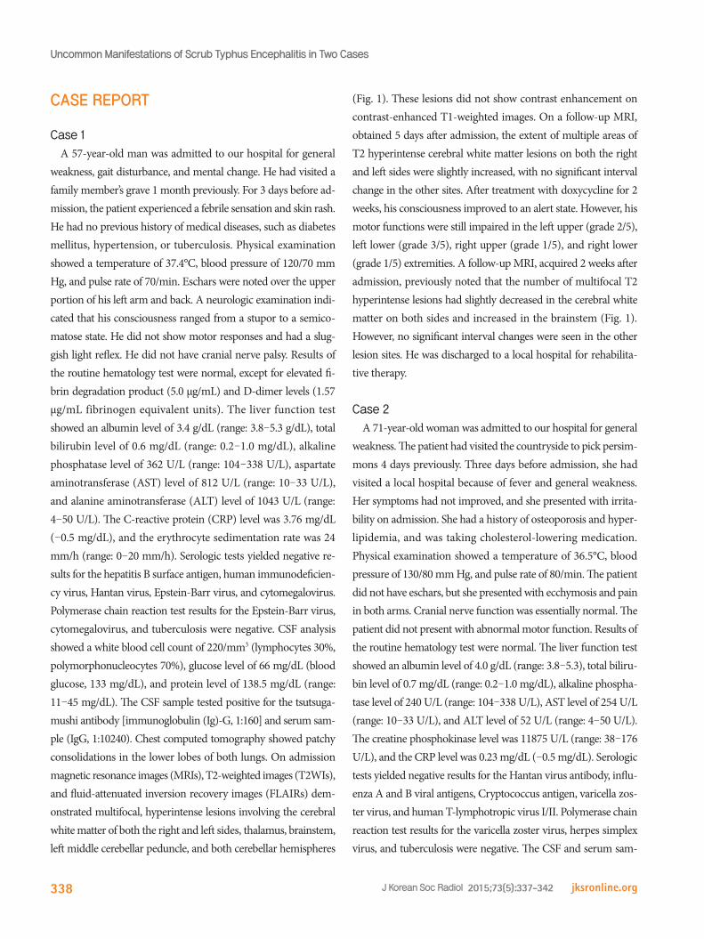
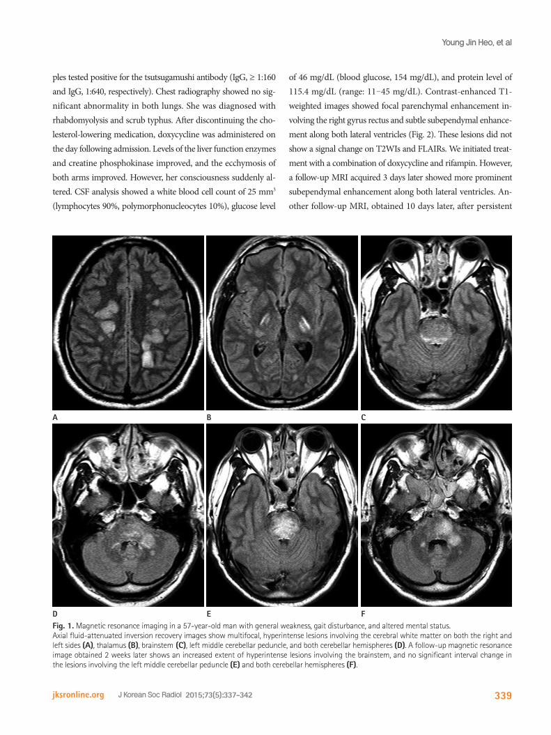
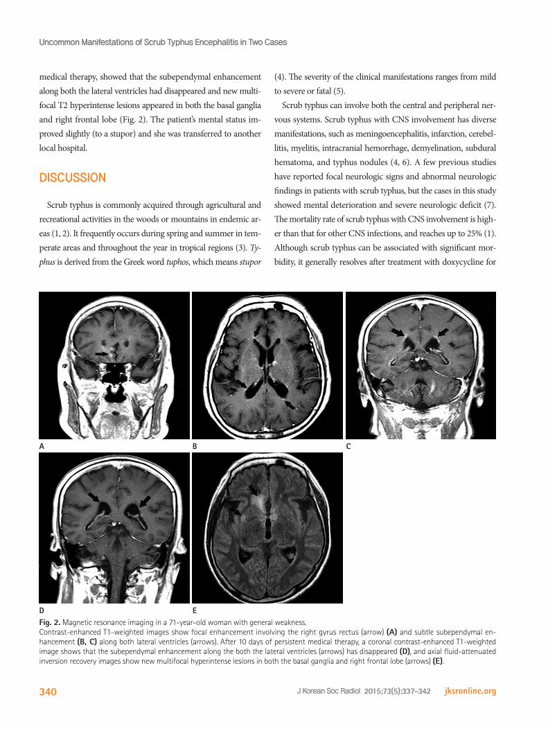
of 46 mg/dL (blood glucose, 154 mg/dL), and protein level of 115.4 mg/dL (range: 11-45 mg/dL). Contrast-enhanced T1-weighted images showed focal parenchymal enhancement in-volving the right gyrus rectus and subtle subependymal enhance-ment along both lateral ventricles (Fig. 2). These lesions did not show a signal change on T2WIs and FLAIRs. We initiated treat-ment with a combination of doxycycline and rifampin. However, a follow-up MRI acquired 3 days later showed more prominent subependymal enhancement along both lateral ventricles. An-other follow-up MRI, obtained 10 days later, after persistent
Fig. 1. Magnetic resonance imaging in a 57-year-old man with general weakness, gait disturbance, and altered mental status.Axial fluid-attenuated inversion recovery images show multifocal, hyperintense lesions involving the cerebral white matter on both the right and left sides (A), thalamus (B), brainstem (C), left middle cerebellar peduncle, and both cerebellar hemispheres (D). A follow-up magnetic resonance image obtained 2 weeks later shows an increased extent of hyperintense lesions involving the brainstem, and no significant interval change in the lesions involving the left middle cerebellar peduncle (E) and both cerebellar hemispheres (F).
A
D
B
E
C
F
340
Uncommon Manifestations of Scrub Typhus Encephalitis in Two Cases
jksronline.orgJ Korean Soc Radiol 2015;73(5):337-342
medical therapy, showed that the subependymal enhancement along both the lateral ventricles had disappeared and new multi-focal T2 hyperintense lesions appeared in both the basal ganglia and right frontal lobe (Fig. 2). The patient’s mental status im-proved slightly (to a stupor) and she was transferred to another local hospital.
DISCUSSION
Scrub typhus is commonly acquired through agricultural and recreational activities in the woods or mountains in endemic ar-eas (1, 2). It frequently occurs during spring and summer in tem-perate areas and throughout the year in tropical regions (3). Ty-phus is derived from the Greek word tuphos, which means stupor
(4). The severity of the clinical manifestations ranges from mild to severe or fatal (5).
Scrub typhus can involve both the central and peripheral ner-vous systems. Scrub typhus with CNS involvement has diverse manifestations, such as meningoencephalitis, infarction, cerebel-litis, myelitis, intracranial hemorrhage, demyelination, subdural hematoma, and typhus nodules (4, 6). A few previous studies have reported focal neurologic signs and abnormal neurologic findings in patients with scrub typhus, but the cases in this study showed mental deterioration and severe neurologic deficit (7). The mortality rate of scrub typhus with CNS involvement is high-er than that for other CNS infections, and reaches up to 25% (1). Although scrub typhus can be associated with significant mor-bidity, it generally resolves after treatment with doxycycline for
Fig. 2. Magnetic resonance imaging in a 71-year-old woman with general weakness.Contrast-enhanced T1-weighted images show focal enhancement involving the right gyrus rectus (arrow) (A) and subtle subependymal en-hancement (B, C) along both lateral ventricles (arrows). After 10 days of persistent medical therapy, a coronal contrast-enhanced T1-weighted image shows that the subependymal enhancement along the both the lateral ventricles (arrows) has disappeared (D), and axial fluid-attenuated inversion recovery images show new multifocal hyperintense lesions in both the basal ganglia and right frontal lobe (arrows) (E).
A
D
B
E
C
341
Young Jin Heo, et al
jksronline.org J Korean Soc Radiol 2015;73(5):337-342
an appropriate duration (8).Scrub typhus is usually limited to the gray matter because,
pathologically, it involves small vessels throughout the body (7, 9). Thus, scrub typhus rarely involves the white matter, and only a few reports have described cases of scrub typhus involving the white matter (3, 7). According to a previous case report, the white matter can be destroyed because of a blood-brain barrier break-down, microinfarction, and edema (7). The first patient in this study showed extensive white matter involvement, which mim-icked radiologic findings of acute disseminated encephalomyeli-tis. The second patient presented with subependymal enhance-ment along both lateral ventricles. To the best of our knowledge, subependymal enhancement associated with scrub typhus has not been reported in previous case reports. Considering the CSF findings, we presumed that the subependymal enhancement is associated with ventriculitis.
Pathologic findings of scrub typhus with CNS involvement in-clude diffuse or focal histiocytes, lymphocytes and plasma cell infiltration of the leptomeninges, typhus nodules (i.e., clusters of microglial cells), and brain hemorrhage (6, 10). The diagnosis of scrub typhus is usually dependent on a history of exposure, clini-cal features including eschars, and serological test results. The pa-tients in this study showed typical findings, such as CSF pleocy-tosis, and both patients had an elevated CSF protein level (> 60 mg). Liver enzyme levels are usually elevated in scrub typhus, and the patients showed elevated liver enzyme levels. However, a radiologic diagnosis based on MRI findings is not easy because scrub typhus does not present typical features. In the cases of this study, the diagnosis was apparent because the first patient pre-sented with eschars, and both patients had a history of agricul-tural activity and positive serologic test results.
Scrub typhus responds well to antibiotics, and the treatment of scrub typhus should be started as soon as possible after diagnosis. The patients in this study did not show complete recovery, but their symptoms resolved with treatment. A previous study re-ported that CSF results of scrub typhus meningitis normalized within a shorter time than other types of meningitis (7). The pa-tients’ follow-up CSF results normalized after 5 and 9 days, re-spectively.
CNS involvement is not rare in scrub typhus; thus, clinicians
should carefully consider a patient’s complaints of neurologic symptoms, including headache, dizziness, and drowsiness. Rec-ognizing atypical radiologic findings of scrub typhus may be helpful in the early diagnosis of scrub typhus with CNS involve-ment, which may alter the prognoses of patients.
REFERENCES
1. Kim HC, Yoon KW, Yoo DS, Cho CS. Hemorrhagic Transfor-
mation of Scrub Typhus Encephalitis: A Rare Entity. Clin
Neuroradiol 2014 Nov 6 [Epub]
2. Chua CJ, Tan KS, Ramli N, Devi S, Tan CT. Scrub typhus with
central nervous system involvement: a case report with CT
and MR imaging features. Neurol J Southeast Asia 1999;
4:53-57
3. Kim DE, Lee SH, Park KI, Chang KH, Roh JK. Scrub typhus
encephalomyelitis with prominent focal neurologic signs.
Arch Neurol 2000;57:1770-1772
4. Sood S, Sharma S, Khanna S. Role of advanced MRI brain
sequences in diagnosing neurological complications of
scrub typhus. J Clin Imaging Sci 2015;5:11
5. Choi YH, Kim SJ, Lee JY, Pai HJ, Lee KY, Lee YS. Scrub ty-
phus: radiological and clinical findings. Clin Radiol 2000;55:
140-144
6. Viswanathan S, Muthu V, Iqbal N, Remalayam B, George T.
Scrub typhus meningitis in South India--a retrospective
study. PLoS One 2013;8:e66595
7. Yum KS, Na SJ, Lee KO, Ko JH. Scrub typhus meningo-en-
cephalitis with focal neurologic signs and associated brain
MRI abnormal findings: literature review. Clin Neurol Neu-
rosurg 2011;113:250-253
8. Raoult D, Drancourt M. Antimicrobial therapy of rickettsial
diseases. Antimicrob Agents Chemother 1991;35:2457-2462
9. Kar A, Dhanaraj M, Dedeepiya D, Harikrishna K. Acute en-
cephalitis syndrome following scrub typhus infection. In-
dian J Crit Care Med 2014;18:453-455
10. Jeong YJ, Kim S, Wook YD, Lee JW, Kim KI, Lee SH. Scrub
typhus: clinical, pathologic, and imaging findings. Radio-
graphics 2007;27:161-172
342
Uncommon Manifestations of Scrub Typhus Encephalitis in Two Cases
jksronline.orgJ Korean Soc Radiol 2015;73(5):337-342
흔하지 않은 소견을 보였던 두 증례의 쯔쯔가무시 뇌염: 임상적, 자기공명영상 소견
허영진 · 정해웅*
쯔쯔가무시병은 Orientia tsutsugamushi에 의해 유발되는 급성 열성 질환으로 잘 알려져 있다. 이 질환은 폐, 심장, 간, 비장,
중추와 말초 신경계와 같은 여러 장기를 침범할 수 있다. 쯔쯔가무시병의 중추신경계 침범은 드물지 않게 발생한다. 그러
나, 뇌수막염과 뇌염은 정신 상태의 변화와 죽음까지 초래할 수 있어서 나쁜 예후와도 관련이 있다. 본 저자들은 연속하여
경험한 두 증례의 중추신경계 침범을 보인 쯔쯔가무시병을 보고하고자 한다. 한 환자는 급성산재성뇌척수염과 유사한 광범
위한 백질 침범을 보였으며 다른 환자는 양측 측뇌실을 따라 뇌실막하 조영증강 소견을 보였다. 저자들이 알기로는 광범
위한 백질 침범과 뇌실막하 조영증강 소견을 보인 쯔쯔가무시 뇌염은 현재까지 매우 드물게 보고되었다. 우리 환자들은
완전한 회복을 보이지는 않았으나, 치료 이후에 증상 호전을 보였다. 흔하지 않은 쯔쯔가무시 뇌염의 영상의학적 소견을
아는 것은 쯔쯔가무시 감염의 빠른 진단뿐 아니라 환자의 예후를 바꾸는 데도 도움이 된다.
인제대학교 부산백병원 영상의학과