Malpighian Tubules in Larvae of Diatraea saccharalis (Lepidoptera ...

Ultrastructure of the ovariole sheath in Diatraea saccharalis(Lepidoptera: Pyralidae)
DANIELA CARVALHO DOS SANTOS AND ELISA APARECIDA GREGÓRIO
Centro de Microscopia Eletrônica, Instituto de Biociências, Universidade Estadual Paulista - UNESP, Campus de Botucatu,Rubião Junior, Botucatu, S.P., 18618-000, Brazil.
Keywords: epithelial sheath; tunica propria; lumen cells; electron microscopy.
ABSTRACT: The ultrastructure of the ovariole sheath along the Diatraea saccharalis ovariole was studiedby scanning and transmission electron microscopy. Each ovariole is surrounded by an epithelial sheath, atunica propria and scattered lumen cells. These three components of the ovariole sheath show different ultra-structural features along the ovariole, in the germarium or in the vitellarium; these differences are moreevident in the epithelial sheath cells. The epithelial sheath is composed by two layers of cells, the external onerunning longitudinally and the internal one running circularly in the ovariole. These cells, in vitellarium,present cytoplasmic bundles of myofilaments that are arranged parallel to the long axis of the cells; thesemyofilaments are apparently related to the contraction movements of the follicles within the ovariole. Theacellular tunica propria, composed of finely f ilamentous material, is attached to the adjacent follicle cells byadhesive dense plates. Between the epithelial sheath and the tunica propria there is a population of lumencells, with morphological features of secretory activity.
BIOCELL2002, 26(2): 229-235
ISSN 0327 - 9545PRINTED IN ARGENTINA
Introduction
The great majority of insects has each of their twoovaries encompassed by an ovarian capsule, in the be-ginning of the female reproductive system development(Machida, 1926; Miya et al., 1970b; Büning, 1994).
During the growth of the ovaries, this ovarian cap-sule tears at its attachment points to the oviduct and theovarioles emerge gradually by elongation into the ab-dominal cavity of the insect (Machida, 1926; Snodgrass,
1935; Koch et al., 1967; King and Aggarwal, 1965; Miyaet al., 1970a, b; Büning, 1994).
Each ovariole is enclosed separately by an ovariolesheath, which is composed by: the cellular epithelialsheath on the outside, the amorphous tunica propria onthe inside and a layer of isolated lumen cells betweenthe epithelial sheath and tunica propria. (Bonhag andArnold, 1961; King and Koch, 1963; King andAggarwal, 1965; Koch and King, 1966; Miya et al.,1970a, b; Cruickshank, 1973; Gregório et al., 1990;Büning, 1994). In this paper, we describe the ultrastruc-tural organization of the ovariole sheath along thegermarium and vitellarium of Diatraea saccharalisovarioles. There are no ultrastructural studies on ova-rian sheath organization in Diatraea saccharalis, al-though this insect has long been considered the mostdestructive pest attacking sugarcane in several LatinAmerican countries.
Address correspondence to: Dra. Daniela Carvalho dosSantos. Centro de Microscopia Eletrônica, Instituto deBiociências, Universidade Estadual Paulista - UNESP,Campus de Botucatu, Rubião Junior, Botucatu, S.P., 18618-000, BRAZIL.Tel: (+55 14) 6802 6149; Fax: (+55 14) 6821 3744. E-mail:[email protected] on September 4, 2001. Accepted on May 13, 2002
230 D. CARVALHO DOS SANTOS and E.A. GREGÓRIO
Material and Methods
The larvae of Diatraea saccharalis were reared onan artificial diet (Hensley and Hammond, 1968), andthe pupae were maintained in recipient, without the ar-tificial diet, until the emergence as adults. Both larvaeand pupae were maintained under controlled tempera-ture (25-27°C) and humidity (70%).
Ovaries removed from insects of the last larval in-star and from pupae (5-7 days) were fixed for 24h in2% glutaraldehyde - 4% paraformaldehyde solutionbuffered in 0.1M buffer phosphate (pH 7.3).
For the transmission electron microscopy (TEM)observations, the ovaries were post-fixed in 1% osmiumtetroxide for 2h, dehydrated through a graded series ofacetone and embedded in Araldite. Ultra-thin sectionswere double stained with uranyl acetate and lead citrate.
For the scanning electron microscopy (SEM) ob-servations, the pre-fixed ovarioles were immersed in10% NaOH solution for 17h, post-fixed in 1% osmiumtetroxide for 2h, dehydrated through a graded series ofacetone, critical point dried with liquid CO2 and sput-tered gold-coated (10nm thick).
Results
Each ovariole of Diatraea saccharalis is surroundedby a continuous sheath composed of the epithelialsheath, the lumen cells and the tunica propria. Thesethree components of the ovariole sheath show differentultrastructural features along the ovariole.
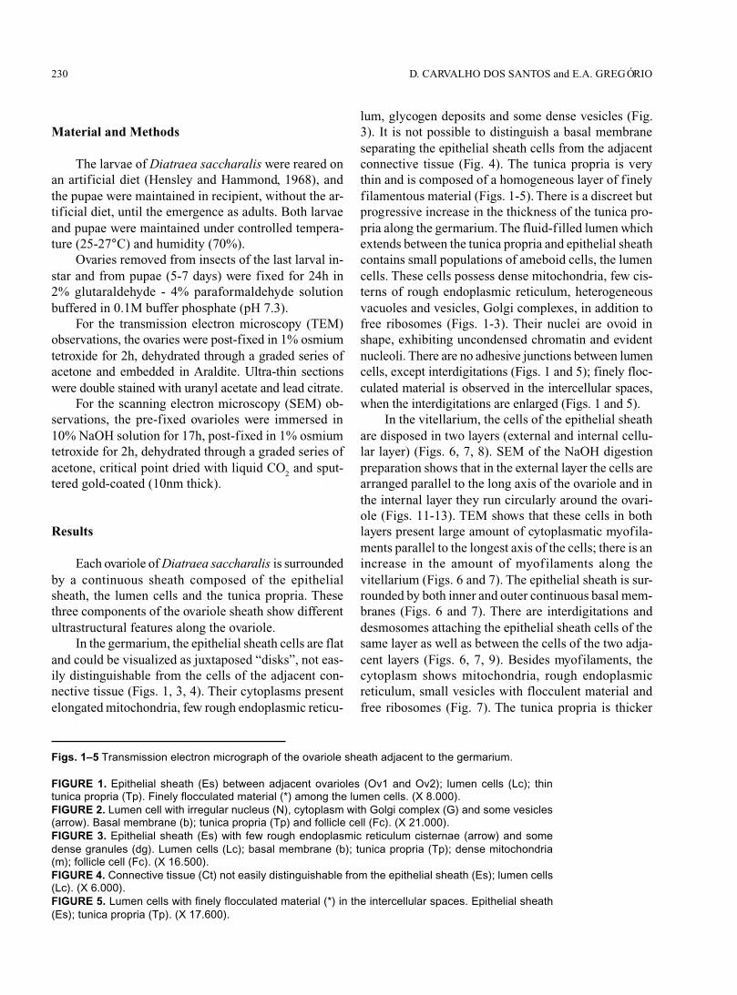
In the germarium, the epithelial sheath cells are flatand could be visualized as juxtaposed “disks”, not eas-ily distinguishable from the cells of the adjacent con-nective tissue (Figs. 1, 3, 4). Their cytoplasms presentelongated mitochondria, few rough endoplasmic reticu-
lum, glycogen deposits and some dense vesicles (Fig.3). It is not possible to distinguish a basal membraneseparating the epithelial sheath cells from the adjacentconnective tissue (Fig. 4). The tunica propria is verythin and is composed of a homogeneous layer of finelyfilamentous material (Figs. 1-5). There is a discreet butprogressive increase in the thickness of the tunica pro-pria along the germarium. The fluid-filled lumen whichextends between the tunica propria and epithelial sheathcontains small populations of ameboid cells, the lumencells. These cells possess dense mitochondria, few cis-terns of rough endoplasmic reticulum, heterogeneousvacuoles and vesicles, Golgi complexes, in addition tofree ribosomes (Figs. 1-3). Their nuclei are ovoid inshape, exhibiting uncondensed chromatin and evidentnucleoli. There are no adhesive junctions between lumencells, except interdigitations (Figs. 1 and 5); finely floc-culated material is observed in the intercellular spaces,when the interdigitations are enlarged (Figs. 1 and 5).
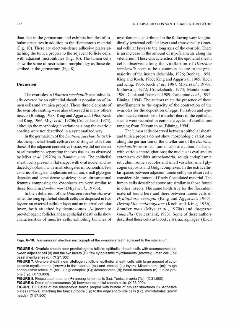
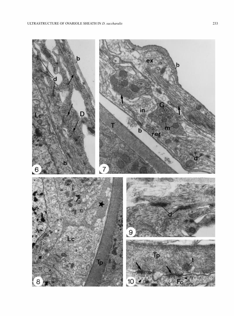
In the vitellarium, the cells of the epithelial sheathare disposed in two layers (external and internal cellu-lar layer) (Figs. 6, 7, 8). SEM of the NaOH digestionpreparation shows that in the external layer the cells arearranged parallel to the long axis of the ovariole and inthe internal layer they run circularly around the ovari-ole (Figs. 11-13). TEM shows that these cells in bothlayers present large amount of cytoplasmatic myofila-ments parallel to the longest axis of the cells; there is anincrease in the amount of myofilaments along thevitellarium (Figs. 6 and 7). The epithelial sheath is sur-rounded by both inner and outer continuous basal mem-branes (Figs. 6 and 7). There are interdigitations anddesmosomes attaching the epithelial sheath cells of thesame layer as well as between the cells of the two adja-cent layers (Figs. 6, 7, 9). Besides myofilaments, thecytoplasm shows mitochondria, rough endoplasmicreticulum, small vesicles with flocculent material andfree ribosomes (Fig. 7). The tunica propria is thicker
Figs. 1–5 Transmission electron micrograph of the ovariole sheath adjacent to the germarium.
FIGURE 1. Epithelial sheath (Es) between adjacent ovarioles (Ov1 and Ov2); lumen cells (Lc); thintunica propria (Tp). Finely flocculated material (*) among the lumen cells. (X 8.000).FIGURE 2. Lumen cell with irregular nucleus (N), cytoplasm with Golgi complex (G) and some vesicles(arrow). Basal membrane (b); tunica propria (Tp) and follicle cell (Fc). (X 21.000).FIGURE 3. Epithelial sheath (Es) with few rough endoplasmic reticulum cisternae (arrow) and somedense granules (dg). Lumen cells (Lc); basal membrane (b); tunica propria (Tp); dense mitochondria(m); follicle cell (Fc). (X 16.500).FIGURE 4. Connective tissue (Ct) not easily distinguishable from the epithelial sheath (Es); lumen cells(Lc). (X 6.000).FIGURE 5. Lumen cells with finely flocculated material (*) in the intercellular spaces. Epithelial sheath(Es); tunica propria (Tp). (X 17.600).

231ULTRASTRUCTURE OF OVARIOLE SHEATH IN D. saccharalis
232 D. CARVALHO DOS SANTOS and E.A. GREGÓRIO
than that in the germarium and exhibits bundles of tu-bular structures in addition to the filamentous material(Fig. 10). There are electron-dense adhesive plates at-taching the tunica propria to the adjacent follicle cells,with adjacent microtubules (Fig. 10). The lumen cellsshow the same ultrastructural morphology as those de-scribed in the germarium (Fig. 8).
Discussion
The ovarioles in Diatraea saccharalis are individu-ally covered by an epithelial sheath, a population of lu-men cells and a tunica propria. These three elements ofthe ovariole coating were also observed in many othersinsects (Bonhag, 1958; King and Aggarwal, 1965; Kochand King, 1966; Miya et al., 1970b; Cruickshank, 1973),although the morphologic variations along the ovariolecoating were not described in a systematized way.
In the germarium of the Diatraea saccharalis ovari-ole, the epithelial sheath cells are not distinguishable fromthose of the adjacent connective tissue; we did not detectbasal membrane separating the two tissues, as observedby Miya et al. (1970b) in Bombyx mori. The epithelialsheath cells present a flat shape, with oval nuclei and re-duced cytoplasm, with small elongated mitochondria, fewcisterns of rough endoplasmic reticulum, small glycogendeposits and some dense vesicles; these ultrastructuralfeatures composing the cytoplasm are very similar tothose found in Bombyx mori (Miya et al., 1970b).
In the vitellarium of the Diatraea saccharalis ova-riole, the long epithelial sheath cells are disposed in twolayers: an external cellular layer and an internal cellularlayer, both attached by desmosomes. Adjacent toprevitellogenic follicles, these epithelial sheath cells showcharacteristics of muscles cells, exhibiting bunches of
myofilaments, distributed in the following way: longitu-dinally (external cellular layer) and transversally (inter-nal cellular layer) to the long axis of the ovariole. Thereis an increase in the amount of myofilaments along thevitellarium. These characteristics of the epithelial sheathcells observed along the vitellarium of Diatraeasaccharalis seem to be a common feature in the greatmajority of the insects (Machida, 1926; Bonhag, 1958;King and Koch, 1963; King and Aggarwal, 1965; Kochand King, 1966; Koch et al., 1967; Miya et al., 1970a;Mahowald, 1972; Cruickshank, 1973; Mandelbaum,1980; Cook and Peterson, 1989; Carcupino et al., 1992;Büning, 1994). The authors relate the presence of thesemyofilaments to the capacity of the contraction of theovarioles for the deposition of eggs. Pulsation and syn-chronized contractions of muscle fibers of the epithelialsheath were recorded in complete cycles of oscillationsranging from 200mm to 4s (Büning, 1994).
The lumen cells observed between epithelial sheathand tunica propria do not show morphologic variationsalong the germarium or the vitellarium of the Diatraeasaccharalis ovarioles. Lumen cells are cuboid in shape,with various interdigitations; the nucleus is oval and itscytoplasm exhibits mitochondria, rough endoplasmicreticulum, some vacuoles and small vesicles, small gly-cogen deposits and Golgi complexes. In the extracellu-lar spaces between adjacent lumen cells, we observed aconsiderable amount of finely flocculated material. Thelumen cells described above are similar to those foundin other insects. The same holds true for the flocculentmaterial found here and there between lumen cells ofHyalophora cecropia (King and Aggarwal, 1965),Drosophila melanogaster (Koch and King, 1966),Bombyx mori (Miya et al., 1970a) and Anagastakuhniella (Cruickshank, 1973). Some of these authorsdescribed these cells as blood cells (macrophages) (Koch
Figs. 6–10. Transmission electron micrograph of the ovariole sheath adjacent to the vitellarium.
FIGURE 6. Ovariole sheath near previtellogenic follicle: epithelial sheath cells with desmosomes be-tween adjacent cell (d) and the two layers (D); few cytoplasmic myofilaments (arrows); lumen cell (Lc);basal membranes (b). (X 27.500).FIGURE 7. Ovariole sheath near vitellogenic follicle: epithelial sheath cells with large amount of cyto-plasmic myofilaments (arrows) in the external (ex) and internal (in) layers. Mitochondria (m); roughendoplasmic reticulum (rer); Golgi complex (G); desmosomes (d); basal membranes (b); tunica pro-pria (Tp). (X 13.000).FIGURE 8. Flocculated material (★) among lumen cells (Lc). Tunica propria (Tp). (X 31.500).FIGURE 9. Detail of desmosomes (d) between epithelial sheath cells. (X 36.000).FIGURE 10. Detail of the filamentous tunica propria with bundle of tubular structures (t). Adhesiveplates (arrows) attaching the tunica propria (Tp) to the adjacent follicle cells (Fc); microtubules (arrowheads). (X 57.500).
233ULTRASTRUCTURE OF OVARIOLE SHEATH IN D. saccharalis

234 D. CARVALHO DOS SANTOS and E.A. GREGÓRIO
Figs. 11–13. Scanning electron micrograph of NaOH digested ovariole sheath in thevitellarium.
FIGURE 11. General view of three ovarian follicles (F). Cells of the epithelial sheath ex-ternal layer (arrows) connecting adjacent follicles. (X 90).FIGURE 12. Cleft between adjacent follicles with the epithelial sheath cells (arrows) ofthe external layer; tracheole (T) with branches (*). (X 330).FIGURE 13. Detail of the epithelial sheath surface. The cells of the circular internal layer(in) are partially hidden by the ones of the longitudinal external layer (ex). The cells ofexternal layer are intensively anastomosed (arrows). Traqueole (T) with branch (★) goingthrough the epithelial sheath cells. (X 1.200).
235ULTRASTRUCTURE OF OVARIOLE SHEATH IN D. saccharalis
and King, 1966; Cruickshank, 1973), capable of secret-ing material to repair the tunica propria at its torn pointsdue to ovariole growth and to the moment of eggs depo-sition; Cruickshank (1973) confirms this function forthe lumen cells, suggesting that there seems to be a cor-relation between its secretory activity and the increasein thickness of the tunica propria during development.Although we did not observe significant modificationsin the ultrastructural organization of the lumen cells,their morphological features indicate its contribution tothe tunica propria thickness, that occurs along theDiatraea saccharalis ovariole.
In the germarium, the tunica propria is extremelythin, composed of finely filamentous material. There isan increase in the tunica propria thickness from thegermarium to the vitellarium, where some dense struc-tures were visualized among its filamentous material.The ultrastructural aspect of the tunica propria is verysimilar in many insects (Machida, 1926; Bonhag, 1958;King and Koch, 1963; King and Aggarwal, 1965; Kochand King, 1966; Koch et al., 1967; Miya et al., 1970a;Mahowald, 1972; Cruickshank, 1973; Gregório et al.,1990; Büning, 1994). According to these authors, thenature of the tunica propria material is still uncertain. Itis known that this acellular layer presents proteins andcarbohydrate complexes (Bonhag and Arnold, 1961) andthat these proteins are rich in S-S groups (Cruickshank,
1973). Büning (1994) suggests that collagen type I,laminine and proteoglycans compose the tunica propriaof insects. This author describes that, adjacent to thevitelogenic follicles, the tunica propria exhibits struc-tures in stick shape, oriented parallel to the long axis ofthe ovariole, as we saw in Diatraea saccharalis at theend of previtellogenesis. However, Büning (1994) doesnot suggest a function for these structures. The basalmembranes (tunica propria) of the insect ovariolespresent three functions, as proposed by Büning (1994):(1) they serve as a mechanical structure to keep the elon-gated structure of the ovarioles stable; (2) they serve asphysical sieves against the haemolymph; (3) they havesome chemical selectivity for macromolecules whichplay a role in hormone access during previtellogenesis.The presence of adhesive plates attaching the tunicapropria to the follicle cells, and their relation to the cy-toplasmic microtubules, suggest that the tunica propriabecomes stretched during the oogenesis.
Acknowledgements
We gratefully acknowledge the EntomologicalLaboratory of Usina Barra Grande, Lençóis Paulista,S.P., Brasil, for rearing the insects. This work was par-tially supported by CNPq (Masters Degree fellowship).
References
BONHAG PF (1958). Ovarian structure and vitellogenesis in insects. Annu Rev Entomol 3: 137-160.BONHAG PF, ARNOLD WJ (1961). Histology, histochemistry and tracheation of the ovariole sheaths in the american cockroach Periplaneta
americana ( L.). J Morphol 108: 107-129.BÜNING J (1994). The insect ovary. Ultrastructure, previtellogenic growth and evolution. London, Chapman and Hall, pp. 40-61.CARCUPINO M, YIN CM, STOFFOLANO JG, SCAPIGLIATI JG, MAZZINI MF (1992). Actin distribution in the ovaries of
previtellogenic and vitellogenic black blowflies, Phormia regina (Meigen) (Diptera: Calliphoridae). Int J Insect Morphol Embryol21: 77-86.
COOK BJ, PETERSON T (1989). Ovarian muscularis of the stable fly Stomoxys calcitrans: its strudtural, motile, and pharmacologicalproperties. Arch Insect Biochem Physiol 12: 15-30.
CRUICKSHANK WJ (1973). The ultrastructure and functions of the ovariole sheath and tunica propria in the flour moth. J Insect Physiol19: 577-592.
GREGÓRIO EA, SECCO VNDP, TOLEDO LA, LELLO E (1990). Ultrastructure of the ovary of Dermatobia hominis (Diptera: Cutere-bridae). II. Origin of the tunica propria in ovarioles. Mem Inst Oswaldo Cruz Rio J 85: 315-320.
HENSLEY SD, HAMMOND AM (1968). Laboratory tecniques for rearing the sugar cane borer on an artif icial diet. J Econ Entomol 61:1742-1743.
KING RC, AGGARWAL SK (1965). Oogenesis in Hyalophora cecropia. Growth 29: 17-83.KING RC, KOCH EA (1963). Studies on ovarian follicle cells of Drosophila. Q J Microsc Sci 104: 297-320.KOCH EA, KING RC (1966). The origin and early differentiation of the egg chamber of Drosophila melanogaster. J Morphol 119: 283-304.KOCH EA, SMITH PA, KING RC (1967). The division and differentiation of Drosophila cystocytes. J Morphol 121: 55-70.MACHIDA J (1926). The development of the ovary in the silkworm (Bombyx mori). J Coll Dairy Agric 7: 293-351.MAHOWALD AP (1972). Ultrastructural observations on oogenesis in Drosophila. J Morphol 137: 29-48.MANDELBAUM I (1980). Intercellular bridges and the fusome in the germ cells of the Cecropia moth. J Morphol 166: 37-50.MIYA K, KURIHARA M, TANIMURA I (1970a). Electron microscope studies on the oogenesis of the silkworm, Bombyx mori L. II.
(Fine structure of follicle cells and ovarial sheath). J Fac Agric Iwate Univ 10: 1-17.MIYA K, KURIHARA M, TANIMURA I (1970b). Electron microscope studies on the oogenesis of the silkworm, Bombyx mori L. III.
(Fine structure of ovary in the early stage of fifth instar larva). J Fac Agric Iwate Univ 10: 59-83.SNODGRASS RE (1935). Principles of Insect Morphology. McGraw-Hill Book Company, New York, pp. 535.