Ultrastructure of diploid and haploid cell of s Xenopus laevis larvae · Embryol. exp. Morph. Vol....
18
/. Embryol. exp. Morph. Vol. 26, 1, pp. 81-98, 1971 Printed in Great Britain Ultrastructure of diploid and haploid cells of Xenopus laevis larvae By H. FOX 1 AND LOUIE HAMILTON 2 SUMMARY A study was made of tissues of diploid and haploid Xenopus laevis larvae by electron microscopy. The comparison of the two types of larvae was both quantitative and qualitative. Haploid cells contain fewer similarly sized mitochondria and/or cilia (compared with the diploid), commensurate with their smaller cell size. Cellular differentiation is retarded in haploids and the degree of retardation is reflected in the retention of cellular lipid and yolk. It is suggested that the failure to differentiate adequately is an important component in the development of the haploid syndrome. INTRODUCTION When haploid amphibian larvae develop they usually become abnormal and are said to show the haploid syndrome (Fankhauser, 1945; Hamilton, 1963; Gallien, 1967). Abnormality is first seen at gastrulation, which does not start in the haploids until their cells have the same nucleo-cytoplasmic ratio as diploid cells. The delay in the onset of haploid gastrulation approximates to the time taken for all the cells to divide once, so that from the beginning of gastrulation onwards a haploid embryo contains about twice as many cells, stage for stage, as a diploid embryo. Even though all Xenopus haploid embryos are morphologically different from diploids during embryonic stages of development about 5 % will develop into slightly abnormal swimming feeding tadpoles. The remaining 95 % develop the haploid syndrome (Hamilton, 1963) and are blocked in their development at stage 43+ (Nieuwkoop & Faber, 1956). They have, in addition to shortened axial structures and microcephaly, a poorly formed gut and oedema, which latter two signs of the haploid syndrome cannot easily be related to reduced cell size per se. In this study we have examined both qualitatively and quantitatively the fundamental cellular architecture of haploid and diploid cells, particularly in tissues which might affect the development of oedema (Fox & Hamilton, 1964#). For this reason our ultrastructural investigation concentrated on different regions of the pronephros, the skin and cloaca - together with the pharynx. The 1 Author's address: Department of Zoology, University College London, Gower Street London WC1, U.K. 2 Author's address: The Middlesex Hospital Medical School, London Wl, U.K. 6 EMB 26
Transcript of Ultrastructure of diploid and haploid cell of s Xenopus laevis larvae · Embryol. exp. Morph. Vol....

/ . Embryol. exp. Morph. Vol. 26, 1, pp. 81-98, 1971
Printed in Great Britain
Ultrastructure of diploid and haploid cells ofXenopus laevis larvae
By H. FOX1 AND LOUIE HAMILTON2
SUMMARYA study was made of tissues of diploid and haploid Xenopus laevis larvae by electron
microscopy. The comparison of the two types of larvae was both quantitative and qualitative.Haploid cells contain fewer similarly sized mitochondria and/or cilia (compared with thediploid), commensurate with their smaller cell size. Cellular differentiation is retarded inhaploids and the degree of retardation is reflected in the retention of cellular lipid and yolk.It is suggested that the failure to differentiate adequately is an important component in thedevelopment of the haploid syndrome.
INTRODUCTION
When haploid amphibian larvae develop they usually become abnormal andare said to show the haploid syndrome (Fankhauser, 1945; Hamilton, 1963;Gallien, 1967). Abnormality is first seen at gastrulation, which does not start inthe haploids until their cells have the same nucleo-cytoplasmic ratio as diploidcells. The delay in the onset of haploid gastrulation approximates to the timetaken for all the cells to divide once, so that from the beginning of gastrulationonwards a haploid embryo contains about twice as many cells, stage for stage,as a diploid embryo.
Even though all Xenopus haploid embryos are morphologically different fromdiploids during embryonic stages of development about 5 % will develop intoslightly abnormal swimming feeding tadpoles. The remaining 95 % develop thehaploid syndrome (Hamilton, 1963) and are blocked in their development atstage 43+ (Nieuwkoop & Faber, 1956). They have, in addition to shortened axialstructures and microcephaly, a poorly formed gut and oedema, which latter twosigns of the haploid syndrome cannot easily be related to reduced cell size per se.
In this study we have examined both qualitatively and quantitatively thefundamental cellular architecture of haploid and diploid cells, particularly intissues which might affect the development of oedema (Fox & Hamilton, 1964#).For this reason our ultrastructural investigation concentrated on differentregions of the pronephros, the skin and cloaca - together with the pharynx. The
1 Author's address: Department of Zoology, University College London, Gower StreetLondon WC1, U.K.
2 Author's address: The Middlesex Hospital Medical School, London Wl, U.K.6 EMB 26

82 H. FOX AND L. HAMILTON
electron micrographs relating to the cloaca have been published by Fox (1970 a)to which reference should be made when this region is described in the presentpaper.
MATERIALS AND METHODS
Androgenetic haploid eggs of Xenopus laevis were obtained using ultravioletirradiation (Gurdon, 1960; Hamilton, 1963). Growth of larvae was in de-chlorinated tap water at about 16-20°C. Specimens were fixed whole in ice-coldosmic acid (Palade, 1952) or occasionally in glutaraldehyde-osmic acid (Hirsch& Fedorko, 1968). (For further details of electron-microscopic methods used,see Fox, 1970#, b; Fox, Mahoney & Bailey, 1970.) Larvae staged according tothe scheme of Nieuwkoop & Faber (1956) were investigated at stages 38/39, 43,44 and 47 for diploids and at stage 43+ and stage 43+ + for haploids. [Owing tothe impossibility of staging haploid Xenopus beyond stage 43, the authors havedecided to call haploid larvae in advance of stage 43, 43+ and 43++, which areequivalent in age to diploids of stage 47 and 50 respectively. Haploid stage 43+
and stage 43+ + are equivalent to haploid stages 43 and 43-45 of previous workby Fox (1970a).]
Araldite sections (1-2 jum thick) and paraffin-embedded transverse serialsections (8 jum and 10 /im thick) were examined by phase contrast and lightmicroscopy, in addition to the ultrathin, silver grey sections (60-100 nm thick)usedTor electron microscopy. Thus in the two types of larvae comparisons ofspecific regions of selected tissues were made. Ultrathin sections were stained byuranyl acetate and lead citrate and viewed under an AEI EM6B electronmicroscope. Mitochondrial profiles were measured with a planimeter. The levelof statistical significance used was P < 0-05.
RESULTS
The pronephros and duct
For morphological and quantitative information on the amphibian (larval)pronephros, including that of Xenopus, reference may be made to papers byFox (1962, 1963, 19706), Fox & Hamilton (1964a, b) and Christensen (1964).
The pronephros of Xenopus includes three nephrostomial tubules, each ofwhich opens at one end into the coelom (nephrostome) and at the other into theproximal, convoluted microvillous tubule (Figs. 3, 4, 5). During later larvaldevelopment the first two nephrostomial tubules lead into a common nephro-stome, with the third not far behind. The lumen is lined by ciliated cells, whichpresumably sweep coelomic fluid (derived by diffusion through the glomerularmembranes of the paired glomi, or from fluid of tissues adjacent to the coelom)into the pronephros. Thenceforth it travels along the pronephric duct to theexterior via the cloaca.

EM of haploid and diploid cells 83
A considerable number of nephrostomial tubule cells of diploids at stage 47,and also those of haploids at stage 43+ of about the same age and older ones atstage 43+ + include a large lipid droplet, often as large or larger than the nucleus(Figs. 1-5). Most of the lipid and practically all the yolk, however, have dis-appeared from the rest of the pronephros and duct in stage 47 diploids, thoughlipid is still plentiful in these regions in stage 43+ + haploids. Though haploidnephrostomial tubule cells are generally smaller than diploids they are howeverfrequently longer (sometimes twice as long) and considerably narrower. Diploidnephrostomial tubule walls vary in thickness from 8 /«n to 16 /on and maximumnuclear measurements were 12 /im longx 8/OTL wide. In haploids comparablemeasurements of walls and nuclei were 5 ju>m to 10 jum and 9 /im x 5 /*m respec-tively. Diploid tubule walls often appear more than one cell across, in contrastto a wall only one cell thick in haploids.
In both types the basal lamina of the nephrostomial tubule is envelopedexternally by a similar delicate collagenous sheath. Intercellular junctions arestraight (Figs. 1, 2), not highly folded, nor are there any elaborate infoldings ofthe plasma membrane as in the rest of the pronephros (Fox, 1970&). Mito-chondria are irregular in shape and variable in numbers in nephrostomial tubulecells; on the whole they are more numerous in the diploid than in comparablehaploid areas.
Within the nephrostomial tubule lumen groups of cilia are recognizable, ineach of which the cilia are usually sectioned in a similar orientation. It is likelythat each ciliary grouping originates from one particular cell (see Figs. 1, 2, 8).In diploids six groups were sufficiently discrete to be measured. They were madeup of 85-100 cilia (mean 89/group). In haploids three comparable discretegroups of nephrostomial cilia each comprised 44,60 and 51 cilia (mean 51 /group),a little over half the diploid number. In both haploids and diploids a length ofcell margin about 1200 nm long in each case included three ciliary bases, or400nm/cilium (Figs. 6, 7). Measurements of 12 cilia in transverse section, fromeach of a diploid and haploid nephrostomial tubule showed their mean areasnot to differ significantly; they average about 250 nm in diameter in both formsand have a similar structure (Table 1).
The pronephric microvillous tubules are well developed in diploids at stage 47(Fig. 9 a, b). A fine collagen layer surrounds it and the plasma membrane hasdeveloped some modest peripheral infoldings; intercellular junctions are like-wise infolded. Nuclei and nucleoli are prominent. Mitochondria are large,irregular in shape, some extremely elongated, and well differentiated. The cyto-plasmic matrix often includes large, roundish less-dense vesicles possibly con-taining mucous-like substances. Well-developed Golgi cisternae are present.The endoplasmic reticulum comprises a number of irregularly shaped smoothand rough vesicles, and groups of polysomes in small rosettes extend throughoutthe cell. Large numbers of pinocytotic vesicles are seen, either at their sites oforigin at the margin of the tubule between the microvilli, or further within the
6-2

84 H. FOX AND L. HAMILTON
cell (Fig. 9b). The whole inner apical margin is bounded by microvilli whoselengths vary between 2 [im and 2-7 /tm.
In contrast stage 43+ and stage 43+ + haploid microvillous tubules are variablein their degree of development, often less well differentiated than in stage 44diploids and far less than in stage 47 diploids. Sometimes some cells, or portionsof them, appear almost as well differentiated as in diploids (especially in the olderhaploids), though lipid and occasional small yolk bodies are usually still retained(Fig. 11). In most cases, however, a basal complex of Moldings is not developed,intercellular junctions are less infolded and the mitochondrial populations ofthese retarded tissues are not as dense as in the diploids. Furthermore, yolkbodies show variable profiles (Fig. 10) and there are numerous large lipiddroplets, often situated close together (Fig. 12), or in groups of smaller dropletsof varying sizes situated around dense tissue, which may well be remnants oflipid digestion (Fig. 10). Microvilli appear of similar length to those in diploidsbut pinocytotic vesicles, present at the lumen margins of the cell, are not asprofuse in numbers.
Diploid distal pronephric tubules at stage 47 differ from its microvilloustubules of the same stage in the greater complexity of the plasma membranousinfoldings, some of which penetrate deeply into the tubule cell (Fig. 13). Inter-cellular junctions are often straight, sometimes highly interfolded or irregularalong their course. Mitochondria are large, variable in shape and often extremelyelongated (up to 4 jim long); very often they are partially enclosed within thesurface infoldings. The inner (apical) lumen margin is bordered by short, stub-like processes, shorter and thicker than the microvilli and fewer in number,though usually they appear transverse in section. No pinocytotic vesicles werefound. Occasional lipid droplets are recognized but practically all the yolk hasdisappeared. Golgi networks are present. On the whole the cytoplasmic matrix
For description of abbreviations on figs, see page 98.Fig. 1. Haploid specimen, stage 43+. Nephrostomial tubule of pronephros withcilia inside the lumen.Fig. 2. Diploid specimen, stage 47. Nephrostomial tubule. The blocks of cilia,sectioned at different orientations, probably are each derived from different indi-vidual nephrostomial cells and clearly show the greater cilia population per groupin the diploid, compared with the haploid of the previous figure.Fig. 3. Diploid specimen, stage 47. Thick Araldite section, picture by phase contrast.Longitudinal section of a nephrostomial tubule leading from the coelom via thenephrostome. The large round lipid droplets are easily seen in the tubule cells.Fig. 4. Haploid specimen, stage 43+. A nephrostomial tubule. The illustration wasprepared in the same way as in Fig. 3.Fig. 5. Haploid specimen, stage 43++ (about 13 days old). Junction of the ciliatednephrostomial tubule distally and the microvillous proximal convoluted tubuleproximally. Note the large lipid droplets retained in the nephrostomial tubule cells.Normally diploids (and probably haploids too) retain lipid in the nephrostomialtubule longer than in the rest of the pronephros.

EM ofhaploid and diploid cells 85

86 H. FOX AND L. HAMILTON
1 //m
Fig. 6. Diploid specimen, stage 47. Cilia and their rootlets from cells of thenephrostomial tubule.Fig. 7. Haploid specimen, stage 43+. Cilia and rootlets (from nephrostomial tubulecells), of similar age, size and appearance as in the diploid of the previous figure.The number of rootlets per unit cell length is generally similar in diploids andhaploids.Fig. 8. Diploid specimen stage 47. Ciliary bases and rootlets, in transverse section,of a nephrostomial tubule cell.

EM of haploid and diploid cells 87
is similar to that of the microvillous tubule, though no mucous vesicles arepresent.
The ultrastructure of the distal pronephric tubules of stage 43+ haploids isgenerally similar to that of the stage 47 diploid and their walls are of similarthickness; 8 /.tm to 10 jam across (Fig. 15). The main differences relate to theinferior haploid nuclear and cell volumes, their retention of lipid (see also instage 44 diploids) and the frequent appearance of cavities between the plasmamembranous infoldings. These could well be artifacts, though they were notfound in comparable diploid tubules, nor in nephrostomial, microvillous orduct tubules of both types of larvae. Fewer haploid mitochondria appear to beenclosed by the membranous folds. In the older stage 43+ + haploids the arrange-ment is more like that of the diploid.
The hinder pronephric duct in the stage 47 diploid is remarkable for the highdegree of infolding or branching of the plasma membrane (Figs. 16, 17),a feature similarly found in Triturus larvae (Lehmann, 1967). Infolding is morehighly developed in stage 47 diploids than in younger stage 44 diploids orstage 43+ haploids of the same age, or older stage 43++ haploids (Figs. 14, 18).The diploid duct is well developed and only occasionally are lipid droplets found,though large ones and often yolk bodies are still present in the comparable agedor older haploids and in stage 44 diploids (Figs. 17, 18). Intercellular junctionsmay be straight but usually they are highly folded. The thickness of the wall isvariable ranging from 6 fim to 12 /.tm in diploids and 5 jim to 15 jum in haploidsand the lumen wall is almost smooth with merely a few cellular projections. Thesmaller nuclear areas of haploids, compared with diploids, are clearly seen inlow resolution ultrasections of the entire duct (Figs. 17, 18). Golgi networks arepresent in both types of cell. In general, apart from the differences described, theduct is similar in appearance to that of the distal tubule.
The cloaca (and hind rectum) (see Figs. 1-13; Fox, \910a)
At diploid stages 38/39 the cloacal cells were partially differentiated, in thatmuch yolk and lipid were present but few signs of ciliation. By stage 44 ciliawere well developed but some yolk and lipid remained. In stage 47 larvalcloacae, most of the lipid and all of the yolk had disappeared. These latter cellsmay contain up to 150 cilia, which total when compared with the 85-100 in thediploid pronephric nephrostomial cells suggests that in different tissues ciliatedcells have a specific mean number of cilia. Non-ciliated vesicular cells, similarto epidermal cells, were also present in the cloaca. Haploid cloacal cells at stages43+ and 43+ + are similar to diploid ones at stages 38/39 and 47 respectively.
The skin
In normal (diploid) Xenopus larvae the first epidermal cilia appear at stages20-21, following fusion of the neural folds (Steinman, 1968). In the present workdiploids at stages 43 and 44 still retain some cilia in tail fin epidermal cells though
H. FOX AND L. HAMILTON
9 (a)

EM of haploid and diploid cells 89
the typical non-ciliated vesicular cells are well developed and now preponderate(Fig. 22). Occasional yolk bodies and large lipid droplets still abound howeverin these cells as in the case of stage 43+ haploids. Lipids, moreover, are stillrecognized in stage 47 diploid epidermal cells (Fig. 21). Epidermal cilia areoccasionally found in stage 43+ haploids, usually in a degenerate condition,though some normal ones may often be present (Fig. 19). In stage 47 diploids ofthe same age and older stage 43+ + haploids they have disappeared entirely fromthe skin (Figs. 20, 21). In general the skin ultrastructure of this stage agrees withthat described by Steinman (1968). The well-developed surface mucous vesiclesof stages 43+ and 43+ + haploids and stages 43 and 47 diploids do not seem todiffer markedly in size, number or appearance.
The pharynx
The haploid stage 43+ pharynx is only partially differentiated, and the cellsusually include one or several large yolk bodies and numerous lipid droplets, ofvarying sizes (Fig. 23). Nuclei and nucleoli are well developed and some mito-chondria have differentiated. The appearance is generally similar in youngerstage 43 diploids. In stage 47 diploids, at the same age as the stage 43+ haploids,the pharynx is highly differentiated (Fig. 25). Cell components include thosewith apical vesicles and in specific regions, particularly laterally in dorsalpouches, there are well-developed ciliated cells (Fig. 26) which are equally well-developed in diploids at stage 44. Goblet cells were recognized in the posteriorpharyngeal region where it grades into the oesophagus (Fig. 24).
Quantitative data on mitochondria in pronephros of diploid and haploid larvae(Table 1)
Where there is sufficient pronephric cellular differentiation, so that well-differentiated mitochondria populate a specific region of a cell, the mito-
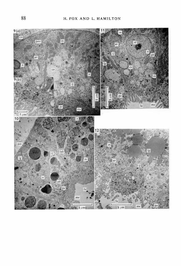
Fig. 9. (a) Diploid specimen, stage 47. Transverse section of pronephric microvilloustubule. The less-dense vesicles may possibly represent areas which previously con-tained lipid granules. Cellular structure is quite well differentiated, (b) Diploid speci-men, stage 47. Pinocytotic vesicles at microvillous margin.Fig. 10. Haploid specimen, stage 43+. A similarly orientated section of a microvilloustubule as in Fig. 9. Compared with the diploid of similar age there is less develop-ment and differentiation in the haploid, and many lipid granules and yolk bodies arestill retained. Some small pinocytotic vesicles are recognized at the lumen margin(they are numerous in the diploid stage 47), which suggests that in these haploidssome incipient functional absorption occurs.Fig. 11. Haploid specimen, stage 43++. Microvillous tubule older than in specimensof Figs. 10 and 12, showing a greater degree of development and differentiation; theintracellular organelles have profiles similar to those in the microvillous tubule of theyounger diploid of Fig. 9.Fig. 12. Haploid specimen, stage 43+. Microvillous tubule of similar age to that inFig. 10, showing a greater degree of differentiation but not as much as in the diploidof the same age in Fig. 9, or in the older haploid of Fig. 11.

H. FOX AND L. HAMILTON
15!
Fig. 13. Diploid specimen, stage 47. Distal pronephric tubule showing highdevelopment of the infoldings of the plasma membrane.Fig. 14. Haploid specimen, stage 43+. Pronephric duct (hinder region) with lipidstill present and only modest infoldings of the plasma membrane.Fig. 15. Haploid specimen, stage 43+. Distal pronephric tubule. Note the cavitiesbetween the cell plasma membranes and the presence of lipid granules, the level ofdifferentiation here being less than in the comparable diploid of Fig. 13.Fig. 16. Diploid specimen, stage 47. Pronephric duct (hinder region). Note the com-plex infoldings of the plasma membrane; generally there is an absence of lipid andyolk and a greater degree of cellular differentiation compared with the comparablehaploid duct of Fig. 14.

EM of haploid and diploid cells 91
chondrial population density per unit area of cell surface (MPD) in diploidnephrostomial tubules, is generally superior to that of haploids. In microvillousand distal tubules and the hind duct however the MPD of haploids and diploidsis practically the same but the MPD of the distal tubule and hind duct is signifi-cantly superior to that in the microvillous tubule.
17
Fig. 17. Diploid specimen, stage 47. Pronephric duct in transverse section.Fig. 18. Haploid specimen, stage 43+. The haploid duct shows smaller nuclearprofiles, less infolding of the cell surface and retains lipid granules compared withthe diploid of the same age. The ducts (as are the distal tubules), however, are quitewell differentiated in both haploid and diploid specimens, compared with the moreslowly developing microvillous tubules.
The mean mitochondrial profile area (MPA) in microvillous tubules of stage 47diploids is significantly superior, by about 29 %, to that of stage 44 diploids andstage 43+ haploids. The MPA superiority of 18% of stage 43+ + haploids over

92 H. FOX A N D L. H A M I L T O N
Fig. 19. Haploid specimen, stage 43+. Tail-skin with some vestigial cilia stillrecognizable.
Fig. 20. Haploid specimen, stage 43+ + . Tail-skin. Cilia are absent and the apicalmucous vesicles are highly developed.
Fig. 21. Diploid specimen, stage 47. Tail-skin. Cilia are absent; the apical vesiclesare well developed and occasional lipid granules and numerous pigment bodiesare present.
Fig. 22. Diploid specimen, stage 43. Tail-skin. Cilia are still present in thisspecimen which is younger than those in Figs. 23-25.

EM of haploid and diploid cells 93
Fig. 23. Haploid specimen, stage 43+. Inner region of the pharynx showing theundifferentiated structure of these cells.Fig. 24. Diploid specimen, stage 47. Outer wall of pharynx-oesophageal region.Note the high degree of differentiation compared with the haploid specimen ofsimilar age in Fig. 23.
Fig. 25. Diploid specimen, stage 47. Pharynx, in region between the centre and side;the upper region is uppermost. Compare the development with the haploid speci-men of Fig. 23.Fig. 26. Diploid specimen, stage 47. Dorsal wall of the pharynx in the lateralciliary groove, showing the highly developed cilia at this stage.

94 H. FOX AND L. HAMILTON
Table 1. Comparison of the numbers of mitochondrial profiles (from comparableareas) and of cilia (per cell) in diploid and haploid pronephric cells of Xenopuslaevis, at various stages of development (stages according to Nieuwkoop & Faber,1956): Age in days (approximately) of the different groups is recorded.
Mitochondria Cilia
Diploid Haploid Diploid Haploid Diploid Haploid(stage 44) (stage 43+) (stage 47) (stage 43++) (stage 47) (stage 43+)
Region of pronephros (6 days) (8 days) (8 days) (13 days) (8 days) (8 days)
Nephrostomial tubuleMean no. organelles/
cellGenerally higher population density 89 (6 cells) 51 (3 cells)in diploid nephrostomial tubules cells (mean diameter cilium
250nm in both groups)
3-5 3-5 4-5 5-3 —±0-24 ±0-28 ±0-33 ±0-50 —
87-5 1260 116-6
Microvillous tubuleTotal no. profiles/ 29 25 28 22
100 /tm2 cell surface(from 400 /tm2)
Mean and standarderror of individualmitochondrial profilearea (x 105nm2)
Total mitochondrial 101-5area/100/tm2 cellsurface (x 105nm2)
Total no. profiles 112 95 101 75actually measured
Distal tubuleTotal no. profiles/ 77 39 51 53
100/tm2 cell surface(from 400 /tm2)
Mean and standarderror of individualmitochondrial profilearea (x 105nm2)
Total mitochondrialarea/100/tm2 cellsurface (x 105nm2)
Total no. profiles 291 137 191 198actually measured
Hind pronephric ductTotal no. profiles/
100/tm2 cell surface
Mean and standarderror of individualmitochondrial profilearea (x 105nm2)
Total mitochondrial 170-5 136-4 184-5 145-7area/100/tm2 cellsurface (x 105nm2)
Total no. profiles 109 243 219 274actually measured
2-7 3-5 3-5 3-5±0-10 ±0-32 ±0-17 ± 0 2 0
207-9 136-5 178-5 185-5
55 44 41 47(from (from (from (from
200/tm2) 600/tm2) 600 /tm2) 600/tm2)31 3-1 4-5 3-1
± 0 1 9 ± 0 1 5 ±0-23 ± 0 1 4

EM of haploid and diploid cells 95
stage 47 diploids was not significant. The mitochondria of such haploids oftenappear somewhat abnormally distorted in shape (though cristae are generallyunaltered), amid the relatively under-differentiated tubule. This feature may,however, be a fixation artifact, but again could well represent abnormal structureor function.
The total mitochondrial area per 100 jam2 of tubule cell surface (TMA) ofhaploids is inferior to that of diploids of comparable age, though in the stage43++ haploids (about 13 days old) this measurement, apart from that of the duct,is now similar to that of the younger stage 47 diploids (8 days old).
In distal pronephric tubules the MPA of stage 44 diploids is significantlyinferior by 22% to that of stage 47 diploids and all the haploids. Presumablythese early diploid mitochondria are in the process of enlarging to the sizereached in the older diploid stage. In the hind duct the MPA of the latter issignificantly superior by 45 % to that in the hind ducts of all the other groups.
During development within the diploid groups significant increases in theMPA in the microvillous tubule and hind duct show this measurement to reach450000 nm2 at stage 47. In the latter the MPA of the distal tubule of 3-5 x 10⁵ nm2
may be its final size but more likely an intermediate stage, perhaps revealing aslower rate of size increase. Within the haploid groups the MPA is unchangedin the distal tubule (3-5 x 10⁵ nm2) and hind duct (3-1 x 10⁵ nm2) or significantlylarger than the others in the microvillous tubules (5-3 x 10⁵ nm2).
It is likely therefore that mitochondria of different regions of the pronephricsystem have different rates of development. The differentiation of the micro-villous tubule seems to be slower than in other pronephric regions and even innormal diploids large lipid droplets are retained within nephrostomial tubulecells far longer than in the rest of the pronephros and duct.
In general in Xenopus larvae, within the range studied, analysis of the limiteddata on pronephric mitochondria reveals a slower rate of development anddifferentiation in haploids. Ultimately from stages 43++ haploid mitochondriaattain a similar size (distal tubule) or are smaller (hind duct) or are somewhatlarger (microvillous tubule) than in the generally younger diploid stage 47.
In each differentiated specific region the numbers of mitochondrial profilesper unit area are similar in haploids and diploids and thus in the entire haploidcell (half the volume of a comparable diploid cell) the diploid number is reducedby half.
DISCUSSION
In assessing the observations made on haploid and diploid Xenopus larvaeone may pose the following questions.
First, are the cell organelles in a haploid cell scaled down and if so is itvolumetrically or numerically, in comparison with those in the larger diploidcell?
Secondly, macroscopic and microscopic studies suggested that the develop-

96 H. FOX AND L. HAMILTON
ment of abnormal haploid embryos is blocked just beyond stage 43 and thatafter stage 43 is reached, advancing oedema and gradual reduction of yolk arethe only observable changes. Can one detect further cell differentiation, afterstage 43, using ultra-structural techniques?
Thirdly, is there any evidence from the study of the pronephric system orepidermis to implicate them in any component of the haploid syndrome, i.e.oedema?
1. The effect of cell size on organelle distributionFankhauser (1945) demonstrated convincingly that amphibian haploids con-
tain more cells than diploid individuals, and that the individual haploid cellswere half the size of their diploid counterparts. The haploid nucleus is also half-sized. In this study we have evidence to suggest that lipid or yolk contained ina cell effectively reduces the amount of cytoplasm available to house mito-chondria. From Table 1 one can see the effect of residual lipid and yolk, for inall cases (except in the distal tubule where the measurement in the haploid stage43++ reaches that in the diploid stage 47) standard areas of haploid cells containa smaller total area of mitochondrial profiles than equivalent diploid areas. Asthe haploids which are blocked at stage 43++ get older, and the lipid and yolkdisappear, this parameter approaches the diploid one.
Cilia, where present, had identical structures and were similarly spaced in thetwo types of animal but the numbers per cell were not the same. The ratio ofsurface area of half-sized cells (haploid) to full-sized cells (diploid) is 0-63 to 1,so one might predict that any reduction in cilia number per haploid cell wouldbe 0-63 of the diploid number rather than 0-5. The ratio of numbers of ciliaobserved was 51 per haploid cell to 89 per diploid cell: that is 0-57:1.
In conclusion one would say that organelle reduction has occurred by numberrather than by individual volume, and that this reduction is in proportion to thegeometry of the cell. The single haploid nucleus is, of course, volumetricallyreduced compared with the diploid nucleus.
2. Cellular differentiation and larval development
From the results reported here and by Fox (1970«) it appears that some cellsof haploid larvae are retarded in their differentiation even when compared withdiploids of a similar stage. Other haploid cell types may approach the level ofdifferentiation seen in diploids of similar age but advanced stage. The key tocellular differentiation appears to lie in the assimilation of lipid and yolk, forthe yolkiest cells examined (pharynx) showed the greatest discrepancy betweenhaploids and diploids and the least yolky (epidermis) showed the least difference.The pronephric system approaches the epidermal situation.
From these observations the authors would speculate that the typical Xenopushaploid larva is trapped in a vicious circle where for some reason yolk is notmetabolized, differentiation is slowed, mitochondrial development is restricted,yolk is not metabolized, and so on.

EM of haploid and diploid cells 97
3. The cause of oedema
Throughout the haploid pronephric system the cells are somewhat retardedin their rate of development and there would appear to be less mitochondrialsubstance per unit volume of tissue compared with that of the diploid. It isknown from studies on mammalian kidneys that efficient tubular resorption ofwater is highly dependent on the provision of sufficient energy. It may well bethe case that owing to mitochondrial deficiency haploids never possess sufficientenergy to osmo-regulate in the normal manner. Dalcq's (1932) original view ofpronephric abnormality and thus its malfunction being related to oedema inhaploid larvae might well be true, though other factors doubtless also contribute.The suggestion that haploids may be unusually permeable to water (Fox& Hamilton, 1964 a) remains unanswered by this investigation, since there areno apparent differences between haploid and diploid epidermal cells exceptin size.
In conclusion the results clearly show that the majority of haploid cells arestrikingly retarded in their rate of development, compared with diploids, afeature manifest in many cells by the retention of large numbers of lipid dropletsand yolk bodies, when comparable diploid cells of similar or even younger agehave already digested them. In addition haploids show delay in developing or inother cases in discarding, specific cellular structures. Within the range of stagesstudied the half-sized haploid cells, when they include specifically recognizable,well-differentiated organelles such as mitochondria and cilia, and have greatlyreduced yolk and lipid content, possess about half the number of mitochondria(or cilia) of similar size compared with diploid cells.
We appreciate the facilities made available to us by Professor M. Abercrombie. Excellenttechnical assistance was provided by Mrs Elaine Bailey and Mr R. Mahoney; Mr R. P.Gould kindly read the manuscript and made valuable suggestions. The investigation wassupported by the Science Research Council.
REFERENCES
CHRISTENSEN, A. K. (1964). The structure of the functional pronephros in larvae of Ambystomaopacum as studied by light and electron microscopy. Am. J. Anat. 115, 257-278.
DALCQ, A. (1932). Contribution a l'analyse des fonctions nucleaires dans Pontogenese de lagrenouille. IV. Modifications de la formule chromosomiale. Archs Biol. (Liege) 43,343-366.
FANKHAUSER, G. (1945). The effect of changes in chromosome number on amphibiandevelopment. Q. Rev. Biol. 20, 20-78.
Fox, H. (1962). Growth and degeneration of the pronephric system of Rana temporaria.J. Embryol. exp. Morph. 10, 103-114.
Fox, H. (.1963). The amphibian pronephric system. Q. Rev. Biol. 38, No. 1, 1-25.Fox, H. (1970a). Cilia in the cloaca and hind gut of Xenopus larvae, seen by electron micro-
scopy. Archs Biol. (Liege) 81, 1-20.Fox, H. (19706). Tissue degeneration: an electron microscopic study of the pronephros of
Rana temporaria. J. Embryol. exp. Morph. 23, 139-157.Fox, H., MAHONEY, R. & BAILEY, E. (1970). Aspects of the ultrastructure of the alimentary
canal and associated glands of the Xenopus laevis larva. Archs Biol. (Liege) 81, 21-50.7 EMB 26

98 H. FOX AND L. HAMILTON
Fox, H. & HAMILTON, L. (1964a). Pronephric system in haploid and diploid larvae ofXenopus laevis. Experientia 20, 289.
Fox, H. & HAMILTON, L. (19646). Origin of the pronephric duct in Xenopus laevis. Archs Biol.{Liege) 75, 245-251.
GALLIEN, L. (1967). Developpement d'individus haploides adultes eleves en parabiose chez letriton Pleurodeles waltlii Michah: Syndrome de l'haploidie et differenciation sexuelle./ . Embryol. exp. Morph. 18, 401-426.
GURDON, J. B. (1960). The effects of ultraviolet irradiation on uncleaved eggs of Xenopuslaevis. Q. Jl microsc. Sci. 101, 299-311.
HAMILTON, L. (1963). An experimental analysis of the development of the haploid syndromein embryos of Xenopus laevis. J. Embryol. exp. Morph. 11, 267-278.
HIRSCH, J. & FEDORKO, M. E. (1968). Ultrastructure of human leucocytes after simultaneousfixation with glutaraldehyde and osmium tetroxide and post-fixation in uranyl acetate.J. Cell Biol. 3$, 615-627.
NiEUWKOOP, P. D. & FABER, J. (1956). Normal table of Xenopus laevis (Daudin). Amsterdam:North Holland Publishing Company.
PALADE, G. (1952). A Study Of fixation for electron microscopy. / . exp. Med. 95, 285-298.STEINMAN, R. M. (1968). An electronmicroscopic study of ciliogenesis in developing epi-
dermis and trachea in the embryo of Xenopus laevis. Am J. Anat. 122, 19-56.
{Manuscript received 16 December 1970)
DESCRIPTION OF ABBREVIATIONS ON FIGURES
bv, blood capillary; c, cilium; cb, ciliary base; en, collagen layer; co, coelom;cr, ciliary rootlet; cv, cellular vesicle of microvillous tubule; d, desmosome; er,endoplasmic reticulum; gc, goblet cell; go, Golgi complex; ic, inner cell (skin);ij, intercellular junction; is, inner surface of cell (nephrostomial tubule); Ig, lipiddroplet (granule); Is, lower side (pharynx); lu, lumen; lyb, lipid-yolk complex; m,mitochondrion; mu, mucous vesicles (skin); mv, microvilli; mvt, microvillous tubuleof pronephros; n, nucleus; nl, nucleolus; nlu, lumen of nephrostomial tubule; np,nucleopore; nst, nephrostome; nt, nephrostomial tubule of pronephros; oc, outercell (skin); os, outer surface of cell (nephrostomial tubule);/#, pigment granule;pm, plasma membrane; pmc, cavity between plasma membranes; pmi, infoldingsof plasma membrane; pne, perinuclear cisterna; pt, pronephric tubule; pv,pinocytotic vesicle; pxl, pharyngeal lumen; r, ribosomes; tb, terminal bar; us,upper side (pharynx); yb, yolk body.