Ultrastructural Study of Poxvirus Causing Myxomatosis in ... · Ultrastructural study of poxvirus...
10
543 Int. J. Morphol., 27(2):543-552, 2009. Ultrastructural Study of Poxvirus Causing Myxomatosis in Rabbits, in São Paulo and Santa Catarina, Brazil Estudio Ultraestructural del Poxvirus Causal de Mixomatosis en Conejos, en São Paulo y Santa Catarina, Brasil * Catroxo, M. H. B.; * Bersano, J. G.; * Martins, A. M. C. P. F.; ** Petrella, S.; * Portugal, M. A. S. C. & *** Souza, O. S. CATROXO, M. H. B.; BERSANO, J. G.; MARTINS, A. M. C. P. F.; PETRELLA, S.; PORTUGAL, M. A. S. C. & SOUZA, O. S. Ultrastructural study of poxvirus causing myxomatosis in rabbits, in São Paulo, and Santa Catarina, Brazil. Int. J. Morphol., 27(2):543- 552, 2009. SUMMARY: The myxomatosis is a contagious worldwide disease caused by poxvirus which infects domestic and wild rabbits. In the present study we present two distinct outbreaks of myxomatosis when raising rabbits, one for commercial purpose of production of meat and skins and, another one for the commercialization of ornamental rabbits. The observed signs were ocular, auricular, nasal, testis lesions and many times scattered throughout the body of the animals. The lesions were characterized by formation of nodules that by palpation disclosed gummy or gelatinous aspect. At the transmission electron microscopy, all the skin and crust samples were analyzed by negative staining technique. A great number of particles with morphology similar to the poxvirus, some enveloped in a brick-shaped and irregular disposition of tubules on the external membrane, measuring 300x240 nm on the average were visualized. Ultrathin sections revealed the presence of intracytoplasmic inclusion bodies surrounded by membrane containing oval particles, measuring 270 x 130 nm, containing nucleus or an internal biconcave (dumbbell-shaped) core. Immature particles (empty), surrounded by membrane were also observed. In addition, intracytoplasmic electron dense inclusion bodies containing viral particles budding of dense amorphous material and intranuclear fibrillar or “digital” inclusions showing a regular striation and arranged in groups were found in the middle of granular material. The nuclei were deformed with densely condensed chromatin forming amorphous and electron dense inclusion bodies. In the immunocytochemistry technique, the antigen-antibody reaction was strongly marked by the particles of colloidal gold, emphasizing the viral particles. The techniques used in this study were important in the diagnosis of the affected animals. KEY WORDS: Myxomatosis; Poxvirus; Rabbits; Transmission electron microscopy. INTRODUCTION Myxomatosis is a severely debilitating infectious- contagious disease that affects the wild and domestic rabbits (Fenner et al., 1992). Etiological agent was first isolated from a colony of laboratory rabbits, in Uruguay, 1898 and characterized as a poxvirus only in1927. In natural hosts of the leporid species, Sylvilagus brasiliensis in South America and Sylvilagus bachmani in California, the viral strains pro- duce in adult animals benign fibroma and generalized disease in young animals. In the European rabbits, two forms of the disease have been identified, the nodular classic and the amyxomatous or respiratory form (Fenner & Hatclife, 1965). In nodular myxomatosis, after incubation period of 2-10 days, the first sign is the appearance of a nodule, in general, in the ocular region which increases in size, becomes protuberant and ulcerates evolving to blepharoconjunctivitis and mucoid subcutaneous tumor formation in the face and ear, originating deformities that confer to the animal a leonine aspect. Additionally, small nodule tumors appear in extremities and genital organs (Fenner et al .) and orchiepididymitis causing infertility (Ferreira & Ferreira, 1990; Fenner & Fantini, 1994; Fountain et al., 1997). The disease may evolve, between 48 hours and 12 days, to paralysis and death (Ferreira & Ferreira), being the estimated death rate ranging from 20 to 100% depending on the viral strain. In the recovery process, the lesions gradually heal (Arthur & Louzis, 1988). There are several vectors of transmission in the nodular form (Beer, 1999). In England, * Laboratory of Electron Microscopy, Research and Development Center in Animal Health, Institute Biological of São Paulo, SP, Brazil. ** Medical Biology Division, Adolfo Lutz Institute, São Paulo, SP, Brazil. *** Veterinary Doctor, Santa Catarina, SC, Brazil.
Transcript of Ultrastructural Study of Poxvirus Causing Myxomatosis in ... · Ultrastructural study of poxvirus...

543
Int. J. Morphol.,27(2):543-552, 2009.
Ultrastructural Study of Poxvirus Causing Myxomatosis in Rabbits, in São Paulo and Santa Catarina, Brazil
Estudio Ultraestructural del Poxvirus Causal de Mixomatosis en Conejos,
en São Paulo y Santa Catarina, Brasil
*Catroxo, M. H. B.; *Bersano, J. G.; *Martins, A. M. C. P. F.; **Petrella, S.; *Portugal, M. A. S. C. & ***Souza, O. S.
CATROXO, M. H. B.; BERSANO, J. G.; MARTINS, A. M. C. P. F.; PETRELLA, S.; PORTUGAL, M. A. S. C. & SOUZA, O. S.Ultrastructural study of poxvirus causing myxomatosis in rabbits, in São Paulo, and Santa Catarina, Brazil. Int. J. Morphol., 27(2):543-552, 2009.
SUMMARY: The myxomatosis is a contagious worldwide disease caused by poxvirus which infects domestic and wild rabbits.In the present study we present two distinct outbreaks of myxomatosis when raising rabbits, one for commercial purpose of production ofmeat and skins and, another one for the commercialization of ornamental rabbits. The observed signs were ocular, auricular, nasal, testislesions and many times scattered throughout the body of the animals. The lesions were characterized by formation of nodules that bypalpation disclosed gummy or gelatinous aspect. At the transmission electron microscopy, all the skin and crust samples were analyzedby negative staining technique. A great number of particles with morphology similar to the poxvirus, some enveloped in a brick-shapedand irregular disposition of tubules on the external membrane, measuring 300x240 nm on the average were visualized. Ultrathin sectionsrevealed the presence of intracytoplasmic inclusion bodies surrounded by membrane containing oval particles, measuring 270 x 130 nm,containing nucleus or an internal biconcave (dumbbell-shaped) core. Immature particles (empty), surrounded by membrane were alsoobserved. In addition, intracytoplasmic electron dense inclusion bodies containing viral particles budding of dense amorphous materialand intranuclear fibrillar or “digital” inclusions showing a regular striation and arranged in groups were found in the middle of granularmaterial. The nuclei were deformed with densely condensed chromatin forming amorphous and electron dense inclusion bodies. In theimmunocytochemistry technique, the antigen-antibody reaction was strongly marked by the particles of colloidal gold, emphasizing theviral particles. The techniques used in this study were important in the diagnosis of the affected animals.
KEY WORDS: Myxomatosis; Poxvirus; Rabbits; Transmission electron microscopy.
INTRODUCTION
Myxomatosis is a severely debilitating infectious-contagious disease that affects the wild and domestic rabbits(Fenner et al., 1992). Etiological agent was first isolated froma colony of laboratory rabbits, in Uruguay, 1898 andcharacterized as a poxvirus only in1927. In natural hosts ofthe leporid species, Sylvilagus brasiliensis in South Americaand Sylvilagus bachmani in California, the viral strains pro-duce in adult animals benign fibroma and generalized diseasein young animals. In the European rabbits, two forms of thedisease have been identified, the nodular classic and theamyxomatous or respiratory form (Fenner & Hatclife, 1965).
In nodular myxomatosis, after incubation period of2-10 days, the first sign is the appearance of a nodule, in
general, in the ocular region which increases in size, becomesprotuberant and ulcerates evolving to blepharoconjunctivitisand mucoid subcutaneous tumor formation in the face andear, originating deformities that confer to the animal a leonineaspect. Additionally, small nodule tumors appear inextremities and genital organs (Fenner et al.) andorchiepididymitis causing infertility (Ferreira & Ferreira,1990; Fenner & Fantini, 1994; Fountain et al., 1997). Thedisease may evolve, between 48 hours and 12 days, toparalysis and death (Ferreira & Ferreira), being the estimateddeath rate ranging from 20 to 100% depending on the viralstrain. In the recovery process, the lesions gradually heal(Arthur & Louzis, 1988). There are several vectors oftransmission in the nodular form (Beer, 1999). In England,
* Laboratory of Electron Microscopy, Research and Development Center in Animal Health, Institute Biological of São Paulo, SP, Brazil.** Medical Biology Division, Adolfo Lutz Institute, São Paulo, SP, Brazil.*** Veterinary Doctor, Santa Catarina, SC, Brazil.

544
the rabbit’s flea is the mayor transmitting agent, in someareas of Europe, the mosquitoes (Fenner & Fantini), inMexico, the mosquitoes of the Ceratopogonidae andAnthomyiidae family (Luna, 2000), in Brazil, flies, fleas andmosquitoes Aedes aegypti, Anopheles and Culex (Ferreira& Ferreira). Airborne transmission is also reported in theliterature, as well as by direct contact among enclosedrabbitries (Farsang et al., 2003).
In the amyxomatous form, the main symptom is inthe respiratory tract which may evolve to pneumonia orbronchopneumonia besides the presence of small nodules inthe skin (Fenner et al.; Marlier et al., 2000). An outbreak ofatypical myxomatosis characterized for respiratorysymptoms of the superior tracheal tract associated to theconjunctivitis and high mortality was reported in the Hungry(Farsang et al.). The transmission of this form of diseaseoccurs by direct contact among enclosed rabbitries. It isimportant to emphasize that this type of transmission wasalso detected in wild rabbits (OIE, 2008). As well, inmountain hares a contagious mucocutaneous dermatitiscaused by a poxvirus was found (Saari et al., 2005).
The myxomatosis was reported in various parts ofthe world, as Mexico, Australia, Spain, France and England(Shepherd et al., 1978; Ross et al., 1989; Ghram et al., 1996;Gelfi et al., 1999; Luna; Calvete et al., 2002; Merchant etal., 2003). In Brazil, myxomatosis occurs periodically and,in the last 20 years, only two outbreaks had been reported,both in Rio de Janeiro state, in rabbitries of small domesticproducers, with high mortality (Barbosa et al., 1996; Brunoet al., 2004).
The poxvirus, member of the Poxviridae family,Leporipoxvirus genus is among the largest and most complexof all animal viruses. They do not possess nucleocapsid andthe external membrane contains a central zone dumbbell-shaped and two lateral bodies of unknown nature. The irre-gular disposition of the tubules on the external lipoproteicbilayer confers it a textured characteristic appearance. Itsreplication occurs in the cytoplasm and the particles releasedby rupture of the cell acquired an envelope form (Doane &Anderson, 1987; Fenner et al.; Fenner, 1996, 2000; Diven,2001).
The genome of myxoma virus, DNA of double strandof 162 Kbp (Cameron et al., 1999) has a central region withstructural and enzymatic genes highly conserved, essentialto the maintenance of the functions of viruses (Upton et al.,1990). The capsid is composed for 17 different proteins(Zachertowska et al., 2006), being that the (MV) M-T5 genecodifies an important protein in the viral replication (Johnston
et al., 2005) and the M141 R gene a necessary protein to thedevelopment of a lethal infection in vivo (Cameron et al.,2005).
The transmission electron microscopy has beenwidely used for many authors to detect typical particles ofpoxvirus (Tektoff et al., 1971; Purcell & Clarke, 1972; Yuill,1972; Marcato & Simoni, 1977; Barbosa et al.; Luna; Farsanget al.; Saari et al.; Ganière et al., 2008), being one of thetechniques more recommended by the OIE for theaccomplishment of the laboratorial diagnosis of the virus, insuspension of skin lesion.
Considering effectiveness of this technique, ourproposal was to detect the etiological agent in skin lesionsof rabbits regarding the two outbreaks.
MATERIAL AND METHOD
Description of the outbreaks. The first outbreak wasdetected in 1978, in São Paulo, in the city of Itapecirica daSerra in a rabittrie with 340 rabbits of Flandres Giant NewZealand White strain, whose production was for consumeand the animal skin for industry. Among those, 26 adultanimals presented the disease with conjunctivitis and rhinitiswith a sanguineous mucous fluid and when dry it formedadherent crusts to nasal orifices (Fig. 1). The ocular statusevolved to panophthalmitis with accentuated palpebral ede-ma and by palpation, the periorbital region revealed agelatinous texture. Some animals presented lesions on thepaws and ear extremities with became edemaciated (Fig. 2).The testis of four male reproductive rabbits, were swollenand with a gelatinous texture (Fig. 3). Eleven out of the 26sick rabbits went to death.
The second outbreak, in 2002, occurred in SantaCatarina, in the city of Sangão (Jaguariúna), in a rabbitriewith approximately 150 ornamental rabbits. Animalspresented nystagmus, motor incoordination followed by ano-rexia, depression, opaque pelage, subcutaneous edema,conjunctivitis, otitis, rhinitis, face malformation, caquexyand death. Most of the young animals (0 to 60 days) went todeath without showing signs of the disease. Some adultanimals recovery but anorexia and cutaneous folds in theneck (chin) area remained. The mortality rate initially low,increased over the months, and only 20 animals remainedbut with contagious symptoms.
Electron microscopy techniques. Fragments of skin lesionsand dry crusts were collected and sent to the ElectronMicroscopy Laboratory of the Biologic Institute, São Paulo,
CATROXO, M. H. B.; BERSANO, J. G.; MARTINS, A. M. C. P. F.; PETRELLA, S.; PORTUGAL, M. A. S. C. & SOUZA, O. S. Ultrastructural study of poxvirus causing myxomatosis in rabbits,in São Paulo, and Santa Catarina, Brazil. Int. J. Morphol., 27(2):543-552, 2009.

545
methodology described by Brenner & Horne (1959), Hayat& Miller (1990) and Madeley (1997). In this technique theclinical samples were suspended in phosphate buffer 0.1 Mand pH 7.0, placed in contact with metallic copper grids withcarbon stabilized supporting film of 0.5% collodium in amylacetate. Next, the grids were drained with filter paper andnegatively stained at 2% ammonium molybdate, pH 5.0.
Resin embedding technique. Fragments of skin lesions anddry crusts were fixed in 2.5% glutaraldehyde in 0.1M, pH7.0phosphate buffer and pos-fixed in 1% osmium tetroxide inthe same buffer. After dehydration in cetonic series, thefragments were embedded in Spurr resin (González-Santander, 1969; Luft, 1961). Ultrathin sections were cut onthe LKB ultratome and mounted on copper grids. Thesections were contrasted with uranyl acetate-lead citrate(Watson, 1958; Reinolds, 1963).
Immnunocitochemistry technique. At the immunolabelingtechnique with colloidal gold particles for negative staining,the copper grids were placed in contact with viral suspensionand, after removing excess with filter paper, the same were
Brazil. They were processed for transmission electronmicroscopy using negative staining (rapid preparation), resinembedding and immunocytochemistry techniques.
Negative staining technique (rapid preparation). Thenegative staining process was performed according to the
put on specific primary antibody drops. After successivewashings in PBS drops, the grids were incubated in proteinA drops in association with 10 nm gold particles (secondaryantibody). Grids were then contrasted at 2% ammoniummolybdate, pH 5.0 (Knutton, 1995). Observations were madein a Philips EM 208 electron microscope, at 80 kV.
RESULTS
Negative staining technique (rapid preparation). A greatnumber of particles with morphology similar to the poxvirus,
Fig.1. Animal showing conjunctivitis and rhinitis and abundantmucosanguineous fluid and crust formations.
Fig.2. Animal with panophthalmia and rhinitis. Observe the earmarkedly edemaciated with lesions along the border, edema in pawsand lesion in the fingers
Fig. 3. Testis impairment where we can observe increase in volume.
CATROXO, M. H. B.; BERSANO, J. G.; MARTINS, A. M. C. P. F.; PETRELLA, S.; PORTUGAL, M. A. S. C. & SOUZA, O. S. Ultrastructural study of poxvirus causing myxomatosis in rabbits,in São Paulo, and Santa Catarina, Brazil. Int. J. Morphol., 27(2):543-552, 2009.
546
some enveloped (Fig. 5, arrow) in a brick-shaped and irre-gular disposition of tubules on the external membrane (Fig.4, arrow), measuring 280x230 nm on the average werevisualized in all the samples of the fragments of skin lesionsand dry crusts negatively stained at 2% ammoniummolybdate.
Resin embedding technique. Ultrathin sections of thefragments of skin and crusts, positively stained bycombination of uranyl acetate and lead citrate, revealed thepresence of intracytoplasmic inclusion bodies (Fig. 6),surrounded by membrane and containing oval viral particles,
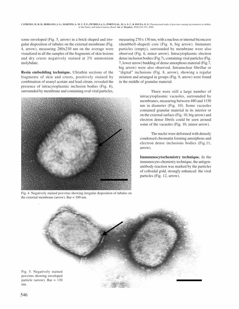
measuring 270 x 130 nm, with a nucleus or internal biconcave(dumbbell-shaped) core (Fig. 6, big arrow). Immatureparticles (empty), surrounded by membrane were alsoobserved (Fig. 6, minor arrow). Intracytoplasmic electrondense inclusion bodies (Fig.7), containing viral particles (Fig.7, lower arrow) budding of dense amorphous material (Fig.7,big arrow) were also observed. Intranuclear fibrillar or“digital” inclusions (Fig. 8, arrow), showing a regularstriation and arranged in groups (Fig. 9, arrow) were foundin the middle of granular material.
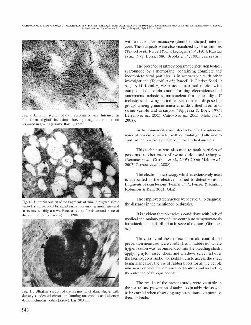
There were still a large number ofintracytoplasmic vacuoles, surrounded bymembranes, measuring between 480 and 1150nm in diameter (Fig. 10). Some vacuolescontained granular material in its interior oron the external surface (Fig. 10, big arrow) andelectron dense fibrils could be seen aroundsome of the vacuoles (Fig. 10, minor arrow).
The nuclei were deformed with denselycondensed chromatin forming amorphous andelectron dense inclusions bodies (Fig.11,arrow).
Immnunocytochemistry technique. In theimmunocyto-chemistry technique, the antigen-antibody reaction was marked by the particlesof colloidal gold, strongly enhanced the viralparticles (Fig. 12, arrow).
Fig. 4. Negatively stained poxvirus showing irregular disposition of tubules onthe external membrane (arrow). Bar = 100 nm.
Fig. 5. Negatively stainedpoxvirus showing envelopedparticle (arrow). Bar = 130nm.
CATROXO, M. H. B.; BERSANO, J. G.; MARTINS, A. M. C. P. F.; PETRELLA, S.; PORTUGAL, M. A. S. C. & SOUZA, O. S. Ultrastructural study of poxvirus causing myxomatosis in rabbits,in São Paulo, and Santa Catarina, Brazil. Int. J. Morphol., 27(2):543-552, 2009.

547
DISCUSSION
In this study, rabbits with poxvirus presented signs andsymptoms, such as conjunctivitis, subcutaneous and perinealedema, and facial malformation conferring the animal a leonineaspect, as reported in the literature (Ferreira & Ferreira; Corrêa& Corrêa, 1992; Fenner et al.; Barbosa et al.; Bruno et al.).
It was observed that the periorbital region as well astestis of the studied animals had a gelatinous texture.
The mucoid nature of the lesion surface caused bypoxvirus confers the name of myxomatosis to the disease(Fenner & Ross, 1994).
Increase in volume of testis observed by us in somereproductive animals was also found by other authors (Ferreira& Ferreira; Fountain et al.; Bruno et al.).
Some animals of our study showed neurological signsas nystagmus and motor incoordination. Studies indicated thatanimals may present paralysis as well (Fenner et al.).
No typical respiratory symptoms of the myxomatosisform described by Farsang et al. could be observed as well aspulmonary lesions verified by Bruno et al.
The two outbreaks occurred in the summer time whenthere is a bigger proliferation of transmitting insects as fleas,flies and mosquitoes, as analyzed by Corrêa & Corrêa, Fenneret al. and Bruno et al. Mosquitoes as Aedes aegypti, Anophelesand Culex may act as virus reservoirs (Ferreira & Ferreira).Airborne transmission had already been reported by Farsanget al.
A great number of typical poxvirus particles, measuringapproximately 300 x 240nm was detected by negative contrasttechnique for transmission electron microscopy (rapidpreparation) in all the analyzed samples.
These morphological features are in accordance withother myxomatosis studies (Padgett et al., 1964; Tektoff etal.; Strayer et al., 1983; Barbosa et al.; Luna).
The use of 2% ammonium molibdate allowed the idealelectronic contrast to viral identification by the negativestaining technique. This stain is also indicated by OIE.
By means of the resin embedding technique, weobserved oval poxvirus particles, measuring 270 x 130 nm,
Fig. 6. Ultrathin section of the fragments of skin. Intracytoplasmicinclusion bodies containing oval viral particles with a nucleus or internalbiconcave (dumbbell-shaped) core (big arrow). Immature particles(empty), surrounded by membrane (minor arrow). Bar = 200 nm.
CATROXO, M. H. B.; BERSANO, J. G.; MARTINS, A. M. C. P. F.; PETRELLA, S.; PORTUGAL, M. A. S. C. & SOUZA, O. S. Ultrastructural study of poxvirus causing myxomatosis in rabbits,in São Paulo, and Santa Catarina, Brazil. Int. J. Morphol., 27(2):543-552, 2009.
Fig. 7. Ultrathin section of the fragments of skin. Intracytoplasmicelectron dense inclusion bodies, containing viral particles (lower arrow)budding of dense amorphous material (big arrow). Bar = 320 nm.
Fig. 8. Ultrathin section of the fragments of skin. Intranuclear fibrillaror “digital” inclusions (arrow). Bar = 600 nm.
548
with a nucleus or biconcave (dumbbell-shaped) internalcore. These aspects were also visualized by other authors(Tektoff et al.; Purcell & Clarke; Ogier et al., 1974; Karstadet al., 1977; Bohn, 1980; Brooks et al., 1995; Saari et al.).
The presence of intracytoplasmatic inclusion bodies,surrounded by a membrane, containing complete andincomplete viral particles is in accordance with otherinvestigations (Tektoff et al.; Purcell & Clarke; Saari etal.). Additionally, we noted deformed nuclei withcompacted dense chromatin forming electrodense andamorphous inclusions, intranuclear fibrilar or “digital”inclusions, showing periodical striation and disposed ingroups among granular material as described in cases ofswine variole and avianpox (Teppema & Boer, 1975;Bersano et al., 2003; Catroxo et al., 2005; Melo et al.,2008).
In the immunocitochemistry technique, the intensivemark of poxvirus particles with colloidal gold allowed toconfirm the poxvirus presence in the studied animals.
This technique was also used to mark particles ofpoxvirus in other cases of swine variole and avianpox(Bersano et al.; Catroxo et al., 2005; 2006; Melo et al.,2007; Catroxo et al., 2008).
The electron microscopy which is extensively usedis advocated as the elective method to detect virus infragments of skin lesions (Fenner et al.; Fenner & Fantini;Robinson & Kerr, 2001; OIE).
The employed techniques were crucial to diagnosethe diseases in the mentioned outbreaks.
It is evident that precarious conditions with lack ofmedical and sanitary procedures contribute to myxomatosisintroduction and distribution in several regions (Ghram etal.).
Thus, to avoid the disease outbreak, control andprevention measures were established in rabbitries, wherehygienization was recommended into the breeding sheds,applying nylon insect doors and windows screen all overthe facility, construction of pedilavium to access the shed,being mandatory the use of rubber boots for all the peoplewho work or have free entrance to rabbitries and restrictingthe entrance of foreign people.
The results of the present study were valuable inthe control and prevention of outbreaks in rabbitries as wellto be careful when observing any suspicious symptom onthese animals.
Fig. 10. Ultrathin section of the fragments of skin. Intracytoplasmicvacuoles, surrounded by membranes contained granular materialin its interior (big arrow). Electron dense fibrils around some ofthe vacuoles (minor arrow). Bar 1200 nm.
Fig. 11. Ultrathin section of the fragments of skin. Nuclei withdensely condensed chromatin forming amorphous and electrondense inclusions bodies (arrow). Bar: 900 nm.
Fig. 9. Ultrathin section of the fragments of skin. Intranuclearfibrillar or “digital” inclusions showing a regular striation andarranged in groups (arrow). Bar: 170 nm.
CATROXO, M. H. B.; BERSANO, J. G.; MARTINS, A. M. C. P. F.; PETRELLA, S.; PORTUGAL, M. A. S. C. & SOUZA, O. S. Ultrastructural study of poxvirus causing myxomatosis in rabbits,in São Paulo, and Santa Catarina, Brazil. Int. J. Morphol., 27(2):543-552, 2009.

549
CATROXO, M. H. B.; BERSANO, J. G.; MARTINS, A. M. C.P. F.; PETRELLA, S.; PORTUGAL, M. A. S. C. & SOUZA, O.S. Estudio ultraestructural del poxvirus causal de mixomatosis enconejos, en São Paulo y Santa Catarina, Brasil. Int. J. Morphol.,27(2):543-552, 2009.
RESUMEN: La mixomatosis es una enfermedad conta-giosa de distribución mundial, causada por poxvirus que infecta co-nejos domésticos y salvajes. En este estudio presentamos dos distin-tos surtos por mixomatosis que ocurrieron en producciones de cone-jos, una para fines comerciales de producción de carne y pieles yotra para el comercio de conejos domésticos. Las señales observa-das fueron afecciones oculares, nasales, testiculares y, a veces, tam-bién distribuida por todo el cuerpo de los animales. Estas se caracte-rizaban por formación de nódulos que a la palpación tenían un as-pecto gelatinoso o gomoso. En la microscopía electrónica de trans-misión, por la técnica de contrastación negativa, se pudo observaren todas las muestras examinadas de piel y de costras, un gran nú-mero de partículas típicas de poxvirus, con envoltura y forma deladrillo, mostrando disposición irregular de los túbulos sobre la mem-brana externa, midiendo 300 x 240 nm en el promedio. Cortesultrafinos de fragmentos de piel y de costras revelaron la presenciade cuerpos de inclusión intracitoplasmáticas, envueltos por mem-brana y conteniendo partículas ovales, midiendo 270 x 130 nm, con-teniendo núcleo o centro interno bicóncavo (forma de mancuernas).Partículas inmaduras (vacías) envueltas por membrana fueron ob-servadas. También fueron analizados cuerpos de inclusiónintracitoplasmáticos, electrodensos, conteniendo partículas viralesbrotando del material denso y amorfo. Fueron observadas inclusio-nes intranucleares fibrilares o “digitales” mostrando una estriaciónperiódica y disposición en grupos en medio del material granular.Los núcleos estaban deformados con cromatina densamente con-densada formando cuerpos de inclusiones electrodensas y amorfas.En la técnica de imunocitoquímica la reacción antígeno-anticuerpofue intensamente marcada por las partículas de oro coloidal realzan-do fuertemente las partículas virales.
PALABRAS CLAVE: Mixomatosis; Poxvirus; Conejo;Microscopía electrónica de transmisión.
Fig. 12. Immunocytochemistry technique. Poxvirus marked by theparticles of colloidal gold (arrow). Bar = 120 nm.
REFERENCES
Arthur, C. P. & Louzis, C. A review of myxomatosis amongrabbits in France. Rev. Sci. Tech., 7:937-76, 1988.
Barbosa, G. A.; Rangel Filho, F. B.; Andrade, C. M.;Mazur, C.; Fontana, C. A. P.; Gripp, R. G.; Praxedes,C. S. & Ribeiro, U. A. Mixomatose: infecção naturale experimental de uma amostra autóctone da área deinfluência da UFRRJ em coelhos domésticos(Oryctolagus cuniculus). Rev. Bras. Med. Vet.,18(6):256-9, 1996.
Beer, J. Doenças Infecciosas em Animais Domésticos.1ª. Ed. São Paulo, Roca, 1999. V.1. p. 380.
Bersano, J. G.; Catroxo, M. H. B.; Villalobos, E. M. C.; Leme,M. C. M.; Martins, A. M. C. R. P. F.; Peixoto, Z. M. P.;Portugal, M. A. S. C.; Monteiro, R. M.; Ogata, R. A. &Curi, N. A. Varíola suína: Estudo sobre a ocorrência desurtos nos Estados de São Paulo e Tocantins, Brasil. Arq.Inst. Biol., 70(3):269-78, 2003.
Bohn, W. Electron microscopic immunoperoxidase studieson the accumulation of virus antigen in cells infected withShope fibroma virus. J. Gen. Virol., 46:439-47, 1980.
Brenner, S. & Horne, R. W. A negative staining method forhigh resolution electron microscopy of viruses. Biochim.Biophys. Acta, 34:103-10, 1959.
Brooks, M. A.; Ali, A. N.; Turner, P. C. & Moyer, R. W. Arabbitpox virus serpin gene controls host range byinhibiting apoptosis in restrictive cells. J. Virol.,69(12):7688-98, 1995.
Bruno, S. F.; Lopes-Júnior, S. V. S.; Demarque, K. C.; Viei-ra, T. B. & Tortelly, R. Achados clínicos eanatomopatológicos em um surto de mixomatose no Riode Janeiro (relato de caso). Arq. Ciênc. Vet. Zool.,7:(1):85-8, 2004.
Calvete, C.; Estrada, R.; Villafuerte, R.; Osácar, J. J. & Lu-cientes, J. Epidemiology of viral haemorrhagic diseaseand myxomatosis in a free-living population of wildrabbits. Vet. Rec., 150(25):776-82, 2002.
Cameron, C. M.; Barrett, J. W.; Liu, L.; Lucas, A. R. &McFadden, G. Myxoma virus M141R expresses a viralCD200 (vOX-2) that is responsible for down-regulationof macrophage and T-cell activation in vivo. J. Virol.,79(10):6052-67, 2005.
CATROXO, M. H. B.; BERSANO, J. G.; MARTINS, A. M. C. P. F.; PETRELLA, S.; PORTUGAL, M. A. S. C. & SOUZA, O. S. Ultrastructural study of poxvirus causing myxomatosis in rabbits,in São Paulo, and Santa Catarina, Brazil. Int. J. Morphol., 27(2):543-552, 2009.
550
Cameron, C.; Hota-Mitchell, S.; Chen, L.; Barrett, J.; Cao,J. X.; Macaulay, C.; Willer, D.; Evans, D. & McFadden,G. The complete DNA sequence of myxoma virus. Virol.,264(2):298-318, 1999.
Catroxo, M. H. B.; Pongiluppi, T.; Milanelo, L.; Godoy, S.N. & Petrella, S. Identification of poxvirus undertransmission electron microscopy during outbreak in wildbirds. XXIII Congresso Brasileiro de Microbiologia,Santos, SP, 2005.
Catroxo, M. H. B.; Pongiluppi, T.; Milanelo, L.; Rebouças,M. M.; Jesus, T. H.; Cardoso, G. H. M. & Petrella, S.Detection of poxvirus by transmission electronmicroscopy using negative staining (rapid preparation)and immunolabelling with colloidal gold particlestechniques during outbreak in bay-winged cowbird(Gnorimopsar chopi). Virus Reviews & Research,11(suppl. 01):101, 2006.
Catroxo, M. H. B.; Melo, N. A.; Milanelo, L.; Rebouças, M.M.; Martins, A. M. C. R. P. F.& Petrella, S. Detecção depoxvírus aviário em lesões cutâneas de bico-de-pimenta(Saltator atricollis). XXXII Congresso Anual daSociedade de Zoológicos do Brasil, Sorocaba, SP, 2008.
Corrêa, W. M. & Corrêa, C. N. M. Enfermedades infeccio-sas dos mamíferos domésticos. 2ª. Ed. Rio de Janeiro,Médica e Científica, 1992.
Diven, D. G. An overview of poxviruses. J. Am. Acad.Dermatol., 44:1-16, 2001.
Doane, F. W. & Anderson, M. Electron microscopy indiagnostic virology. In: A pratical guide and atlas.Cambridge, Cambridge University Press, 1987.
Farsang, A.; Makranszki, L.; Dobos-Kovács, M.; Virág, G.;Fabián K.; Barna, T.; Kulcsár, G.; Kucsera, L. & Vetési,F. Occurrenceof atypical myxomatosis in Central Europe:clinical and virological examinations. Acta Vet. Hung.,51(4):493-501, 2003.
Fenner, F. Poxviruses. In: Fields, B. N.; Knife, D. M. &Howley, P. M. (Eds.). Fields' Virology. Philadelphia,Lippincott-Raven, 1996. V.2. pp. 2673-702.
Fenner, F. Adventures with poxviruses of vertebrates. FEMSMicrobiol. Rev., 24(2):123-33, 2000.
Fenner, F., Bachmann, P. A.; Gibbs, E. P. J.; Murphy, F. A.;Studdert, M. J. & White, D. O. Virología Veterinaria.Zaragoza, Acribia, 1992.
Fenner, F. & Fantini, B. Myxomatosis. In: The Europeanrabbit. The history and biology of a succesful colonizer.Oxford, Ed. University Press, 1994. pp. 205-35.
Fenner, F. & Hatclife, F. N. Myxomatosis. Cambridge. Ed.Cambridge University Press, 1965.
Fenner F. & Ross J. Myxomatosis. In: The European Rabbit.The history and biology of a successful colonizer.Thompson, H. V. & King, C. M. (Eds). Oxford, U. K.,Oxford University Press, 1994. pp.205-40.
Ferreira, A. J. & Ferreira, C. Doenças infecto-contagiosasdos animais domésticos. 4ª. Ed. Lisboa, FundaçãoCalouste Gulbenkian, 1990.
Fountain, S.; Holland, M. K.; Hinds, L. A.; Janssens, P. A.& Kerr, P. J. Interstitial orchitis with impairedsteroidogenesis and spermatogenesis in the testes ofrabbits infected with an attenuated strain of myxomavirus. J. Reprod. Fertil., 110:161-9, 1997.
Ganière, J-P.; Gourreau, J-M.; Montabord, D.; Rive, M. &Chantal, J. Myxomatosis of the depilated Angora rabbit.A preliminary study. Vet. Dermatol., 2(1):11-5, 2008.
Gelfi, J.; Chantal, J.; Phong, T. T.; Py, R. & Boucraut-Baralon, C. Development of an ELISA for detectionof myxoma virus specific rabbit antibodies: testevaluation for diagnostic application on vaccinated andwild rabbit sera. J. Vet. Diagn. Invest., 11(3):240-45,1999.
González-Santander, R. Técnicas de microscopia eletrónicaen biología. Madrid, Ed. Aguilar, 1969. p. 666.
Ghram, A.; Benzarti, M.; Amira, A. & Amara, A.Myxomatosis in Tunisia: seroepidemiological study inthe Monastir region (Tunisia). Arch. Inst. Pasteur Tunis,73(3-4):167-72, 1996.
Hayat, M. A. & Miller, S. E. Negative Staining. New York,McGraw Hill Publ. Company, 1990. p.235.
Johnston, J. B.; Wang, G.; Barrett, J. W.; Nazarian, S. H.;Colwill, K.; Moran, M. & McFadden, G. Myxoma vi-rus M-75 protects infected cells from the stress of cellcycle arrest through its interaction with host cell cullin-1. J. Virol., 79(16):10750-63, 2005.
Karstad, L.; Thorsen, J.; Davies, G. & Kaminjolo, J. S.Poxvirus fibromas on African hares. J. Wildl. Dis.,13:245-7, 1977.
CATROXO, M. H. B.; BERSANO, J. G.; MARTINS, A. M. C. P. F.; PETRELLA, S.; PORTUGAL, M. A. S. C. & SOUZA, O. S. Ultrastructural study of poxvirus causing myxomatosis in rabbits,in São Paulo, and Santa Catarina, Brazil. Int. J. Morphol., 27(2):543-552, 2009.

551
Knutton, S. Electron microscopical methods in adhesion.Methods Enzymol., 253:145-58, 1995.
Luft, J. H. Improvements in an epoxy resin embeddingmethods. J. Biophys. Biochem. Cytol., 9:409-14, 1961.
Luna, R. M. L. First report of myxomatosis in Mexico. J.Wildl. Dis., 36(3):580-3, 2000.
Madeley, C. R. Electron microscopy and virus diagnosis.J. Clin. Pathol., 50:454-56, 1997.
Marcato, P. S. & Simoni, P. Ultrastructural researches onrabbit myxomatosis. Lymphnodal lesions. Vet. Pathol.,14(4):361-7, 1977.
Marlier, D.; Mainil, J.; Linde, A. & Vindevogel, H.Infectious agents associated with rabbit pneumonia:isolation of amyxomatous myxoma virus strains. Vet.J., 159(2):171-8, 2000.
Melo, N. A.; Lopes; D.; Milanelo, L.; Rebouças; M. M.;Jesus, T. H.; Petrella, S. & Catroxo, M. H. B. Presençade poxvírus em lesões cutâneas de pata do cardeal(Paroaria coronata). Biológico, 67(1):29, 2007.
Melo, N. A.; Milanelo, L.; Oliveira, A. C.; Rebouças, M.M.; Martins, A. M. C. R. P. F.; Petrela, S. & Catroxo,M. H. B. Isolation of poxvírus in cutaneous lesions ofthe cowled-cardinal (Paroaria dominicana) bytransmission electron microscopy techniques. In: XIVCongresso da Sociedade Brasileira de Biologia Celu-lar, 2008, São Paulo, SP.
Merchant, J. C.; Kerr, P. J.; Simms, N. G.; Hood, G. M.;Pech, R. P. & Robinson, A. J. Monitoring the spread ofmyxoma virus in rabbit Oryctolagus cuniculuspopulations on the southern tablelands of New SouthWales, Australia. III. Release, persistence and rate ofspread of an identifiable strain of myxoma virus.Epidemiol. Infect. 130(1):135-47, 2003.
OIE. Manual of Diagnostic Tests & Vaccines for TerrestrialAnimal. In: Lagomorpha. 6ª. Ed. France, Paris, 2008.pp. 937-46.
Ogier, G.; Chardonnet, Y. & Gazzolo, L. Role of lysosomesduring infection with Shope fibroma virus of primaryrabbit kidney tissue culture cells. J. Gen. Virol., 22:249-53, 1974.
Padgett, B. L; Wright, M. J.; Jayne, A. & Walker, D. L.Electron microscopic structure of mixoma virus and
some reactivable derivatives. J. Bacteriol., 87(2):454-60, 1964.
Purcell, D. A. & Clarke, J. K. Some aspects of themorphogenesis of mixoma virus in vivo. Arch. Virol.,39(4):369-75, 1972.
Reinolds, E. S. The use of lead citrate at high pH as anelectron-opaque stain in electron microscopy. J. Cell.Biol., 17:208-12, 1963.
Robinson, A. J. & Kerr, P. J. Poxvirus Infections. In:Williams, E. S. & Barker, I. K. (eds.). Infectiousdiseases of wild mammals. London, U.K., MansonPublishing Ltd., 2001. pp.179-201.
Ross, J.; Tittensor, A. M.; Fox, A. P. & Sanders, M. F.Myxomatosis in farmland rabbit populations inEngland and Wales. Epidemiol.Infect., 103(2):333-57,1989.
Saari, S. A. M.; Rudbäck, E.; Niskanen, M.; Syrjälä, P.;Nylund, M. & Anttila, M. Contagious mucocutaneousdermatitis of the mountain hare (Lepus timidus):pathology and cause. J. Wildl. Dis., 41(4):775-82, 2005.
Shepherd, R. C. H.; Edmonds, J. W.; Nolan, I. F. & Gocs,A. Myxomatosis in the Mallee region of Victoria, Aus-tralia. J. Hyg., 81(2):239-43, 1978.
Strayer, D. S.; Cabirac, G.; Sell, S. & Leibowitz, J. L.Malignant rabbit fibroma virus: observations on theculture and histopathologic characteristics of a new vi-rus-induced rabbit tumor. J. Natl. Cancer. Inst.,71(1):91-104, 1983.
Tektoff, J.; Gazzolo, L. & Leftheriotis, E. Morphogènèsedu virus de la myxomatose du lapin. Pathol. Biol.,19(23-24):1045-54, 1971.
Teppema, J. S. & de Boer, G. F. Ultrastructural aspects ofexperimental swinepox with special reference toinclusion bodies. Arch. Virol., 49(2-3):151-63, 1975.
Upton, C.; Macen, J. L.; Wishart, D. S. & McFadden, G.Myxoma virus and malignant rabbit fibroma virusencode a serpin-like protein important for virusvirulence. Virology, 179(2):618-31, 1990.
Yuill, T. M. Mixomatosis y Fibromatosis. In: Davis. J. W.;Karstad. L. & Trainer, D. Õ. Enfermedades Infeccio-sas de los Mamíferos Salvajes. Zaragoza, Ed. Acribia,1972.
CATROXO, M. H. B.; BERSANO, J. G.; MARTINS, A. M. C. P. F.; PETRELLA, S.; PORTUGAL, M. A. S. C. & SOUZA, O. S. Ultrastructural study of poxvirus causing myxomatosis in rabbits,in São Paulo, and Santa Catarina, Brazil. Int. J. Morphol., 27(2):543-552, 2009.
552
Watson, M. L. Staining of tissue section for electronmicroscopy with heavy metals. J. Biophys. & Biochem.Cytol., 4:475-8, 1958.
Zachertowska, A.; Brewer, D. & Evans, D. H.Characterization of the major capsid proteins of myxomavirus particles using MALDI-TOF mass spectrometry.J. Virol. Methods., 132(1-2):1-12, 2006.
Correspondence to:Prof. Dr. Marcia CatroxoElectron Microscopy LaboratoryResearch and Development Center in Animal HealthBiological Institute of São PauloAv. Conselheiro Rodrigues Alves, 1252CEP 04014-002Vila Mariana, São Paulo, SPBRAZIL
E-mail: [email protected] Received: 10-02-2009Accepted: 14-03-2009
CATROXO, M. H. B.; BERSANO, J. G.; MARTINS, A. M. C. P. F.; PETRELLA, S.; PORTUGAL, M. A. S. C. & SOUZA, O. S. Ultrastructural study of poxvirus causing myxomatosis in rabbits,in São Paulo, and Santa Catarina, Brazil. Int. J. Morphol., 27(2):543-552, 2009.