Ultrastructural Alterations and Quantitative Changes in ...4)15/6.pdf · Accumulation of collagen...
11
World Journal of Zoology 10 (4): 274-284, 2015 ISSN 1817-3098 © IDOSI Publications, 2015 DOI: 10.5829/idosi.wjz.2015.10.4.95312 Corresponding Author: Hussein M Ageely, Department of Internal Medicine, Faculty of Medicine, Jazan University, Jazan, Kingdom of Saudi Arabia. Tel: +(966) 505769344. 274 Ultrastructural Alterations and Quantitative Changes in the Mineral Content of Colonic Mucosa in a Diabetic Rat Model Hussein M. Ageely Department of Internal Medicine, Faculty of Medicine, Jazan University, Jazan, Kingdom of Saudi Arabia Abstract: Background: Scarce information is available regarding the effects of diabetes on the colonic mucosa. Objective: The present study was undertaken to investigate the effect of diabetes on the ultrastructure and trace elements of the rat colonic mucosa. Methods: Twenty adult male Wister albino rats were randomly divided into two groups; control and diabetic, 10 animals each. The diabetic group received intravenous injection of a single dose of streptozotocin (STZ) (30 mg/ml). Rats were sacrificed at the end of 8 weeks with an overdose of ether anesthesia. The proximal colon was immediately removed and processed for scanning electron microscope (SEM) and energy dispersive X-ray analysis (EDAX) using Quanta 250 FEG-EDAX for element detection. Results: revealed that colonic crypts of the diabetic animals showed enlarged, irregular crypt openings, with excessive exfoliation at the periphery. Numerous lymphocytes and activated monocytes were observed on the crypt surfaces and in the lamina propria. Accumulation of collagen fibres in the sub-epithelial layer and degranulation of apical mucin secreting goblet cells could be seen. Enterocytes showed significant decrease in P and Ca and significant increase in Na, K and Si (P < 0.05). The Goblet cells showed decrease in P, Ca and Cl while there was significant increase in Na and K (P < 0.05). Conclusions: It could be concluded that using the scanning electron microscope (SEM) with energy dispersive X-ray analysis (EDAX) opens new perspective to study the pathophysiological effect of diabetes on colonic mucosa. Key words: Diabetes Mucosa Mineral content and rat model INTRODUCTION changes of the cells of the colonic mucosa. The present Up to 60% of the patients with diabetes mellitus on the ultrastructure and trace elements of the rat colonic develop gastrointestinal (GI) symptoms, such as mucosa. constipation and diarrhea [1, 2]. Diarrhea was noted also in rats with streptozotocin-induced chronic diabetes [3]. MATERIALS AND METHODS The occurrence of colonic disturbance in diabetes was accompanied with thickened subepithelial collagen layer Induction of Diabetes: A total of 20 male Wistar rats, of [4], impaired mucosal absorption of fluid and electrolytes 200-250g body weight, were randomly divided into control [3] and autonomic neuropathy of enterocytes [5]. and diabetic groups, 10 animals each. The control group The present study was undertaken to investigate the was injected with the vehicle (phosphate-buffered saline, effect of diabetes on rat colonic mucosa using scanning PBS). The diabetic group was made diabetic by electron microscope (SEM) with energy dispersive X-ray intravenous injection of a single dose of streptozotocin analysis (EDAX). The latter is a powerful analytical tool (STZ). The injection was freshly prepared immediately that allows elemental identification and quantitative before use by dissolving STZ (30 mg/ml) in (PBS) and compositional information and characterization of injected into the penile vein of the recipient rats, under biological specimens [6]. This will allow synchronous light ether anesthesia, at a dose of 40 mg/kg body weight. examination of the ultrastructure and trace elements Rats were maintained with free access to standard diet study was undertaken to investigate the effect of diabetes
Transcript of Ultrastructural Alterations and Quantitative Changes in ...4)15/6.pdf · Accumulation of collagen...
World Journal of Zoology 10 (4): 274-284, 2015ISSN 1817-3098© IDOSI Publications, 2015DOI: 10.5829/idosi.wjz.2015.10.4.95312
Corresponding Author: Hussein M Ageely, Department of Internal Medicine, Faculty of Medicine, Jazan University, Jazan, Kingdom of Saudi Arabia. Tel: +(966) 505769344.
274
Ultrastructural Alterations and Quantitative Changes in theMineral Content of Colonic Mucosa in a Diabetic Rat Model
Hussein M. Ageely
Department of Internal Medicine, Faculty of Medicine, Jazan University, Jazan, Kingdom of Saudi Arabia
Abstract: Background: Scarce information is available regarding the effects of diabetes on the colonic mucosa.Objective: The present study was undertaken to investigate the effect of diabetes on the ultrastructure andtrace elements of the rat colonic mucosa. Methods: Twenty adult male Wister albino rats were randomly dividedinto two groups; control and diabetic, 10 animals each. The diabetic group received intravenous injection ofa single dose of streptozotocin (STZ) (30 mg/ml). Rats were sacrificed at the end of 8 weeks with an overdoseof ether anesthesia. The proximal colon was immediately removed and processed for scanning electronmicroscope (SEM) and energy dispersive X-ray analysis (EDAX) using Quanta 250 FEG-EDAX for elementdetection. Results: revealed that colonic crypts of the diabetic animals showed enlarged, irregular cryptopenings, with excessive exfoliation at the periphery. Numerous lymphocytes and activated monocytes wereobserved on the crypt surfaces and in the lamina propria. Accumulation of collagen fibres in the sub-epitheliallayer and degranulation of apical mucin secreting goblet cells could be seen. Enterocytes showed significantdecrease in P and Ca and significant increase in Na, K and Si (P < 0.05). The Goblet cells showed decrease inP, Ca and Cl while there was significant increase in Na and K (P < 0.05). Conclusions: It could be concluded thatusing the scanning electron microscope (SEM) with energy dispersive X-ray analysis (EDAX) opens newperspective to study the pathophysiological effect of diabetes on colonic mucosa.
Key words: Diabetes Mucosa Mineral content and rat model
INTRODUCTION changes of the cells of the colonic mucosa. The present
Up to 60% of the patients with diabetes mellitus on the ultrastructure and trace elements of the rat colonicdevelop gastrointestinal (GI) symptoms, such as mucosa.constipation and diarrhea [1, 2]. Diarrhea was noted alsoin rats with streptozotocin-induced chronic diabetes [3]. MATERIALS AND METHODSThe occurrence of colonic disturbance in diabetes wasaccompanied with thickened subepithelial collagen layer Induction of Diabetes: A total of 20 male Wistar rats, of[4], impaired mucosal absorption of fluid and electrolytes 200-250g body weight, were randomly divided into control[3] and autonomic neuropathy of enterocytes [5]. and diabetic groups, 10 animals each. The control group
The present study was undertaken to investigate the was injected with the vehicle (phosphate-buffered saline,effect of diabetes on rat colonic mucosa using scanning PBS). The diabetic group was made diabetic byelectron microscope (SEM) with energy dispersive X-ray intravenous injection of a single dose of streptozotocinanalysis (EDAX). The latter is a powerful analytical tool (STZ). The injection was freshly prepared immediatelythat allows elemental identification and quantitative before use by dissolving STZ (30 mg/ml) in (PBS) andcompositional information and characterization of injected into the penile vein of the recipient rats, underbiological specimens [6]. This will allow synchronous light ether anesthesia, at a dose of 40 mg/kg body weight.examination of the ultrastructure and trace elements Rats were maintained with free access to standard diet
study was undertaken to investigate the effect of diabetes

World J. Zool., 10 (4): 274-284, 2015
275
and water ad libitum, without any attempt to control Statistical Analysis: Results were presented as means ±diabetes. Animals were housed individually in specialclear sided cages at controlled temperature (22°C) with a12:12-h light: dark cycle for 2-wk adaptation period withgood ventilation. Animals weren’t exposed to painfultreatment. The plan of the study conducted in accordancewith the principles, guide lines and procedures of CARE(Committee of Animal Research Ethics) of faculty ofmedicine Jazan University. The concept of 3R (Reduction,Replacement and Refinement) was addressed. Thediabetic status of the rats was assessed by weeklymeasurement of blood glucose level using (Accu-ChekActive Blood Glucose Meter Germany). Animals wereconsidered diabetic if they show repeated non-fastingblood glucose level above 16.7 mmol/l (300 mg/dl).
Specimen Preparation: Rats were sacrificed from eachgroup, at the end of 8 weeks after the injection of STZ orPBS. The rats were sacrificed by an overdose of etheranesthesia. The whole-mounts from the proximal colonwere immediately removed and processed for SEMexamination. Colonic rat mucosa washed twice in sterile(PBS) and then the tissues were fixed for 60 minutes in 1%glutraldehyde, 2% paraformaldehyde in phosphate -bufferat room temperature (pH 7.4). The mucosa was thenwashed twice in buffered sucrose (0.1 M phosphatebuffer, 5% sucrose solution) for 5 min. each. Post fixationwas performed at 4°C for 60 min in phosphate-buffered 2%osmium. The mucosa was dehydrated in a graded seriesof ethanol’s (40, 50, 70, 80, 90 and twice in 100%) afterrinsing in several changes in cold distilled water. Thetissues were then dehydrated in ethanol - acetone (1:1)absolute solution for 30 minutes, then in absolute acetone100% for additional 30 minutes 3 times 10 minutes each.The tissues were critically point dried in CO drying2
apparatus CPD030 and mounted on stubs, then coatedwith gold sputter coater SCD005.
SEM Examination with EDAX: For the ultrastructureobservation and determination of the mineral composition,Quanta 250 FEG-EDAX was used. It was equipped withlarge field gaseous SE detector (LFD), gaseous SEdetector, (GSED) and gaseous BS detector (GBSD) forchemical analysis that detects all the elements excepthydrogen. The observations were carried out in variablevacuum pressure modalities (0.2-6 Torr). Detection of eachelement and the respective average weight (WT) % wastaken as its actual WT%. In every specimen, elementssuch as C, O, Na, Si, K, Ca, S, P, Au etc were detected.
SEM. The differences between the two groups weretested by the Student's t test. The differences wereconsidered significant at P < 0.05.
RESULTS
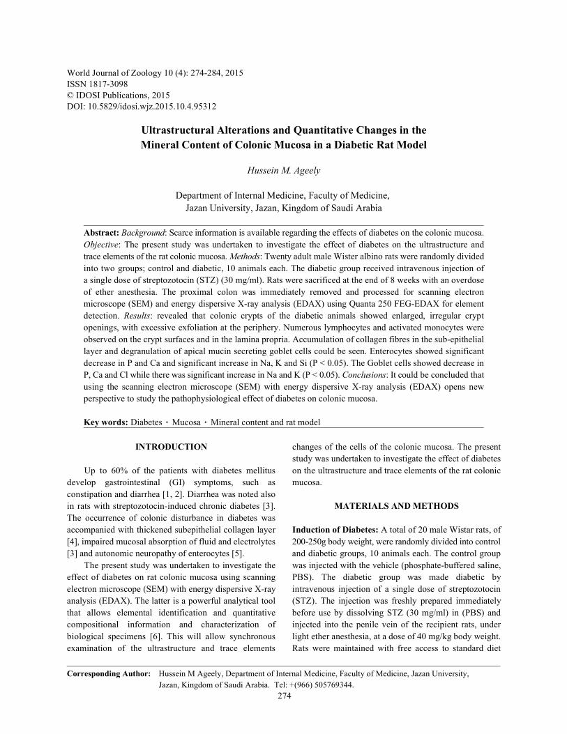
SEM of Normal Colonic Rat Mucosa: The mucosalsurface of the normal proximal colon is composed oforderly and closely packed rounded or polygonal cryptalunits with centrally located oval or rounded openings.Some of these cryptal mouths were seen thrusting outmucus. Each cryptal unit is being delineated by a furrowor cleft (intercryptal cleft) (Fig. 1). Each unit consists ofconcentrically arranged colonic absorptive epithelial cellsand mucous–secreting goblet cells (Fig. 2). Goblet cellswere scattered throughout the surface colonic epithelium.Many of these goblet cells were seen embracing themouth of colonic crypts. Moreover, their luminal surfacesare characterized by short, sparse microvilli and apicalmucigen granules (Fig. 2). In most instant a thick layer ofmucus coat was seen covering the cryptal colonic surface(Fig. 3). The colonic surface epithelial cells werecolumnar with a thick carpet of uniform surface microvilli).The colonic crypt showed minimal collagen fibers andminimal supepithelial collagen deposit (Fig. 4).
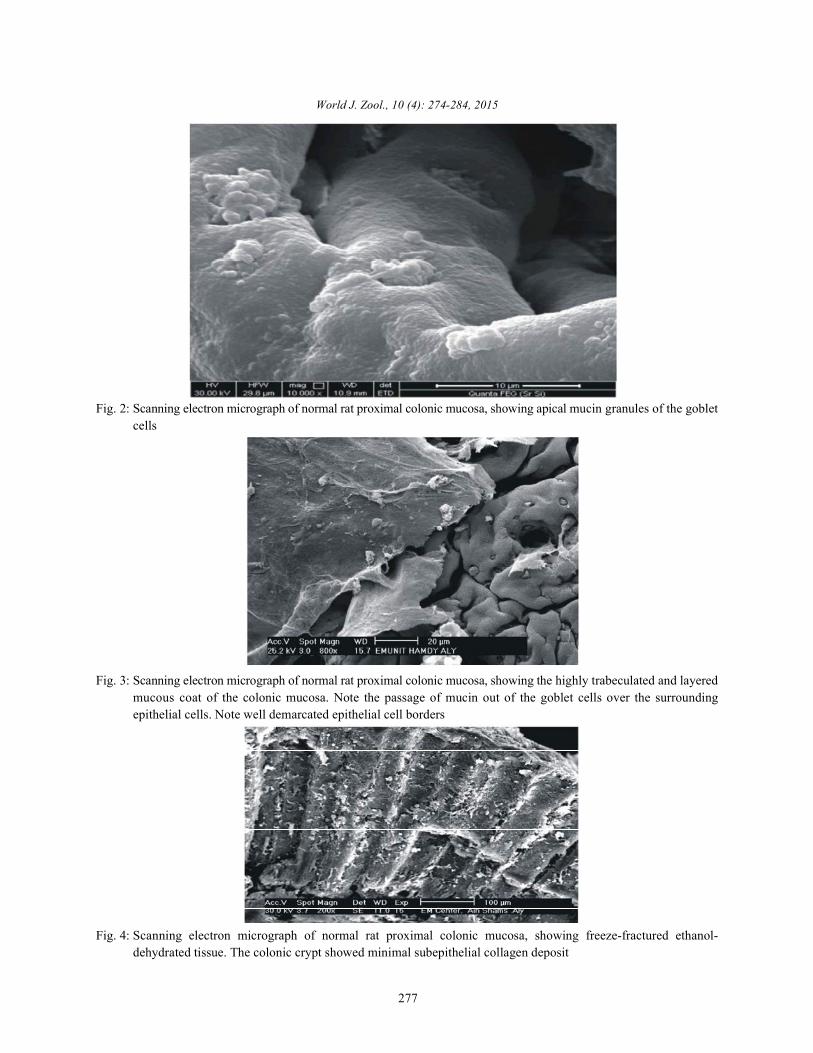
SEM of Diabetic Colonic Rat Mucosa: The shape of cryptopenings in diabetic mucosal surface were distorted,enlarged and they were irregularly spaced (Fig. 5,6).Some cryptal mouths were seen extruding mucin (Fig. 6).Excessive exfoliation was observed at the periphery ofcolonic crypts (Fig. 7). Marked inflammatory changeswere also observed on the cryptal colonic surfaces and inthe lamina propria. Most of cellular infiltrates werenumerous lymphocytes activated monocytes andactivated leucocytes (Fig. 8). Prominent excessivecollagen fibres accumulation could be seen in thesub-epithelial layer of colonic mucosa (Fig. 9). Moderatecollagen fibres accumulation was seen around the coloniccrypts and in-between the crypts (Fig. 10). Widespreaddegranulation of apical mucin-secreting goblet cells(goblet-cell depletion) was observed (Fig. 11).
Mineral Analysis of Enterocytes: The enterocytes ofthe diabetic mucosal surface showed significantdecrease in P and Ca while there was significant increasein Na, K and Si (P < 0.05). The changes in the otherminerals; C, N, O, S & Au (gold coating) were notsignificant (Table 1, Figs. 12 vs. 13).

World J. Zool., 10 (4): 274-284, 2015
276
Table 1: Control vs. diabetic (multi-point analyses) of mineral weight % ratio rat enterocytes of colonic mucosa. Values are presented as mean ± SEM
Element Control Diabetic p
Carbon (C) 47.39833±3.89 57.13833±3.13793 p = 0.06426
Nitrogen (N) 9.15± 0.92824 7.99273±1.76593 p = 0.28417
Oxygen (O) 13.70667±3.02037 13.196±2.49112 p = 0.44848
Sodium (Na) * 1.35333±0.1646 2.099±0.14874 p = 0.00182
Phosphorus (P) * 7.085±1.23525 3.28333±1.06857 p = 0.01711
Sulphur (S) 4.21286±2.63834 7.58571±1.53825 p = 0.14554
Potassium (K) * 1.38667±0.40676 3.771±0.71153 p = 0.00587
Calcium (Ca) * 1.076±0.21753 0.605± 0.13368 p = 0.0408
Gold (Au) 46.75± 1.25 40.5±5.5 p = 0.31025
Silica (Si) * 0.286± 0.03945 0.8588± 0.14806 p = 0.00572
* Significant differences at P < 0.05
Table 2: Control vs. diabetic (multi-point analyses) of mineral weight % ratio rat goblet cells of colonic mucosa. Values are presented as mean ± SEM
Element Control Diabetic p
Carbon (C) 27.825±6.69925 41.33±5.15665 p = 0.07062
Nitrogen (N) 7.69±0.74951 8.39333±0.66953 p = 0.24999
Oxygen (O) 7.45333± 1.75326 7.63333±1.58205 p = 0.47037
Sodium (Na) * 1.14714±.27491 2.03714±0.21482 p = 0.01271
Phosphorus (P) * 4.80667±1.87396 4.40667±1.41931 p = 0.03115
Sulphur (S) 4.675± 0.125 5.1625±0.43367 p = 0.32157
Potassium (K) * 1.172±0.27102 2.492±0.25291 p = 0.0037
Calcium (Ca) * 0.72643 ±0.05575 0.54±0.16074 p = 0.00586
Gold (AU) 58.5 ±5.37742 47.25±3.17214 p = 0.12163
Chlorine (Cl) * 0.4875±0.08892 0.1025±0.03425 p = 0.0034
* Significant differences at P < 0.05
Fig. 1: Scanning electron micrograph of normal rat proximal colonic mucosa, showing some cryptal mouths thrusting outmucous
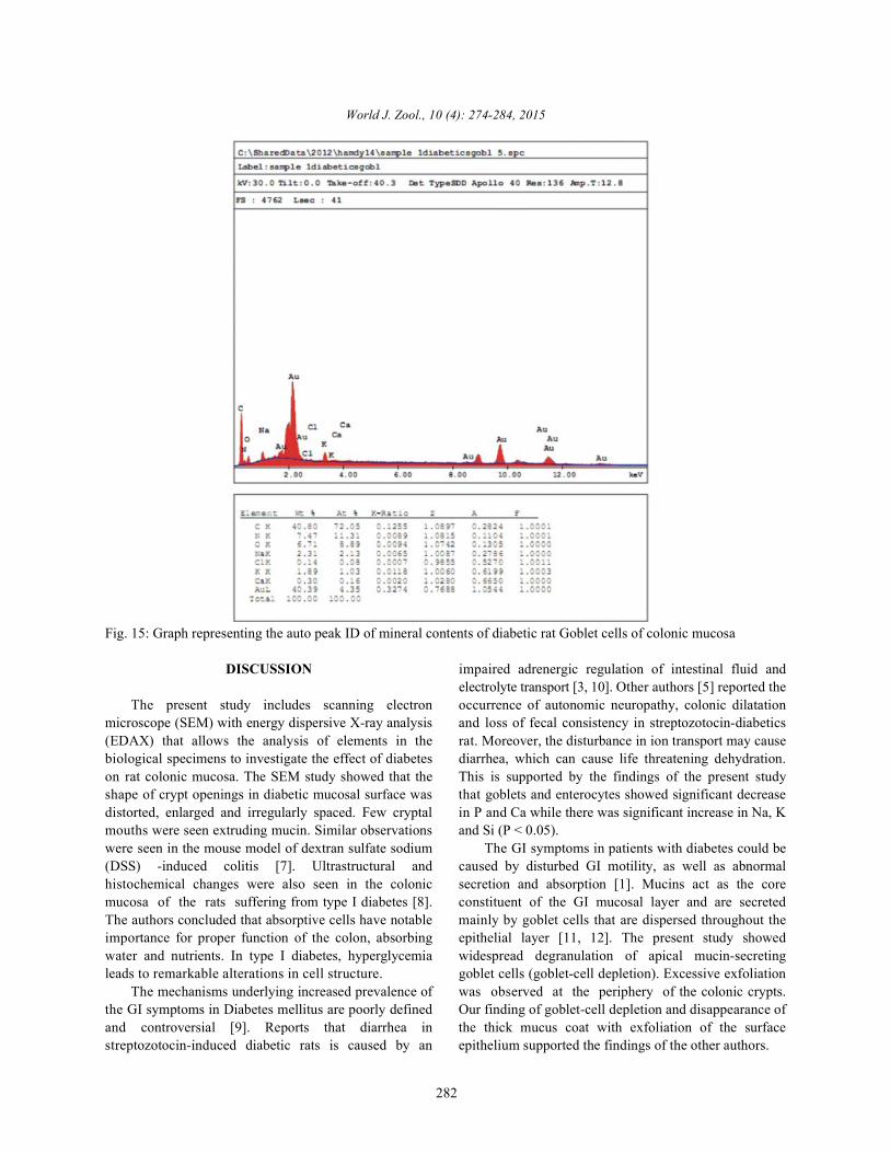
Mineral Analysis of Goblet Cells: The Goblet cells of increase in Na and K (P < 0.05). The changes in the otherthe diabetic mucosal surface showed significant minerals; C, N, O, S & Au (gold coating) were notdecrease in P, Ca and Cl while there was significant significant (Table 2, Figs. 14 vs. 15).
World J. Zool., 10 (4): 274-284, 2015
277
Fig. 2: Scanning electron micrograph of normal rat proximal colonic mucosa, showing apical mucin granules of the gobletcells
Fig. 3: Scanning electron micrograph of normal rat proximal colonic mucosa, showing the highly trabeculated and layeredmucous coat of the colonic mucosa. Note the passage of mucin out of the goblet cells over the surroundingepithelial cells. Note well demarcated epithelial cell borders
Fig. 4: Scanning electron micrograph of normal rat proximal colonic mucosa, showing freeze-fractured ethanol-dehydrated tissue. The colonic crypt showed minimal subepithelial collagen deposit
World J. Zool., 10 (4): 274-284, 2015
278
Fig. 5: Scanning electron micrograph of the diabetic rat proximal colonic mucosa, showing that the shape of cryptopening of the diabetic mucosal surface are distorted, collapsed and irregularly spaced
Fig. 6: Scanning electron micrograph of the diabetic rat proximal colonic mucosa, showing obliteration of crypts openingtogether with loss of inter-cryptal clefts. Note remnants of mucosal coat
Fig. 7: Scanning electron micrograph of the diabetic rat proximal colonic mucosa, showing diffuse surface edema of thecryptal cell lining and numerous exfoliations

World J. Zool., 10 (4): 274-284, 2015
279
Fig. 8: Scanning electron micrograph of the diabetic rat proximal colonic mucosa, showing marked inflammatory infiltratesin the lamina propria
Fig. 9: Scanning electron micrograph of rat diabetics’ colonic proximal mucosa, showing prominent excessive collagenfibres accumulation in the sub-epithelial layer of colonic mucosa
Fig. 10: Scanning electron micrograph of rat diabetics’ colonic proximal mucosa, showing excessive collagen fibresaccumulation around the colonic crypts and in-between the crypts

World J. Zool., 10 (4): 274-284, 2015
280
Fig. 11: Scanning electron micrograph of the diabetic rat proximal colonic mucosa, showing widespread de granulationof the apical mucin secreting goblet cells (goblet cell depletion)
Fig. 12: Graph representing the auto peak ID of mineral contents of control rat enterocytes of colonic mucosa

World J. Zool., 10 (4): 274-284, 2015
281
Fig. 13: Graph representing the auto peak ID of mineral contents of diabetic rat enterocytes of colonic mucosa
Fig. 14: Graph representing the auto peak ID of mineral contents of control rat Goblet cells of colonic mucosa
World J. Zool., 10 (4): 274-284, 2015
282
Fig. 15: Graph representing the auto peak ID of mineral contents of diabetic rat Goblet cells of colonic mucosa
DISCUSSION impaired adrenergic regulation of intestinal fluid and
The present study includes scanning electron occurrence of autonomic neuropathy, colonic dilatationmicroscope (SEM) with energy dispersive X-ray analysis and loss of fecal consistency in streptozotocin-diabetics(EDAX) that allows the analysis of elements in the rat. Moreover, the disturbance in ion transport may causebiological specimens to investigate the effect of diabetes diarrhea, which can cause life threatening dehydration.on rat colonic mucosa. The SEM study showed that the This is supported by the findings of the present studyshape of crypt openings in diabetic mucosal surface was that goblets and enterocytes showed significant decreasedistorted, enlarged and irregularly spaced. Few cryptal in P and Ca while there was significant increase in Na, Kmouths were seen extruding mucin. Similar observations and Si (P < 0.05).were seen in the mouse model of dextran sulfate sodium The GI symptoms in patients with diabetes could be(DSS) -induced colitis [7]. Ultrastructural and caused by disturbed GI motility, as well as abnormalhistochemical changes were also seen in the colonic secretion and absorption [1]. Mucins act as the coremucosa of the rats suffering from type I diabetes [8]. constituent of the GI mucosal layer and are secretedThe authors concluded that absorptive cells have notable mainly by goblet cells that are dispersed throughout theimportance for proper function of the colon, absorbing epithelial layer [11, 12]. The present study showedwater and nutrients. In type I diabetes, hyperglycemia widespread degranulation of apical mucin-secretingleads to remarkable alterations in cell structure. goblet cells (goblet-cell depletion). Excessive exfoliation
The mechanisms underlying increased prevalence of was observed at the periphery of the colonic crypts.the GI symptoms in Diabetes mellitus are poorly defined Our finding of goblet-cell depletion and disappearance ofand controversial [9]. Reports that diarrhea in the thick mucus coat with exfoliation of the surfacestreptozotocin-induced diabetic rats is caused by an epithelium supported the findings of the other authors.
electrolyte transport [3, 10]. Other authors [5] reported the

World J. Zool., 10 (4): 274-284, 2015
283
Marked inflammatory changes were also observed on patients. Restoration of colonic mineral contents and thethe cryptal colonic surfaces and in the lamina propria. colon osmolality may ameliorate the effect on diabeticMost of the cellular infiltrates were numerous colitis.lymphocytes and activated monocytes. Prominentexcessive accumulation of collagen fibres could be seen ACKNOWLEDGMENTin the sub-epithelial layer of colonic mucosa. Uncontrolledstreptozotocin-induced diabetes leads to accumulation of This research was supported by grants numbercollagen [13]. This is supported by the finding of other 3034 from the Medical Research Center, Jazan University.authors [4,14,15] who reported a marked thickening of The author would like to thank Prof. Hamdy Mohammadythe sub-epithelial collagen layer (SCL) in diabetic patients. Aly, Department of Anatomy, Faculty of Medicine,In agreement of previous studies [4,13-15], the present Ain-Shams University, Cairo, Egypt for the electronwork confirmed a prominent excessive collagen fibres microscopic work and Prof. Mostafa M. El-Naggar,accumulation which was seen in the sub-epithelial layer of Department of Anatomy, Faculty of Medicine, Jazancolonic mucosa, mainly around colonic crypts, in-between University for revising the manuscript.the crypts and the bases of the crypts.
Calcium is one of the most important minerals in Abbreviations:vertebrate physiology. The ionic form of calcium (Ca2+)is also an intracellular messenger that mediates aspects of STZ Streptozotocinmuscle contraction, nerve transmission, enzyme and SEM Scanning electron microscopehormone secretion and many other biological processes, EDAX Energy dispersive X-ray analysissuch as cell cycle regulation and programmed cell death PBS Phosphate-buffered saline[16]. The reported decrease in Ca2+ in goblet cells and GI Gastrointestinalenterocytes emphasis the significant role played by Ca2+ CRC Colorectal cancerin the GI physiological function and in the occurrence of SCL Sub-epithelial collagen layerdigestive disease [17]. Also studies suggested that dairy DSS Dextran sulfate sodiumcalcium and vitamin D inhibit the development ofcolorectal cancer (CRC) [19]. Epidemiological findings and REFERENCESthe results from Ca2+ supplementation trials in patients aswell as in studies in experimental animals showed effective 1. Rossol, S., 2007. Constipation in patients withchemoprevention of cancer colon with Ca2+, alone or in diabetes mellitus. MMW Fortschr Med.,conjunction with vitamin D [20]. Also Calcium deficiency 149(44): 39-42.may cause abnormal epithelial proliferation and increases 2. Touw, K., S. Chakraborty, W. Zhang, A.G. Obukhov,colon cancer risk [21]. Other authors [22] suggested that J.D. Tune, S.J. Gunst and B.P. Herring, 2012. Alteredcalcium signaling had a vital role in the proliferation and calcium signaling in colonic smooth muscle of typemigration of SW620 colon cancer cells. 1 diabetic mice. Am J Physiol Gastrointest Liver
CONCLUSIONS 3. Chang, E.B., R.N. Fedorak and M. Field, 1986.
It could be concluded that using the scanning mucosal denervation hypersensitivity and treatmentelectron microscope (SEM) with Energy dispersive X-ray with clonidine. Gastroenterology, 91(3): 564-91. analysis (EDAX) opens new perspective to study the 4. Kandemir, O., C. Utas, O. Gönen, T.E. Patiroglu,effect of diabetes on the individual cells of the colonic O. Ozbakir, F. Kelestimur and M. Yücesoy, 1995.mucosa. Colonic mucosa, mucus secretion of the goblet Colonic subepithelial collagenous thickening incells, as well as the submucosa, is extremely important for diabetic patients. Dis Colon Rectum.,the function and transit of substances in this organ. 38(10): 1097-1100. However, the damage arising from STZ-induced diabetes, 5. Schmidt, R.E., D.A. Dorsey, L.N. Beaudet,in the form of alteration of the mineral contents seen in the C.A. Parvin, W. Zhang and A.A. Sima, 2004.enterocytes and goblet cells of the diabetic colonic Experimental rat models of types 1 and 2 diabetesmucosa may be one of the earliest mechanisms of differ in sympathetic neuroaxonal dystrophy. J.aberrations of bowel mucosa cells seen in diabetic Neuropathol. Exp. Neurol., 63(5): 450-460.
Physiol., 302(1): G66-G76.
Experimental diabetic diarrhea in rats. Intestinal
World J. Zool., 10 (4): 274-284, 2015
284
6. Goldstein, J., D.E. Newbury, D.C. Joy and 16. Zhuang, L., J.B. Peng, L. Tou, H. Takanaga,C.E. Lyman, 2003. Scanning Electron Microscopy and R.M. Adam, M.A. Hediger and M.R. Freeman, 2002.X-Ray Microanalysis. 3 Ed. New York: Kluwer Calcium-selective ion channel, CaT1, is apicallyrd
Academic/Plenum. localized in gastrointestinal tract epithelia and is7. Ono, K., S. Nimura, T. Nishinakagawa, Y. Hideshima, aberrantly expressed in human malignancies. Lab
M. Enjyoji, K. Nabeshima and M. Nakashima, 2013. Invest., 82: 1755-1764.Sodium 4-phenylbutyrate suppresses the 17. Xie, R., B. Tang, X. Yong, G. Luo and S.M. Yang,development of dextran sulfate sodium-induced 2014. Roles of the calcium sensing receptor incolitis in mice. Experimental and Therapeutic digestive physiology and pathophysiology (review).Medicine, 7(3): 573-578. Biomed Rep., 2(4): 559-563.
8. Remedio, R.N., R.A. Barbosa, A. Castellar, R.J. Gomes 18. Celasco, G., L. Moro, C. Aiello, K. Mangano,and F.H. Caetano, 2012. Ultrastructural alterations in A. Milasi, C. Quattrocchi and R. DI Marco, 2004.colon absorptive cells of alloxan-induced diabetic Calcium butyrate: Anti-inflammatory effect onrats submitted to long-term physical training. experimental colitis in rats and antitumor properties.Microsc Res Tech., 75(10): 1305-1312. Biomed Rep., 2(4): 559-563.
9. Bytzer, P., N.J. Talley, J. Hammer, L.J. Young, 19. Huncharek, M.I., J. Muscat and B. Kupelnick, 2009.M.P. Jones and M.Horowitz, 2002. GI symptoms in Colorectal cancer risk and dietary intake of calcium,diabetes mellitus are associated with both poor vitamin D and dairy products: a meta-analysis ofglycemic control and diabetic complications. Am. J. 26,335 cases from 60 observational studies. Nutr.Gastroenterol., 97(3): 604-611. Cancer., 61(1): 47-69.
10. Chang, E.B., D.R. Brown, M. Field and R.J. Miller, 20. Dame, M.K., I. Veerapaneni, N. Bhagavathula,1984. An antiabsorptive basis for precipitated M. Naik and J. Varani, 2011. Human colon tissue inwithdrawal diarrhea in morphine-dependent rats. J. organ culture: calcium and multi-mineral-inducedPharmacol. Exp. Ther., 228(2): 364-9. mucosal differentiation. In vitro Cell Dev. Biol. Anim.,
11. Hasnain, S.Z., A.L. Gallagher, R.K. Grencis and 20; 47(1): 32-38. D.J. Thornton, 2013. A new role for mucins in 21. Dai, W., Y. Bai, L. Hebda, X. Zhong, J. Liu, J. Kao andimmunity: Insights from gastrointestinal nematode C. Duan, 2013. Calcium deficiency-induced and TRPinfection. Int J. Biochem. Cell Biol., 45: 364-374. channel-regulated IGF1R-PI3K-Akt signaling
12. Strugnell, R.A. and O.L. Wijburg, 2010. The role of regulates abnormal epithelial cell proliferation. Cellsecretory antibodies in infection immunity. Nat Rev Death Differ, 21: 568-581.Microbiol., 8: 656-667. 22. Wu, Y., J. Wang, H. Zhou, X. Yu, L. Hu, F. Meng and
13. Verhofstad, M.H., R.M. Lomme, B.M. De Man and S. Jiang, 2014. Effects of calcium signaling onT. Hendriks, 2002. Intestinal anastomoses from coagulation factor VIIa-induced proliferation anddiabetic rats contain supranormal levels of gelatinase migration of the SW620 colon cancer cell line.activity. Dis Colon Rectum., 45(4): 554-561. Molecular Medicine Reports., 10(6): 3021-3026.
14. Verhofstad, M.H.J., W.P. Lange, J.A. Van Der Laak,A.A.J. Verhofstad and T. Hendriks, 2001.Microscopic analysis of anastomotic healing in theintestine of normal and diabetic rats. Diseases of theColon & Rectum., 44(3): 423-431.
15. Unal, A., K. Guven, A. Yurci, E. Torun, S. Gursoy,M. Baskol, F. Ozturk and V. Arsav, 2008. Is increasedcolon subepithelial collagen layer thickness indiabetic patients related to collagenous colitis? Animmunohistochemical study. Pathol Res Pract.,204(8): 537-544.