Ultrasound in the diagnosis of papillary breast lesions€¦ · Malignant papillary lesions...
6
Pictorial essay Med Ultrason 2015, Vol. 17, no. 3, 392-397 DOI: 10.11152/mu.2013.2066.173.aci Abstract Papillary lesions are defined as an amalgam of various lesions which represent a diagnostic challenge, for both the radiolo- gist and pathologist. They are diagnosed on the basis of a combination of clinical, imaging, and pathological findings and their features carry differing prognostic implications for the affected patients. The aim of this pictorial essay is to review the clinical and pathological findings in papillary breast lesions, to show the advantages and limits of each imaging method in their diag- nosis with emphasis on the importance of ultrasound in the differential diagnosis. Also, the spectrum of ultrasound features (conventional, Doppler ultrasound and elastography) of papillary lesions of the breast will be illustrated. Keywords: breast, papillary lesion, ultrasound, elastography. Ultrasound in the diagnosis of papillary breast lesions Anca Ciurea¹, Adriana Călin², Cristiana Ciortea², Sorin M. Dudea¹ ¹Department of Radiology, University of Medicine and Pharmacy “Iuliu Hatieganu” Cluj-Napoca, ²Department of Radiology, Emergency University County Hospital Cluj, Cluj-Napoca, Romania Received 15.06.2015 Accepted 30.06.2015 Med Ultrason 2015, Vol. 17, No 3, 392-397 Corresponding author: Anca Ciurea Department of Radiology, University of Medicine and Pharmacy “Iuliu Hatieganu” Cluj-Napoca, 3-5 Clinicilor Street, 400006 Cluj-Napoca, Romania E-mail: [email protected] Introduction Papillary lesions are defined as an amalgam of vari- ous lesions which represent a diagnostic challenge, for both the radiologist and pathologist. Because of their wide morphologic spectrum, they are difficult to charac- terize as malignant or benign. They are diagnosed on the basis of a combination of clinical, imaging and patholog- ical findings and their features carry differing prognostic implications for the affected patients [1,2]. Papillary benign lesions include papillomas. The ma- lignant papillary lesions encountered are micropapillary ductal carcinoma in situ (DCIS), noninvasive papillary carcinoma, invasive papillary carcinoma, and invasive micropapillary carcinoma [1,2]. The main histopathologic feature of papillary lesions is represented by a finger-like projection composed by a central fibrovascular stalk that supports an arborescent epitelial proliferation, without an intervening myoepithe- lial (ME) cell layer. The absence of ME cells is charac- teristic for carcinomas, but its presence does not exclude malignancy [3,4]. Cytologic findings are poorly correlated with histo- logical findings, while core needle biopsy (CNB) plays an important role in the diagnosis of papillary lesions, de- spite its limits [2,3]. A correlation between CNB results and mammographic and sonographic findings is crucial in the diagnosis. In this pictorial essay we will review the clinical, pathological and imaging findings in papillary breast le- sions, and we shall illustrate the spectrum of ultrasound features (conventional, Doppler ultrasound and elastog- raphy) of these lesions. Mammography Mammography has a low sensitivity and specificity in the diagnosis of papillary lesions since they may be ocult (especially benign papillomas) or may present with discrete or nonspecific findings: area of increased density (micropapillary DCIS), dilated duct (intraductal papil- loma or malignancy), or a solitary mass with benign or malignant features [1,5]. Its value consists of depicting the malignant calcifications that can accompany a papil- lary malignant lesion [5] (fig 1).
Transcript of Ultrasound in the diagnosis of papillary breast lesions€¦ · Malignant papillary lesions...
Pictorial essay Med Ultrason 2015, Vol. 17, no. 3, 392-397DOI: 10.11152/mu.2013.2066.173.aci
AbstractPapillary lesions are defined as an amalgam of various lesions which represent a diagnostic challenge, for both the radiolo-
gist and pathologist. They are diagnosed on the basis of a combination of clinical, imaging, and pathological findings and their features carry differing prognostic implications for the affected patients. The aim of this pictorial essay is to review the clinical and pathological findings in papillary breast lesions, to show the advantages and limits of each imaging method in their diag-nosis with emphasis on the importance of ultrasound in the differential diagnosis. Also, the spectrum of ultrasound features (conventional, Doppler ultrasound and elastography) of papillary lesions of the breast will be illustrated.
Keywords: breast, papillary lesion, ultrasound, elastography.
Ultrasound in the diagnosis of papillary breast lesions
Anca Ciurea¹, Adriana Călin², Cristiana Ciortea², Sorin M. Dudea¹
¹Department of Radiology, University of Medicine and Pharmacy “Iuliu Hatieganu” Cluj-Napoca, ²Department of Radiology, Emergency University County Hospital Cluj, Cluj-Napoca, Romania
Received 15.06.2015 Accepted 30.06.2015 Med Ultrason 2015, Vol. 17, No 3, 392-397 Corresponding author: Anca Ciurea Department of Radiology, University of Medicine and Pharmacy “Iuliu Hatieganu” Cluj-Napoca, 3-5 Clinicilor Street, 400006 Cluj-Napoca, Romania E-mail: [email protected]
Introduction
Papillary lesions are defined as an amalgam of vari-ous lesions which represent a diagnostic challenge, for both the radiologist and pathologist. Because of their wide morphologic spectrum, they are difficult to charac-terize as malignant or benign. They are diagnosed on the basis of a combination of clinical, imaging and patholog-ical findings and their features carry differing prognostic implications for the affected patients [1,2].
Papillary benign lesions include papillomas. The ma-lignant papillary lesions encountered are micropapillary ductal carcinoma in situ (DCIS), noninvasive papillary carcinoma, invasive papillary carcinoma, and invasive micropapillary carcinoma [1,2].
The main histopathologic feature of papillary lesions is represented by a finger-like projection composed by a central fibrovascular stalk that supports an arborescent
epitelial proliferation, without an intervening myoepithe-lial (ME) cell layer. The absence of ME cells is charac-teristic for carcinomas, but its presence does not exclude malignancy [3,4].
Cytologic findings are poorly correlated with histo-logical findings, while core needle biopsy (CNB) plays an important role in the diagnosis of papillary lesions, de-spite its limits [2,3]. A correlation between CNB results and mammographic and sonographic findings is crucial in the diagnosis.
In this pictorial essay we will review the clinical, pathological and imaging findings in papillary breast le-sions, and we shall illustrate the spectrum of ultrasound features (conventional, Doppler ultrasound and elastog-raphy) of these lesions.
Mammography
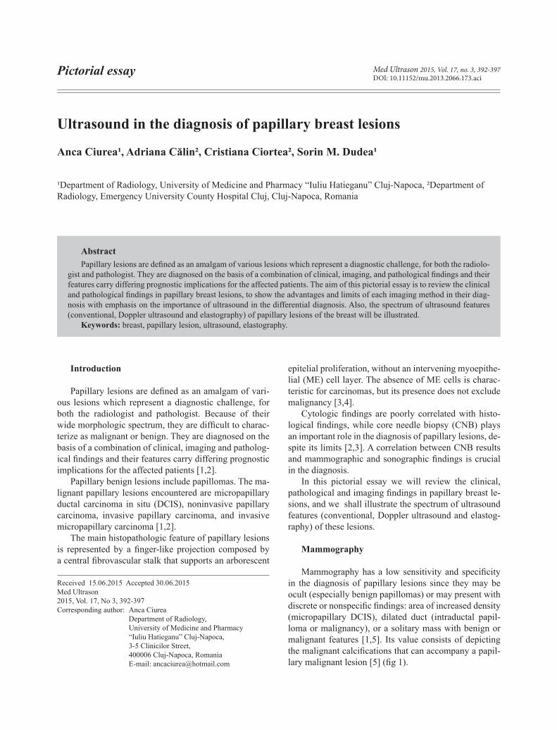
Mammography has a low sensitivity and specificity in the diagnosis of papillary lesions since they may be ocult (especially benign papillomas) or may present with discrete or nonspecific findings: area of increased density (micropapillary DCIS), dilated duct (intraductal papil-loma or malignancy), or a solitary mass with benign or malignant features [1,5]. Its value consists of depicting the malignant calcifications that can accompany a papil-lary malignant lesion [5] (fig 1).
393Med Ultrason 2015; 17(3): 392-397
Fig 1. Mammography of a patient with invasive papillary car-cinoma, showing multiple calcifications with heterogeneous morphology.
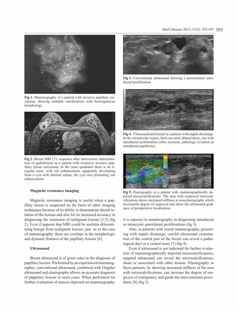
Fig 3. Conventional ultrasound showing a parenchimal intra-ductal proliferation.
Fig 4. Ultrasound performed in a patient with nipple discharge. In the retroareolar region, there are more dilated ducts, one with intraductal proliferation (after excision, pathology revealed an intraductal papilloma).
Fig 5. Elastography in a patient with mammographically de-picted microcalcifications. The area with suspected microcal-cifications shows increased stiffness at sonoelastography which increasethe degree of suspicion and allow the ultrasound guid-ance of preoperative localization.
Fig 2. Breast MRI (T1 sequence after intravenous administra-tion of gadolinium) in a patient with extensive invasive pap-illary breast carcinoma. In the outer quadrants there is an ir-regular mass, with rim enhancement, apparently developing from a cyst with internal septae, the cyst also presenting rim enhancement.
Magnetic resonance imaging
Magnetic resonance imaging is useful when a pap-illary lesion is suspected on the basis of other imaging techniques because of its ability to demonstrate ductal re-lation of the lesions and also for its increased accuracy in diagnosing the extension of malignant lesions [1,5] (fig 2). Even if appears that MRI could be usefulin diferenti-ating benign from malignant lesions, just as in the case of mammography, there are overlaps in the morphologic and dynamic features of the papillary lesions [6].
Ultrasound
Breast ultrasound is of great value in the diagnosis of papillary lesions. Performed by an experienced mammog-rapher, conventional ultrasound, combined with Doppler ultrasound and elastography allows an accurate diagnosis of pappilary lesions in most cases. When performed for further evaluation of masses depicted on mammography,
it is superior to mammography in diagnosing intraductal or intracystic parechimal proliferations (fig 3).
Also, in patients with occult mammography, present-ing with nipple discharge, careful ultrasound examina-tion of the central part of the breast can reveal a patho-logical duct or a central mass [7] (fig 4).
Even if ultrasound is not indicated for further evalua-tion of mammographically depicted microcalcifications, targeted ultrasound can reveal the microcalcifications, alone or associated with other lesions. Elastography in these patients, by showing increased stiffness of the area with microcalcifications, can increase the degree of sus-picion of malignancy and guide the interventional proce-dures [8] (fig 5).
394 Anca Ciurea et al Ultrasound in the diagnosis of papillary breast lesions
It is important, when suspecting a papillary lesion, to keep in mind that benign and malignant lesions have overlapping features and even if ultrasound can rise or infirm the suspicion of malignancy, the final diagnosis is based on core or excisional biopsy [9].
Benign papillary lesions
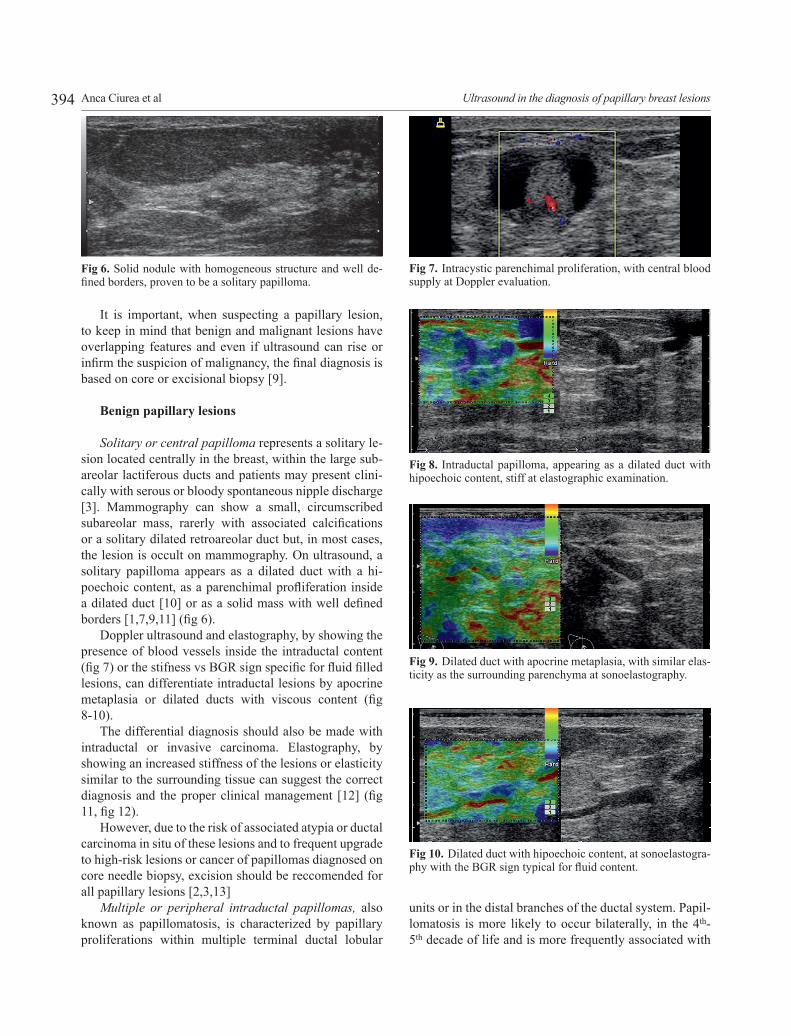
Solitary or central papilloma represents a solitary le-sion located centrally in the breast, within the large sub-areolar lactiferous ducts and patients may present clini-cally with serous or bloody spontaneous nipple discharge [3]. Mammography can show a small, circumscribed subareolar mass, rarerly with associated calcifications or a solitary dilated retroareolar duct but, in most cases, the lesion is occult on mammography. On ultrasound, a solitary papilloma appears as a dilated duct with a hi-poechoic content, as a parenchimal profliferation inside a dilated duct [10] or as a solid mass with well defined borders [1,7,9,11] (fig 6).
Doppler ultrasound and elastography, by showing the presence of blood vessels inside the intraductal content (fig 7) or the stifness vs BGR sign specific for fluid filled lesions, can differentiate intraductal lesions by apocrine metaplasia or dilated ducts with viscous content (fig 8-10).
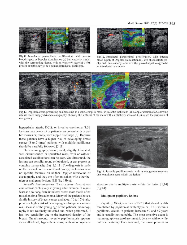
The differential diagnosis should also be made with intraductal or invasive carcinoma. Elastography, by showing an increased stiffness of the lesions or elasticity similar to the surrounding tissue can suggest the correct diagnosis and the proper clinical management [12] (fig 11, fig 12).
However, due to the risk of associated atypia or ductal carcinoma in situ of these lesions and to frequent upgrade to high-risk lesions or cancer of papillomas diagnosed on core needle biopsy, excision should be reccomended for all papillary lesions [2,3,13]
Multiple or peripheral intraductal papillomas, also known as papillomatosis, is characterized by papillary proliferations within multiple terminal ductal lobular
Fig 6. Solid nodule with homogeneous structure and well de-fined borders, proven to be a solitary papilloma.
Fig 7. Intracystic parenchimal proliferation, with central blood supply at Doppler evaluation.
Fig 8. Intraductal papilloma, appearing as a dilated duct with hipoechoic content, stiff at elastographic examination.
Fig 9. Dilated duct with apocrine metaplasia, with similar elas-ticity as the surrounding parenchyma at sonoelastography.
Fig 10. Dilated duct with hipoechoic content, at sonoelastogra-phy with the BGR sign typical for fluid content.
units or in the distal branches of the ductal system. Papil-lomatosis is more likely to occur bilaterally, in the 4th-5th decade of life and is more frequently associated with
395Med Ultrason 2015; 17(3): 392-397
hyperplasia, atypia, DCIS, or invasive carcinoma [1,3]. Lesions may be occult or patients can present with palpa-ble masses or, rarely, with nipple discharge [3]. Because these patients have a higher risk of developing breast cancer (3 to 7 times) patients with multiple papillomas should be carefully followed [3,11].
On mammography, round, oval, slightly lobulated, well-circumscribed or spiculated mass, with or without associated calcifications can be seen. On ultrasound, the lesions can be solid, round or lobulated, or can present as complex masses (fig 13a) [1,5,11]. The diagnosis is made on the basis of core or excisional biopsy; the lesions have no specific features, on neither Doppler ultrasound or elastography and they are often mistaken with other be-nign or malignant lesions [12] (fig 13b,c).
Juvenile Papillomatosis (Swiss cheese disease) oc-curs almost exclusively in young adult women. It mani-fests as a solitary, firm, unilateral breast mass that is often mistaken for a fibroadenoma. Many of the patients have a family history of breast cancer and about 10 to 15% also present a higher risk of developing a subsequent carcino-ma. Because of the young age of the patients, mammog-raphy is not routinely indicated and, when performed, it has low sensibility due to the increased density of the breast. On ultrasound, juvenile papillomatosis appears as an illdefined, hypoechoic mass, with inhomogeneus
structure due to multiple cysts within the lesion [1,14] (fig 14).
Malignant papillary lesions
Papillary DCIS, a variant of DCIS that should be dif-ferentiated by papillomas with atypia or DCIS within a papilloma, occurs in patients between 50 and 59 years and is usually not palpable. The most sensitive exam is mammography (area of asymmetric density, with or with-out calcifications). On ultrasound, the lesion presents as
Fig 11. Intraductal parenchimal proliferation, with intense blood supply at Doppler examination (a) but elasticity similar with the surrounding tissue, with an elasticity score of 1 (b), proved at pathology to be a benign intraductal papilloma.
Fig 12. Intraductal parenchimal proliferation, with intense blood supply at Doppler examination (a), stiff at sonoelastogra-phy, with an elasticity score of 4 (b), proved at pathology to be an intraductal carcinoma.
Fig 14. Juvenile papillomatosis, with inhomogeneus structure due to multiple cysts within the lesion.
Fig 13. Papillomatosis, presenting on ultrasound as a solid, complex mass, with cystic inclusions (a); Doppler examination, showing intense blood supply (b) and elastography, showing the stiffness of the mass with an elasticity score of 4 (c) raised the suspicion of malignancy.
396 Anca Ciurea et al Ultrasound in the diagnosis of papillary breast lesions
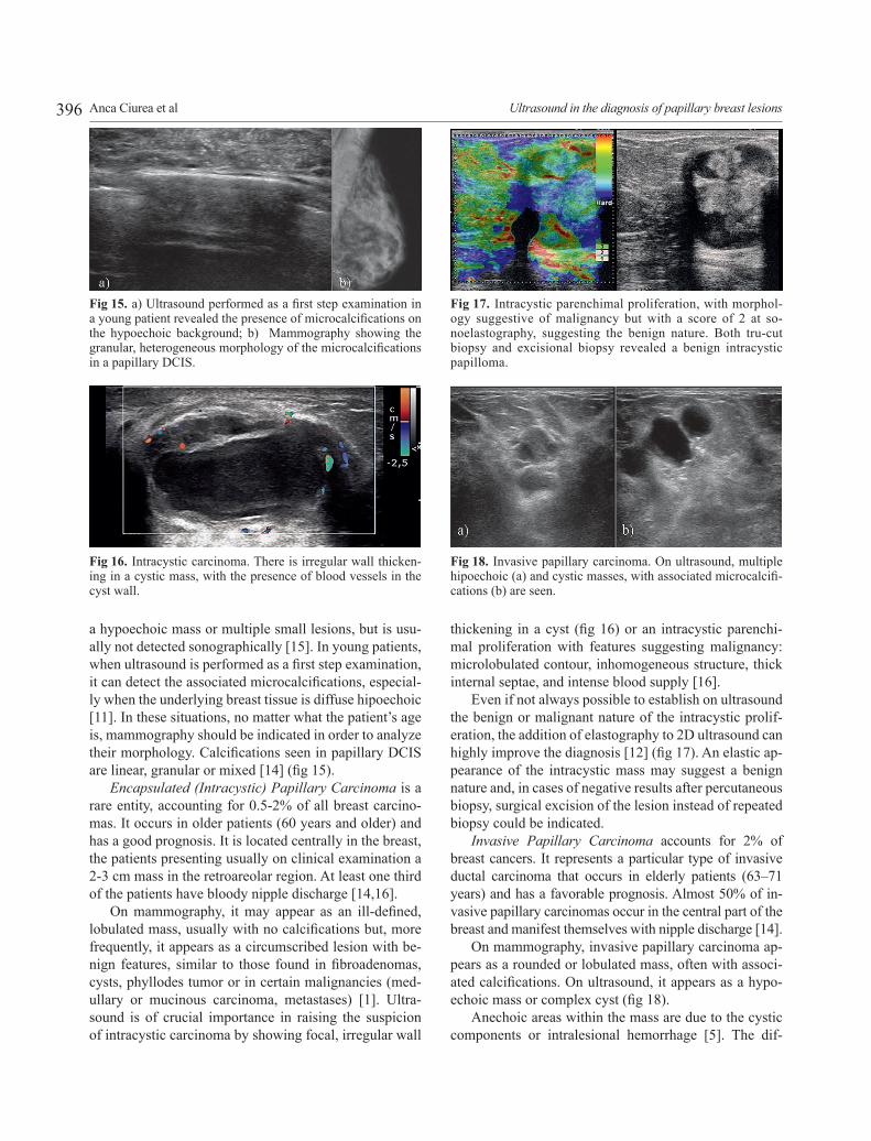
a hypoechoic mass or multiple small lesions, but is usu-ally not detected sonographically [15]. In young patients, when ultrasound is performed as a first step examination, it can detect the associated microcalcifications, especial-ly when the underlying breast tissue is diffuse hipoechoic [11]. In these situations, no matter what the patient’s age is, mammography should be indicated in order to analyze their morphology. Calcifications seen in papillary DCIS are linear, granular or mixed [14] (fig 15).
Encapsulated (Intracystic) Papillary Carcinoma is a rare entity, accounting for 0.5-2% of all breast carcino-mas. It occurs in older patients (60 years and older) and has a good prognosis. It is located centrally in the breast, the patients presenting usually on clinical examination a 2-3 cm mass in the retroareolar region. At least one third of the patients have bloody nipple discharge [14,16].
On mammography, it may appear as an ill-defined, lobulated mass, usually with no calcifications but, more frequently, it appears as a circumscribed lesion with be-nign features, similar to those found in fibroadenomas, cysts, phyllodes tumor or in certain malignancies (med-ullary or mucinous carcinoma, metastases) [1]. Ultra-sound is of crucial importance in raising the suspicion of intracystic carcinoma by showing focal, irregular wall
thickening in a cyst (fig 16) or an intracystic parenchi-mal proliferation with features suggesting malignancy: microlobulated contour, inhomogeneous structure, thick internal septae, and intense blood supply [16].
Even if not always possible to establish on ultrasound the benign or malignant nature of the intracystic prolif-eration, the addition of elastography to 2D ultrasound can highly improve the diagnosis [12] (fig 17). An elastic ap-pearance of the intracystic mass may suggest a benign nature and, in cases of negative results after percutaneous biopsy, surgical excision of the lesion instead of repeated biopsy could be indicated.
Invasive Papillary Carcinoma accounts for 2% of breast cancers. It represents a particular type of invasive ductal carcinoma that occurs in elderly patients (63–71 years) and has a favorable prognosis. Almost 50% of in-vasive papillary carcinomas occur in the central part of the breast and manifest themselves with nipple discharge [14].
On mammography, invasive papillary carcinoma ap-pears as a rounded or lobulated mass, often with associ-ated calcifications. On ultrasound, it appears as a hypo-echoic mass or complex cyst (fig 18).
Anechoic areas within the mass are due to the cystic components or intralesional hemorrhage [5]. The dif-
Fig 15. a) Ultrasound performed as a first step examination in a young patient revealed the presence of microcalcifications on the hypoechoic background; b) Mammography showing the granular, heterogeneous morphology of the microcalcifications in a papillary DCIS.
Fig 16. Intracystic carcinoma. There is irregular wall thicken-ing in a cystic mass, with the presence of blood vessels in the cyst wall.
Fig 17. Intracystic parenchimal proliferation, with morphol-ogy suggestive of malignancy but with a score of 2 at so-noelastography, suggesting the benign nature. Both tru-cut biopsy and excisional biopsy revealed a benign intracystic papilloma.
Fig 18. Invasive papillary carcinoma. On ultrasound, multiple hipoechoic (a) and cystic masses, with associated microcalcifi-cations (b) are seen.
397Med Ultrason 2015; 17(3): 392-397
ferentiation from benign lesions relies on its irregular contours and on the detection of suspect axillary lymph nodes on mammography or ultrasound [15].
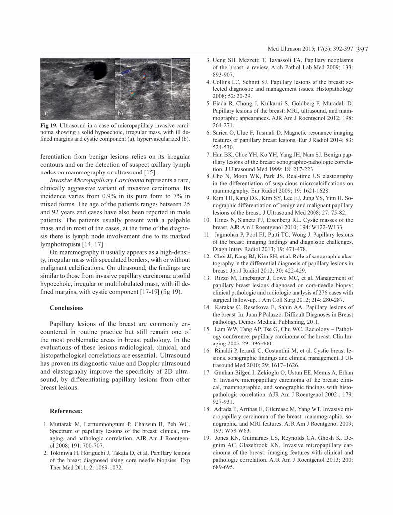
Invasive Micropapillary Carcinoma represents a rare, clinically aggressive variant of invasive carcinoma. Its incidence varies from 0.9% in its pure form to 7% in mixed forms. The age of the patients ranges between 25 and 92 years and cases have also been reported in male patients. The patients usually present with a palpable mass and in most of the cases, at the time of the diagno-sis there is lymph node involvement due to its marked lymphotropism [14, 17].
On mammography it usually appears as a high-densi-ty, irregular mass with speculated borders, with or without malignant calcifications. On ultrasound, the findings are similar to those from invasive papillary carcinoma: a solid hypoechoic, irregular or multilobulated mass, with ill de-fined margins, with cystic component [17-19] (fig 19).
Conclusions
Papillary lesions of the breast are commonly en-countered in routine practice but still remain one of the most problematic areas in breast pathology. In the evaluations of these lesions radiological, clinical, and histopathological correlations are essential. Ultrasound has proven its diagnostic value and Doppler ultrasound and elastography improve the specificity of 2D ultra-sound, by differentiating papillary lesions from other breast lesions.
References:
1. Muttarak M, Lerttumnongtum P, Chaiwun B, Peh WC. Spectrum of papillary lesions of the breast: clinical, im-aging, and pathologic correlation. AJR Am J Roentgen-ol 2008; 191: 700-707.
2. Tokiniwa H, Horiguchi J, Takata D, et al. Papillary lesions of the breast diagnosed using core needle biopsies. Exp Ther Med 2011; 2: 1069-1072.
3. Ueng SH, Mezzetti T, Tavassoli FA. Papillary neoplasms of the breast: a review. Arch Pathol Lab Med 2009; 133: 893-907.
4. Collins LC, Schnitt SJ. Papillary lesions of the breast: se-lected diagnostic and management issues. Histopathology 2008; 52: 20-29.
5. Eiada R, Chong J, Kulkarni S, Goldberg F, Muradali D. Papillary lesions of the breast: MRI, ultrasound, and mam-mographic appearances. AJR Am J Roentgenol 2012; 198: 264-271.
6. Sarica O, Uluc F, Tasmali D. Magnetic resonance imaging features of papillary breast lesions. Eur J Radiol 2014; 83: 524-530.
7. Han BK, Choe YH, Ko YH, Yang JH, Nam SJ. Benign pap-illary lesions of the breast: sonographic-pathologic correla-tion. J Ultrasound Med 1999; 18: 217-223.
8. Cho N, Moon WK, Park JS. Real-time US elastography in the differentiation of suspicious microcalcifications on mammography. Eur Radiol 2009; 19: 1621-1628.
9. Kim TH, Kang DK, Kim SY, Lee EJ, Jung YS, Yim H. So-nographic differentiation of benign and malignant papillary lesions of the breast. J Ultrasound Med 2008; 27: 75-82.
10. Hines N, Slanetz PJ, Eisenberg RL. Cystic masses of the breast. AJR Am J Roentgenol 2010; 194: W122-W133.
11. Jagmohan P, Pool FJ, Putti TC, Wong J. Papillary lesions of the breast: imaging findings and diagnostic challenges. Diagn Interv Radiol 2013; 19: 471-478.
12. Choi JJ, Kang BJ, Kim SH, et al. Role of sonographic elas-tography in the differential diagnosis of papillary lesions in breast. Jpn J Radiol 2012; 30: 422-429.
13. Rizzo M, Linebarger J, Lowe MC, et al. Management of papillary breast lesions diagnosed on core-needle biopsy: clinical pathologic and radiologic analysis of 276 cases with surgical follow-up. J Am Coll Surg 2012; 214: 280-287.
14. Karakas C, Resetkova E, Sahin AA. Papillary lesions of the breast. In: Juan P Palazzo. Difficult Diagnoses in Breast pathology. Demos Medical Publishing, 2011.
15. Lam WW, Tang AP, Tse G, Chu WC. Radiology – Pathol-ogy conference: papillary carcinoma of the breast. Clin Im-aging 2005; 29: 396-400.
16. Rinaldi P, Ierardi C, Costantini M, et al. Cystic breast le-sions. sonographic findings and clinical management. J Ul-trasound Med 2010; 29: 1617–1626.
17. Günhan-Bilgen I, Zekioglu O, Ustün EE, Memis A, Erhan Y. Invasive micropapillary carcinoma of the breast: clini-cal, mammographic, and sonographic findings with histo-pathologic correlation. AJR Am J Roentgenol 2002 ; 179: 927-931.
18. Adrada B, Arribas E, Gilcrease M, Yang WT. Invasive mi-cropapillary carcinoma of the breast: mammographic, so-nographic, and MRI features. AJR Am J Roentgenol 2009; 193: W58-W63.
19. Jones KN, Guimaraes LS, Reynolds CA, Ghosh K, De-gnim AC, Glazebrook KN. Invasive micropapillary car-cinoma of the breast: imaging features with clinical and pathologic correlation. AJR Am J Roentgenol 2013; 200: 689-695.
Fig 19. Ultrasound in a case of micropapillary invasive carci-noma showing a solid hypoechoic, irregular mass, with ill de-fined margins and cystic component (a), hypervascularized (b).


![Recalling Cohnheim's Theory: Papillary Renal Cell Tumor as a … · 2018-05-19 · papillary renal cell tumor (PRCT) may also arise from nephrogenic rest-like lesions [5]. Small tubular-](https://static.fdocuments.net/doc/165x107/5ed58e6be4e9005a3e7b0aa2/recalling-cohnheims-theory-papillary-renal-cell-tumor-as-a-2018-05-19-papillary.jpg)