UltrasoUnd gUided dorsal ramUs nerve block for redUction ... Guided Dorsal Ramus...Ultrasound-...
10
251 M.E.J. ANESTH 23 (2), 2015 CASE REPORTS ULTRASOUND GUIDED DORSAL RAMUS NERVE BLOCK FOR REDUCTION OF POSTOPERATIVE PAIN IN PATIENTS UNDERGOING LUMBAR SPINE SURGERY: A CASE SERIES IMAGING STUDY ACHIR AL-ALAMI * , ASHRAF ABOU EL EZZ ** AND F ARID KASSAB *** Abstract In patients undergoing spine surgery postoperative pain management can often be complicated with side effects associated with high dose narcotic such as respiratory depression and those associated with non-steroidal anti-inflammatory drugs such as interference with bone healing process. Local anesthetics can help in both decreasing postoperative pain and minimizing side effects associated with systematically administered analgesics. This report describes the use of preoperative ultrasound guided dorsal ramus nerve block to reduce postoperative pain in six patients undergoing lumbar spine surgery under general anesthesia. Introduction Patients undergoing spine surgery usually experience severe postoperative pain 1-3 . In the literature there are many reports describing different techniques of pain relief ranging from systemically administered analgesia to acute pain procedures such as neuraxial analgesia, paravertebral block and local anesthesia infiltration. Intrachecal morphine showed good results in postoperative analgesia for multilevel spine surgery. Epidural opioids in form of either single shot extended-release morphine or catheter placement had better safety margin in terms of respiratory depression and urinary retention. Ultrasound- guided bilateral paravertebral catheters recently have been described for postoperative analgesia in patients undergoing lumbar and thoracic laminectomy. Finally intraoperative infiltrations of local anesthesia into the wound, onto the neural root sheath or at the facet joint have also been described 4-10 . * MD, Title: Associate Consultant, Affiliation: Anesthesia, International Medical Center, Jeddah, Saudi Arabia. E-mail: [email protected] Conflicts of Interest: None ** MD, Consultant & chairman, Affiliation: Anesthesia, International Medical Center, Jeddah, Saudi Arabia. E-mail:elezzay@ aol.com Conflicts of Interest: None *** Consultant & Chairman, Affiliation: Musculoskeletal Center, International Medical Center, Jeddah, Saudi Arabia. E-mail: [email protected] Conflicts of Interest: None Anesthesia and musculoskeletal departments, International Medical Center, Jeddah, Saudi Arabia Corresponding author: Name Achir Al-alami, Department of Anesthesia, Institution International Medical Center, Mailing address, 9414 Crestwood Dr Parma Heights, OH 44130. Tel: 2672105822. E-mail [email protected]
Transcript of UltrasoUnd gUided dorsal ramUs nerve block for redUction ... Guided Dorsal Ramus...Ultrasound-...
251 M.E.J. ANESTH 23 (2), 2015
CASE REPORTS
UltrasoUnd gUided dorsal ramUs nerve block for redUction of postoperative
pain in patients Undergoing lUmbar spine sUrgery: a case series imaging stUdy
Achir Al-AlAmi*, AshrAf Abou El Ezz** And fArid KAssAb***
Abstract
in patients undergoing spine surgery postoperative pain management can often be complicated with side effects associated with high dose narcotic such as respiratory depression and those associated with non-steroidal anti-inflammatory drugs such as interference with bone healing process. local anesthetics can help in both decreasing postoperative pain and minimizing side effects associated with systematically administered analgesics. this report describes the use of preoperative ultrasound guided dorsal ramus nerve block to reduce postoperative pain in six patients undergoing lumbar spine surgery under general anesthesia.
Introduction
patients undergoing spine surgery usually experience severe postoperative pain1-3. in the literature there are many reports describing different techniques of pain relief ranging from systemically administered analgesia to acute pain procedures such as neuraxial analgesia, paravertebral block and local anesthesia infiltration. Intrachecal morphine showed good results in postoperative analgesia for multilevel spine surgery. epidural opioids in form of either single shot extended-release morphine or catheter placement had better safety margin in terms of respiratory depression and urinary retention. Ultrasound- guided bilateral paravertebral catheters recently have been described for postoperative analgesia in patients undergoing lumbar and thoracic laminectomy. Finally intraoperative infiltrations of local anesthesia into the wound, onto the neural root sheath or at the facet joint have also been described4-10.
* MD, Title: Associate Consultant, Affiliation: Anesthesia, International Medical Center, Jeddah, Saudi Arabia. E-mail: [email protected]
Conflicts of Interest: none** md, Consultant & chairman, Affiliation: Anesthesia, International Medical Center, Jeddah, Saudi Arabia. E-mail:elezzay@
aol.com Conflicts of Interest: None*** Consultant & Chairman, Affiliation: Musculoskeletal Center, International Medical Center, Jeddah, Saudi Arabia. E-mail:
[email protected] Conflicts of Interest: None Anesthesia and musculoskeletal departments, International Medical Center, Jeddah, Saudi Arabia Corresponding author: name achir al-alami, department of anesthesia, institution international medical center, mailing
address, 9414 crestwood dr parma Heights, oH 44130. tel: 2672105822. e-mail [email protected]
252 acHir al-alami et. al
Case Description
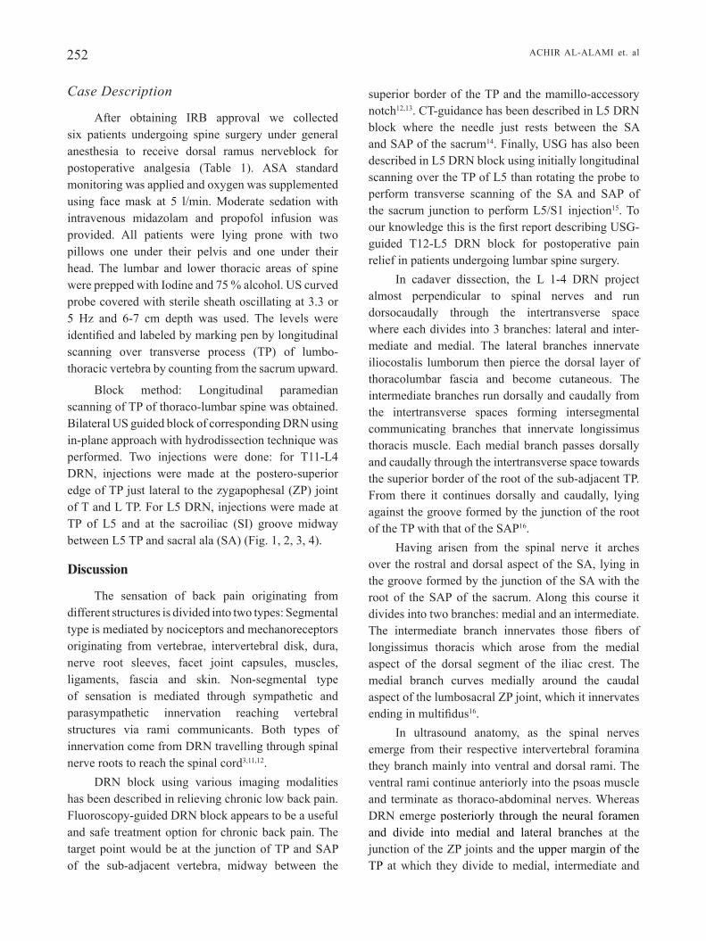
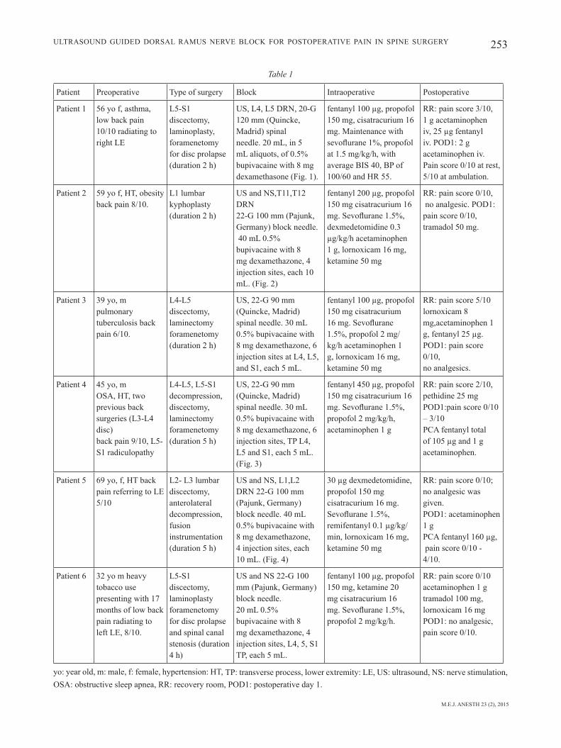
after obtaining irb approval we collected six patients undergoing spine surgery under general anesthesia to receive dorsal ramus nerveblock for postoperative analgesia (table 1). asa standard monitoring was applied and oxygen was supplemented using face mask at 5 l/min. moderate sedation with intravenous midazolam and propofol infusion was provided. all patients were lying prone with two pillows one under their pelvis and one under their head. the lumbar and lower thoracic areas of spine were prepped with iodine and 75 % alcohol. Us curved probe covered with sterile sheath oscillating at 3.3 or 5 Hz and 6-7 cm depth was used. the levels were identified and labeled by marking pen by longitudinal scanning over transverse process (tp) of lumbo-thoracic vertebra by counting from the sacrum upward.
block method: longitudinal paramedian scanning of tp of thoraco-lumbar spine was obtained. bilateral Us guided block of corresponding drn using in-plane approach with hydrodissection technique was performed. two injections were done: for t11-l4 drn, injections were made at the postero-superior edge of tp just lateral to the zygapophesal (Zp) joint of t and l tp. for l5 drn, injections were made at tp of l5 and at the sacroiliac (si) groove midway between l5 tp and sacral ala (sa) (fig. 1, 2, 3, 4).
Discussion
the sensation of back pain originating from different structures is divided into two types: segmental type is mediated by nociceptors and mechanoreceptors originating from vertebrae, intervertebral disk, dura, nerve root sleeves, facet joint capsules, muscles, ligaments, fascia and skin. non-segmental type of sensation is mediated through sympathetic and parasympathetic innervation reaching vertebral structures via rami communicants. both types of innervation come from drn travelling through spinal nerve roots to reach the spinal cord3,11,12.
drn block using various imaging modalities has been described in relieving chronic low back pain. fluoroscopy-guided drn block appears to be a useful and safe treatment option for chronic back pain. the target point would be at the junction of tp and sap of the sub-adjacent vertebra, midway between the
superior border of the tp and the mamillo-accessory notch12,13. ct-guidance has been described in l5 drn block where the needle just rests between the sa and sap of the sacrum14. finally, Usg has also been described in l5 drn block using initially longitudinal scanning over the tp of l5 than rotating the probe to perform transverse scanning of the sa and sap of the sacrum junction to perform l5/s1 injection15. to our knowledge this is the first report describing USG-guided t12-l5 drn block for postoperative pain relief in patients undergoing lumbar spine surgery.
in cadaver dissection, the l 1-4 drn project almost perpendicular to spinal nerves and run dorsocaudally through the intertransverse space where each divides into 3 branches: lateral and inter-mediate and medial. the lateral branches innervate iliocostalis lumborum then pierce the dorsal layer of thoracolumbar fascia and become cutaneous. the intermediate branches run dorsally and caudally from the intertransverse spaces forming intersegmental communicating branches that innervate longissimus thoracis muscle. each medial branch passes dorsally and caudally through the intertransverse space towards the superior border of the root of the sub-adjacent tp. from there it continues dorsally and caudally, lying against the groove formed by the junction of the root of the tp with that of the sap16.
Having arisen from the spinal nerve it arches over the rostral and dorsal aspect of the sa, lying in the groove formed by the junction of the sa with the root of the sap of the sacrum. along this course it divides into two branches: medial and an intermediate. The intermediate branch innervates those fibers of longissimus thoracis which arose from the medial aspect of the dorsal segment of the iliac crest. the medial branch curves medially around the caudal aspect of the lumbosacral Zp joint, which it innervates ending in multifidus16.
in ultrasound anatomy, as the spinal nerves emerge from their respective intervertebral foramina they branch mainly into ventral and dorsal rami. the ventral rami continue anteriorly into the psoas muscle and terminate as thoraco-abdominal nerves. Whereas drn emerge posteriorly through the neural foramen and divide into medial and lateral branches at the junction of the Zp joints and the upper margin of the tp at which they divide to medial, intermediate and
M.E.J. ANESTH 23 (2), 2015
253UltrasoUnd gUided dorsal ramUs nerve block for postoperative pain in spine sUrgery
Table 1
patient preoperative type of surgery block intraoperative postoperative
patient 1 56 yo f, asthma, low back pain 10/10 radiating to right le
l5-s1 discectomy, laminoplasty, foramenetomy for disc prolapse (duration 2 h)
Us, l4, l5 drn, 20-g 120 mm (Quincke, madrid) spinal needle. 20 ml, in 5 ml aliquots, of 0.5% bupivacaine with 8 mg dexamethasone (fig. 1).
fentanyl 100 µg, propofol 150 mg, cisatracurium 16 mg. maintenance with sevoflurane 1%, propofol at 1.5 mg/kg/h, with average bis 40, bp of 100/60 and Hr 55.
rr: pain score 3/10, 1 g acetaminophen iv, 25 µg fentanyl iv. pod1: 2 g acetaminophen iv. pain score 0/10 at rest, 5/10 at ambulation.
patient 2 59 yo f, Ht, obesityback pain 8/10.
l1 lumbar kyphoplasty (duration 2 h)
Us and ns,t11,t12 drn22-g 100 mm (pajunk, germany) block needle. 40 ml 0.5% bupivacaine with 8 mg dexamethazone, 4 injection sites, each 10 ml. (fig. 2)
fentanyl 200 µg, propofol 150 mg cisatracurium 16 mg. Sevoflurane 1.5%, dexmedetomidine 0.3 µg/kg/h acetaminophen 1 g, lornoxicam 16 mg, ketamine 50 mg
rr: pain score 0/10, no analgesic. pod1:pain score 0/10, tramadol 50 mg.
patient 3 39 yo, mpulmonary tuberculosis back pain 6/10.
l4-l5 discectomy, laminectomy foramenetomy (duration 2 h)
Us, 22-g 90 mm (Quincke, madrid) spinal needle. 30 ml 0.5% bupivacaine with 8 mg dexamethazone, 6 injection sites at l4, l5, and s1, each 5 ml.
fentanyl 100 µg, propofol 150 mg cisatracurium 16 mg. Sevoflurane 1.5%, propofol 2 mg/kg/h acetaminophen 1 g, lornoxicam 16 mg, ketamine 50 mg
rr: pain score 5/10 lornoxicam 8 mg,acetaminophen 1 g, fentanyl 25 µg.pod1: pain score 0/10,no analgesics.
patient 4 45 yo, mosa, Ht, two previous back surgeries (l3-l4 disc)back pain 9/10, l5-s1 radiculopathy
l4-l5, l5-s1 decompression, discectomy, laminectomy foramenetomy (duration 5 h)
Us, 22-g 90 mm (Quincke, madrid) spinal needle. 30 ml 0.5% bupivacaine with 8 mg dexamethazone, 6 injection sites, tp l4, l5 and s1, each 5 ml. (fig. 3)
fentanyl 450 µg, propofol 150 mg cisatracurium 16 mg. Sevoflurane 1.5%, propofol 2 mg/kg/h, acetaminophen 1 g
rr: pain score 2/10, pethidine 25 mg pod1:pain score 0/10 – 3/10 pca fentanyl total of 105 µg and 1 g acetaminophen.
patient 5 69 yo, f, Ht back pain referring to le 5/10
l2- l3 lumbar discectomy, anterolateral decompression, fusion instrumentation (duration 5 h)
Us and ns, l1,l2 drn 22-g 100 mm (pajunk, germany) block needle. 40 ml 0.5% bupivacaine with 8 mg dexamethazone, 4 injection sites, each 10 ml. (fig. 4)
30 µg dexmedetomidine, propofol 150 mg cisatracurium 16 mg. Sevoflurane 1.5%, remifentanyl 0.1 µg/kg/min, lornoxicam 16 mg, ketamine 50 mg
rr: pain score 0/10; no analgesic was given. pod1: acetaminophen 1 g pca fentanyl 160 µg, pain score 0/10 - 4/10.
patient 6 32 yo m heavy tobacco use presenting with 17 months of low back pain radiating to left le, 8/10.
l5-s1 discectomy, laminoplasty foramenetomy for disc prolapse and spinal canal stenosis (duration 4 h)
Us and ns 22-g 100 mm (pajunk, germany) block needle.20 ml 0.5% bupivacaine with 8 mg dexamethazone, 4 injection sites, l4, 5, s1 tp, each 5 ml.
fentanyl 100 µg, propofol 150 mg, ketamine 20 mg cisatracurium 16 mg. Sevoflurane 1.5%, propofol 2 mg/kg/h.
rr: pain score 0/10 acetaminophen 1 gtramadol 100 mg, lornoxicam 16 mg pod1: no analgesic, pain score 0/10.
yo: year old, m: male, f: female, hypertension: Ht, tp: transverse process, lower extremity: le, Us: ultrasound, ns: nerve stimulation, osa: obstructive sleep apnea, rr: recovery room, pod1: postoperative day 1.
254 acHir al-alami et. al
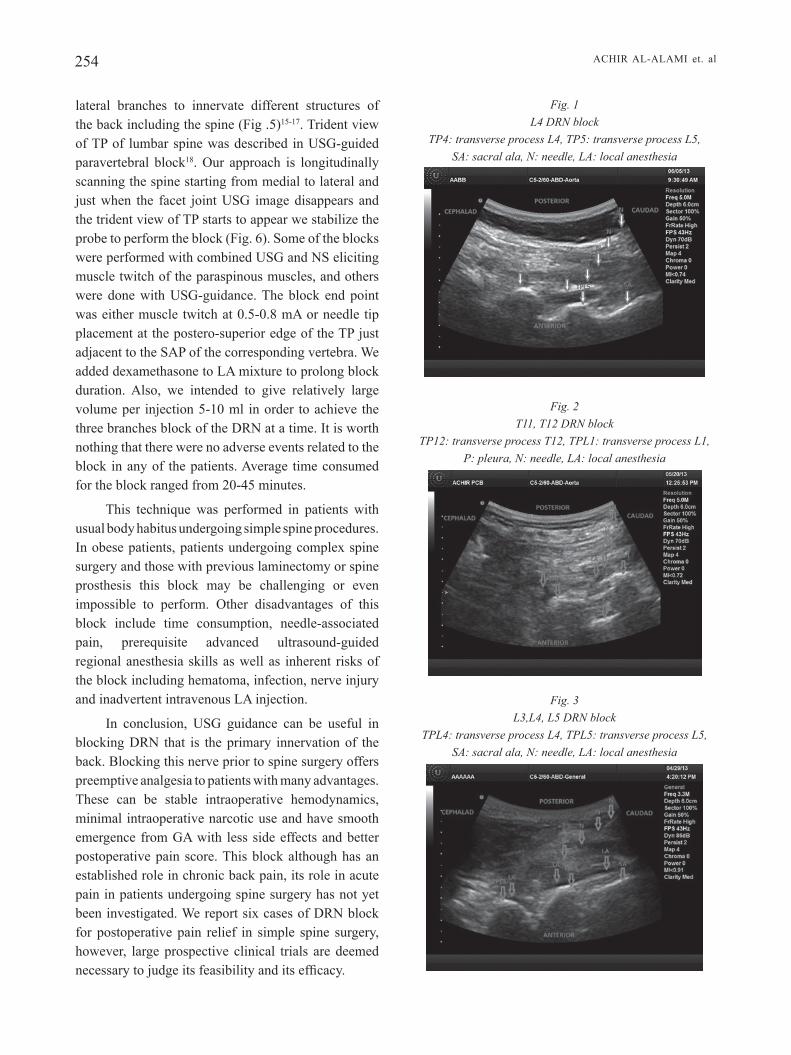
lateral branches to innervate different structures of the back including the spine (fig .5)15-17. trident view of tp of lumbar spine was described in Usg-guided paravertebral block18. our approach is longitudinally scanning the spine starting from medial to lateral and just when the facet joint Usg image disappears and the trident view of tp starts to appear we stabilize the probe to perform the block (fig. 6). some of the blocks were performed with combined Usg and ns eliciting muscle twitch of the paraspinous muscles, and others were done with Usg-guidance. the block end point was either muscle twitch at 0.5-0.8 ma or needle tip placement at the postero-superior edge of the tp just adjacent to the sap of the corresponding vertebra. We added dexamethasone to la mixture to prolong block duration. also, we intended to give relatively large volume per injection 5-10 ml in order to achieve the three branches block of the drn at a time. it is worth nothing that there were no adverse events related to the block in any of the patients. average time consumed for the block ranged from 20-45 minutes.
this technique was performed in patients with usual body habitus undergoing simple spine procedures. in obese patients, patients undergoing complex spine surgery and those with previous laminectomy or spine prosthesis this block may be challenging or even impossible to perform. other disadvantages of this block include time consumption, needle-associated pain, prerequisite advanced ultrasound-guided regional anesthesia skills as well as inherent risks of the block including hematoma, infection, nerve injury and inadvertent intravenous la injection.
in conclusion, Usg guidance can be useful in blocking drn that is the primary innervation of the back. blocking this nerve prior to spine surgery offers preemptive analgesia to patients with many advantages. these can be stable intraoperative hemodynamics, minimal intraoperative narcotic use and have smooth emergence from ga with less side effects and better postoperative pain score. this block although has an established role in chronic back pain, its role in acute pain in patients undergoing spine surgery has not yet been investigated. We report six cases of drn block for postoperative pain relief in simple spine surgery, however, large prospective clinical trials are deemed necessary to judge its feasibility and its efficacy.
Fig. 1 L4 DRN block
TP4: transverse process L4, TP5: transverse process L5, SA: sacral ala, N: needle, LA: local anesthesia
Fig. 2 T11, T12 DRN block
TP12: transverse process T12, TPL1: transverse process L1, P: pleura, N: needle, LA: local anesthesia
Fig. 3 L3,L4, L5 DRN block
TPL4: transverse process L4, TPL5: transverse process L5, SA: sacral ala, N: needle, LA: local anesthesia

M.E.J. ANESTH 23 (2), 2015
255
Fig.5DRN L3,4,5 anatomy
With permission from Lipincot William and Wilkins
UltrasoUnd gUided dorsal ramUs nerve block for postoperative pain in spine sUrgery
Fig. 4L2 DRN block,
TPL1: transverse process L1, TPL2: transverse process L2,TPL3: transverse process L3, D5W: Dextrose 5 % in water,
N: needle, LA: local anesthesia
Fig.6Water skeletal scanning of lumbar spine
FJ: facet jointTP: transverse process

256 acHir al-alami et. al
References
1. biAnconi m, fErrAro l, ricci r, zAnoli G, AntonElli t, GiuliA b, GubErti A, mAssAri l, Et Al: The pharmacokinetics and efficacy of ropivacaine continuous wound installation after spine fusion surgery. Anesth Analg; 98:166-72, 2004.
2. cohEn bE, hArtmAn mb, WAdE Jt, millEr Js, GilbErt r, chApmAn tm: postoperative pain control after lumbar spine fusion: patient-controlled analgesia versus continuous epidural analgesia. Spine; 22:1892-7, 1997.
3. lAi lt, ortiz-cArdonA Jr, bEndo AA: perioperative pain management in the neurosurgical patient. Anesthesiol Clin; 30:347-67, 2012.
4. schEnK mr, putziEr m, KuGlEr b, tohtz s, VoiGt K, schinK t, Kox WJ, spiEs c, VolK t: postoperative analgesia after major spine surgery: patient-controlled analgesia versus patient-controlled intravenous analgesia. Anesth analg; 103:1311-7, 2006.
5. KlubA t, hofmAnn f, brEdAnGEr s, blumEnstocK G, niEmEyEr t: Efficacy of postoperative analgesia after posterior lumbar instrumented fusion for degenerative disc disease: a prospective randomized comparison of epidural catheter and intravenous administration of analgesics. Orthop Rev; 20:2(1), 2010.
6. pAndin p, rEttAb s, lubAnsu A: Ultrasound guidance is Helpful for paravertebral block performance and catheter placement in patients with laminectomy after thoracotomy or lumbotomy: a case series imaging study. Open Journal of Anesthesiology; 3:67-73, 2013.
7. GurbEt A, bEKAr A, bilGin h, KorfAli G, yilmAzlAr s, tErcAn m: Pre-emptive infiltration of levobupivacaine is superior to at closure administration in lumbar laminectomy patients. Eur Spine J; 17:1237-41, 2008.
8. bAhAri s, El-dAhAb m, clEAry m, spArKEd J: Efficacy of triamcinolone acetonide and bupivacaine for pain after lumbar discectomy. Eur Spine J; 19:1099-103, 2010.
9. bAdEmci G, bAsAr h, sAhin s, ozcAKir s, AnbArci h, EVliyAoGlu c, KEsKil s: Can facet joint infiltrative analgesia reduce postoperative pain in degenerative lumbar disc surgery? Neurocirugia; 19:45-9, 2008.
10. hiGuchi K, sAto t: anatomical study of lumbar spine innervation. Folia Morphol; 61:71-9, 2002.
11. ohtori s, yAmAshitA m, inouE G, yAmAuchi K, suzuKi m, oritA s, EGuchi y, ochiAi n, KishidA s, tAKAso m, tAKAhAshi K: the l2 spinal verve block effects on acute low back pain from osteoporotic vertebral fracture. Journal of Pain; 10:870-875, 2009.
12. miyAKoshi n, shimAdA y, KAsuKAWA y, sAito h, KodAmA h, itoi E: total dorsal ramus block for the treatment of chronic low back pain: a preliminary study. Joint Bone Spine; 74:270-4, 2007.
13. KyounG-tAE K, sEunG-Won p, tAEK-Kyun n: lumbar dorsal ramus block for low back pain. J Kor Neurotraumatol Soc; 3:82-86, 2007.
14. stoJAnoVic mp: basic pain management interventions using fluoroscopy: targets and optimal imaging of lumbar spine. Techniques in Regional Anesthesia and Pain Management; 11:55-62, 2007.
15. biGElEisEn p, orEbAuGh s: Ultrasound-guided regional anesthesia and pain medicine. 1st ed. baltimore, Wolters kluwer, lippincott Williams &Wilkins, 2010.
16. boGduK n, Wilson As, tynAn W: the human lumbar dorsal rami. J Anat; 2:383-397, 1982.
17. zhou l, schnEcK cd, shAo z: the anatomy of dorsal ramus nerves and its implications in lower back pain. Neuroscience & Medicine; 3:192-201, 2012.
18. KArmAKAr mK, ho Am, li x, KWoKWh, tsAnG K, nGAn KEE Wd: Ultrasound-guided lumbar plexus block through the acoustic window of the lumbar ultrasound trident. Br J Anaesth; 100:533-7, 2008.

B. Braun Melsungen AG | Hospital Care | 34209 Melsungen | GermanyTel. +49 5661 71-0 | www.bbraun.com
Thanks to AirStop in the drip chamber - the sight of a container running empty is no longer cause for alarm and no reason for energy and time to be wasted rushing around because the patient gets upset.
When the container is empty, AirStop maintains a constant � uid level. No air can get through to the patient.
Thanks to the PrimeStop at the patient connector - you can now prepare several infusions at once, quicker and more hygienic than ever before. Right away your hands are free to prepare the next infusion.
Intra� x® SafeSet The � rst IV administration set with AirStop and PrimeStop
Gives every ward that extra measure of safety while providing higher e� ciency.
Prime Stop
AirStop
Prime Stop
AirStop
www.safeinfusiontherapy.com
For more information about Intrafix® SafeSet and Safe Infusion Therapy:
Risk Prevention in Infusion TherapyParticulate Contamination
www.safeinfusiontherapy.com
Chemical Contamination
Risk Prevention in Infusion Therapy
www.safeinfusiontherapy.com
Risk Prevention in Infusion TherapyAir Embolism
www.safeinfusiontherapy.com
Risk Prevention in Infusion TherapyMedication Error
www.safeinfusiontherapy.com
www.safeinfusiontherapy.com
Risk Prevention in Infusion TherapyDrug Incompatibility
www.safeinfusiontherapy.com
www.safeinfusiontherapy.comwww.safeinfusiontherapy.com
www.safeinfusiontherapy.com
Risk Prevention in Infusion TherapyMicrobiological Contamination
www.safeinfusiontherapy.com

.
References: 1. Talon M. et al., J Burn Care Research 2009; 30: 599-605. 2. MAD (Mucosal Atomization Device) Medical Atomizer in Vitro Spray Characterization, 2011