
Ultrasound Committee Activities - QIBA Wikiqibawiki.rsna.org/images/6/6f/QIBA_Ultrasound... ·...
16
7/23/2014 1 Ultrasound Committee Activities Anthony E. Samir MD MPH Clinical Director MGH/MIT Center for Ultrasound Research & Translation PINTAD 2014 Disclosures • Grant Support – Harvard Catalyst: Pilot Grant (NIBIB subcontract) – American Institute of Ultrasound in Medicine – MGH: Clinical Innovation Award – American Roentgen Ray Society: ARRS Scholarship 2011-2013 – NIBIB subcontract via RSNA/QIBA 2013- – Partners Innovation Development Grant 2014- • Research support (equipment): – Supersonic Imagine, General Electric, Hitachi, Toshiba, Siemens • Consulting (via MGH) – Multiple CROs
Transcript of Ultrasound Committee Activities - QIBA Wikiqibawiki.rsna.org/images/6/6f/QIBA_Ultrasound... ·...
7/23/2014
1
Ultrasound Committee Activities
Anthony E. Samir MD MPH Clinical Director
MGH/MIT Center for Ultrasound Research & Translation
PINTAD 2014
Disclosures
• Grant Support – Harvard Catalyst: Pilot Grant (NIBIB subcontract) – American Institute of Ultrasound in Medicine – MGH: Clinical Innovation Award – American Roentgen Ray Society: ARRS Scholarship 2011-2013 – NIBIB subcontract via RSNA/QIBA 2013- – Partners Innovation Development Grant 2014-
• Research support (equipment): – Supersonic Imagine, General Electric, Hitachi, Toshiba, Siemens
• Consulting (via MGH) – Multiple CROs

7/23/2014
2
Premise
• Variation in clinical practice results in poorer outcomes and higher costs.
• One approach to reduce variability in radiology is to extract objective, quantitative data from scans.
Slide obtained from Dan Sullivan MD
Why Must Imaging Become More
Quantitative?
• Molecular medicine (precision medicine) requires quantitative test results.
• Evidence-based medicine & QA Programs depend on objective data.
• Decision-support tools (CADx, CDSS) need quantitative input.
Slide obtained from Dan Sullivan MD
7/23/2014
3
RSNA’s Perspective:
• Extracting objective, quantitative results
from imaging studies will improve the
value of imaging in clinical practice.
Slide obtained from Dan Sullivan MD
QIBA
• Mission: To improve the value and practicality of quantitative biomarkers by reducing variability across devices, patients and time.
• Relevance to ultrasound:
– US data acquisition is subject to operator-related variability
– Acquired US data is complex and influenced by attenuation, refraction, reflections, tissue properties, temperature etc.
7/23/2014
4
QIBA Criteria for Biomarker
Selection • Transformational
– addresses a significant medical need
• Translational
– will likely result in significant improvement in the development, approval,
or delivery of care to patients.
• Feasible
– end goals can likely be achieved in a specific timeframe
• Practical
– leverages preexisting resources (e.g., intellectual capital, personnel,
facilities, specimens, reagents, data) wherever possible; warrants
access to RSNA resources and support.
• Collaborative
– the biomarker has the support of the stakeholder community and the
organizational impetus to sustain continued efforts.
Slide obtained from Dan Sullivan MD
Ultrasound Biomarker: SWS
• Tissue shear wave speed
• What is elastography?
• Why is elastography transformational?
7/23/2014
5
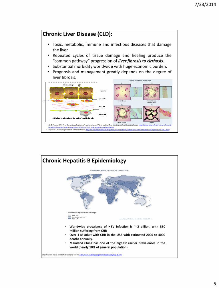
Chronic Liver Disease (CLD):
• Toxic, metabolic, immune and infectious diseases that damage the liver.
• Repeated cycles of tissue damage and healing produce the “common pathway” progression of liver fibrosis to cirrhosis.
• Substantial morbidity worldwide with huge economic burden. • Prognosis and management greatly depends on the degree of
liver fibrosis.
• Ch.S. Pavlov, D.V. Et al, Current applications of elastometry and Fibro- and ActiTest for diagnostics of hepatic fibrosis. http://www.biopredictive.com/ru/current-applications-of-elastometry-and-fibro-and-acti-test-for-diagnostics-of-hepatic-fibrosis
• Hepatitis C New Drug Research And Liver Health, http://www.hepatitiscnewdrugresearch.com/starting-hepatitis-c-treatment-tips-and-information-2011.html
Chronic Hepatitis B Epidemiology
• Worldwide prevalence of HBV infection is ~ 2 billion, with 350 million suffering from CHB
• Over 1 M adult with CHB in the USA with estimated 2000 to 4000 deaths annually.
• Mainland China has one of the highest carrier prevalences in the world (nearly 10% of general population).
The National Travel Health Network and Centre, http://www.nathnac.org/travel/factsheets/hep_b.htm

7/23/2014
6
Chronic Hepatitis C Epidemiology
• Worldwide prevalence of HCV infection is 170-200 M. • Prevalence of HCV in the USA is around 2-3 M. HCV prevalence in Mainland China
is also substantial (estimated 3.2% of general population) • Rate of cirrhosis in HCV:10% in 20 years and 20% in 30 years. • Mortality in cirrhotic: 1-5 % per year. • HCC in cirrhotic: 1-4% per year.
US Centers for Disease Control and Prevention, 2011 , http://wwwnc.cdc.gov/travel/pdf/yellowbook-2012-map-03-05-prevalence-chronic-hepatitis-c.pdf
Nonalcoholic Fatty Liver Disease (NAFLD): • NAFLD is no longer a disease exclusive to developed Western
countries. It should be regarded as a global problem. Among more affluent regions of China, its prevalence is ~ 15%.
• May progress to NASH in a minority, with risk of cirrhosis.
• Prognosis strongly depends on histologic severity.
Next Level Nutrition, http://next-level-nutrition.com/?p=1448
7/23/2014
7
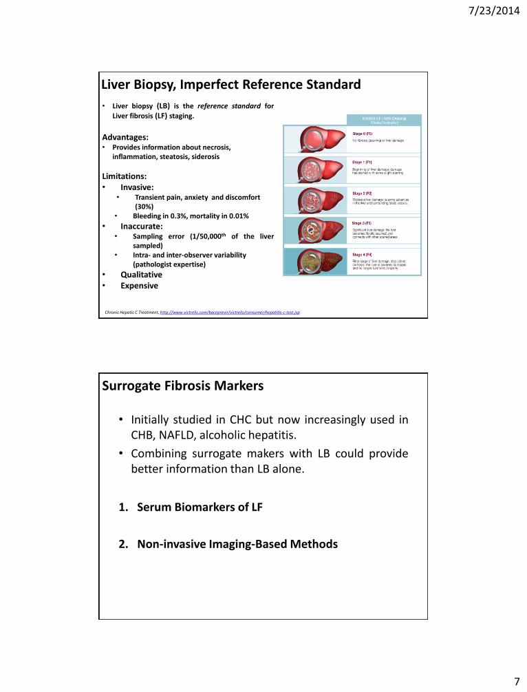
Liver Biopsy, Imperfect Reference Standard
• Liver biopsy (LB) is the reference standard for
Liver fibrosis (LF) staging.
Advantages: • Provides information about necrosis,
inflammation, steatosis, siderosis
Limitations: • Invasive:
• Transient pain, anxiety and discomfort (30%)
• Bleeding in 0.3%, mortality in 0.01%
• Inaccurate: • Sampling error (1/50,000th of the liver
sampled) • Intra- and inter-observer variability
(pathologist expertise)
• Qualitative • Expensive
Chronic Hepatic C Treatment, http://www.victrelis.com/boceprevir/victrelis/consumer/hepatitis-c-test.jsp
Surrogate Fibrosis Markers
• Initially studied in CHC but now increasingly used in CHB, NAFLD, alcoholic hepatitis.
• Combining surrogate makers with LB could provide better information than LB alone.
1. Serum Biomarkers of LF
2. Non-invasive Imaging-Based Methods
7/23/2014
8
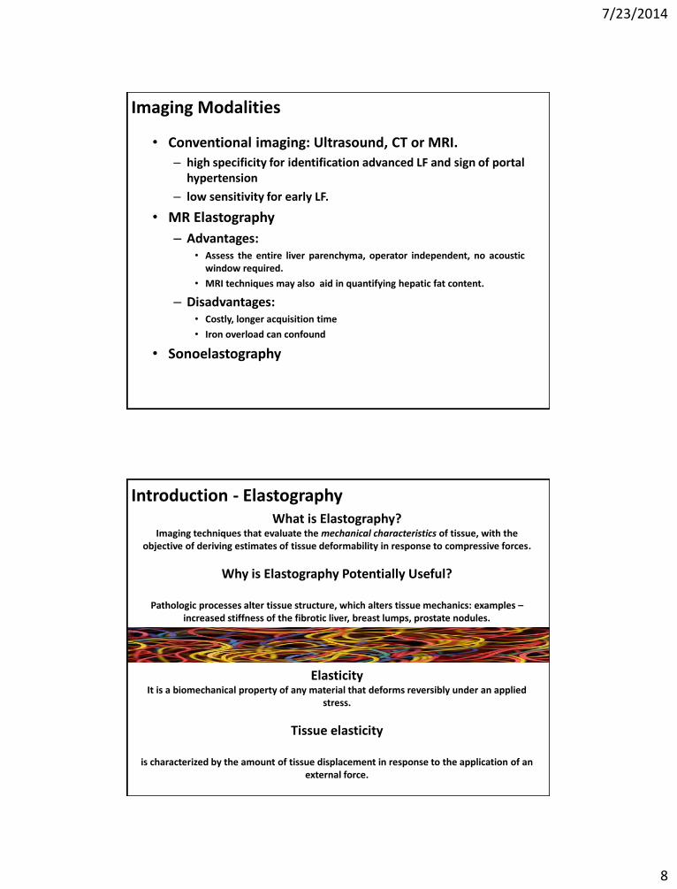
Imaging Modalities
• Conventional imaging: Ultrasound, CT or MRI.
– high specificity for identification advanced LF and sign of portal hypertension
– low sensitivity for early LF.
• MR Elastography
– Advantages: • Assess the entire liver parenchyma, operator independent, no acoustic
window required.
• MRI techniques may also aid in quantifying hepatic fat content.
– Disadvantages: • Costly, longer acquisition time
• Iron overload can confound
• Sonoelastography
Introduction - Elastography What is Elastography?
Imaging techniques that evaluate the mechanical characteristics of tissue, with the objective of deriving estimates of tissue deformability in response to compressive forces.
Why is Elastography Potentially Useful?
Pathologic processes alter tissue structure, which alters tissue mechanics: examples – increased stiffness of the fibrotic liver, breast lumps, prostate nodules.
Elasticity
It is a biomechanical property of any material that deforms reversibly under an applied stress.
Tissue elasticity
is characterized by the amount of tissue displacement in response to the application of an external force.

7/23/2014
9
Physical Principles: Key Concepts
• Stress: The force divided by the cross-sectional area. (Unit is kPa).
• Strain: Deformation quantified as relative displacement of particles in the
material. (No unit)
Non Destructive Testing: Topics: Physical and Chemical Properties, Stress and Strain. http://www.ndted.org/EducationResources/CommunityCollege/Materials/Mechanical/StressStrain.htm
Elastic Modulus
The description of a material’s tendency to be non-permanently deformed under stress. It can be depicted on a stress-strain curve.
Principles of ultrasound elastography, Peter R Hoskins, Ultrasound February 2012 vol. 20 no. 1 8-15

7/23/2014
10
Young’s Modulus (Tensile Modulus)
Young’s Modulus = E = Stress/Strain
Every material has a specific Young’s modulus. Values for healthy soft tissues (liver, prostate, kidney and breast) range from 0.5-70 kPa, whereas the Young’s modulus of cancer is typically higher, ranging from 20-560 kPa.
Steps in the Estimation of Elastic Modulus in Shear Wave Sonoelastography:
Induce shear waves
Measure shear wave speed
Estimate the tissue stiffness & differentiate normal from
pathologic tissues
This velocity is directly related to tissue stiffness. The harder the tissue, the faster the shear wave propagates.
7/23/2014
11
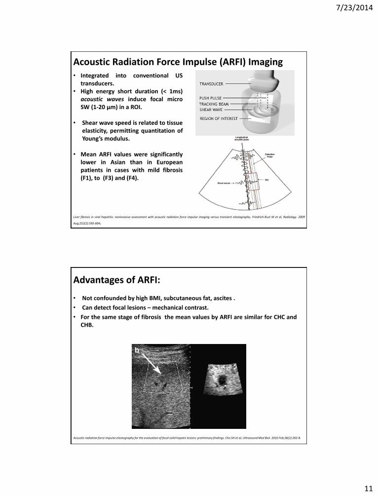
Acoustic Radiation Force Impulse (ARFI) Imaging • Integrated into conventional US
transducers. • High energy short duration (< 1ms)
acoustic waves induce focal micro SW (1-20 µm) in a ROI.
• Shear wave speed is related to tissue
elasticity, permitting quantitation of Young’s modulus.
• Mean ARFI values were significantly
lower in Asian than in European patients in cases with mild fibrosis (F1), to (F3) and (F4).
Liver fibrosis in viral hepatitis: noninvasive assessment with acoustic radiation force impulse imaging versus transient elastography. Friedrich-Rust M et al, Radiology. 2009
Aug;252(2):595-604.
Advantages of ARFI:
• Not confounded by high BMI, subcutaneous fat, ascites .
• Can detect focal lesions – mechanical contrast.
• For the same stage of fibrosis the mean values by ARFI are similar for CHC and CHB.
Acoustic radiation force impulse elastography for the evaluation of focal solid hepatic lesions: preliminary findings. Cho SH et al, Ultrasound Med Biol. 2010 Feb;36(2):202-8.
7/23/2014
12
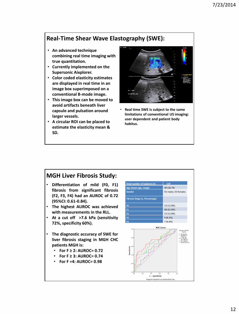
Real-Time Shear Wave Elastography (SWE):
• An advanced technique combining real time imaging with true quantitation.
• Currently implemented on the Supersonic Aixplorer.
• Color coded elasticity estimates are displayed in real time in an image box superimposed on a conventional B-mode image.
• This image box can be moved to avoid artifacts beneath liver capsule and pulsation around larger vessels.
• A circular ROI can be placed to estimate the elasticity mean & SD.
• Real time SWE is subject to the same limitations of conventional US imaging: user dependent and patient body habitus.
MGH Liver Fibrosis Study:
• Differentiation of mild (F0, F1) fibrosis from significant fibrosis (F2, F3, F4) had an AUROC of 0.72 (95%CI: 0.61-0.84).
• The highest AUROC was achieved with measurements in the RLL.
• At a cut off >7.6 kPa (sensitivity 72%, specificity 60%).
• The diagnostic accuracy of SWE for
liver fibrosis staging in MGH CHC patients MGH is: • For F ≥ 2: AUROC= 0.72 • For F ≥ 3: AUROC= 0.74 • For F =4: AUROC= 0.98
Total number of Subjects (n) 110
Age (mean age, range) 49 (18-79)
Gender 55 males; 55 females
Fibrosis Stage (n, Percentage)
F0 13 (11.8%)
F1 68 (61.8%)
F2 13 (11.8%)
F3 9 (8.2%)
F4 7 (6.4%)

7/23/2014
13
Detection of Portal Hypertension and Esophageal Varices
PH & EV:
• Estimated prevalence of EV in cirrhotic patients : 50% • Mortality rate of VB ranges from 20% to 35%. • American and European guidelines recommend screening of upper GI
endoscopy for early detection of EV in all patients with compensated cirrhosis.
• Effective prophylactic treatment (β- blocker) of EV in decompensated cirrhosis : • Improve patient prognosis. • Reduce hospitalization cost.
7/23/2014
14
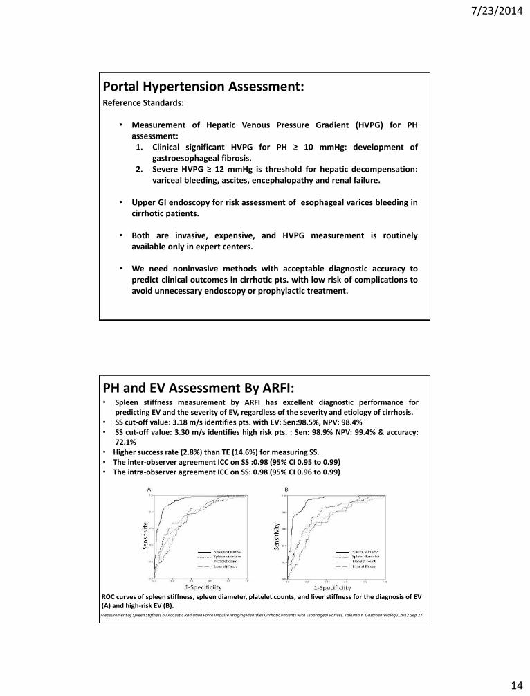
Portal Hypertension Assessment: Reference Standards:
• Measurement of Hepatic Venous Pressure Gradient (HVPG) for PH assessment: 1. Clinical significant HVPG for PH ≥ 10 mmHg: development of
gastroesophageal fibrosis. 2. Severe HVPG ≥ 12 mmHg is threshold for hepatic decompensation:
variceal bleeding, ascites, encephalopathy and renal failure. • Upper GI endoscopy for risk assessment of esophageal varices bleeding in
cirrhotic patients. • Both are invasive, expensive, and HVPG measurement is routinely
available only in expert centers. • We need noninvasive methods with acceptable diagnostic accuracy to
predict clinical outcomes in cirrhotic pts. with low risk of complications to avoid unnecessary endoscopy or prophylactic treatment.
PH and EV Assessment By ARFI: • Spleen stiffness measurement by ARFI has excellent diagnostic performance for
predicting EV and the severity of EV, regardless of the severity and etiology of cirrhosis. • SS cut-off value: 3.18 m/s identifies pts. with EV: Sen:98.5%, NPV: 98.4% • SS cut-off value: 3.30 m/s identifies high risk pts. : Sen: 98.9% NPV: 99.4% & accuracy:
72.1% • Higher success rate (2.8%) than TE (14.6%) for measuring SS. • The inter-observer agreement ICC on SS :0.98 (95% CI 0.95 to 0.99) • The intra-observer agreement ICC on SS: 0.98 (95% CI 0.96 to 0.99)
Measurement of Spleen Stiffness by Acoustic Radiation Force Impulse Imaging Identifies Cirrhotic Patients with Esophageal Varices. Takuma Y, Gastroenterology. 2012 Sep 27
ROC curves of spleen stiffness, spleen diameter, platelet counts, and liver stiffness for the diagnosis of EV (A) and high-risk EV (B).

7/23/2014
15
QIBA: US Activities
• Technical and Clinical Subcommittees
• Technical subcommittee focuses on sources of variability originating within the equipment chain.
• Clinical subcommittee focuses on sources of variability originating within clinical data acquisition and interpretation.
• Phantom construction – Simple linear phantoms
– Nonlinear phantoms approximating tissue (2014)
• Simulations
• Clinical guidelines – Elastography case report forms (version 5)
– Pathology case report forms (version 2)
• UPICT protocol
• QIDW uploads
QIBA: US Activities

7/23/2014
16
MGH Clinical Ultrasound Research Fellowship
Arash Anvari M.D. Research Fellow
Qingli Zhu M.D. Research Fellow
Priyanush Kandakatla M.D. Research Fellow
• 1-2 years of customizable practical research experience and coursework and clinical observation.
Manish Dhyani M.D. Research Fellow
THANK YOU


