ULTRASHORT-PULSE LASERS TREATING THE … · It had been a long-held belief in ophthalmology that...
36
ULTRASHORT-PULSE LASERS TREATING THE CRYSTALLINE LENS: WILL THEY CAUSE VISION- THREATENING CATARACT? (AN AMERICAN OPHTHALMOLOGICAL SOCIETY THESIS) By Ronald R. Krueger MD MSE, Harvey Uy MD, Jared McDonald, and Keith Edwards FCOptom ABSTRACT Purpose: To demonstrate that ultrashort-pulse laser treatment in the crystalline lens does not form a focal, progressive, or vision- threatening cataract. Methods: An Nd:vanadate picosecond laser (10 ps) with prototype delivery system was used. Primates: 11 rhesus monkey eyes were prospectively treated at the University of Wisconsin (energy 25-45 μJ/pulse and 2.0-11.3M pulses per lens). Analysis of lens clarity and fundus imaging was assessed postoperatively for up to 4½ years (5 eyes). Humans: 80 presbyopic patients were prospectively treated in one eye at the Asian Eye Institute in the Philippines (energy 10 μJ/pulse and 0.45-1.45M pulses per lens). Analysis of lens clarity, best-corrected visual acuity, and subjective symptoms was performed at 1 month, prior to elective lens extraction. Results: Bubbles were immediately seen, with resolution within the first 24 to 48 hours. Afterwards, the laser pattern could be seen with faint, noncoalescing, pinpoint micro-opacities in both primate and human eyes. In primates, long-term follow-up at 4½ years showed no focal or progressive cataract, except in 2 eyes with preexisting cataract. In humans, <25% of patients with central sparing (0.75 and 1.0 mm radius) lost 2 or more lines of best spectacle-corrected visual acuity at 1 month, and >70% reported acceptable or better distance vision and no or mild symptoms. Meanwhile, >70% without sparing (0 and 0.5 mm radius) lost 2 or more lines, and most reported poor or severe vision and symptoms. Conclusions: Focal, progressive, and vision-threatening cataracts can be avoided by lowering the laser energy, avoiding prior cataract, and sparing the center of the lens. Trans Am Ophthalmol Soc 2012;110:130-165 INTRODUCTION THINKING OUTSIDE THE BOX Looking for a Paradigm Shift in Innovation In 1983, Stephen Trokel, MD, took note of the published observation of Air Force researcher John Taboada, who reported that excimer laser light striking the cornea would cause a small depression in the epithelium. 1 Being an expert in laser-tissue interaction, he believed that lasers could be used to reshape the cornea, but all the lasers he previously investigated were thermal in their interaction and would produce a scar. It had been a long-held belief in ophthalmology that any type of surgery in the center of the cornea would produce a scar and impair vision. Radial keratotomy was popular but controversial, 2 and cryolathe keratomileusis was uncommonly performed in the hands of only a few surgeons. 3 Trokel reasoned that a laser causing a depression in the cornea could be used as a surgical tool and perhaps overcome the taboo of treating the center of the cornea. He contacted IBM photochemist R. Srinivasan, PhD, who had shown that excimer lasers could sculpt plastics using a new interaction called photoablative decomposition. 4 He visited him in Yorktown Heights, New York, to test his hypothesis with a series of cow eyes and, in turn, showed that the 193-nm wavelength argon-fluoride excimer laser could sculpt the cornea without forming a scar. Trokel patented and published his findings in the American Journal of Ophthalmology, 5 and now, more than 25 years later, excimer laser in situ keratomileusis (LASIK) is the most popular elective surgical procedure in the world. 6 This account of the development of the excimer laser for LASIK is one of the classic stories of a major innovation created by overcoming a paradigm in ophthalmology. A paradigm shift is a change in the way of thinking and is usually discovered by noting a taboo in any profession and thinking outside the box to ask “Why?” Overcoming the Taboos in Ophthalmology In ophthalmology, taboos exist beyond the field of laser vision correction in many subspecialties. Submacular surgery is an example of a procedure overcoming the taboo of surgery on the macula. Optic nerve sheath decompression and radial optic neurotomy for central retinal vein occlusion 7 are further examples of overcoming the taboo of optic nerve surgery. Even vitrectomy is an innovation developed during the time when surgeons feared and avoided handling the vitreous during cataract extraction. Why do the taboos in ophthalmology exist? Because surgical interventions must first ensure safety, and the fear of worsening vision is difficult to overcome. Historically, it is easier to justify a surgical intervention when facing a vision-limiting or vision- threatening disease. However, operating on an otherwise healthy eye with 20/20 acuity was long considered a taboo until LASIK, as the most common elective procedure, changed our thinking, hence paving the way for overcoming the other vision-threatening taboos in elective refractive surgery. Refractive lens exchange is an example of another refractive procedure overcoming the taboo of operating on an eye with 20/20 acuity, but this time with intraocular surgery. The superior outcomes of modern-day phacoemulsification make this procedure reasonably safe, leading to the growing expansion of refractive surgery to the lens. 8 While extracting the clear crystalline lens is not of greater risk than extracting a mature cataract, the justification for operating on an otherwise healthy eye must first be weighed and Trans Am Ophthalmol Soc / 110 / 2012 130 From the Cole Eye Institute & Cleveland Clinic Lerner College of Medicine of Case Western Reserve University, Cleveland Clinic, Cleveland, Ohio (Dr Krueger); Pacific Eye and Laser Institute, Makati City, Manila, The Philippines (Dr Uy); University of Wisconsin, Madison, Wisconsin (Mr McDonald); and LensAR, Inc, Orlando, Florida (Dr Edwards).
Transcript of ULTRASHORT-PULSE LASERS TREATING THE … · It had been a long-held belief in ophthalmology that...

ULTRASHORT-PULSE LASERS TREATING THE CRYSTALLINE LENS: WILL THEY CAUSE VISION-THREATENING CATARACT? (AN AMERICAN OPHTHALMOLOGICAL SOCIETY THESIS) By Ronald R. Krueger MD MSE, Harvey Uy MD, Jared McDonald, and Keith Edwards FCOptom
ABSTRACT Purpose: To demonstrate that ultrashort-pulse laser treatment in the crystalline lens does not form a focal, progressive, or vision-threatening cataract.
Methods: An Nd:vanadate picosecond laser (10 ps) with prototype delivery system was used. Primates: 11 rhesus monkey eyes were prospectively treated at the University of Wisconsin (energy 25-45 µJ/pulse and 2.0-11.3M pulses per lens). Analysis of lens clarity and fundus imaging was assessed postoperatively for up to 4½ years (5 eyes). Humans: 80 presbyopic patients were prospectively treated in one eye at the Asian Eye Institute in the Philippines (energy 10 µJ/pulse and 0.45-1.45M pulses per lens). Analysis of lens clarity, best-corrected visual acuity, and subjective symptoms was performed at 1 month, prior to elective lens extraction.
Results: Bubbles were immediately seen, with resolution within the first 24 to 48 hours. Afterwards, the laser pattern could be seen with faint, noncoalescing, pinpoint micro-opacities in both primate and human eyes. In primates, long-term follow-up at 4½ years showed no focal or progressive cataract, except in 2 eyes with preexisting cataract. In humans, <25% of patients with central sparing (0.75 and 1.0 mm radius) lost 2 or more lines of best spectacle-corrected visual acuity at 1 month, and >70% reported acceptable or better distance vision and no or mild symptoms. Meanwhile, >70% without sparing (0 and 0.5 mm radius) lost 2 or more lines, and most reported poor or severe vision and symptoms.
Conclusions: Focal, progressive, and vision-threatening cataracts can be avoided by lowering the laser energy, avoiding prior cataract, and sparing the center of the lens.
Trans Am Ophthalmol Soc 2012;110:130-165
INTRODUCTION
THINKING OUTSIDE THE BOX Looking for a Paradigm Shift in Innovation
In 1983, Stephen Trokel, MD, took note of the published observation of Air Force researcher John Taboada, who reported that excimer laser light striking the cornea would cause a small depression in the epithelium.1 Being an expert in laser-tissue interaction, he believed that lasers could be used to reshape the cornea, but all the lasers he previously investigated were thermal in their interaction and would produce a scar. It had been a long-held belief in ophthalmology that any type of surgery in the center of the cornea would produce a scar and impair vision. Radial keratotomy was popular but controversial,2 and cryolathe keratomileusis was uncommonly performed in the hands of only a few surgeons.3 Trokel reasoned that a laser causing a depression in the cornea could be used as a surgical tool and perhaps overcome the taboo of treating the center of the cornea. He contacted IBM photochemist R. Srinivasan, PhD, who had shown that excimer lasers could sculpt plastics using a new interaction called photoablative decomposition.4 He visited him in Yorktown Heights, New York, to test his hypothesis with a series of cow eyes and, in turn, showed that the 193-nm wavelength argon-fluoride excimer laser could sculpt the cornea without forming a scar. Trokel patented and published his findings in the American Journal of Ophthalmology,5 and now, more than 25 years later, excimer laser in situ keratomileusis (LASIK) is the most popular elective surgical procedure in the world.6
This account of the development of the excimer laser for LASIK is one of the classic stories of a major innovation created by overcoming a paradigm in ophthalmology. A paradigm shift is a change in the way of thinking and is usually discovered by noting a taboo in any profession and thinking outside the box to ask “Why?”
Overcoming the Taboos in Ophthalmology In ophthalmology, taboos exist beyond the field of laser vision correction in many subspecialties. Submacular surgery is an example of a procedure overcoming the taboo of surgery on the macula. Optic nerve sheath decompression and radial optic neurotomy for central retinal vein occlusion7 are further examples of overcoming the taboo of optic nerve surgery. Even vitrectomy is an innovation developed during the time when surgeons feared and avoided handling the vitreous during cataract extraction.
Why do the taboos in ophthalmology exist? Because surgical interventions must first ensure safety, and the fear of worsening vision is difficult to overcome. Historically, it is easier to justify a surgical intervention when facing a vision-limiting or vision-threatening disease. However, operating on an otherwise healthy eye with 20/20 acuity was long considered a taboo until LASIK, as the most common elective procedure, changed our thinking, hence paving the way for overcoming the other vision-threatening taboos in elective refractive surgery.
Refractive lens exchange is an example of another refractive procedure overcoming the taboo of operating on an eye with 20/20 acuity, but this time with intraocular surgery. The superior outcomes of modern-day phacoemulsification make this procedure reasonably safe, leading to the growing expansion of refractive surgery to the lens.8 While extracting the clear crystalline lens is not of greater risk than extracting a mature cataract, the justification for operating on an otherwise healthy eye must first be weighed and
Trans Am Ophthalmol Soc / 110 / 2012 130
From the Cole Eye Institute & Cleveland Clinic Lerner College of Medicine of Case Western Reserve University, Cleveland Clinic, Cleveland, Ohio (Dr Krueger); Pacific Eye and Laser Institute, Makati City, Manila, The Philippines (Dr Uy); University of Wisconsin, Madison, Wisconsin (Mr McDonald); and LensAR, Inc, Orlando, Florida (Dr Edwards).

Krueger, Uy, McDonald, Edwards
Trans Am Ophthalmol Soc / 110 / 2012 131
explained to the patient. This brings us to the latest developments of laser refractive cataract surgery, where pretreatment of a cataractous lens with a laser
can help achieve greater refractive precision while also enhancing safety.9 The lasers that are used in extracting a cataract or precataractous lens nucleus might also be used for intraocular modification and surgical manipulation of the crystalline lens without extraction. In an eye experiencing presbyopia, the lens clarity is still consistent with good vision, whereas the accommodative function of the lens is impaired. Using lasers to treat such a lens in order restore accommodation would be a less invasive approach to that of lens extraction; however, it introduces the taboo of laser surgery inside the lens and its potential for leading to the formation of a cataract.
CRYSTALLINE LENS AS A REFRACTIVE OCULAR STRUCTURE Static Power (Phakia)
Before considering the possibility of laser surgery within the crystalline lens, it is important to consider the refractive structure and function of the lens and how this contributes to the overall refractive power of the eye. As a static structure, the crystalline lens contributes to one-third of the eye’s refractive power, while the cornea covers the other two-thirds. Both the anterior and posterior curvature of the young crystalline lens contributes to the refractive power, as does the graded refractive index of the lens. The graded refractive index makes the calculation of static power complex, but overall the average power of the young lens is ~20 D.
Dynamic Power (Accommodation) In addition to the static refractive power, the crystalline lens dynamically increases in power with accommodation. The most widely accepted theory on the mechanism of accommodation states that ciliary muscle contraction causes the anterior ciliary body to move forward and toward the central axis of the eye. This results in a release in the resting tension on the zonular fibers around the lens equator, allowing the elasticity of the lens proteins and capsule to become more spherical. The resulting increase in the curvature of the anterior and posterior lens surfaces causes an increase in the optical power of the eye.10 When the ciliary muscle relaxes (ceasing the accommodative effort), the elasticity of the posterior attachment of the ciliary muscle and the posterior zonular fibers pulls the ciliary muscle backward into an unaccommodated configuration. This leads to an increased tension in the zonular fibers at the lens equator, resulting in a flattening of the lens and a decrease in the anterior and posterior lens curvature. This complex process of accommodation is a vital part of daily visual function, and when lost with age, leads to a dysfunctional lens.
Age-Dependent Lens Growth As the natural crystalline lens ages, new lens fibers grow inwardly within the lens capsule, increasing both the axial thickness of the lens and the compaction of internal lens fibers. Despite the greater curvature of the anterior and posterior lens surfaces with aging, the static, refractive power of the lens remains relatively constant, presumably due to the concomitant change in the graded refractive index. Although the axial thickness increases, the equatorial diameter of the lens is believed to remain unchanged, so that the dynamic effect of the ciliary body and zonules upon the lens with accommodation remains active.11 The age-dependent growth, however, leads to an anterior shift in the zonular insertion with a narrowing of the circumlental space, which together with a loss of lens nucleus elasticity limits the accommodative range.
Age-Dependent Dynamic Power Change (Presbyopia) Although there are many components of the accommodative apparatus that undergo change with age, such as the change in thickness and elasticity of the capsule and anterior shift of the zonular insertion, the most important component contributing to presbyopia is the stiffening of the lens. According to the Helmholtz theory, the age-related loss of elasticity is responsible for the progressive age-related loss of accommodation.12 In 1971, Ronald Fisher demonstrated this effect by measuring the age-dependent axial deformation of rotating cadaver lenses.13 This was then validated in 1998 by Adrian Glasser, who observed an age-dependent decrease in lens deformation with ex vivo stretching of the lens, zonule, and ciliary body complex.14 In both of these studies, the stiffness of the lens was the limiting factor in lens deformation with age. This was further shown in lens stiffness and density studies in Australia.15
Visual Consequence of Accommodation Loss Presbyopia leads to a reduction in the visual depth of focus, which limits the functionality of the distance-sighted individuals when attempting to read or view near objects. Such a loss generates a great deal of patient frustration in prompting the unwanted use of reading glasses throughout the day. At present, there is a great deal of research investigation looking into surgical methods for expanding the depth of focus, thereby correcting presbyopia.16,17 The major categories of investigation are either corneal, scleral, or lens-based.
The corneal methods involve a static change in the aberration structure of the cornea for inducing an expanded depth of focus. This involves either a laser reshaping procedure,18 an intrastromal laser-induced change in corneal elasticity and shape,19 or a corneal inlay for either adding central power20 or enhancing the diffraction-based depth-of-focus range.21 These corneal procedures are not attempting to restore accommodation and so are potentially limited by the negative effect of higher-order aberrations or diffraction.
The scleral procedures seek to alter the local stiffness and/or shape of the sclera overlying the ciliary body to enhance the geometry of accommodation. This is done either by implanting scleral expansion bands (segments)22 or by circumferentially placing incisions, laser excisions, or laser thermal shrinkage spots.23 These scleral methods, attempting to restore accommodation, are limited by the absence of change in the major cause of presbyopia, that of increasing lens stiffness.
The lens-based methods are principally seeking to replace the aging lens with a new multifocal or pseudo-accommodating lens implant. As with the corneal procedures, the multifocal implants are limited by higher-order aberrations and diffractive effects, which
Ultrashort-Pulse Lasers Treating The Crystalline Lens
Trans Am Ophthalmol Soc / 110 / 2012 132
are further dependent on good centration.24,25 Meanwhile, the pseudo-accommodating lenses are limited by the magnitude and consistency of lens movement, both acutely after surgery and chronically over time.26 The concept of a lens-based procedure that modifies rather than replaces the natural crystalline lens has not yet been aggressively pursued. The major obstacles toward this pursuit are the questions of how to surgically modify the lens in order to restore accommodation and whether cataractogenesis might be experienced with such a modification.
CATARACTOGENESIS Lenses must be transparent in order to function properly. Simplistically stated, when divergent rays of light bouncing off near and distant objects are refracted by a lens into a concentrated or focused beam to be transmitted onto the retina for image processing, it is transparent and working properly. But, when divergent rays are diffracted by a lens into innumerable scattered or diffuse beams that cannot be transmitted onto the retina, the site of scatter is opaque, the lens is not completely transparent, and it is not working properly. Technically, any site of opacity or light scatter in a lens is a cataract. However, by convention, from a clinical standpoint, an opacity is considered to be a cataract only if it impairs vision. Thus, if the cataract (site of excessive scatter) either is too small to significantly hinder vision or is located off the visual axis such that it does adversely affect vision, it is generally referred to simply as an opacity.
While any lens opacification could be considered a cataract, most small, focal opacities have little, if any, visual effect. On the other extreme, progressive cataracts are vision-threatening and ultimately must be extracted or stopped. The visual consequences of focal opacities are specific to size, density, and location within the lens and may not significantly affect vision or require extraction. As with the cornea, certain focal opacities of the lens may have no adverse visual side effect, so long as they do not locally alter the refractive and aberration status of the lens or induce significant light scatter. The size, density, and location of the opacities have a multifactorial effect on visual symptoms and are generally more profound when blocking the visual axis or posterior region of the lens. As an example, anterior polar cataracts are often seen throughout life, with no visual decrement because of their well-circumscribed, central, yet anterior lens position, whereas posterior subcapsular cataracts are more visually disabling because of their more diffuse, posterior location.
While a single, well-circumscribed, anterior polar cataract can remain intact without visual decrement, the impact of multiple, focal cataracts close to or within the visual axis has not yet been optically or visually studied. The effect of laser photodisruption or other laser modification effects within the lens requires investigation, which is the focus of this thesis.
INTRAOCULAR LASER PHOTODISRUPTION Mechanisms of Tissue Separation
In 1990, George Eisner published his book Eye Surgery, in which he described the three mechanisms of tissue separation.27 Although most of ophthalmology is familiar with “cutting” as the predominant method, “cleaving” and “ablation” are two other methods that are gaining an increasing impact within the field. Within corneal surgery, cleaving is employed when separating the epithelium from the Bowman layer with the blunt wedge of an epikeratome in Epi-LASIK, and the same principle is also used in deep anterior lamellar keratoplasty (DALK) with the big bubble technique using forced air. Although ablation is historically most noted in corneal surgery when using the excimer laser in LASIK, femtosecond lasers are gaining an increasing prominence as a highly precise, tissue-separating tool. The unique mechanism of femtosecond lasers, however, implements not just tissue ablation, but also tissue cleaving by the rapid expansion of cavitation bubble associated with the photodisruption process. The cleaving effect of photodisruption is dependent on the sectility of the tissue it impacts, and this is what helps to make femtosecond lasers such good tools for creating lamellar flaps for LASIK. This same tissue sectility also resides in the crystalline lens, where overlapping lens fibers make up the complex lens structure . Hence, both tissue ablation and cleaving are used with laser photodisruption in separating highly sectile ocular structures.
Delivery of Laser Pulses Beyond the Cornea The unique mechanism of photodisruption is further accentuated by the precise localization of the laser pulse within the ocular structure. Intracorneal or intraocular tissue separation without an externalizing entry wound is a superior feature of this laser and ideal for modifying the internal structure of the crystalline lens. Although Nd:YAG laser photodisruption has long been known to separate and “open” the center of an opacified posterior capsule, the intraocular delivery and localized disruption of much shorter and lower-energy femtosecond laser pulses within the lens still needed to be further investigated. The delivery of femtosecond laser pulses deep within the crystalline lens was first investigated and reported for the purpose of laser safety in 2005.28 Herein, a series of several hundred thousand laser pulses were effectively delivered inside rabbit lenses in sequential spirals without creating obvious cataract. The uniqueness of laser delivery is what makes lens modification surgery possible.
Imaging and Localization of Intraocular Laser Pulses The precise localization of pulses for gaining a therapeutic effect in the lens is much more challenging than localization for femtosecond laser flap creation or any other corneal application. Knowing the precise pattern and ensuring its effective delivery are not possible without high-resolution, anterior segment imaging and image-guided surgical delivery. This component is an essential part of modern-day laser refractive cataract surgery and is commercially available using either high-resolution optical coherence tomography (OCT)9,29 or a newly developed method of three-dimensional confocal structured illumination (3D-CSI) (Olmstead T, et al. IOVS 2007;48:ARVO E-Abstract 3835). Although the exact pattern for accommodation restoration or any other lens modification has not yet been determined, image-guided surgery will be essential for the further development and implementation of these patterns.

Krueger, Uy, McDonald, Edwards
Trans Am Ophthalmol Soc / 110 / 2012 133
Concept and Modeling of Laser Refractive Lens Surgery The earliest concept of treating the crystalline lens with ultrashort-pulse lasers for accommodation restoration was proposed by Myers and Krueger in 1998.30 Three years later, they conducted experiments on human cadaver lenses using the rotational deformation method first reported by Fisher in 1971.13,31 They verified the age-dependent decline of rotational deformation, described by Fisher, and then showed that when paired lenses were treated with Nd:YAG laser photodisruption in an annular pattern (Figure 1), the treated lens showed greater rotational deformation than the paired, untreated lens. From this observation, safety studies and modeling were pursued.
FIGURE 1 Nd:YAG laser photodisruption within the dissected lens of a human cadaver eye using 100 laser pulses (2.5-7.0 mJ/ pulse) in a ~3-mm diameter annular pattern. Left, frontal view; right, side view. The intralenticular bubbles resorbed without opacity within 24 hours.
In determining the possible patterns for effective laser lens delivery, complex finite element modeling is needed to best simulate
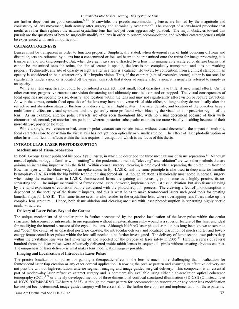
the internal workings of the crystalline lens. In 2006, the most sophisticated finite element model of the crystalline lens that had been created to date was the Burd lens model.32 This simple axisymmetric model had no boundaries between the capsule, cortex, and nucleus, and excluded the fiber structure of the lens with a constant age-independent nucleus and no consideration of ciliary muscle forces, just zonular displacement. With the complex shape and age-dependent lens fiber geometry already established by Kuszak and colleagues,33 a more sophisticated human lens model was created involving zonules (64 springs), a capsule (3-24 µm thick), multilayered cortex with its natural fiber orientation (90 µm/fiber), and a central nucleus (300 µm thick) (Kuszak JR, et al. IOVS 2007;48:ARVO E-Abstract 998). The anterior and posterior radii of curvature were defined by the age-matched lens shape when accommodated and when under resting tension, and Lagrangian brick elements were used to create the multiple nodes of the capsule, and for each of the individual fibers following the age-dependent opposite-end curvature and suture formation of the natural cortex and nucleus. The model with the zonular connections was stretched, and the resulting lens capsule surface profile was analyzed to determine refractive lens power as a function of zonular forces. The zonular forces converted the outward equatorial forces into inward polar forces to compress the fiber mass within the model. The model more closely follows the age-dependent loss of accommodation characterized by Duane34 in comparison to the model of Burd32 without requiring a change in material modulus of elasticity to explain this loss, because the curved fibers straighten and slide over each other with increasing compaction to reflect this age-dependent change.35 The model is figuratively displayed in Figure 2.
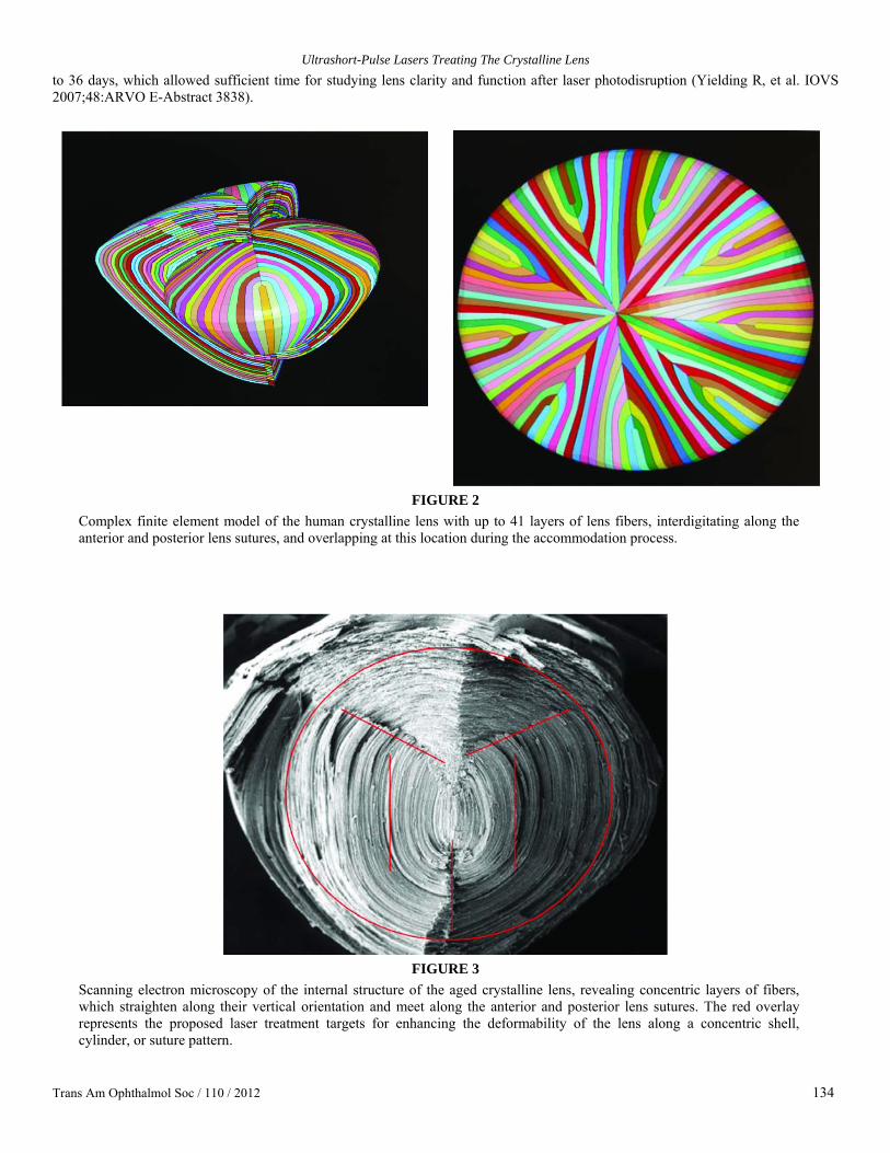
The model, which used up to 41 layers of lens fibers, was tested with known elastic moduli and used to simulate patterns for experimentation. In assessing the nominal sliding of up to 20 layers compared to no sliding in a 45-year-old lens, the model was shown to lead to a 3.4 diopter change in power with simulated accommodation in comparison to the 1.0 diopter change with no sliding. The methods to achieve this sliding surgically were proposed along three different laser patterns that would follow the microanatomy of the crystalline lens, as shown in Figure 3. These included the concentric shell, cylinder, and radial suture patterns.
Early Experimental Studies of Laser-Induced Accommodation Restoration The precursor experimental work in laser-induced accommodation restoration began at LensAR, Inc (Winter Park, Florida) in 2005 and involved ex vivo crystalline lens studies before moving on to living animal eyes. The first step was developing a method for preserving ex vivo porcine, bovine, and human cadaver lenses for experimental photodisruption testing. A lens culture system was created using a culture medium (M199) and incubator at 37°C. The lenses were tested for viability using an alamar blue assay by quantitating their metabolic activity as well as by observing lens clarity. Within the culture medium, lenses would remain clear for up
Ultrashort-Pulse Lasers Treating The Crystalline Lens
Trans Am Ophthalmol Soc / 110 / 2012 134
to 36 days, which allowed sufficient time for studying lens clarity and function after laser photodisruption (Yielding R, et al. IOVS 2007;48:ARVO E-Abstract 3838).
FIGURE 2
Complex finite element model of the human crystalline lens with up to 41 layers of lens fibers, interdigitating along the anterior and posterior lens sutures, and overlapping at this location during the accommodation process.
FIGURE 3
Scanning electron microscopy of the internal structure of the aged crystalline lens, revealing concentric layers of fibers, which straighten along their vertical orientation and meet along the anterior and posterior lens sutures. The red overlay represents the proposed laser treatment targets for enhancing the deformability of the lens along a concentric shell, cylinder, or suture pattern.
Krueger, Uy, McDonald, Edwards
Trans Am Ophthalmol Soc / 110 / 2012 135
The patterns of photodisruption studied included mostly the multilayered concentric shell pattern but also included the cylinder pattern and radial suture pattern. Once the ex vivo lenses were irradiated, they would be tested for accommodative potential by one of two main methods, either the rotation or the compression method. The rotation method, introduced by Fisher in 1971,13 and consisted of putting the lens on a rotating pedestal to simulate the pulling force of the zonules. The reduction of lens thickness with rotation (polar strain) would define the deformability of the lens, which was dependent on age. The compression method was introduced by Glasser and Campbell in 1998 and 199914,36 and consisted of placing the lens on a mechanical “squidger” device, applying gradient steps of compressive force on the lens, and measuring the relative lens resistance to displacement along a fixed distance. A greater force meant the lens was less deformable, and this was also nicely correlated to age.
A third method, the stretching method,14,37 required dissecting out not only the lens, but also the zonules, ciliary body, and overlying band of sclera so that the complex could be attached to a stretching device that would facilitate measurement of the curvature and focusing power of the lens under different degrees of stretching. Although this method is more physiologic because it includes most of the eye’s accommodative apparatus, it was not pursued in these studies because the complex dissection and preparation made it impractical for laser pattern testing.
Each of the former two testing methods was used in ex vivo experimental studies to show an improvement in the deformability of the lens with intralenticular laser treatment (Krueger RR, et al. IOVS 2010;51:ARVO E-Abstract 810). With the rotational studies, there was nearly a twofold reduction of lens thickness (and lens curvature) with spinning after the laser treatment of human cadaver lenses. The laser pattern of multiple concentric shells led to a mean central curvature change of 5.8 ± 2.8 diopters after spinning when compared to controls. Similarly, with the compression studies, there was a twofold or greater reduction in lens stiffness (relative lens resistance, RLR) when using the concentric shells laser pattern in human cadaver lenses (aged 55-77 years) compared to controls. This twofold reduction of RLR was calculated to improve the deformability of a 45- and 55-year-old lens by ~3 diopters and a 65-year-old lens by ~2 diopters, based on the studies of Glasser and Campbell.36 On the basis of these two sets of data and preliminary work in living rabbits, the primate studies were later pursued.
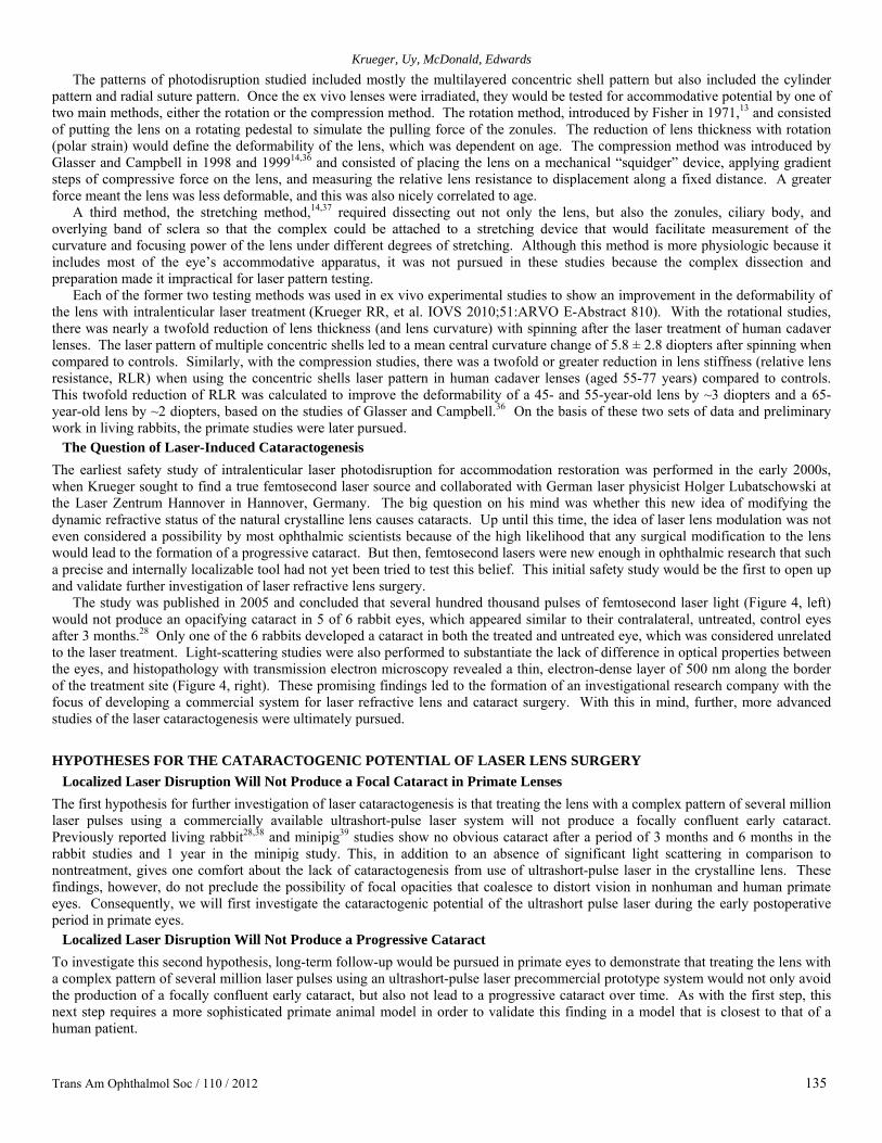
The Question of Laser-Induced Cataractogenesis The earliest safety study of intralenticular laser photodisruption for accommodation restoration was performed in the early 2000s, when Krueger sought to find a true femtosecond laser source and collaborated with German laser physicist Holger Lubatschowski at the Laser Zentrum Hannover in Hannover, Germany. The big question on his mind was whether this new idea of modifying the dynamic refractive status of the natural crystalline lens causes cataracts. Up until this time, the idea of laser lens modulation was not even considered a possibility by most ophthalmic scientists because of the high likelihood that any surgical modification to the lens would lead to the formation of a progressive cataract. But then, femtosecond lasers were new enough in ophthalmic research that such a precise and internally localizable tool had not yet been tried to test this belief. This initial safety study would be the first to open up and validate further investigation of laser refractive lens surgery.
The study was published in 2005 and concluded that several hundred thousand pulses of femtosecond laser light (Figure 4, left) would not produce an opacifying cataract in 5 of 6 rabbit eyes, which appeared similar to their contralateral, untreated, control eyes after 3 months.28 Only one of the 6 rabbits developed a cataract in both the treated and untreated eye, which was considered unrelated to the laser treatment. Light-scattering studies were also performed to substantiate the lack of difference in optical properties between the eyes, and histopathology with transmission electron microscopy revealed a thin, electron-dense layer of 500 nm along the border of the treatment site (Figure 4, right). These promising findings led to the formation of an investigational research company with the focus of developing a commercial system for laser refractive lens and cataract surgery. With this in mind, further, more advanced studies of the laser cataractogenesis were ultimately pursued.
HYPOTHESES FOR THE CATARACTOGENIC POTENTIAL OF LASER LENS SURGERY Localized Laser Disruption Will Not Produce a Focal Cataract in Primate Lenses
The first hypothesis for further investigation of laser cataractogenesis is that treating the lens with a complex pattern of several million laser pulses using a commercially available ultrashort-pulse laser system will not produce a focally confluent early cataract. Previously reported living rabbit28,38 and minipig39 studies show no obvious cataract after a period of 3 months and 6 months in the rabbit studies and 1 year in the minipig study. This, in addition to an absence of significant light scattering in comparison to nontreatment, gives one comfort about the lack of cataractogenesis from use of ultrashort-pulse laser in the crystalline lens. These findings, however, do not preclude the possibility of focal opacities that coalesce to distort vision in nonhuman and human primate eyes. Consequently, we will first investigate the cataractogenic potential of the ultrashort pulse laser during the early postoperative period in primate eyes.
Localized Laser Disruption Will Not Produce a Progressive Cataract To investigate this second hypothesis, long-term follow-up would be pursued in primate eyes to demonstrate that treating the lens with a complex pattern of several million laser pulses using an ultrashort-pulse laser precommercial prototype system would not only avoid the production of a focally confluent early cataract, but also not lead to a progressive cataract over time. As with the first step, this next step requires a more sophisticated primate animal model in order to validate this finding in a model that is closest to that of a human patient.
Ultrashort-Pulse Lasers Treating The Crystalline Lens
Trans Am Ophthalmol Soc / 110 / 2012 136
FIGURE 4
Left, Annular patterns of femtosecond laser pulses within a rabbit crystalline lens, showing microbubbles without coalescence into larger bubbles. Right, Electron microscopy of the treated lens reveals a sharp border with a 0.5-µm electron dense border layer (×5000).
Localized Laser Disruption Will Not Produce a Vision-Threatening Cataract
The final hypothesis regarding the threat of visual decline or symptoms requires a clinical model with subjective vision testing. Here the treatment of the lens with a complex pattern of several million laser pulses using a commercially available ultrashort-pulse laser system is proposed to not produce a vision-threatening cataract in both appearance and visual symptoms. This final step would move beyond primates to human clinical observation.
METHODS
DEVELOPMENT OF A LASER SYSTEM FOR IMAGE-GUIDED LASER LENS PHOTODISRUPTION Intraocular vs Intracorneal Laser Delivery
The laser prototype used during the experimental studies was progressively modified through the various stages of investigation, but basically consists of the following core elements: The laser cavity is a Nd:vanadate (Nd:YVO4) laser with a 10-picosecond pulse width and a 1064-nm wavelength of emission. The laser uses a regenerative amplifier to achieve therapeutic pulse energies, ranging from 5 µJ to 50 µJ. The delivery system involves x,y,z scanning to deliver the programmed laser pattern into the crystalline lens through the user interface at a pulse repetition rate between 20 kHz and 200 kHz. The most recently introduced commercially available laser system has since been upgraded to a femtosecond laser pulse with lower energies and faster pulse repetition rate, but this was not used in these preclinical and early clinical studies.
The ideal user interface with this system involves saline fluid as a coupling medium, together with 3D-CSI as the means for image guidance of the laser delivery. This prototype device did not fully implement the fluid interface optics, but utilized a suction ring with a flat applanation plate in the rabbits and primates, due to its ease of use for early experimentation, and a curved applanation system (radius of curvature of 8 mm) for early clinical investigation, due to the historical ease in patient docking. In both the flat and curved applanating systems, the limitation of posterior corneal folds made this less than ideal for the final commercial system but was acceptable for these early investigations. The numerical aperture (NA) of the focusing optics in the system is not disclosed, but is lower than that seen in systems used purely for corneal applications, so that a deep, intralenticular delivery of the laser could be achieved. This means that a higher-pulse energy is necessary in the lens than that used in the cornea. The flat applanation system used in the primate studies also required an even higher-pulse energy to achieve the therapeutic laser effect in the lens.
Krueger, Uy, McDonald, Edwards
Trans Am Ophthalmol Soc / 110 / 2012 137
Image-Guided Laser Delivery Is Clinically Necessary An essential component in the laser system for use inside the crystalline lens is the use of a high-resolution imaging system for image-guided surgery. In the clinical prototype system, the anterior and posterior cornea and anterior lens surface were imaged with off-axis slit laser imaging, but the location was provided by manual placement of reticules on the imaged surface (Olmstead T, et al. IOVS 2007;48:ARVO E-Abstract 3835). The posterior lens surface position was not calculated from the image, and lens thickness was therefore provided by ultrasonographic methods. In the fully implemented commercial system, a more advanced imaging system using 3D-CSI was built on the early clinical and preclinical experience. This system uses a rotating camera to produce multiple images that automatically locate the corneal and lenticular surfaces, develop a 3-dimensional reconstruction of the anterior eye by a ray-tracing method, and provide biometric data on anterior and posterior corneal curvature, corneal thickness, anterior chamber depth, anterior and posterior lens curvature, and lens thickness. Alternatively, a high-resolution optical coherence tomographer or other similar device could be used in a comparable laser system, so long as adequate imaging of the anterior and posterior lens surfaces is achieved in relation to the other structures of the eye and laser delivery system.29
Early Laser Prototype Is Not the Fully Developed Version The new commercial unit for laser refractive cataract and lens surgery is not the same system as what we used in these experiments, but was developed from these preclinical and clinical prototypes. The commercial system uses a lower pulse width (femtoseconds, rather than picoseconds), superior 3D-CSI imaging, and fluid-based laser coupling to the eye. The femtosecond laser pulse provides a more localized therapeutic effect with less energy,28 while the 3D-CSI imaging provides more accurate biometry and laser guidance. The low-pressure suction ring and fluid-filled, no-corneal-touch patient interface decrease distortion of the cornea and improve image quality and femtosecond laser delivery.40
LONG-TERM PROGRESSIVE CLARITY EVALUATION OF PRIMATE LENSES Experimental Subjects and Preparation
Because of the complexity of management and experimentation in dealing with primate eyes, the experimental laser was transported to and set up at the University of Wisconsin in connection with their ophthalmology research team and the primate center. Dr Paul Kaufman’s laboratory team was responsible for the handling of the animals during the laser treatments and evaluations, as well as in preparing for the experimental setup of the primates, which were tested not only for cataractogenesis, but also for accommodative potential.
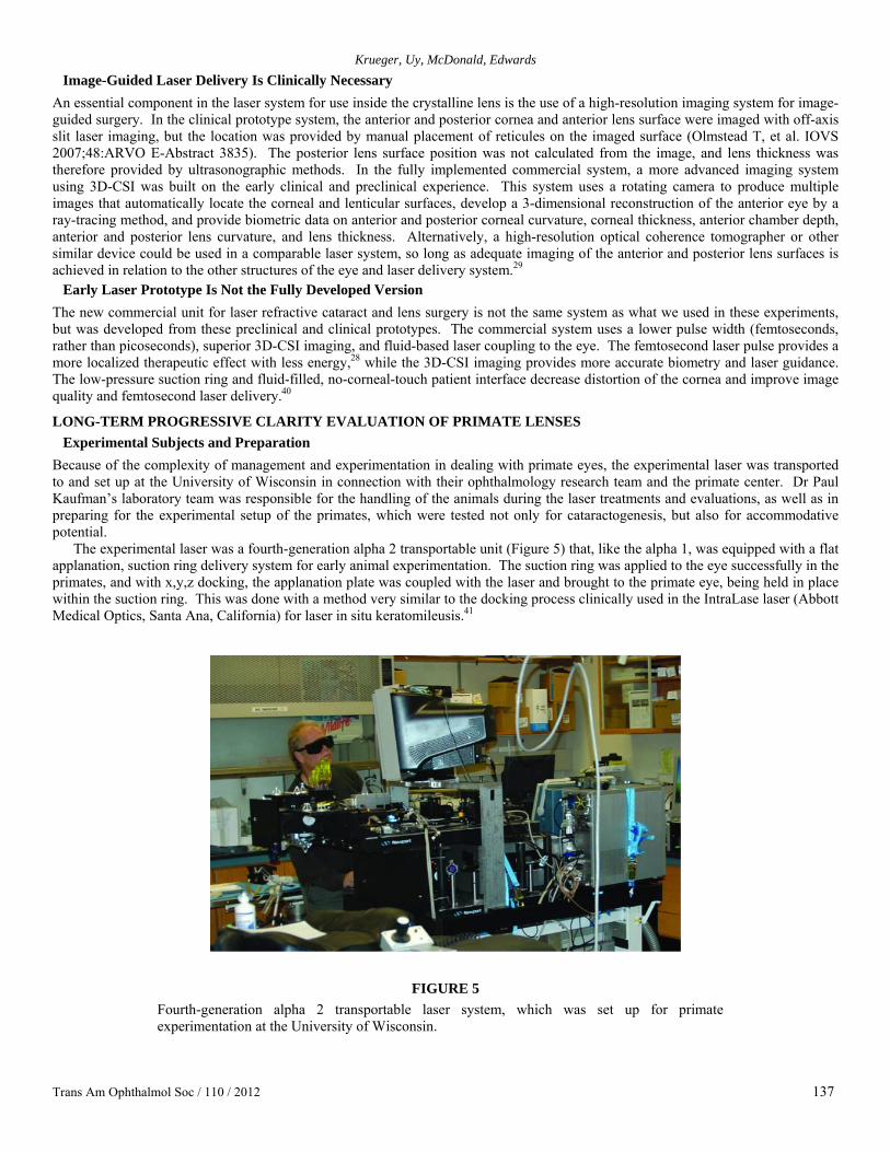
The experimental laser was a fourth-generation alpha 2 transportable unit (Figure 5) that, like the alpha 1, was equipped with a flat applanation, suction ring delivery system for early animal experimentation. The suction ring was applied to the eye successfully in the primates, and with x,y,z docking, the applanation plate was coupled with the laser and brought to the primate eye, being held in place within the suction ring. This was done with a method very similar to the docking process clinically used in the IntraLase laser (Abbott Medical Optics, Santa Ana, California) for laser in situ keratomileusis.41
FIGURE 5 Fourth-generation alpha 2 transportable laser system, which was set up for primate experimentation at the University of Wisconsin.

Ultrashort-Pulse Lasers Treating The Crystalline Lens
Trans Am Ophthalmol Soc / 110 / 2012 138
All monkeys (aged 6 to 20 years) underwent ocular screening grossly and biomicroscopically and exhibited no preexisting ocular abnormalities. The monkeys were housed in a stainless steel cage in an Association for Assessment and Accreditation of Laboratory Animal Care International accredited facility in accordance with the US Department of Agriculture Animal Welfare Act and National Institutes of Health Guide for Care and Use of Laboratory Animals in Research. Tap water was offered ad libitum. There were no contaminants expected in the diet or water that would interfere with the conduct of this study.
A total of 7 animals were enrolled, but ultimately 6 were treated in two groups of 3 each. They included rhesus monkeys between 6 and 20 years of age, with the younger ones (aged 6-10 years) in the first group for the initial pilot investigation, and the older ones (aged 19-20 years) in the second group for more advanced investigation of midbrain electrically stimulated accommodation. The age of the younger primates was equivalent to human subjects in their early 20s, and the older primates were of an equivalent human age of greater than 50 years. The preparation of the headcap through which the primates could be electrically stimulated was already performed prior to enrollment of these latter 3 primates. It involved complex surgical steps in which a bipolar stimulating electrode was implanted into the Edinger-Westphal (E-W) nucleus of the brain to stimulate the accommodation center of the brain.42,43
To facilitate a thorough testing of accommodation, prior to the laser treatment, each of the primates’ eyes underwent surgical removal of the iris (total iridectomy), so that the movement of the lens, zonules, and ciliary body could be fully visualized and the change in refractive power fully analyzed during pharmacologic stimulation with corneal iontophoresis of 40% carbachol in agar (CARB; a supramaximal dose for inducing accommodation)42,43 or during midbrain electrical stimulation.
Anesthesia was induced prior to all experimental procedures with the following medications: (a) In performing the total iridectomy and slit-lamp examinations, we used ketamine, 10 mg/kg intramuscularly, supplemented by ketamine, 5 mg/kg every 20 to 30 minutes as needed. (b) In the midbrain electrode implantation, we used ketamine, 10 mg/kg intramuscularly, and inhalant isofluorane 1% to 2%. (c) In the central electrical stimulation, video recording of accommodative apparatus, and lens lasering procedures, we used ketamine, 10 mg/kg intramuscularly, plus pentobarbital sodium intravenously (15mg/kg, supplemented by pentobarbital sodium, 10 mg/kg per hour intravenously, beginning at 2 to 3 hours, as needed), or inhalant isofluorane 1% to 3%.
Laser Treatment Parameters A minified suction ring and applanation device was coupled to the eye using low-pressure suction. In the event that the suction ring did not adhere properly, four sutures were placed in each quadrant of the ciliary muscle to sustain immobility. The diagnostic laser was used to scan the eye, and based on the diagnostic data the picosecond laser delivered ~30 µJ/pulse at a wavelength of 1064 nm to the lens. It was expected that for each laser pulse, an air bubble approximately three times the initial laser point would be created. Following treatment, a diagnostic scan was performed to determine the outcome of the laser pattern. The laser pattern used for each animal is outlined in Table 1, together with the number of pulses and pulse energy.
TABLE 1. LASER LENS TREATMENT PARAMETERS IN PRIMATE EYES SHAKEDOWN RHESUS HEADCAP RHESUS
IDENTIFIER IDENTIFIER PARAMETER
AY 45 AX 04 AV 42 AN 74 AO 22 AN 89 OD Combo NA Combo Cylinder Spokes Cylinders Laser pattern OS Cylinders NA No laser Shells NA Combo OD 25-45 uJ NA 25 µJ 25-45 µJ 30 µJ 35 µJ Pulse energy OS 26 µJ NA — 31 µJ NA 30-32 µJ OD 0.5+2.0M NA 9.5+1.8M ~2.5 M 2.0 M 7.6 M Total No. pulses OS 5.6 M NA — 3.15 M NA 3.6+1.4M
NA, not available; —, no treatment.
Evaluation and Documentation Over Time The time course of treatment and examination of each rhesus monkey is shown in Table 2. Since the first group was used for early pilot investigation of the laser interaction in the primate lens before treating the older monkeys with the headcap for midbrain electrical stimulation, formalized imaging of the lenses was not performed preoperatively. The second group, however, received imaging 1 to 3 months before the laser treatment. This pre-imaging included a voltage response in midbrain electrical stimulation, gonioscopic imaging of the nasal and temporal quadrants, infrared photo refraction, and ultrasound biomicroscopy (UBM). Originally, fundus photos were not part of the initial protocol but were included with OCT imaging during the postoperative period when concern was expressed regarding the possibility of untoward laser effects at the level of the retina. During these imaging sessions and all the postoperative examinations, qualitative description of the appearance of the eye was reported by Dr Paul Kaufman or his senior laboratory technician, Jared McDonald. Tonometry (Tonopen; Reichert, Inc, Buffalo, New York) was also performed and documented for each examination. During some of these examinations, a slit-lamp video camera was used to record the exam, and details of these exams were extrapolated from the recording. At various times during the postoperative period, pharmacologic evaluation of accommodative amplitude was performed in the first group and midbrain stimulation was performed to elicit the accommodative amplitude in the second group with the headcap. The details of the magnitude of the accommodative amplitude
Krueger, Uy, McDonald, Edwards
Trans Am Ophthalmol Soc / 110 / 2012 139
before and after laser treatment are mentioned only as a brief summary, as these details are beyond the scope of the content of this thesis, and for the most part were noncontributory to its conclusions.
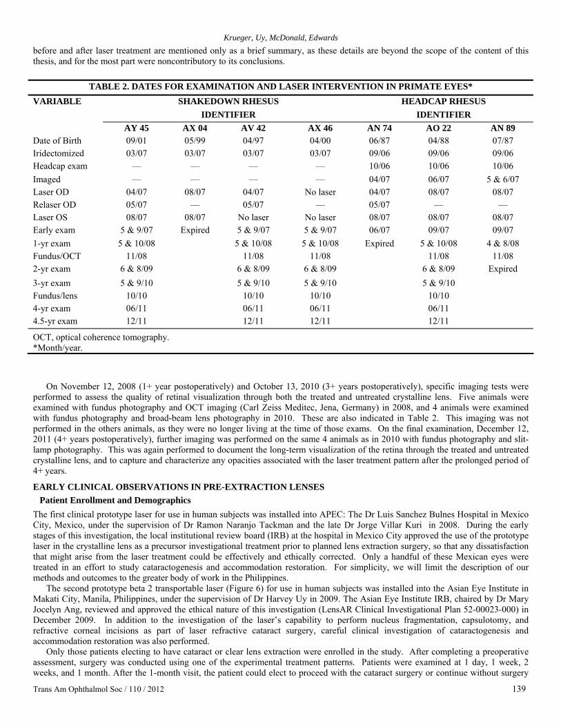
TABLE 2. DATES FOR EXAMINATION AND LASER INTERVENTION IN PRIMATE EYES* SHAKEDOWN RHESUS HEADCAP RHESUS
IDENTIFIER IDENTIFIER VARIABLE
AY 45 AX 04 AV 42 AX 46 AN 74 AO 22 AN 89 Date of Birth 09/01 05/99 04/97 04/00 06/87 04/88 07/87 Iridectomized 03/07 03/07 03/07 03/07 09/06 09/06 09/06 Headcap exam — — — — 10/06 10/06 10/06 Imaged — — — — 04/07 06/07 5 & 6/07 Laser OD 04/07 08/07 04/07 No laser 04/07 08/07 08/07 Relaser OD 05/07 — 05/07 — 05/07 — — Laser OS 08/07 08/07 No laser No laser 08/07 08/07 08/07 Early exam 5 & 9/07 Expired 5 & 9/07 5 & 9/07 06/07 09/07 09/07 1-yr exam 5 & 10/08 5 & 10/08 5 & 10/08 Expired 5 & 10/08 4 & 8/08 Fundus/OCT 11/08 11/08 11/08 11/08 11/08 2-yr exam 6 & 8/09 6 & 8/09 6 & 8/09 6 & 8/09 Expired 3-yr exam 5 & 9/10 5 & 9/10 5 & 9/10 5 & 9/10 Fundus/lens 10/10 10/10 10/10 10/10 4-yr exam 06/11 06/11 06/11 06/11 4.5-yr exam 12/11 12/11 12/11 12/11
OCT, optical coherence tomography. *Month/year.
On November 12, 2008 (1+ year postoperatively) and October 13, 2010 (3+ years postoperatively), specific imaging tests were performed to assess the quality of retinal visualization through both the treated and untreated crystalline lens. Five animals were examined with fundus photography and OCT imaging (Carl Zeiss Meditec, Jena, Germany) in 2008, and 4 animals were examined with fundus photography and broad-beam lens photography in 2010. These are also indicated in Table 2. This imaging was not performed in the others animals, as they were no longer living at the time of those exams. On the final examination, December 12, 2011 (4+ years postoperatively), further imaging was performed on the same 4 animals as in 2010 with fundus photography and slit-lamp photography. This was again performed to document the long-term visualization of the retina through the treated and untreated crystalline lens, and to capture and characterize any opacities associated with the laser treatment pattern after the prolonged period of 4+ years.
EARLY CLINICAL OBSERVATIONS IN PRE-EXTRACTION LENSES Patient Enrollment and Demographics
The first clinical prototype laser for use in human subjects was installed into APEC: The Dr Luis Sanchez Bulnes Hospital in Mexico City, Mexico, under the supervision of Dr Ramon Naranjo Tackman and the late Dr Jorge Villar Kuri in 2008. During the early stages of this investigation, the local institutional review board (IRB) at the hospital in Mexico City approved the use of the prototype laser in the crystalline lens as a precursor investigational treatment prior to planned lens extraction surgery, so that any dissatisfaction that might arise from the laser treatment could be effectively and ethically corrected. Only a handful of these Mexican eyes were treated in an effort to study cataractogenesis and accommodation restoration. For simplicity, we will limit the description of our methods and outcomes to the greater body of work in the Philippines.
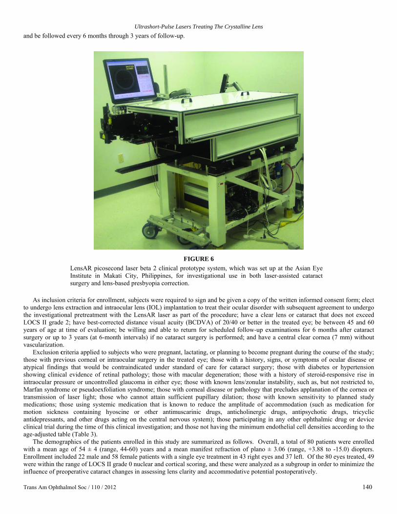
The second prototype beta 2 transportable laser (Figure 6) for use in human subjects was installed into the Asian Eye Institute in Makati City, Manila, Philippines, under the supervision of Dr Harvey Uy in 2009. The Asian Eye Institute IRB, chaired by Dr Mary Jocelyn Ang, reviewed and approved the ethical nature of this investigation (LensAR Clinical Investigational Plan 52-00023-000) in December 2009. In addition to the investigation of the laser’s capability to perform nucleus fragmentation, capsulotomy, and refractive corneal incisions as part of laser refractive cataract surgery, careful clinical investigation of cataractogenesis and accommodation restoration was also performed.
Only those patients electing to have cataract or clear lens extraction were enrolled in the study. After completing a preoperative assessment, surgery was conducted using one of the experimental treatment patterns. Patients were examined at 1 day, 1 week, 2 weeks, and 1 month. After the 1-month visit, the patient could elect to proceed with the cataract surgery or continue without surgery
Ultrashort-Pulse Lasers Treating The Crystalline Lens
Trans Am Ophthalmol Soc / 110 / 2012 140
and be followed every 6 months through 3 years of follow-up.
FIGURE 6 LensAR picosecond laser beta 2 clinical prototype system, which was set up at the Asian Eye Institute in Makati City, Philippines, for investigational use in both laser-assisted cataract surgery and lens-based presbyopia correction.
As inclusion criteria for enrollment, subjects were required to sign and be given a copy of the written informed consent form; elect
to undergo lens extraction and intraocular lens (IOL) implantation to treat their ocular disorder with subsequent agreement to undergo the investigational pretreatment with the LensAR laser as part of the procedure; have a clear lens or cataract that does not exceed LOCS II grade 2; have best-corrected distance visual acuity (BCDVA) of 20/40 or better in the treated eye; be between 45 and 60 years of age at time of evaluation; be willing and able to return for scheduled follow-up examinations for 6 months after cataract surgery or up to 3 years (at 6-month intervals) if no cataract surgery is performed; and have a central clear cornea (7 mm) without vascularization.
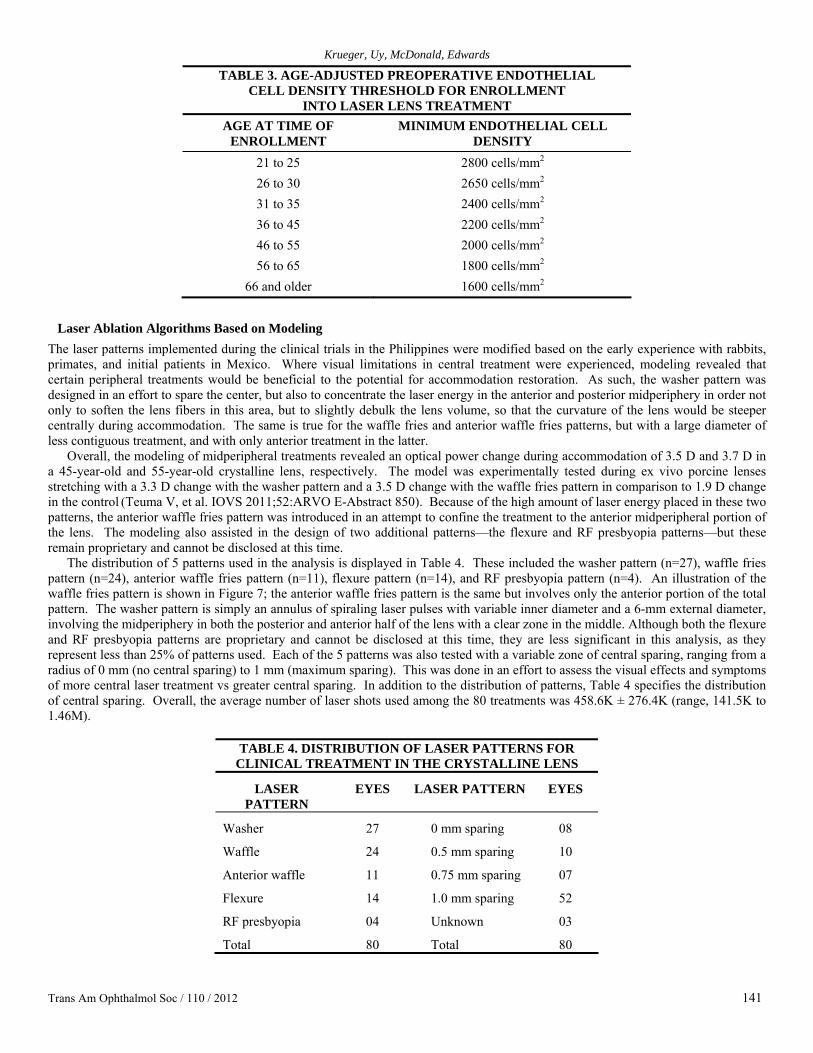
Exclusion criteria applied to subjects who were pregnant, lactating, or planning to become pregnant during the course of the study; those with previous corneal or intraocular surgery in the treated eye; those with a history, signs, or symptoms of ocular disease or atypical findings that would be contraindicated under standard of care for cataract surgery; those with diabetes or hypertension showing clinical evidence of retinal pathology; those with macular degeneration; those with a history of steroid-responsive rise in intraocular pressure or uncontrolled glaucoma in either eye; those with known lens/zonular instability, such as, but not restricted to, Marfan syndrome or pseudoexfoliation syndrome; those with corneal disease or pathology that precludes applanation of the cornea or transmission of laser light; those who cannot attain sufficient pupillary dilation; those with known sensitivity to planned study medications; those using systemic medication that is known to reduce the amplitude of accommodation (such as medication for motion sickness containing hyoscine or other antimuscarinic drugs, anticholinergic drugs, antipsychotic drugs, tricyclic antidepressants, and other drugs acting on the central nervous system); those participating in any other ophthalmic drug or device clinical trial during the time of this clinical investigation; and those not having the minimum endothelial cell densities according to the age-adjusted table (Table 3).
The demographics of the patients enrolled in this study are summarized as follows. Overall, a total of 80 patients were enrolled with a mean age of 54 ± 4 (range, 44-60) years and a mean manifest refraction of plano ± 3.06 (range, +3.88 to -15.0) diopters. Enrollment included 22 male and 58 female patients with a single eye treatment in 43 right eyes and 37 left. Of the 80 eyes treated, 49 were within the range of LOCS II grade 0 nuclear and cortical scoring, and these were analyzed as a subgroup in order to minimize the influence of preoperative cataract changes in assessing lens clarity and accommodative potential postoperatively.
Krueger, Uy, McDonald, Edwards
Trans Am Ophthalmol Soc / 110 / 2012 141
TABLE 3. AGE-ADJUSTED PREOPERATIVE ENDOTHELIAL CELL DENSITY THRESHOLD FOR ENROLLMENT
INTO LASER LENS TREATMENT AGE AT TIME OF
ENROLLMENT MINIMUM ENDOTHELIAL CELL
DENSITY
21 to 25 2800 cells/mm2 26 to 30 2650 cells/mm2 31 to 35 2400 cells/mm2 36 to 45 2200 cells/mm2 46 to 55 2000 cells/mm2 56 to 65 1800 cells/mm2
66 and older 1600 cells/mm2
Laser Ablation Algorithms Based on Modeling
The laser patterns implemented during the clinical trials in the Philippines were modified based on the early experience with rabbits, primates, and initial patients in Mexico. Where visual limitations in central treatment were experienced, modeling revealed that certain peripheral treatments would be beneficial to the potential for accommodation restoration. As such, the washer pattern was designed in an effort to spare the center, but also to concentrate the laser energy in the anterior and posterior midperiphery in order not only to soften the lens fibers in this area, but to slightly debulk the lens volume, so that the curvature of the lens would be steeper centrally during accommodation. The same is true for the waffle fries and anterior waffle fries patterns, but with a large diameter of less contiguous treatment, and with only anterior treatment in the latter.
Overall, the modeling of midperipheral treatments revealed an optical power change during accommodation of 3.5 D and 3.7 D in a 45-year-old and 55-year-old crystalline lens, respectively. The model was experimentally tested during ex vivo porcine lenses stretching with a 3.3 D change with the washer pattern and a 3.5 D change with the waffle fries pattern in comparison to 1.9 D change in the control (Teuma V, et al. IOVS 2011;52:ARVO E-Abstract 850). Because of the high amount of laser energy placed in these two patterns, the anterior waffle fries pattern was introduced in an attempt to confine the treatment to the anterior midperipheral portion of the lens. The modeling also assisted in the design of two additional patterns—the flexure and RF presbyopia patterns—but these remain proprietary and cannot be disclosed at this time.
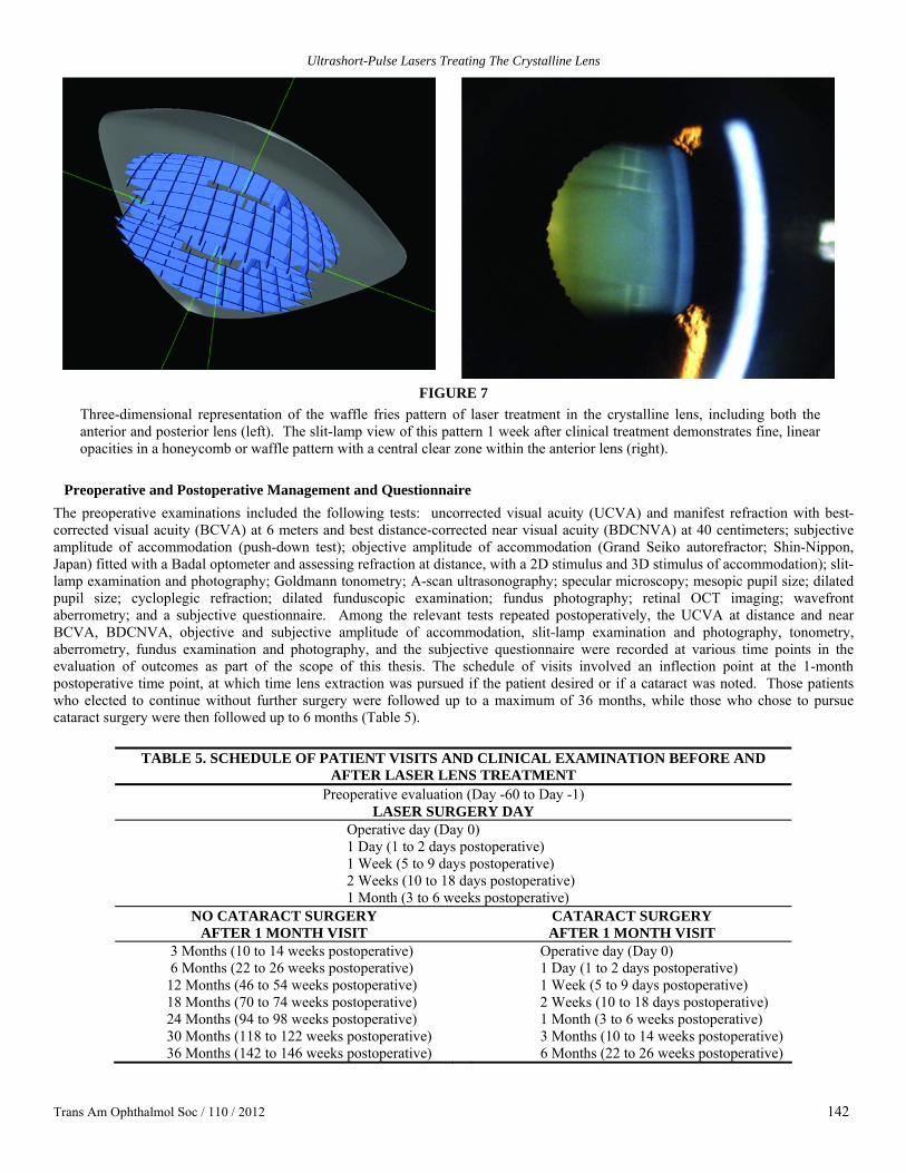
The distribution of 5 patterns used in the analysis is displayed in Table 4. These included the washer pattern (n=27), waffle fries pattern (n=24), anterior waffle fries pattern (n=11), flexure pattern (n=14), and RF presbyopia pattern (n=4). An illustration of the waffle fries pattern is shown in Figure 7; the anterior waffle fries pattern is the same but involves only the anterior portion of the total pattern. The washer pattern is simply an annulus of spiraling laser pulses with variable inner diameter and a 6-mm external diameter, involving the midperiphery in both the posterior and anterior half of the lens with a clear zone in the middle. Although both the flexure and RF presbyopia patterns are proprietary and cannot be disclosed at this time, they are less significant in this analysis, as they represent less than 25% of patterns used. Each of the 5 patterns was also tested with a variable zone of central sparing, ranging from a radius of 0 mm (no central sparing) to 1 mm (maximum sparing). This was done in an effort to assess the visual effects and symptoms of more central laser treatment vs greater central sparing. In addition to the distribution of patterns, Table 4 specifies the distribution of central sparing. Overall, the average number of laser shots used among the 80 treatments was 458.6K ± 276.4K (range, 141.5K to 1.46M).
TABLE 4. DISTRIBUTION OF LASER PATTERNS FOR
CLINICAL TREATMENT IN THE CRYSTALLINE LENS
LASER PATTERN
EYES LASER PATTERN EYES
Washer 27 0 mm sparing 08
Waffle 24 0.5 mm sparing 10
Anterior waffle 11 0.75 mm sparing 07
Flexure 14 1.0 mm sparing 52
RF presbyopia 04 Unknown 03
Total 80 Total 80
Ultrashort-Pulse Lasers Treating The Crystalline Lens
Trans Am Ophthalmol Soc / 110 / 2012 142
FIGURE 7
Three-dimensional representation of the waffle fries pattern of laser treatment in the crystalline lens, including both the anterior and posterior lens (left). The slit-lamp view of this pattern 1 week after clinical treatment demonstrates fine, linear opacities in a honeycomb or waffle pattern with a central clear zone within the anterior lens (right).
Preoperative and Postoperative Management and Questionnaire
The preoperative examinations included the following tests: uncorrected visual acuity (UCVA) and manifest refraction with best-corrected visual acuity (BCVA) at 6 meters and best distance-corrected near visual acuity (BDCNVA) at 40 centimeters; subjective amplitude of accommodation (push-down test); objective amplitude of accommodation (Grand Seiko autorefractor; Shin-Nippon, Japan) fitted with a Badal optometer and assessing refraction at distance, with a 2D stimulus and 3D stimulus of accommodation); slit-lamp examination and photography; Goldmann tonometry; A-scan ultrasonography; specular microscopy; mesopic pupil size; dilated pupil size; cycloplegic refraction; dilated funduscopic examination; fundus photography; retinal OCT imaging; wavefront aberrometry; and a subjective questionnaire. Among the relevant tests repeated postoperatively, the UCVA at distance and near BCVA, BDCNVA, objective and subjective amplitude of accommodation, slit-lamp examination and photography, tonometry, aberrometry, fundus examination and photography, and the subjective questionnaire were recorded at various time points in the evaluation of outcomes as part of the scope of this thesis. The schedule of visits involved an inflection point at the 1-month postoperative time point, at which time lens extraction was pursued if the patient desired or if a cataract was noted. Those patients who elected to continue without further surgery were followed up to a maximum of 36 months, while those who chose to pursue cataract surgery were then followed up to 6 months (Table 5).
TABLE 5. SCHEDULE OF PATIENT VISITS AND CLINICAL EXAMINATION BEFORE AND AFTER LASER LENS TREATMENT
Preoperative evaluation (Day -60 to Day -1) LASER SURGERY DAY
Operative day (Day 0) 1 Day (1 to 2 days postoperative) 1 Week (5 to 9 days postoperative) 2 Weeks (10 to 18 days postoperative) 1 Month (3 to 6 weeks postoperative)
NO CATARACT SURGERY AFTER 1 MONTH VISIT
CATARACT SURGERY AFTER 1 MONTH VISIT
3 Months (10 to 14 weeks postoperative) Operative day (Day 0) 6 Months (22 to 26 weeks postoperative) 1 Day (1 to 2 days postoperative) 12 Months (46 to 54 weeks postoperative) 1 Week (5 to 9 days postoperative) 18 Months (70 to 74 weeks postoperative) 2 Weeks (10 to 18 days postoperative) 24 Months (94 to 98 weeks postoperative) 1 Month (3 to 6 weeks postoperative) 30 Months (118 to 122 weeks postoperative) 3 Months (10 to 14 weeks postoperative) 36 Months (142 to 146 weeks postoperative) 6 Months (22 to 26 weeks postoperative)

Krueger, Uy, McDonald, Edwards
Trans Am Ophthalmol Soc / 110 / 2012 143
A patient questionnaire was also administered preoperatively to assess (1) patients’ overall vision with and without glasses, (2) their visual experience, including detailed questions regarding symptoms of glare, haziness, halos, and sharpness, and (3) their visual comfort throughout the day and with reading. The questionnaire was then administered again 1 month postoperatively to assess the same questions about overall vision, visual symptoms, and visual comfort, as well as (4) their experience since the surgery. Those who underwent lens extraction surgery were also questioned again 1 month post lens extraction surgery, especially those who received a diffractive multifocal lens implant (ReSTOR 3.0; Alcon Laboratories, Inc, Fort Worth, Texas) as part of their visual rehabilitation.
RESULTS
PRECLINICAL INVESTIGATION IN NONHUMAN PRIMATES Intraoperative and Immediate Postoperative Findings in Primate Eyes
Among the 7 rhesus monkeys that were enrolled in this analysis, one received no laser treatment, one was sacrificed immediately after bilateral laser treatment, one died immediately after laser treatment of the second eye, and one received laser treatment in only one eye. In each of the treated eyes, focal bubbles were noted immediately at the time of laser intervention, which led to a coalescence of bubbles and localized whitening of the treated areas of the lens. A photograph of the immediate appearance of these bubbles in the pattern of treatment is shown in Figure 8. In the 3 older animals treated, multiple fractures within the lens occurred due to the hardness of these older lenses.
FIGURE 8 The coalescence pattern of bubbles and localized whitening noted within 10 minutes following laser pulsing of primate crystalline lenses using a cylindrical (C) (left), spherical shell (S) (middle), and radial spokes (R) (right) pattern.
The pattern of bubbles apparent in the lens prevented measurements of the the eye’s total refractive error; however, slit-lamp
examinations revealed that the gas bubbles caused by the femtosecond laser completely dissipated within 24 hours. A mild inflammatory reaction was observed in each animal eye (n=11) post laser treatment, as evidenced by the observation of cells and flare in the eyes; however, this reaction did not prevent subsequent refraction measurements beyond 24 hours. The animals appeared to tolerate the procedure well, exhibiting normal behavior and no signs of discomfort post laser treatment. Since the eyes appeared to be normal during the first 3 days of clinical observation (observations of awake animals in home cages), further slit-lamp examination was not performed until later in the month (time period we labeled as “early”) and then yearly afterwards (Tables 2 and 6).
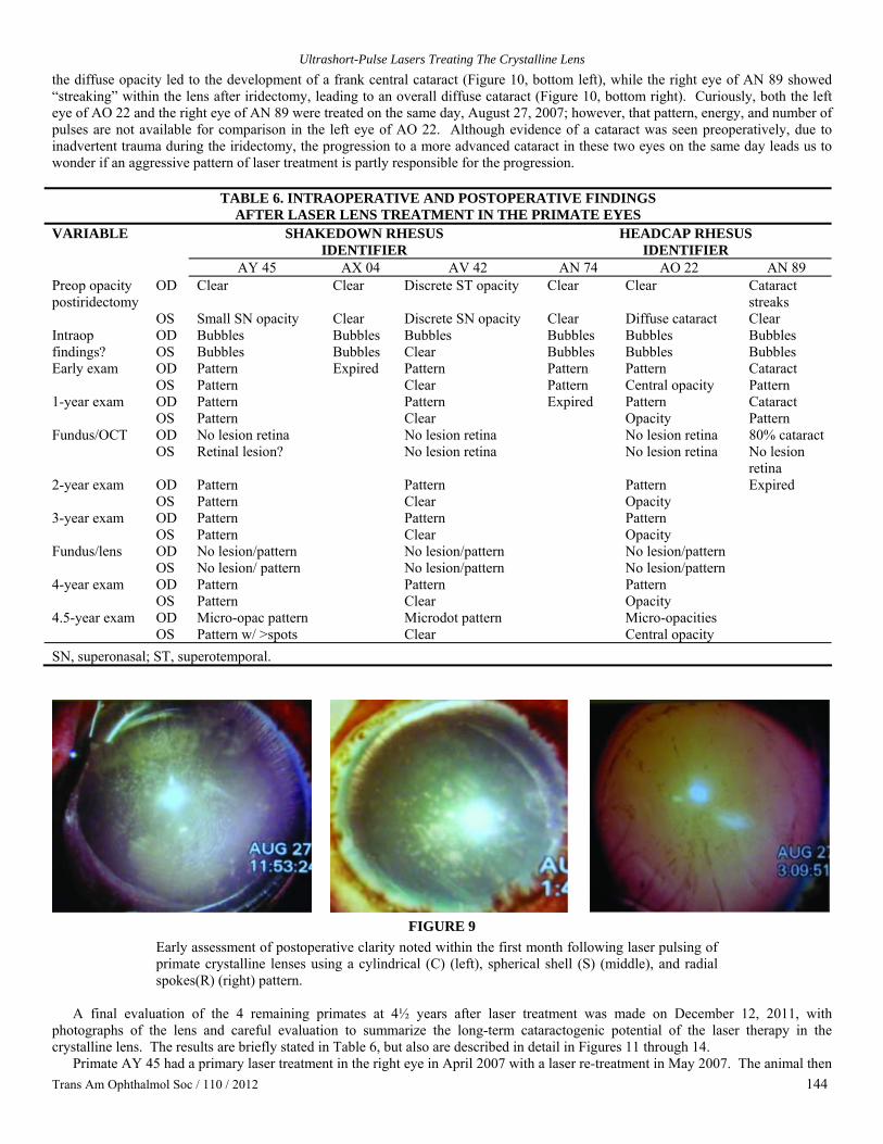
Postoperative Clarity of Primate Lenses Over 4½ Years Overall, a total of 8 eyes received the laser treatment and experienced a period of follow-up for observation of focal lens opacification and cataractogenesis. In each of the 8 eyes, a focal pitting and micro-opacification was noted at the site of each laser pulsing during the earliest postoperative examinations, and this continued throughout the postoperative follow-up period, as described in Table 6. The findings were described as one of two notations—“laser pattern seen” or “pattern clear”—and this was validated by the two examiners, Paul Kaufman and Jared McDonald, as the same finding that was used interchangeably to express that the micro-opacities would define the laser pattern, and yet, beside the observed pattern, the rest of the lens was clear and free of cataract. In Table 6, this is expressed as “pattern” for simplicity. The actual appearance of the 3 patterns of focal micro-opacities is seen in Figure 9.
In two of the lenses examined perioperatively, a regional, focal cataract was noted superiorly and to the left, secondary to the manipulation and unexpected lens trauma of performing the previous iridectomy. This was later photographed and documented as nonprogressive in the right eye of primate AV 42 at 3½ years postoperatively (Figure 10, top left), and in the left eye of animal AY 45 at 1½ years postoperatively (Figure 10, top right). In both the left eye of animal AO 22 and the right eye of animal AN 89, however, a larger area of opacity following the iridectomy resulted in a more diffuse cataract after the laser treatment. In the left eye of AO 22,
Ultrashort-Pulse Lasers Treating The Crystalline Lens
Trans Am Ophthalmol Soc / 110 / 2012 144
the diffuse opacity led to the development of a frank central cataract (Figure 10, bottom left), while the right eye of AN 89 showed “streaking” within the lens after iridectomy, leading to an overall diffuse cataract (Figure 10, bottom right). Curiously, both the left eye of AO 22 and the right eye of AN 89 were treated on the same day, August 27, 2007; however, that pattern, energy, and number of pulses are not available for comparison in the left eye of AO 22. Although evidence of a cataract was seen preoperatively, due to inadvertent trauma during the iridectomy, the progression to a more advanced cataract in these two eyes on the same day leads us to wonder if an aggressive pattern of laser treatment is partly responsible for the progression.
TABLE 6. INTRAOPERATIVE AND POSTOPERATIVE FINDINGS
AFTER LASER LENS TREATMENT IN THE PRIMATE EYES SHAKEDOWN RHESUS HEADCAP RHESUS
IDENTIFIER IDENTIFIER VARIABLE
AY 45 AX 04 AV 42 AN 74 AO 22 AN 89 OD Clear Clear Discrete ST opacity Clear Clear Cataract
streaks Preop opacity postiridectomy
OS Small SN opacity Clear Discrete SN opacity Clear Diffuse cataract Clear OD Bubbles Bubbles Bubbles Bubbles Bubbles Bubbles Intraop
findings? OS Bubbles Bubbles Clear Bubbles Bubbles Bubbles OD Pattern Pattern Pattern Pattern Cataract Early exam OS Pattern
Expired Clear Pattern Central opacity Pattern
OD Pattern Pattern Pattern Cataract 1-year exam OS Pattern
Clear
Expired Opacity Pattern
OD No lesion retina No lesion retina No lesion retina 80% cataract Fundus/OCT OS Retinal lesion?
No lesion retina
No lesion retina No lesion
retina OD Pattern Pattern Pattern 2-year exam OS Pattern
Clear
Opacity
Expired
OD Pattern Pattern Pattern 3-year exam OS Pattern
Clear
Opacity
OD No lesion/pattern No lesion/pattern No lesion/pattern Fundus/lens OS No lesion/ pattern
No lesion/pattern
No lesion/pattern
OD Pattern Pattern Pattern 4-year exam OS Pattern
Clear
Opacity
OD Micro-opac pattern Microdot pattern Micro-opacities 4.5-year exam OS Pattern w/ >spots
Clear
Central opacity
SN, superonasal; ST, superotemporal.
FIGURE 9
Early assessment of postoperative clarity noted within the first month following laser pulsing of primate crystalline lenses using a cylindrical (C) (left), spherical shell (S) (middle), and radial spokes(R) (right) pattern.
A final evaluation of the 4 remaining primates at 4½ years after laser treatment was made on December 12, 2011, with
photographs of the lens and careful evaluation to summarize the long-term cataractogenic potential of the laser therapy in the crystalline lens. The results are briefly stated in Table 6, but also are described in detail in Figures 11 through 14.
Primate AY 45 had a primary laser treatment in the right eye in April 2007 with a laser re-treatment in May 2007. The animal then

Krueger, Uy, McDonald, Edwards
Trans Am Ophthalmol Soc / 110 / 2012 145
had laser treatment in the left eye in August 2007. All along, a faint pattern of laser-induced micro-opacity could be seen in each eye with no progressive cataract. Now, 4½ years later, the right lens again reveals faint, highly localized micro-opacities (Figure 11, left), while the left lens shows larger laser-induced spots that coalesce and are closer to both the anterior and posterior capsule (Figure 11, right). In both eyes the capsules are clear, and the anterior chamber is quiet.
FIGURE 10
Focal opacities photographed superiorly and to the left in the right eye of primate AV 42 (top left) at 3½ years, and in the left eye of primate AY 45 (top right) at 1½ years after laser treatment. The focal opacities existed prior to the laser, due to trauma at the time of iridectomy in both eyes. A more diffuse opacity (cataract) is photographed at 3½ years postoperatively in the left eye of primate AO 22 (bottom left) and at 1½ years postoperatively in the right eye of primate AN 89 (bottom right), but again both existed preoperatively due to trauma at the time of iridectomy. A mild pattern of micro-opacities is also apparent in each of the laser treated eyes.
FIGURE 11
Faint, highly localized micro-opacities in a cylindrical pattern within the right lens (left) with a slighly greater size and axial distribution in the left lens (right) 4½ years after ultrashort-pulse laser therapy inside primate crystalline lenses.

Ultrashort-Pulse Lasers Treating The Crystalline Lens
Trans Am Ophthalmol Soc / 110 / 2012 146
In primate AV 42, the laser treatment and re-treatment in the right eye were performed on the same days as in the previous animal, in April 2007 and May 2007, respectively, but the left eye remained without laser treatment throughout the follow-up period. The right lens revealed a perfect circular pattern of small, discrete dots with no real opacity (Figure 12, left), while the left lens was perfectly clear (Figure 12, right). The rest of each eye was normal.
FIGURE 12 Faint circular pattern of mico-dots without real opacity in the right lens at 4½ years following ultrashort-pulse laser therapy of a primate crystalline lens (left) in comparison to a perfectly clear left lens without laser treatment (right). The two lenses gave an equally clear image when viewing the fundus.
In primate AX 46, no laser treatment was performed in either eye. After nearly 5 years from the time of the iridectomy, only a few
tiny dots were noted in the right lens (Figure 13, left), while the left lens reveals no opacity but only a prominent light reflex (Figure 13, right). The rest of both eyes are normal.
FIGURE 13
Essentially clear primate crystalline lenses without laser therapy; however, small, random dot-like opacities can be seen after follow-up for nearly 5 years.
Finally, in primate AO 22, this older animal was treated in both eyes on two separate days in August 2007. The left eye had
evidence of cataractous opacity prior to the treatment, which remained with some increasing density postoperatively. Now, after 4½ years, the right eye shows a spoked wagon wheel pattern (radial spokes pattern) of faint micro-opacities that appear just beneath the anterior capsule with good clarity of both the anterior and posterior capsule (Figure 14, left). Inferiorly, there is an arc of peripheral opacity that wraps around the equator of the lens, but no new opacity within the laser pattern. In the left eye, a more central pattern of confluent lens opacity is seen within a smaller-diameter area, but with a significantly greater central cloudiness and opacity than in the right eye (Figure 14, right). The anterior and posterior capsule is intact, and the anterior chamber and vitreous are clear.

Krueger, Uy, McDonald, Edwards
Trans Am Ophthalmol Soc / 110 / 2012 147
Accommodative Amplitude in Laser-Treated Primate Lenses Following laser treatment, we were able to obtain refractive measurements using the Hartinger coincidence refractometer (HCR) (Zeiss, Jena, Germany) on each of the monkeys. At various times postoperatively, pharmacologic stimulation with carbachol was performed, mainly among the shakedown monkeys, to assess the refractive status both with and without pharmacologically induced accommodation. The midbrain electrically stimulated voltage response was conducted in the 3 older animals with the headcap to determine the physiologic amplitude of accommodation both preoperatively and postoperatively. An example of the time response of carbachol in pharmacologically inducing accommodation is seen in Figure 15. The left image shows the difference in accommodative response between the laser-treated right eye of animal AV 42 and the untreated left eye. The right image shows the accommodative response of the youngest animal (AY 45) at 3 years after laser lens therapy in both eyes, with the right eye showing a diminished response relative to the left. Although this difference does not reflect a change from the pretreatment value, it does reveal a twofold greater accommodative amplitude in the eye that received a twofold greater magnitude of laser pulsing. Without the preoperative value, however, one can only conclude that there is variability of laser effect and measurement that is beyond the scope of our limited analysis.
FIGURE 14
Faint micro-opacities in a radial sutural pattern 4½ years following ultrashort-pulse laser therapy to an older primate right crystalline lens (left) in comparison to a confluent central opacity (cataract), which existed in part prior to the laser therapy (right). Despite its presence, the fundus was still easily visualized.
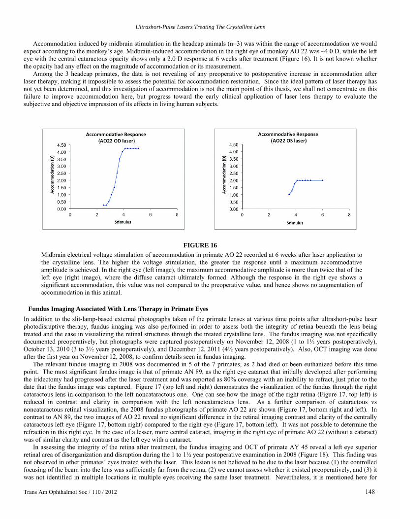
FIGURE 15
Iontophoresis delivered carbechol stimulation of accommodation in primate eyes, revealing a maximum effect after 45 to 60 minutes from topical application. In primate AV 42 (left), the laser treated eye demonstrates greater accommodative amplitude than the nontreated eye. In primate AY 45 (right), the accommodative amplitude in the left eye (treated with a cylindrical pattern of over 5 million pulses) is more than twice that of the right eye (treated with a combination pattern of 2.5 million pulses). In this latter primate, the lens receiving a twofold greater magnitude of laser treatment shows the greater accommodative amplitude.
Ultrashort-Pulse Lasers Treating The Crystalline Lens
Trans Am Ophthalmol Soc / 110 / 2012 148
Accommodation induced by midbrain stimulation in the headcap animals (n=3) was within the range of accommodation we would
expect according to the monkey’s age. Midbrain-induced accommodation in the right eye of monkey AO 22 was ~4.0 D, while the left eye with the central cataractous opacity shows only a 2.0 D response at 6 weeks after treatment (Figure 16). It is not known whether the opacity had any effect on the magnitude of accommodation or its measurement.
Among the 3 headcap primates, the data is not revealing of any preoperative to postoperative increase in accommodation after laser therapy, making it impossible to assess the potential for accommodation restoration. Since the ideal pattern of laser therapy has not yet been determined, and this investigation of accommodation is not the main point of this thesis, we shall not concentrate on this failure to improve accommodation here, but progress toward the early clinical application of laser lens therapy to evaluate the subjective and objective impression of its effects in living human subjects.
FIGURE 16
Midbrain electrical voltage stimulation of accommodation in primate AO 22 recorded at 6 weeks after laser application to the crystalline lens. The higher the voltage stimulation, the greater the response until a maximum accommodative amplitude is achieved. In the right eye (left image), the maximum accommodative amplitude is more than twice that of the left eye (right image), where the diffuse cataract ultimately formed. Although the response in the right eye shows a significant accommodation, this value was not compared to the preoperative value, and hence shows no augmentation of accommodation in this animal.
Fundus Imaging Associated With Lens Therapy in Primate Eyes
In addition to the slit-lamp-based external photographs taken of the primate lenses at various time points after ultrashort-pulse laser photodisruptive therapy, fundus imaging was also performed in order to assess both the integrity of retina beneath the lens being treated and the ease in visualizing the retinal structures through the treated crystalline lens. The fundus imaging was not specifically documented preoperatively, but photographs were captured postoperatively on November 12, 2008 (1 to 1½ years postoperatively), October 13, 2010 (3 to 3½ years postoperatively), and December 12, 2011 (4½ years postoperatively). Also, OCT imaging was done after the first year on November 12, 2008, to confirm details seen in fundus imaging.
The relevant fundus imaging in 2008 was documented in 5 of the 7 primates, as 2 had died or been euthanized before this time point. The most significant fundus image is that of primate AN 89, as the right eye cataract that initially developed after performing the iridectomy had progressed after the laser treatment and was reported as 80% coverage with an inability to refract, just prior to the date that the fundus image was captured. Figure 17 (top left and right) demonstrates the visualization of the fundus through the right cataractous lens in comparison to the left noncataractous one. One can see how the image of the right retina (Figure 17, top left) is reduced in contrast and clarity in comparison with the left noncataractous lens. As a further comparison of cataractous vs noncataractous retinal visualization, the 2008 fundus photographs of primate AO 22 are shown (Figure 17, bottom right and left). In contrast to AN 89, the two images of AO 22 reveal no significant difference in the retinal imaging contrast and clarity of the centrally cataractous left eye (Figure 17, bottom right) compared to the right eye (Figure 17, bottom left). It was not possible to determine the refraction in this right eye. In the case of a lesser, more central cataract, imaging in the right eye of primate AO 22 (without a cataract) was of similar clarity and contrast as the left eye with a cataract.
In assessing the integrity of the retina after treatment, the fundus imaging and OCT of primate AY 45 reveal a left eye superior retinal area of disorganization and disruption during the 1 to 1½ year postoperative examination in 2008 (Figure 18). This finding was not observed in other primates’ eyes treated with the laser. This lesion is not believed to be due to the laser because (1) the controlled focusing of the beam into the lens was sufficiently far from the retina, (2) we cannot assess whether it existed preoperatively, and (3) it was not identified in multiple locations in multiple eyes receiving the same laser treatment. Nevertheless, it is mentioned here for
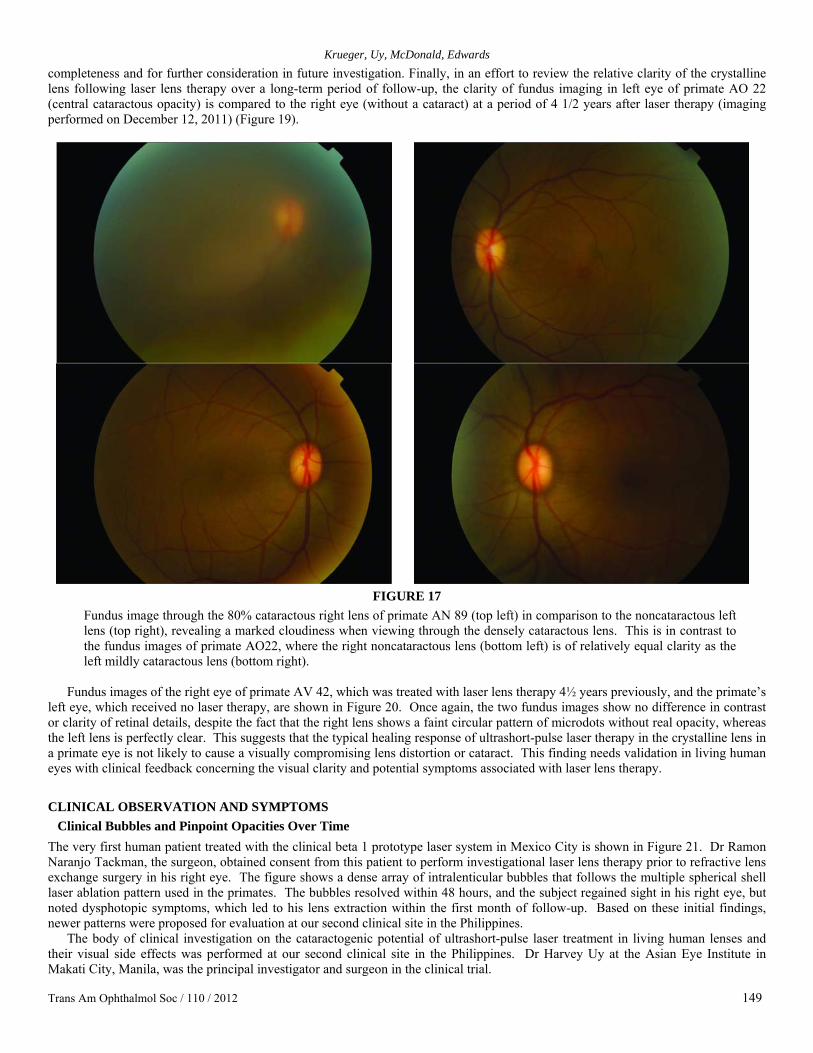
Krueger, Uy, McDonald, Edwards
Trans Am Ophthalmol Soc / 110 / 2012 149
completeness and for further consideration in future investigation. Finally, in an effort to review the relative clarity of the crystalline lens following laser lens therapy over a long-term period of follow-up, the clarity of fundus imaging in left eye of primate AO 22 (central cataractous opacity) is compared to the right eye (without a cataract) at a period of 4 1/2 years after laser therapy (imaging performed on December 12, 2011) (Figure 19).
FIGURE 17
Fundus image through the 80% cataractous right lens of primate AN 89 (top left) in comparison to the noncataractous left lens (top right), revealing a marked cloudiness when viewing through the densely cataractous lens. This is in contrast to the fundus images of primate AO22, where the right noncataractous lens (bottom left) is of relatively equal clarity as the left mildly cataractous lens (bottom right).
Fundus images of the right eye of primate AV 42, which was treated with laser lens therapy 4½ years previously, and the primate’s
left eye, which received no laser therapy, are shown in Figure 20. Once again, the two fundus images show no difference in contrast or clarity of retinal details, despite the fact that the right lens shows a faint circular pattern of microdots without real opacity, whereas the left lens is perfectly clear. This suggests that the typical healing response of ultrashort-pulse laser therapy in the crystalline lens in a primate eye is not likely to cause a visually compromising lens distortion or cataract. This finding needs validation in living human eyes with clinical feedback concerning the visual clarity and potential symptoms associated with laser lens therapy.
CLINICAL OBSERVATION AND SYMPTOMS Clinical Bubbles and Pinpoint Opacities Over Time
The very first human patient treated with the clinical beta 1 prototype laser system in Mexico City is shown in Figure 21. Dr Ramon Naranjo Tackman, the surgeon, obtained consent from this patient to perform investigational laser lens therapy prior to refractive lens exchange surgery in his right eye. The figure shows a dense array of intralenticular bubbles that follows the multiple spherical shell laser ablation pattern used in the primates. The bubbles resolved within 48 hours, and the subject regained sight in his right eye, but noted dysphotopic symptoms, which led to his lens extraction within the first month of follow-up. Based on these initial findings, newer patterns were proposed for evaluation at our second clinical site in the Philippines.
The body of clinical investigation on the cataractogenic potential of ultrashort-pulse laser treatment in living human lenses and their visual side effects was performed at our second clinical site in the Philippines. Dr Harvey Uy at the Asian Eye Institute in Makati City, Manila, was the principal investigator and surgeon in the clinical trial.

Ultrashort-Pulse Lasers Treating The Crystalline Lens
Trans Am Ophthalmol Soc / 110 / 2012 150
FIGURE 18
Fundus red-free photograph and ocular coherence tomography image revealing a superior lesion in the left eye at 1 to 1½ years after laser therapy to the primate crystalline. This lesion reveals retinal disorganization and pigmentary disruption but otherwise appears to be benign. Unfortunately, there are no preoperative fundus photographs to compare, and therefore assigning causation to the laser therapy in the lens cannot be concluded.
FIGURE 19
After 4½ years, the clarity of the fundus image in primate AO 22 is essentially the same in the right eye (left image) as it is in the left eye (right image), even though the left lens has a centrally confluent opacity (cataract).
FIGURE 20
After 4½ years, the clarity of the fundus image in primate AV 42 is the same in the right eye (left image) as it is in the left eye (right image), even though the right eye had laser lens therapy, while the left eye had no laser treatment.

Krueger, Uy, McDonald, Edwards
Trans Am Ophthalmol Soc / 110 / 2012 151
The majority of the 80 eyes treated (>75%) had one of three patterns (washer ring, waffle fries, or anterior waffle fries), as specified in Table 4. All the eyes experienced bubbles intraoperatively, which in some cases were more densely distributed than in others. In a few eyes with more dense nuclei, lens fracturing occurred during the procedure, and this also dissipated within the first days. Figure 22 shows the “down the pipe” view immediately after treatment of a 2+ nuclear sclerotic lens with an anterior waffle fries pattern, where only minimal, small bubbles can be seen along the pattern, but with a large superior paracentral ridge of lens fracturing. This myopic patient had a preoperative BSCVA of 20/40, which remained 20/40 after the mild bubbles and dense fracture ridge disappeared. There were no laser-related complications, but also no change in accommodation after placement of the anterior waffle fries pattern. After 1 month, the patient elected to have cataract surgery with a diffractive multifocal IOL in an effort to improve his near vision.
FIGURE 21
First clinical eye treated with ultrashort-pulse laser therapy in the crystalline lens by Dr Ramon Naranjo Tackman in Mexico City in 2008. Immediately following the treatment, a dense array of microbubbles can be seen, outlining the laser ablation pattern of multiple spherical shells. The bubbles completely resolved within the first 48 hours.
FIGURE 22 Intraoperative photograph of laser lens surgery in a 2+ nuclear sclerotic lens, showing minimal bubble expansion and coalescence along the intersecting squared lines of the laser pattern (anterior waffle fries), but extensive posterior lens fracturing (irregular gray circle with randomly radiating lines) due to the density of the lens. The bubbles and evidence of the lens fracturing disappear within the first days after laser lens surgery. The broken circle of white spots is the light reflex from the laser’s illumination system.
Postoperative slit-lamp photography of the most frequently utilized laser ablation pattern, the washer ring pattern, is shown in
Figure 23. Images of the crystalline lens at 1 hour, 1 day, 1 week, and 1 month are shown, with the annular pattern of laser pulses with central sparing noted. Throughout the brief postoperative follow-up, there is no evidence of a progressive cataract. At the 1-month visit, the 51-year-old patient, who had only a 4-letter improvement in near vision and +0.25 D improvement in objective accommodation, elected to undergo lens extraction surgery with implantation of a multifocal intraocular lens. Postoperatively, the patient’s distance and near visual acuity both improved, although he reported halos and glare at night from the diffractive multifocal intraocular lens.
Figure 24 shows an eye treated with the anterior waffle fries pattern at 1 hour and 1 month after laser lens surgery. The anterior waffle fries pattern with clear zones and central sparing shows persistent clarity in these untreated zones with only faint lines of micro-opacity at the locations of laser impact. The 52-year-old patient reported marked visual disturbances with blurred, hazy vision and glare immediately after the treatment. Within a couple days after laser application, these visual symptoms disappeared concomitant with disappearance of bubbles. After a week, visual acuity returned to preoperative levels with only minimal haziness due to the original mild cataract. The patient elected to have lens extraction surgery with a ReSTOR multifocal lens (Alcon Labs, Inc, Fort Worth, Texas) because of the desire to regain reading vision. After multifocal lens implantation, distance, intermediate, and reading acuity improved with elimination of the mildly hazy vision. However, nocturnal halos and glare were experienced, together with a
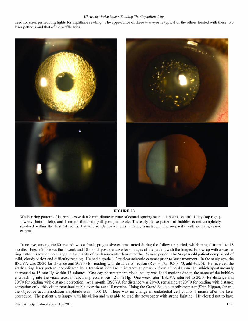
Ultrashort-Pulse Lasers Treating The Crystalline Lens
Trans Am Ophthalmol Soc / 110 / 2012 152
need for stronger reading lights for nighttime reading. The appearance of these two eyes is typical of the others treated with these two laser patterns and that of the waffle fries.
FIGURE 23
Washer ring pattern of laser pulses with a 2-mm-diameter zone of central sparing seen at 1 hour (top left), 1 day (top right), 1 week (bottom left), and 1 month (bottom right) postoperatively. The early dense pattern of bubbles is not completely resolved within the first 24 hours, but afterwards leaves only a faint, translucent micro-opacity with no progressive cataract. In no eye, among the 80 treated, was a frank, progressive cataract noted during the follow-up period, which ranged from 1 to 18
months. Figure 25 shows the 1-week and 18-month postoperative lens images of the patient with the longest follow-up with a washer ring pattern, showing no change in the clarity of the laser-treated lens over the 1½ year period. The 56-year-old patient complained of mild, cloudy vision and difficulty reading. He had a grade 1-2 nuclear sclerotic cataract prior to laser treatment. In the study eye, the BSCVA was 20/20 for distance and 20/200 for reading with distance correction (Rx= +1.75 -0.5 × 70, add +2.75). He received the washer ring laser pattern, complicated by a transient increase in intraocular pressure from 17 to 41 mm Hg, which spontaneously decreased to 15 mm Hg within 15 minutes. One day posttreatment, visual acuity was hand motions due to the some of the bubbles encroaching into the visual axis; intraocular pressure was 12 mm Hg. One week later, BSCVA returned to 20/50 for distance and 20/70 for reading with distance correction. At 1 month, BSCVA for distance was 20/40, remaining at 20/70 for reading with distance correction only; this vision remained stable over the next 18 months. Using the Grand Seiko autorefractometer (Shin-Nippon, Japan), the objective accommodation amplitude was +1.00 D. There was no change in endothelial cell counts 1 month after the laser procedure. The patient was happy with his vision and was able to read the newspaper with strong lighting. He elected not to have
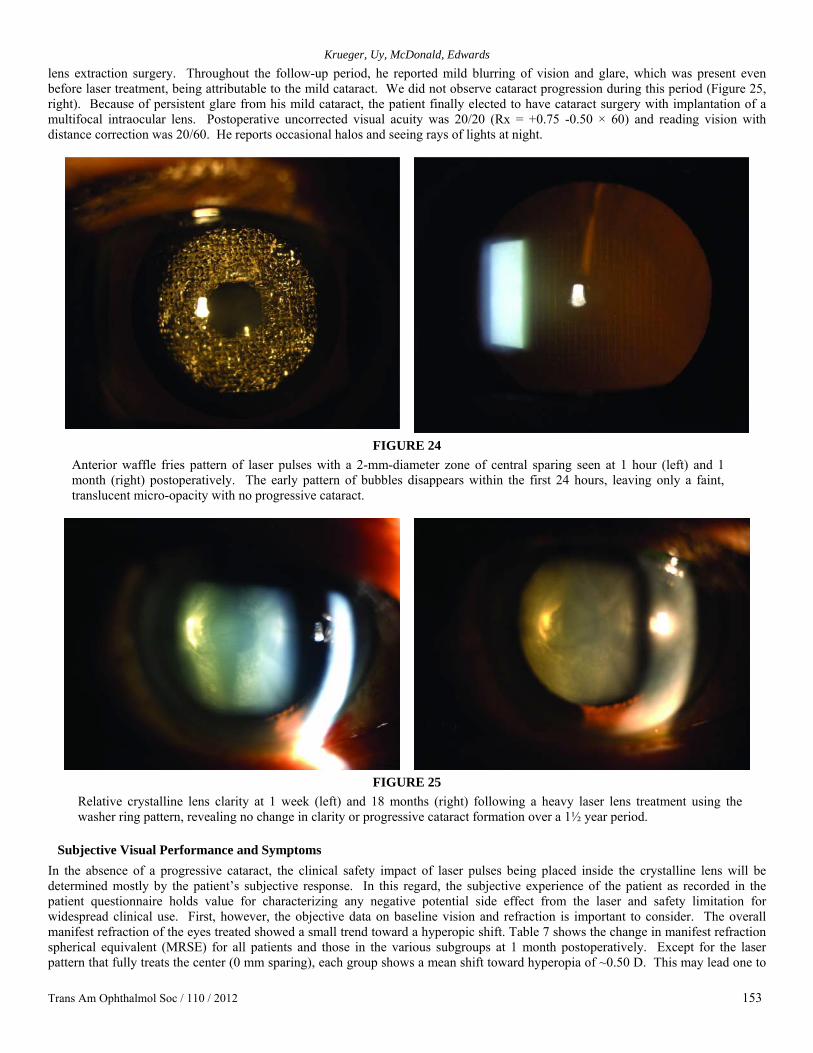
Krueger, Uy, McDonald, Edwards
Trans Am Ophthalmol Soc / 110 / 2012 153
lens extraction surgery. Throughout the follow-up period, he reported mild blurring of vision and glare, which was present even before laser treatment, being attributable to the mild cataract. We did not observe cataract progression during this period (Figure 25, right). Because of persistent glare from his mild cataract, the patient finally elected to have cataract surgery with implantation of a multifocal intraocular lens. Postoperative uncorrected visual acuity was 20/20 (Rx = +0.75 -0.50 × 60) and reading vision with distance correction was 20/60. He reports occasional halos and seeing rays of lights at night.
FIGURE 24
Anterior waffle fries pattern of laser pulses with a 2-mm-diameter zone of central sparing seen at 1 hour (left) and 1 month (right) postoperatively. The early pattern of bubbles disappears within the first 24 hours, leaving only a faint, translucent micro-opacity with no progressive cataract.
FIGURE 25
Relative crystalline lens clarity at 1 week (left) and 18 months (right) following a heavy laser lens treatment using the washer ring pattern, revealing no change in clarity or progressive cataract formation over a 1½ year period.
Subjective Visual Performance and Symptoms
In the absence of a progressive cataract, the clinical safety impact of laser pulses being placed inside the crystalline lens will be determined mostly by the patient’s subjective response. In this regard, the subjective experience of the patient as recorded in the patient questionnaire holds value for characterizing any negative potential side effect from the laser and safety limitation for widespread clinical use. First, however, the objective data on baseline vision and refraction is important to consider. The overall manifest refraction of the eyes treated showed a small trend toward a hyperopic shift. Table 7 shows the change in manifest refraction spherical equivalent (MRSE) for all patients and those in the various subgroups at 1 month postoperatively. Except for the laser pattern that fully treats the center (0 mm sparing), each group shows a mean shift toward hyperopia of ~0.50 D. This may lead one to
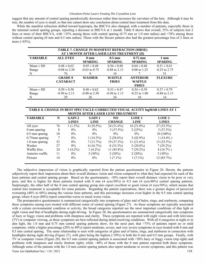
Ultrashort-Pulse Lasers Treating The Crystalline Lens
Trans Am Ophthalmol Soc / 110 / 2012 154
suggest that any amount of central sparing paradoxically decreases rather than increases the curvature of the lens. Although it may be true, the number of eyes is small, so that one cannot draw any conclusion about central laser treatment from this data.
While the manifest refraction shifted toward hyperopia, the BSCVA also changed, with a number of patients, especially those in the minimal central sparing groups, reporting a decrease in BSCVA at 1 month. Table 8 shows that overall, 35% of subjects lost 2 lines or more of their BSCVA, with <25% among those with central sparing (0.75 mm or 1.0 mm radius) and >70% among those without central sparing (0 mm and 0.5 mm radius). Those with the flexure pattern also had the greatest percentage loss of 2 lines or more (>85%).
TABLE 7. CHANGE IN MANIFEST REFRACTION (MRSE)
AT 1 MONTH AFTER LASER LENS TREATMENT (D) VARIABLE ALL EYES 0 mm
SPARING 0.5 mm
SPARING 0.75 mm
SPARING 1 mm
SPARING Mean ± SD 0.48 ± 0.62 0.05 ± 0.48 0.56 ± 0.80 0.64 ± 0.48 0.51 ± 0.61 Range -0.88 to 2.50 -0.63 to 0.75 -0.88 to 2.13 0.00 to 1.25 -0.25 to 2.75 N 76 8 10 7 51 GRADE 0
CATARACT WASHER WAFFLE
FRIES ANTERIOR
WAFFLE FRIES
FLEXURE
Mean ± SD 0.50 ± 0.50 0.48 ± 0.62 0.33 ± 0.47 0.54 ± 0.39 0.37 ± 0.79 Range -0.50 to 2.13 -0.88 to 2.50 -0.50 to 1.13 -0.25 to 1.00 -0.88 to 2.13 N 29 26 24 10 14
TABLE 8. CHANGE IN BEST SPECTACLE-CORRECTED VISUAL ACUITY logMAR LINES AT 1 MONTH AFTER LASER LENS TREATMENT
VARIABLE N GAIN 2 LINES
GAIN 1 LINE
NO CHANGE
LOSE 1 LINE
LOSE 2 LINES
All eyes 76 1 (1.3%) 8 (10.5%) 24 (31.6%) 16 (21.0%) 27 (35.5%) 0 mm sparing 8 0% 0% 3 (37.5%) 2 (25%) 3 (37.5%) 0.5 mm sparing 10 0% 0% 0% 0% 10 (100%) 0.75mm sparing 7 0% 1 (14.3%) 2 (28.6%) 3 (42.9%) 1 (14.4% ) 1.0 mm sparing 52 0% 7 (13.7%) 19 (37.3%) 11 (21.6%) 13 (25.5%) Washer 27 0% 4 (16.7%) 8 (33.3%) 5 (20.8%) 7 (29.2%) Waffle fries 24 1 (4.2%) 1 (4.2%) 11 (45.8%) 7 (29.2%) 4 (16.7% ) Anterior waffle 11 0% 3 (30%) 2 (20%) 2 (20%) 3 (30%) Flexure 14 0% 0% 1 (7.1%) 1 (7.1%) 12 (85.7%)
The subjective impression of vision is graphically reported from the patient questionnaire in Figure 26. Herein, the patients
subjectively report their impression about their overall distance vision and vision compared to what they had expected for each of the laser patterns and central sparing groups. Based on the questionnaire, ~30% report their overall distance vision to be poor or very poor, and this is higher for those patients treated with 0 mm (4 eyes/50%) or 0.5 mm (4 eyes/40%) central sparing patterns. Surprisingly, the other half of the 0 mm central sparing group also report excellent or good vision (4 eyes/50%), which means that central lens treatment is acceptable for some patients. Regarding the patient expectations, there was a greater degree of perceived worsening (40% to 60%) among the various laser patterns, and this percentage increases even higher in the 0.5 mm central sparing group, where 8 eyes (80%) report somewhat worse or much worse vision.
The postoperative questionnaire is summarized categorically into symptoms of glare and of halos, rings, and starbursts, comparing these symptoms among eyes treated with different zones of central sparing (Figure 27). As these symptoms are typically associated with a certain environmental condition or activity, the broad categories reported are the most important conditions of dim light or night vision and that of headlights during night driving. In Figure 28, the questionnaires are summarized categorically into symptoms of hazy or foggy vision and problems with sharpness and clarity. These symptoms are reported with night vision and with television (TV) or computer viewing, as these symptoms are best reflected during detail-resolving conditions. With all 4 categories at night or in dim light, the 1.0 mm and 0.75 mm central sparing patterns show, for the most part, that >75% of patients report no or mild symptoms, while a higher percentage (20% to 80%) report moderate, severe, and very severe symptoms in eyes treated with 0 mm and 0.5 mm central sparing. The same relationship is seen with categories of glare and of halos, rings, and starbursts in connection with headlights during night driving (Figure 27), with glare being the highest (>50%) in both the 0 mm and 0.5 mm groups. Finally, with TV or computer vision (Figure 28), the 0.5 mm central sparing pattern is associated with >50% hazy or foggy vision (top right) and problems with sharpness and clarity (bottom right), while ~40% of those with the 0 mm pattern reported both these symptoms. Although some of the patients with the 1.0 mm central sparing pattern also report moderate or severe symptoms, and this pattern was
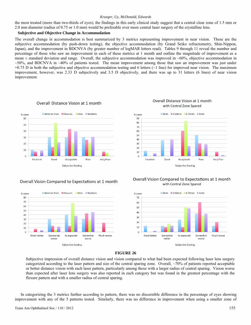
Krueger, Uy, McDonald, Edwards
Trans Am Ophthalmol Soc / 110 / 2012 155
the most treated (more than two-thirds of eyes), the findings in this early clinical study suggest that a central clear zone of 1.5 mm or 2.0 mm diameter (radius of 0.75 or 1.0 mm) would be preferable over more central laser surgery of the crystalline lens.
Subjective and Objective Change in Accommodation The overall change in accommodation is best summarized by 3 metrics representing improvement in near vision. These are the subjective accommodation (by push-down testing), the objective accommodation (by Grand Seiko refractometry, Shin-Nippon, Japan), and the improvement in BDCNVA (by greater number of logMAR letters read). Tables 9 through 11 reveal the number and percentage of those who saw an improvement in each of these metrics at 1 month and outline the magnitude of improvement as a mean ± standard deviation and range. Overall, the subjective accommodation was improved in ~60%, objective accommodation in ~50%, and BDCNVA in ~40% of patients tested. The mean improvement among those that saw an improvement was just under +0.75 D in both the subjective and objective accommodation testing and 6 letters (~1 line) for improved near vision. The maximum improvement, however, was 2.33 D subjectively and 3.5 D objectively, and there was up to 31 letters (6 lines) of near vision improvement.
FIGURE 26
Subjective impression of overall distance vision and vision compared to what had been expected following laser lens surgery categorized according to the laser pattern and size of the central sparing zone. Overall, ~70% of patients reported acceptable or better distance vision with each laser pattern, particularly among those with a larger radius of central sparing. Vision worse than expected after laser lens surgery was also reported in each category but was found in the greatest percentage with the flexure pattern and with a smaller radius of central sparing.
In categorizing the 3 metrics further according to pattern, there was no discernible difference in the percentage of eyes showing improvement with any of the 5 patterns tested. Similarly, there was no difference in improvement when using a smaller zone of
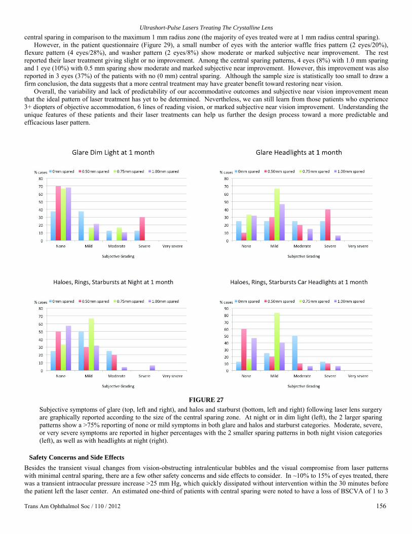
Ultrashort-Pulse Lasers Treating The Crystalline Lens
Trans Am Ophthalmol Soc / 110 / 2012 156
central sparing in comparison to the maximum 1 mm radius zone (the majority of eyes treated were at 1 mm radius central sparing). However, in the patient questionnaire (Figure 29), a small number of eyes with the anterior waffle fries pattern (2 eyes/20%),
flexure pattern (4 eyes/28%), and washer pattern (2 eyes/8%) show moderate or marked subjective near improvement. The rest reported their laser treatment giving slight or no improvement. Among the central sparing patterns, 4 eyes (8%) with 1.0 mm sparing and 1 eye (10%) with 0.5 mm sparing show moderate and marked subjective near improvement. However, this improvement was also reported in 3 eyes (37%) of the patients with no (0 mm) central sparing. Although the sample size is statistically too small to draw a firm conclusion, the data suggests that a more central treatment may have greater benefit toward restoring near vision.
Overall, the variability and lack of predictability of our accommodative outcomes and subjective near vision improvement mean that the ideal pattern of laser treatment has yet to be determined. Nevertheless, we can still learn from those patients who experience 3+ diopters of objective accommodation, 6 lines of reading vision, or marked subjective near vision improvement. Understanding the unique features of these patients and their laser treatments can help us further the design process toward a more predictable and efficacious laser pattern.
FIGURE 27
Subjective symptoms of glare (top, left and right), and halos and starburst (bottom, left and right) following laser lens surgery are graphically reported according to the size of the central sparing zone. At night or in dim light (left), the 2 larger sparing patterns show a >75% reporting of none or mild symptoms in both glare and halos and starburst categories. Moderate, severe, or very severe symptoms are reported in higher percentages with the 2 smaller sparing patterns in both night vision categories (left), as well as with headlights at night (right).
Safety Concerns and Side Effects
Besides the transient visual changes from vision-obstructing intralenticular bubbles and the visual compromise from laser patterns with minimal central sparing, there are a few other safety concerns and side effects to consider. In ~10% to 15% of eyes treated, there was a transient intraocular pressure increase >25 mm Hg, which quickly dissipated without intervention within the 30 minutes before the patient left the laser center. An estimated one-third of patients with central sparing were noted to have a loss of BSCVA of 1 to 3
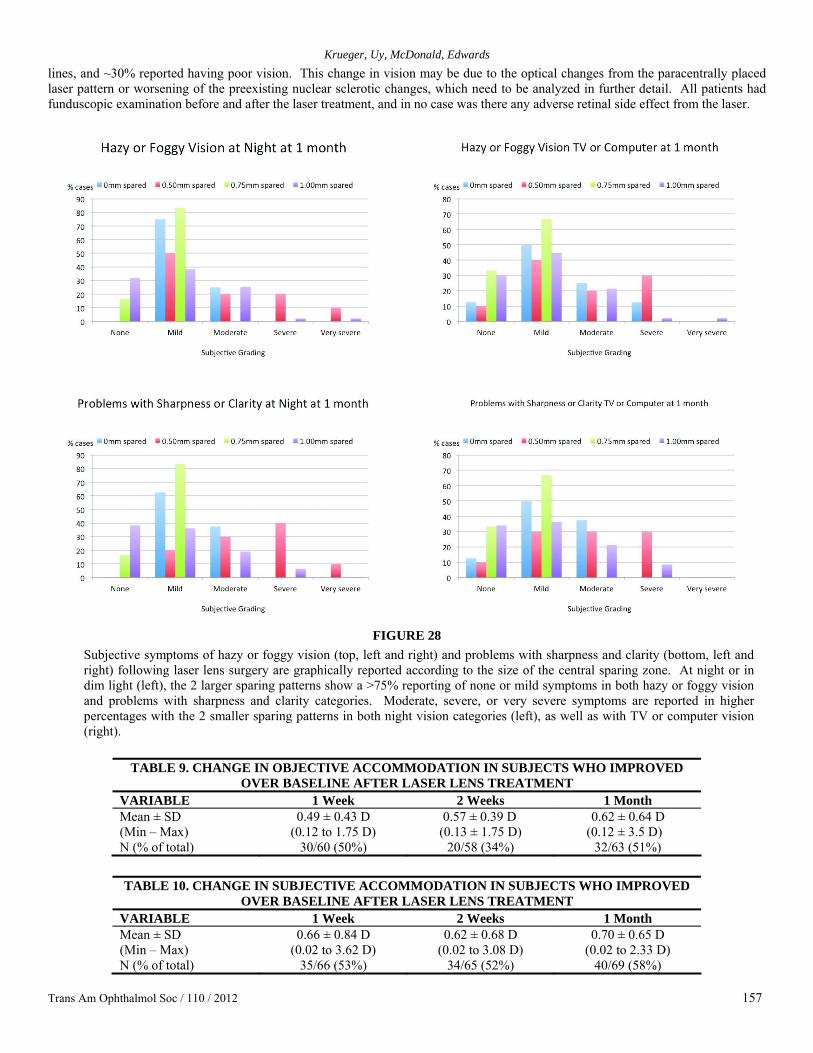
Krueger, Uy, McDonald, Edwards
Trans Am Ophthalmol Soc / 110 / 2012 157
lines, and ~30% reported having poor vision. This change in vision may be due to the optical changes from the paracentrally placed laser pattern or worsening of the preexisting nuclear sclerotic changes, which need to be analyzed in further detail. All patients had funduscopic examination before and after the laser treatment, and in no case was there any adverse retinal side effect from the laser.
FIGURE 28
Subjective symptoms of hazy or foggy vision (top, left and right) and problems with sharpness and clarity (bottom, left and right) following laser lens surgery are graphically reported according to the size of the central sparing zone. At night or in dim light (left), the 2 larger sparing patterns show a >75% reporting of none or mild symptoms in both hazy or foggy vision and problems with sharpness and clarity categories. Moderate, severe, or very severe symptoms are reported in higher percentages with the 2 smaller sparing patterns in both night vision categories (left), as well as with TV or computer vision (right).
TABLE 9. CHANGE IN OBJECTIVE ACCOMMODATION IN SUBJECTS WHO IMPROVED
OVER BASELINE AFTER LASER LENS TREATMENT VARIABLE 1 Week 2 Weeks 1 Month Mean ± SD 0.49 ± 0.43 D 0.57 ± 0.39 D 0.62 ± 0.64 D (Min – Max) (0.12 to 1.75 D) (0.13 ± 1.75 D) (0.12 ± 3.5 D) N (% of total) 30/60 (50%) 20/58 (34%) 32/63 (51%)
TABLE 10. CHANGE IN SUBJECTIVE ACCOMMODATION IN SUBJECTS WHO IMPROVED
OVER BASELINE AFTER LASER LENS TREATMENT VARIABLE 1 Week 2 Weeks 1 Month Mean ± SD 0.66 ± 0.84 D 0.62 ± 0.68 D 0.70 ± 0.65 D (Min – Max) (0.02 to 3.62 D) (0.02 to 3.08 D) (0.02 to 2.33 D) N (% of total) 35/66 (53%) 34/65 (52%) 40/69 (58%)

Ultrashort-Pulse Lasers Treating The Crystalline Lens
Trans Am Ophthalmol Soc / 110 / 2012 158
TABLE 11. CHANGE IN BEST DISTANCE-CORRECTED NEAR VISUAL ACUITY (LETTERS) IN SUBJECTS WHO IMPROVED OVER BASELINE AFTER LASER LENS TREATMENT
VARIABLE 1 Week 2 Weeks 1 Month Mean ± SD 5 ± 4 letters 6 ± 5 letters 6 ± 6 letters (Min – Max) (1 to 17) (1 to 18) (1 to 31) N (% of total) 28/74 (38%) 36/72 (50%) 32/76 (42%)
FIGURE 29
Subjective improvement in near vision following laser lens surgery categorized according to the laser pattern and size of the central sparing zone.
DISCUSSION
SIGNIFICANCE OF THE FINDINGS IN THIS THESIS Lasers and Cataracts
Before addressing the hypotheses presented in the “Introduction,” it seems appropriate to define the terms cataract and laser, from the thesis title, in order to draw the proper conclusions. Not all lasers are the same, and similarly not all cataracts are the same. When using term cataract, we need to be specific in its context.
Defining what we mean by cataract. Webster’s dictionary describes a cataract as a clouding of the lens of the eye that obstructs the passage of light. The word comes from the Latin cataracta, waterfall, and from the Greek kataraktes, to dash or strike down. The word is synonymous with a large, forceful waterfall or cascade. So, as described, it is the whiteness of the waterfall and the forcefulness of the water dashing down on the rock that historically characterizes the cataract.
Inside the crystalline lens, it is not the dashing down of water that produces what we clinically refer to as cataract, but a host of other insults, with age itself being one of the biggest culprits.44-46 Unlike the cornea, which is a living, transparent structure that maintains its clarity by a fine homeostasis of drawing in and pumping out fluid, and new surface cells that push out and slough away the old, dying cells, the crystalline lens is an immunologically privileged organ, separated from the outside, intraocular environment by a largely impermeable capsule. It is this protective capsule that keeps out the negative influence that could alter the delicate, highly organized fiber structure that maintains transparency. The shape and organization of the fiber structure and the lattice-like matrix of the crystallines within keep the living lens clear, even as it grows over time.47
The relative disadvantage of the lens in maintaining its transparency is that the aging epithelial cells do not slough away, as with the cornea, but are sequestered as they lead to the formation of new fibers that grow inwardly, compacting the old fiber cells. Were it not for this inwardly growing compaction process, we would probably not lose the dynamic process of accommodation with aging, and likely, we would not experience the development of nuclear sclerosis that leads to cataract. As a living organ there is still diffusion of nutrients into the lens and waste products out of the lens, and accommodation is the movement that keeps this process functional. As accommodation is lost with the central compaction of dead and dying fibers, so the transport of nutrients and waste products is impacted too.48 Eventually the crystalline lens is destined to develop a cataract, if not from an outward environmental

Krueger, Uy, McDonald, Edwards
Trans Am Ophthalmol Soc / 110 / 2012 159
insult, then inwardly from the dysfunction that comes with aging. In contrast, the relative advantage of the lens over the cornea in maintaining its transparency is the lack of a wound healing
response, which could pathologically impair its function. The beauty of excimer laser photorefractive keratectomy (PRK) in sculpting the cornea is occasionally haunted by an aggressive wound healing response, which leads to a recalcitrant corneal haze and scarring.49 Not only is the refractive effect disturbed and unstable, but the haze obstructs the clear passage of light. Alternatively, the beauty of a laser in the lens is that there is no cellular wound healing response that can become pathologic. Indeed, the capsule could be violated to create a bigger problem, but precise ultrashort-pulse laser delivery can avoid a capsular violation and has the potential to precisely incise and separate lens fiber layers with minimal disturbance to the complex lens ultrastructure.50-52
Understanding differences in lasers. The word laser is an acrostic that stands for light amplification by stimulated emission of radiation. We all fundamentally know what a laser is but technically may have a hard time explaining it to someone. A laser is a device that utilizes the natural oscillation of atoms or molecules between energy levels for generating a beam of electromagnetic radiation, usually in the visible, ultraviolet, or infrared regions of the spectrum. Lasers differ by a host of various distinguishers, such as wavelength, duration, pulse width, energy density, peak power, spot size, pulse frequency, numerical aperture, and absorption coefficient, but, most important, by the fundamental effect on the irradiated tissue.
There are basically five different laser tissue interactions to consider in understanding how therapeutic lasers work, and for the most part they are divided by the intensity of the beam and its interaction time with the tissue (Figure 30). (1) Photovaporization is the physical basis of the early surgical applications with lasers in tissue cutting and removal at relatively high energy density, moderate exposure times (milliseconds to seconds), and the rapid deposition of heat with subsequent vaporization. (2) Photocoagulation, with a relatively long exposure time (from 10 to 100 milliseconds) but lower irradiance (up to 10 W/cm2) and energy density, is typically achieved with a 30- to 100-μm focused spot, derived from a temperature rise and protein denaturation within the tissue. (3) Photoablation uses shorter, nanosecond pulses and high-photon energies (ie, ultraviolet excimer laser wavelength at 193 nm) to deliver single photons that cleave specific chemical bonds, resulting in a strong, nonthermal absorption. (4) Photodisruption, using picosecond and shorter pulses, induces a nonlinear absorption at high peak powers to freely pass through and interact in otherwise transparent tissue. (5) Photochemistry, as the final mechanism, is observed at very low laser-intensity levels delivered over minutes or hours. These interactions are strongly coupled to the host tissue response to photochemistry and remain, as yet, poorly understood.
Of the 5 main laser tissue interaction mechanisms, photodisruption is the one most suited for delivery into the crystalline lens. The mechanism of interaction behind laser photodisruption is best described as plasma-mediated ablation, or optical breakdown. It relies on the nonlinear absorption of laser energy in the target achieved when the material-specific radiant exposure is exceeded. Fundamentally, optical breakdown is characterized by 3 successive major events: plasma formation, shock wave generation, and expanding cavitation (Figure 31). The plasma is a highly ionized state of matter and can be generated by laser pulses (from femtosecond to few nanoseconds) of low energy and high peak power. It has been shown that shortening the pulse duration from nanoseconds to femtoseconds decreases the threshold for plasma formation and reduces mechanical effects.53
For the sake of clinical discussion and characterization, it seems best to define and specifically refer to a cataract as a vision-compromising opacity. Although the scientific purist might wish to call any opacity a cataract, the clinician who is attempting to restore or maintain vision must see the functional component and differentiate a cataract from an insignificant opacity, as will be done here. Similarly, for the clinical discussion of ultrashort-pulse lasers, our prototype system using picosecond laser pulsing is comparable to that of femtosecond lasers, even though the energy and spot size are greater. Hence, as we discuss our findings, we also believe that the results can be improved with even more refined femtosecond laser delivery.
Does Localized Laser Disruption Produce Focal Cataract in Primate Lenses? The previously published studies of potential cataractogenesis with femtosecond lasers nicely report the ability of delivering a therapeutic laser pattern inside the living lenses of rabbits28,38 and minipig,39 and then following these animals for 3 months and 6 months (rabbit studies) and 1 year (minipig). These studies concluded the following: (1) “…no cataract formation with no loss of lens function or induced light scattering after 3 months”28; (2) “…did not induce an increasing opacification (cataract) over a six month follow-up period. However, the incisions were still detectable using Scheimpflug imaging and histopathological techniques, although the visibility of the incisions was declining”38; and (3) “…the laser pattern itself induces light scattering [by Scheimpflug imaging], which needs to be distinguished from cataractous opacities.…light scattering within the laser treated lenses remains on a higher level compared to the controls, but the slit lamp images clearly show that there is neither an indication of cataract formation nor any significant healing process.…Fs-laser treatment of the crystalline lens has not led to significant cataractous opacities in minipigs during the 1 year follow-up….”39
The conclusions drawn in rabbit and minipig studies report a “laser pattern” being distinguished throughout the follow-up period with declining visibility, but no visible opacities. This laser pattern comes from the localized energy of the laser photodisruption with creation of a voided space that locally disrupts the lens microarchitechture. This corresponds with the report of the “laser pattern” being seen throughout the follow-up of our primates, although in our study we also report seeing micro-opacities. This additional finding is not reflective of the difference between primates and rabbits/minipigs. Minipigs are very similar to human primates with regard to their lens protein content (31%)45 and distribution and age dependence of lens crystalline.54 In contrast, rabbits have a higher lens protein content (55%)45 and a unique lens activity that can lead to regeneration.55 The minipig data should be very comparable to that of primates and humans. The difference in the micro-opacities is more explainable from the differences in the energy sources being tested. With our prototype picosecond laser, the pulse energy used in the primates was on the order of 25 to 45 µJ, whereas in
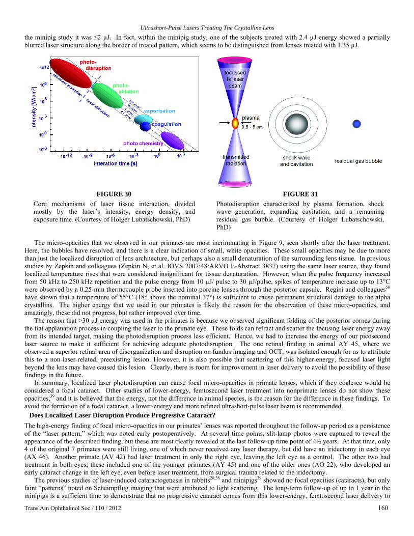
Ultrashort-Pulse Lasers Treating The Crystalline Lens
Trans Am Ophthalmol Soc / 110 / 2012 160
the minipig study it was ≤2 µJ. In fact, within the minipig study, one of the subjects treated with 2.4 µJ energy showed a partially blurred laser structure along the border of treated pattern, which seems to be distinguished from lenses treated with 1.35 µJ.
FIGURE 30
Core mechanisms of laser tissue interaction, divided mostly by the laser’s intensity, energy density, and exposure time. (Courtesy of Holger Lubatschowski, PhD)
FIGURE 31 Photodisruption characterized by plasma formation, shock wave generation, expanding cavitation, and a remaining residual gas bubble. (Courtesy of Holger Lubatschowski, PhD)
The micro-opacities that we observed in our primates are most incriminating in Figure 9, seen shortly after the laser treatment.
Here, the bubbles have resolved, and there is a clear indication of small, white opacities. These small opacities may be due to more than just the localized disruption of lens architecture, but perhaps also a small denaturation of the surrounding lens tissue. In previous studies by Zepkin and colleagues (Zepkin N, et al. IOVS 2007;48:ARVO E-Abstract 3837) using the same laser source, they found localized temperature rises that were considered insignificant for tissue denaturation. However, when the pulse frequency increased from 50 kHz to 250 kHz repetition and the pulse energy from 10 µJ/ pulse to 30 µJ/pulse, spikes of temperature increase up to 13°C were observed by a 0.25-mm thermocouple probe inserted into porcine lenses through the posterior capsule. Regini and colleagues56 have shown that a temperature of 55°C (18° above the nominal 37°) is sufficient to cause permanent structural damage to the alpha crystallins. The higher energy that we used in our primates is likely the reason for the observation of these micro-opacities, and amazingly, these did not progress, but rather improved over time.
The reason that >30 µJ energy was used in the primates is because we observed significant folding of the posterior cornea during the flat applanation process in coupling the laser to the primate eye. These folds can refract and scatter the focusing laser energy away from its intended target, making the photodisruption process less efficient. Hence, we had to increase the energy of our picosecond laser source to make it sufficient for achieving adequate photodisruption. The one retinal finding in animal AY 45, where we observed a superior retinal area of disorganization and disruption on fundus imaging and OCT, was isolated enough for us to attribute this to a non-laser-related, preexisting lesion. However, it is also possible that scattering of this higher-energy, focused laser light beyond the lens may have caused this lesion. Clearly, there is room for improvement in laser delivery to avoid the possibility of these findings in the future.
In summary, localized laser photodisruption can cause focal micro-opacities in primate lenses, which if they coalesce would be considered a focal cataract. Other studies of lower-energy, femtosecond laser treatment into nonprimate lenses do not show these opacities,39 and it is believed that the energy, not the difference in animal species, is the reason for the difference in these findings. To avoid the formation of a focal cataract, a lower-energy and more refined ultrashort-pulse laser beam is recommended.
Does Localized Laser Disruption Produce Progressive Cataract? The high-energy finding of focal micro-opacities in our primates’ lenses was reported throughout the follow-up period as a persistence of the “laser pattern,” which was noted early postoperatively. At several time points, slit-lamp photos were captured to reveal the appearance of the described finding, but these are most clearly revealed at the last follow-up time point of 4½ years. At that time, only 4 of the original 7 primates were still living, one of which never received any laser therapy, but did have an iridectomy in each eye (AX 46). Another primate (AV 42) had laser treatment in only the right eye, leaving the left eye as a control. The other two had treatment in both eyes; these included one of the younger primates (AY 45) and one of the older ones (AO 22), who developed an early cataract change in the left eye, even before laser treatment, from surgical trauma related to the iridectomy.
The previous studies of laser-induced cataractogenesis in rabbits28,38 and minipigs39 showed no focal opacities (cataracts), but only faint “patterns” noted on Scheimpflug imaging that were attributed to light scattering. The long-term follow-up of up to 1 year in the minipigs is a sufficient time to demonstrate that no progressive cataract comes from this lower-energy, femtosecond laser delivery to
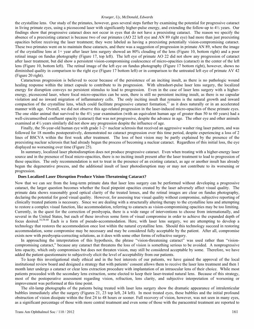
Krueger, Uy, McDonald, Edwards
Trans Am Ophthalmol Soc / 110 / 2012 161
the crystalline lens. Our study of the primates, however, goes several steps further by examining the potential for progressive cataract in living primate eyes, using a picosecond laser with significantly higher-pulse energy, and extending the follow-up to 4½ years. Our findings show that progressive cataract does not occur in eyes that do not have a preexisting cataract. The reason we specify the absence of a preexisting cataract is because two of our primates (AO 22 left eye and AN 89 right eye) had more than just preexisting opacities before receiving the laser treatment; they were labeled as having a preexisting potentially vision-compromising cataract. These two primates went on to maintain these cataracts, and there was a suggestion of progression in primate AN 89, where the image of the crystalline lens at 1+ year after laser lens surgery showed an 80% clouding of the lens (Figure 10, bottom right) and a poor retinal image on fundus photography (Figure 17, top left). The left eye of primate AO 22 did not show any progression of cataract after laser treatment, but did show a persistent vision-compromising coalescence of micro-opacities (cataract) in the center of the left lens (Figure 10, bottom left). The retinal image of the left eye on fundus photography (Figure 17 bottom right), however, shows no diminished quality in comparison to the right eye (Figure 17 bottom left) or in comparison to the untreated left eye of primate AV 42 (Figure 20 right).
Cataractous progression is believed to occur because of the persistence of an inciting insult, as there is no pathologic wound healing response within the intact capsule to contribute to its progression. With ultrashort-pulse laser lens surgery, the localized energy for disruption conveys no persistent stimulus to lead to progression. Even in the case of laser lens surgery with a higher-energy, picosecond laser, where focal micro-opacities can be seen, there is still no persistent inciting insult, as there is no capsular violation and no inward migration of inflammatory cells. The only inciting insult that remains is the natural growth and inward compaction of the crystalline lens, which could facilitate progressive cataract formation,57 as it does naturally or in an accelerated manner with age. Overall, we did not observe this age-related progression in the laser-induced micro-opacities of these primate eyes. The one older animal that survived to the 4½ year examination (with an equivalent human age of greater than 50 to 60 years) had a well-circumscribed confluent opacity (cataract) that was not progressive, despite the advance in age. The other eye and other animals examined at 4½ years similarly did not show any progression despite the influence of age.
Finally, the 56-year-old human eye with grade 1-2+ nuclear sclerosis that received an aggressive washer ring laser pattern, and was followed for 18 months postoperatively, demonstrated no cataract progression over this time period, despite experiencing a loss of 2 lines of BSCVA within the first week after treatment. The loss of best vision may be partly explained by the presence of the preexisting nuclear sclerosis that had already begun the process of becoming a nuclear cataract. Regardless of this initial loss, the eye displayed no worsening over time (Figure 25).
In summary, localized laser photodisruption does not produce progressive cataract. Even when treating with a higher-energy laser source and in the presence of focal micro-opacities, there is no inciting insult present after the laser treatment to lead to progression of these opacities. The only recommendation is not to treat in the presence of an existing cataract, as age or another insult has already begun the degenerative process, and the additional insult of laser photodisruption may or may not contribute to its worsening or progression.
Does Localized Laser Disruption Produce Vision-Threatening Cataract? Now that we can see from the long-term primate data that laser lens surgery can be performed without developing a progressive cataract, the larger question becomes whether the focal pinpoint opacities created by the laser adversely affect visual quality. The primate data shows reasonably good optical clarity of the treated lenses, and the retinal images are clear on fundus photography, declaring the potential for good visual quality. However, for assessing true visual quality without compromise, subjective reporting of clinically treated patients is necessary. Since we are dealing with a structurally altering therapy to the crystalline lens and attempting to restore a complex visual function, like accommodation, referring to cataracts as vision-compromising opacities may be too limiting. Currently, in the quest for the correction of presbyopia, there is a wide range of interventions to choose from internationally, and several in the United States, but each of these involves some form of visual compromise in order to achieve the expanded depth of focus desired.16,58-60 Each is a form of pseudo-accommodation. Here, with laser lens surgery, we are attempting to develop a technology that restores the accommodation once lost within the natural crystalline lens. Should this technology succeed in restoring accommodation, some compromise may be necessary and may be considered fully acceptable by the patient. After all, compromise exists now with presbyopia-correcting solutions, as it does with some other forms of refractive surgery.
In approaching the interpretation of this hypothesis, the phrase “vision-threatening cataract” was used rather than “vision-compromising cataract,” because any cataract that threatens the loss of vision is something serious to be avoided. A nonprogressive lens opacity, which only compromises but does not threaten vision, may still be considered acceptable by some. Therefore, we have added the patient questionnaire to subjectively elicit the level of acceptability from our patients.
To keep this investigational study ethical and in the best interests of our patients, we have gained the approval of the local institutional review board and designed a strategy that with patients’ consent allows them to receive the laser lens treatment and then 1 month later undergo a cataract or clear lens extraction procedure with implantation of an intraocular lens of their choice. While most patients proceeded with the secondary lens extraction, some elected to keep their laser-treated natural lens. Because of this strategy, most of the postoperative assessment regarding vision, refraction, lens clarity, and subjective interpretation of worsening or improvement was performed at this time point.
The slit-lamp photographs of the patients being treated with laser lens surgery show the dramatic appearance of intralenticular bubbles immediately after the surgery (Figures 21, 23 top left, 24 left). In most treated eyes, these bubbles and the initial profound obstruction of vision dissipate within the first 24 to 48 hours or sooner. Full recovery of vision, however, was not seen in many eyes, as a significant percentage of those with more central treatment and even some of those with the paracentral treatment are reported to

Ultrashort-Pulse Lasers Treating The Crystalline Lens
Trans Am Ophthalmol Soc / 110 / 2012 162
have lost 1 to 3 lines of BSCVA. This alone makes laser lens surgery appear to be vision-compromising, if not vision-threatening, intervention that should be avoided.
Yet, when we review the subjective experience, reported in the patient questionnaires, ~70% of patients find their overall distance vision to be acceptable, good, or excellent. Among these, the central sparing patterns, with a centrally spared radius of 0.75 and 1.0 mm, are considerably better than those that avoid central sparing, extending the laser treatment to a diameter less than 1.5 mm of the center. While it is hard to draw any firm conclusions from just these subjective questionnaires, it is important to note that the relationship of improved outcomes with central sparing is consistently reported in each of the graphical Figures 26 through 28 on distance vision and distance visual symptoms. While these Figures represent only the 1-month postoperative experience, and do not directly consider change of vision from preoperative to postoperative, they do represent a level of patient satisfaction that will help with future pattern design considerations.
Of all the symptoms associated with dissatisfaction in refractive surgery, the greatest are those associated with night vision, quality of vision, and dysphotopias. These are the types of symptoms we elicited in our patient questionnaire and reported concerning the perception of our clinically treated subjects. Even more specifically, we also included the two symptoms and complaints most reported by patients experiencing early cataracts: “a hazy veil or fog seems to interfere with my clear vision” and “bright lights such as those from oncoming headlights almost blind me.”61 We recognize that both of these complaints are caused by an increase in light scattering within the lens and, as such, the light rays have a wider angle of dispersion, causing a loss in contrast and waxy, foggy vision. In the patient responses specific to these symptoms, the vast majority of patients with a central sparing pattern report mild or no findings, whereas the majority of those with minimal or no central sparing report moderate, severe, or very severe symptoms. Based on these observations, it is clear that the central sparing patterns with a diameter of 1.5 mm or 2.0 mm are preferable for avoiding the vision-threatening symptom of a cataract.
While the central sparing patterns tend to subjectively show better distance visual outcomes and less severe vision-threatening symptoms, surprisingly, there are a few exceptional patients with central laser treatment (0 sparing) who also report acceptable or good vision. Additionally, 3 of the 8 “no sparing” patients also subjectively report a moderate or marked improvement in near vision. This observation sets up a challenge for future laser pattern design, as central sparing has the greatest potential for avoiding visual compromise, while central treatment may have the greatest potential for achieving a level of success in restoring accommodation. It is important to note that this observation is based only on subjective data, and with a central sparing bias, as the majority (>two-thirds) of patients were treated with the 1.0 mm central sparing radius (Table 4).
Although not the focus of this thesis, the subjective and objective accommodative results and change in BDCNVA reveal nearly as many eyes showing an improvement as those showing a worsening. Although our reporting focused on our results among those eyes that show improvement, the facts show that the laser patterns we have used are not very effective. Despite this overall failure in accommodation restoration, the maximum subjective and objective improvement in accommodation of 2.33 and 3.5 D, and 6 lines improvement of BDCNVA, does give us hope that we may eventually sort out the many variables that would lead to the development of the ideal laser treatment pattern.
When viewing the postoperative change in manifest refraction (MRSE), the trend toward a hyperopic shift was an unexpected finding, as it seems to go against our finite element modeling of the paracentral (central sparing) treatment, which was to increase the central curvature during accommodation. Although this trend may not be valid, due to the significant number of eyes with loss of BSCVA, it does draw attention to the static change in power of the crystalline lens, opening the remote possibility of lens-based laser refractive surgery for ametropias in the future. At present, there is no explanation as to why this change was observed, nor has it been confirmed to be statistically significant in this cohort.
As a final observation, the 56-year-old patient who lost 3 lines of BSCVA and was followed for 18 months initially elected not to have the secondary lens extraction, because his BDCNVA improved from 20/200 to 20/70, so that he needed to wear only his distance spectacles to function. Eventually, when he was ready, he underwent lens extraction with IOL implantation to improve things further. In the meantime, this non–externally invasive laser procedure inside the crystalline lens palliated his difficulty with presbyopia until he was fully ready to undergo a refractive lens exchange procedure. In this case, the laser lens surgery postponed the need for invasive lens removal and, in the interim, did not alter the natural progression of his cataract beyond the initial laser treatment.
In summary, localized laser photodisruption has the potential to cause vision-threatening symptoms of a cataract, especially with central treatment, with high laser energy, and when an incipient cataract already exists. The variability of visual outcomes and symptoms requires further clinical investigation with refinements in laser energy, laser pulse width, laser delivery, and laser treatment patterns to optimize its potential for routine clinical use in the future. Further investigation of central sparing will also need to be reviewed when these refinements have been realized, and it is recommended that a minimum central sparing diameter of 1.5 mm or 2.0 mm be initially pursued.
LIMITATIONS OF THE TECHNOLOGY USED IN THIS ANALYSIS Timing of the Early Laser Prototype Limited the Possible Outcome
The technology used to conduct these studies was still under development and hence introduced specific limitations to the conclusions that could be drawn. The ultrashort-laser system used in this clinical investigation was of a longer, picosecond pulse width than other femtosecond lasers used in laser cataract surgery, which means that it utilized greater pulse energy, with a greater likelihood of laser-induced damage to the crystalline lens. As stated earlier, we used a 10 picosecond (10-12 seconds) laser source, rather than a less than 500 femtosecond (10-15 seconds) laser source, which led to a less refined laser spot and higher energy for threshold photodisruption. Additionally, we used a flat applanation delivery system in the primates and a curved applanation system in the human clinical trial,

Krueger, Uy, McDonald, Edwards
Trans Am Ophthalmol Soc / 110 / 2012 163
both of which led to folds in the posterior cornea and required further energy to overcome scattering and reflective losses. In our primate studies, the typical pulse energy we used for photodisruption in the crystalline lens was ~30 µJ, while in the clinical study it was ~10 µJ. When these energies are compared to that used in the German femtosecond laser study (400 fs, 200 kHz source) conducted in the minipigs,39 the pulse energy there was ~2.0 µJ, which is considerably lower. Consequently, there were no opacities visibly seen and only mild scattering changes noted on Scheimpflug imaging with the German study. When this is compared with the micro-opacities seen in the primates (Figures 11 through 14) as well as those seen clinically (Figures 23 through 25), it is reasonable in the future to consider the use of a femtosecond laser with a comparably low-pulse energy as well as a fluid coupling interface to enhance imaging and minimize compressive and reflective losses.
The imaging we used for surgery in the primates and human clinical subjects was also limited by the need to confirm the posterior capsular distance with ultrasonic biometry, as well as the manual detection and confirmation of refractive surfaces prior to beginning the procedure. The limited resolution and automation made it more difficult to line up the proper orientation of our laser pattern and hence may have introduced centration errors that would affect the visual outcome.
Finally, the recruitment of human subjects with early cataracts made the purity of our data more challenging to analyze, as the nuclear sclerotic process had already begun in some of the eyes that lost BSCVA.
Anticipated Improvement With Commercially Available Laser LensAR is currently releasing their commercially available femtosecond laser with a fluid coupling interface and automated 3-D confocal structured illumination to refine the delivery of photodisruptive laser energy within the crystalline lens for cataract surgery. With the commercial unit, many of the limitations experienced with the prototype laser system will be eliminated. The former difficulty in creating posterior corneal folds that distort the beam delivery does not exist with the fluid interface, since there is no touch and compression to the cornea. The anticipated pulse energies with the femtosecond laser source and no-touch, fluid interface will be significantly lower than 10 µJ and closer to the <2 µJ energy of the German laser treating minipigs.39 As a result, the question of cataractogenesis will no longer be an issue with laser treatment inside the lens, and the likelihood of developing an effective therapeutic pattern will be improved because of the greater precision in localizing the smaller refined beam with the advanced imaging of the 3-D confocal structured illumination system.
CONCLUSION
The use of a prototype laser system with a picosecond laser source of higher threshold energy and a prototype compressive delivery system, leading to an even further increase in energy, due to scattering and reflective losses, was not a disadvantage or limitation for answering the hypotheses of this thesis, but was perhaps the ideal scenario for testing the resilience of the natural crystalline lens to cataract formation using a greater, more threatening, photodisruptive laser insult. This insult was not sufficient to lead to a confluence of micro-opacities that might be considered a focal cataract and also did not lead to a progressively worsening cataract; on the contrary, an improvement in the intralenticular appearance of the laser pattern was shown with time. The percentage of human eyes with a loss in BCVA was high, but despite this finding, greater than 70% of patients found their distance vision and experience with potentially compromising symptoms to be acceptable, good, or excellent. These results should be encouraging as they set thresholds that can be technologically further improved upon with time. The three hypotheses we established and the corresponding recommendations we concluded should be useful in establishing a foundation for further investigation of laser lens surgery in the future.
ACKNOWLEDGMENTS Funding/Support: This work was funded by LensAR, Inc, Orlando, Florida, and by an institutional grant to the Cleveland Clinic by Research to Prevent Blindness.
Financial Disclosures: Dr Krueger is a cofounder and consultant of LensAR, Inc. Dr Uy serves as a paid investigator and consultant with LensAR, Inc. Dr Edwards is an employee of LensAR, Inc.
Other Acknowledgments: The author is indebted to the following individuals for their work in cooperation with this project: Jer Kuszak, PhD, professor emeritus, Rush Medical College, Chicago, Illinois; Paul Kaufman, MD, Peter A. Duehr Professor and Chairman of Ophthalmology, and Mary Ann Croft, senior researcher, University of Wisconsin, Madison, WI..
REFERENCES 1. Taboada J, Archibald CJ. An extreme sensitivity in the corneal epithelium to far UV ArF excimer laser pulses. Proceedings of
the Scientific Program of the Aerospace Medical Association; May 4, 1981; San Antonio, Texas. 2. Teimeier CG, Abbott RL, Ellis JH. Risk management issues in radial keratotomy surgery. Surv Ophthalmol 1994;39(1):52-56. 3. Polit F, Ibrahim O, el Maghraby A, Salah T. Cryolathe keratomileusis for correction of myopia of 4.00 to 8.00 diopters. Refract
Corneal Surg 1993;9(4):259-267. 4. Srinivasan R. Kinetics of the ablative photodecomposition of organic polymers in the far ultraviolet (193 nm). J Vac Sci
Technol B 1983;1(4):923-926. 5. Trokel SL, Srinivasan R, Braren B. Excimer laser surgery of the cornea. Am J Ophthalmol 1983;96(6):710-715.
Ultrashort-Pulse Lasers Treating The Crystalline Lens
Trans Am Ophthalmol Soc / 110 / 2012 164
6. Solomon KD, Fernandez de Castro LE, Sandoval HP, et al. Joint LASIK Study Task Force. LASIK world literature review: quality of life and patient satisfaction. Ophthalmology 2009;116(4):691-701.
7. Le Rouic JF, Becquet F, Zanlonghi X, et al. Radial optic neurotomy for severe central retinal vein occlusion: preliminary results. J Fr Ophthalmol 2003;26(6):577-585.
8. Duffey RJ, Leaming D. US trends in refractive surgery: 2003 ISRS/AAO survey. J Refract Surg 2005;21(1):87-91. 9. Nagy Z, Takacs A, Filkorn T, Sarayba M. Initial clinical evaluation of an intraocular femtosecond laser in cataract surgery. J
Refract Surg 2009;25(12):1053-1060. 10. Glasser A, Kaufman PL. The mechanism of accommodation in primates. Ophthalmology 1999;106(5):863-872. 11. Strenk SA, Strenk LM, Koretz JF. The mechanism of presbyopia. Prog Retin Eye Res 2005;24(3):379-393. 12. Helmholtz HV. Uber die Akkommodation des Auges. Graefes Arch Klin Ophthalmol 1855;1:1-74. 13. Fisher RF. The elastic constants of the human lens. J Physiol 1971;212(1):147-180. 14. Glasser A, Campbell MC. Presbyopia and the optical changes in the human crystalline lens with age. Vision Res
1998;38(2):209-229. 15. Heys KR, Cram SL, Truscott RJ. Massive increase in the stiffness of the human lens with age: the basis for presbyopia? Mol
Vis 2004;10:956-963. 16. Glasser A. Restoration of accommodation: surgical options for correction of presbyopia. Clin Exp Optom 2008;91(3):279-295. 17. Mantry S, Shah S. Surgical management of presbyopia. Cont Lens Anterior Eye 2004;27(4):171-175. 18. Reinstein DZ, Archer TJ, Gobbe M. LASIK for myopic astigmatism and presbyopia using non-linear aspheric micro-
monovision with the Carl Zeiss Meditec MEL 80 platform. J Refract Surg 2011;27(1):23-37. 19. Ruiz LA, Cepeda LM, Fuentes VC. Intrastromal correction of presbyopia using a femtosecond laser system. J Refract Surg
2009;25(10):847-854. 20. Waring GO 4th, Klyce SD. Intracorneal inlays for the treatment of presbyopia. Int Ophthalmol Clin 2011;51(2):51-62. 21. Seyeddain O, Hohensinn M, Riha W, et al. Small aperture corneal inlay for the correction of presbyopia: 3 year follow-up. J
Cataract Refract Surg 2012;38(1);35-45. 22. Qazi MA, Pepose JS, Shuster JJ. Implantation of scleral expansion band segments for the treatment of presbyopia. Am J
Ophthalmol 2002;134(6):808-815. 23. Lin JT, Kadambi V. Update of presbyopia treatment by scleral ablation using Er:YAG and UV lasers. J Refract Surg
2006:22(1):16-17. 24. Alfonso JF, Fernández-Vega L, Valcárcel B, Ferrer-Blasco T, Montés-Micó R. Outcomes and patient satisfaction after bilateral
lens exchange with the ReSTOR IOL in emmetropic patients. J Refract Surg 2010;26(12):927-933. 25. de Vries NE, Webers CA, Touwslager WR. Dissatisfaction after implantation of multifocal intraocular lenses. J Cataract
Refract Surg 2011;37(5):859-865. 26. Findl O, Leydolt C. Meta-analysis of accommodating intraocular lenses. J Cataract Refract Surg 2007;33(3):522-527. 27. Eisner G. Eye Surgery: An Introduction to Operative Technique. Philadelphia: Springer-Verlag, 1990. 28. Krueger RR, Kuszak J, Lubatschowski H, Myers RI, Ripken T, Heisterkamp A. First safety study of femtosecond laser
photodisruption in animal lenses: tissue morphology and cataractogenesis. J Cataract Refract Surg 2005;31(12):2386-2394. 29. Palanker DV, Blumenkranz MS, Andersen D, et al. Femtosecond laser-assisted cataract surgery with integrated optical
coherence tomography. Sci Transl Med 2010;2(58):58ra85. 30. Myers RI, Krueger RR. Novel approaches to correction of presbyopia with laser modification of the crystalline lens. J Refract
Surg 1998;14(2):136-139. 31. Krueger RR, Sun XK, Stroh J, Myers R. Experimental increase in accommodative potential after neodymium:yttrium-aluminum-
garnet laser photodisruption of paired cadaver lenses. Ophthalmology 2001;108(11):2122-2129. 32. Burd HJ, Judge SJ, Cross JA. Numerical modeling of the accommodating lens. Vision Res 2002;42(18):2235-2251. 33. Kuszak JR, Zoltoski RK, Tiedemann CE. Development of lens sutures. Int J Dev Biol 2004;48(8-9):889-902. 34. Duane A. Studies in monocular and binocular accommodation, with their clinical application. Trans Am Ophthalmol Soc
1922;20:132-157. 35. Kuszak JR, Mazurkiewicz M, Zoltoski RK. Computer modeling of secondary fiber development and growth: I. Nonprimate
lenses. Mol Vis 2006;12:251-270. 36. Glasser A, Campbell MC. Biometric, optical and physical changes in the isolated human crystalline lens with age in relation to
presbyopia. Vision Res 1999;39(11):1991-2015. 37. Pierscionek BK. Age-related response of human lenses to stretching forces. Exp Eye Res 1995;60(3):325-332. 38. Lubatschowski H, Schumacher S, Fromm M, et al. Femtosecond lentotomy: generating gliding planes inside the crystalline lens
to regain accommodation ability. J Biophotonics 2010;3(5-6):265-268. 39. Ackermann R, Kunert KS, Kammel R, et al. Femtosecond laser treatment of the crystalline lens: a 1-year study of possible
cataractogenesis in minipigs. Graefes Arch Clin Exp Ophthalmol 2011;249:1567-1573. 40. Sheehy KB, Talamo JH, et al. The patient interface: setting the stage for treatment. In: Krueger RR, Talamo JH, Lindstrom RL,
eds. Textbook of Laser Refractive Cataract Surgery. Philadelphia, PA: Springer; 2013: chap 6. 41. Salomão MQ, Wilson SE. Femtosecond laser in laser in-situ keratomileusis. J Cataract Refract Surg 2010;36(6):1022-1034. 42. Crawford K, Terasawa E, Kaufman PL. Reproducible stimulation of ciliary muscle contraction in the cynomolgus monkey via a
permanent indwelling midbrain electrode. Brain Res 1989;503(2):265-272.
Krueger, Uy, McDonald, Edwards
Trans Am Ophthalmol Soc / 110 / 2012 165
43. Ostrin LA, Glasser A. Edinger-Westphal and pharmacologically stimulated accommodative refractive changes and lens and ciliary process movements in rhesus monkeys. Exp Eye Res 2007;84(2):302-313.
44. Truscott RJ. Human cataract: the mechanisms responsible: light and butterfly eyes. Int J Biochem Cell Biol 2003;35(11):1500-1504.
45. Truscott RJ. Age-related nuclear cataract-oxidation is the key. Exp Eye Res 2005;80(5):709-725. 46. Garner MH, Kuszak JR. Cations, oxidants, light as causative agents in senile cataracts. P R Health Sci J 1993;12(2):115-122. 47. Delaye M, Tardieu A. Short-range order of crystallin proteins accounts for eye lens transarency. Nature 1983;302(5907):415-
417. 48. Koretz JF, Cook CA, Kuszak JR. The zones of discontinuity in the human lens: development and distribution with age. Vision
Res 1994;34(22):2955-2962. 49. Meyer JC, Stulting RD, Thompson KP, Durrie DS. Late onset of corneal scar after excimer laser photorefractive keratectomy.
Am J Ophthalmol 1996;121(5):529-539. 50. Stachs O, Schumacher S, Hovakimyan M, et al. Visualization of femtosecond laser pulse-induced micro incisions inside the
crystalline lens tissue. J Cataract Refract Surg 2009;35:1979-1983. 51. Schumacher S, Fromm M, Oberheide U, Gerten G, Wegener A, Lubatschowski H. In vivo application and imaging of
intralenticular femtosecond laser pulses for the restoration of accommodation. J Refract Surg 2008;24(9):991-995. 52. Lubatschowski H, Schumacher S, Fromm M, Wegener A, Hoffmann H, Oberheide U. Femtosecond lentotomy: generating
sliding planes inside the crystalline lens to regain accommodation ability. J Biophotonics 2010;3(5-6):265-268. 53. Vogel A, Busch S, Jungnickel K, Birngruber R. Mechanisms of intraocular photodisruption with picosecond and nanosecond
laser pulses. Lasers Surg Med 1994;15(1):32-43. 54. Keenan J, Orr DF, Pierscionek BK. Patterns of crystalline distribution in porcine eye lenses. Mol Vis 2008;14:1245-1253. 55. Gwon A. Lens regeneration in mammals: a review. Surv Ophthalmol 2006;51:51-62. 56. Regini JW, Grossmann JG, Burgio MR, et al. Structural changes in alpha-crystallin and whole eye lens during heating, observed
by low-angle X-ray diffraction. J Mol Biol 2001;336(5):1185-1194. 57. Truscott RJ. Age-relation nuclear cataract: a lens transport problem. Ophthalmic Res 2000;32(5):185-194. 58. Erickson P. Potential range of clear vision in monovision. J Am Optom Assoc 1988;59(3):203-205. 59. Lichtinger A, Rootman DS. Intraocular lenses for presbyopia correction: past, present and future. Curr Opin Ophthalmol
2012;23(1):40-46. 60. Alarcón A, Anera RG, Soler M, Del Barco LJ. Visual evaluation of different multifocal corneal models for the correction of
presbyopia by laser ablation. J Refract Surg 2011;27(11):833-836. 61. Miller D, Benedek G. Intraocular Light Scattering: Theory and Clinical Application. Springfield, IL: Charles C Thomas;
1973:62.