

UCLA Clinical Microbiology Laboratory...UCLA Clinical Microbiology Laboratory Types of Tests Methods...
70
UCLA Clinical Microbiology Laboratory 800,000 tests per year Bacteriology, Virology, Mycology, Immunoserology, Parasitology Sample Type: Respiratory, Wound, Urine, Fecal, Blood, Tissue . . . Pneumonia, blood stream infections, Tissue infection (wounds), UTI, URI, Diarrheal disease, STD testing, any other disease of infectious origin
Transcript of UCLA Clinical Microbiology Laboratory...UCLA Clinical Microbiology Laboratory Types of Tests Methods...

UCLA Clinical Microbiology Laboratory
800,000 tests per year Bacteriology, Virology, Mycology, Immunoserology, Parasitology Sample Type: Respiratory, Wound, Urine, Fecal, Blood, Tissue . . .
Pneumonia, blood stream infections, Tissue infection (wounds), UTI, URI, Diarrheal disease, STD testing, any other disease of infectious origin

UCLA Clinical Microbiology Laboratory
Types of Tests


Methods for Lab Diagnosing Infectious Disease 1. Microscopy (Parasitology, Bacteriology, Mycology) Blood smear for malaria, Trichrome stain for Giardia, Gram stain of CSF for N. meningitidis, Chitin stain for Candida Rapid turn around time, low sensitivity 2. Culture (Bacteriology, Mycology, Mycobacteria, Viral Culture) Pseudomonas, Aspergillus, TB, HSV Slow turn around, commensal organism contamination 3. Antigen Recognition (Nucleic Acid, Carbohydrates, Protein) RT-PCR for Bordetella, Mycoplasma, HSV, Group A Strep Rapid test, Trichomonas antigen test Rapid turn around time, little susceptibility information 4. Serology (Antibody response to a pathogen) HIV, Hepatitis, Toxoplasma, Syphilis . . . Hard to find any other way, provides diagnostic information about the course of the disease

Viruses HIV (AIDS) Ebola (Viral hemorrhagic fever) Hepatitis A,B,C (Hepatitis) Influenza (The flu) Adenovirus (Common Cold)
What types of Microbes Exist?
4 Main Classes of Infectious Agents
Bacteria Streptococcus pyogenes (Strep Throat) Neisseria meningitidis (Meningitis) Bordetella pertussis (Whooping Cough) Clostridium perfringens (Gas gangrene) Treponema pallidum (Syphilis)
Fungus Candida albicans (Thrush) Aspergillus fumigatus (Aspergillosis) Dermatophytes (Athlete’s Foot) Coccidioides immitis (Valley Fever) Cryptocoocus neoformans (Meningitis)
Parasites Plasmodium falciparum (Malaria) Trypansoma cruzi (Chagas disease) Ascaris lumbricoides (Intestinal roundworms) Giardia lamblia (Giardiasis) Trichomonas vaginalis (Trichomoniasis)

Virus Size:
0.001 – 0.1mM
Bacteria Size:
1-2mM
Fungus Yeast Size: 5mM
Human Cell Size:
~20mM
.
How big are microbes?
1mM = 1/1,000,000 Meter

How big are microbes?
Parasites

Where do Microbes Live?
The Environment (Parasites, Fungus, Bacteria)
Clostridium tetani (Tetanus) Aspergillus terreus (Aspergillosis) Coccidioides immitis (Valley fever) Bacillus anthracis (Anthrax) Clostridium botulinum (Botulism)
Nagleria fowleri (Primary meningo-encephalitis) Aeromonas hydrophila (Flesh-eating bacteria) Vibirio parahaemolyticus (Sepsis)

Where do Microbes Live? In/On Animals (Parasites, Fungus, Bacteria, Virus)
Plasmodium falciparum (Malaria) Ehrlichia chaffeensis (Ehrlichiosis) Borrelia burgdorferi (Lyme Disease) Dengue Virus (Dengue Hemmorhagic Fever) Chikungunya Virus (Chikungunya)
Influenza virus (Bird flu/swine flu) Bartonella (Cat scratch disease) Chlamydohila psittaci (Psittacosis) Nipah Virus (Encephalitis)
Vector – Any agent that transmits an infectious agent into a living organism Host – Organism that harbors an infectious agent

Where do Microbes Live? On Humans (Fungus, Bacteria)
Commensal Organisms – Microbes that colonize the human body (normal microflora or human microbiome) Symbiosis – Both the organism and the host have a mutually beneficial relationship Number of Human cells in human body = 1013
Number of bacterial cells in/on human body = 1014
Gastrointestinal Tract
Mouth and Nasopharynx
Skin Genitourinary
Tract
Escherichia coli, Viridans group streptococcus, Staphylococcus, Lactobacillus, Candida

Where do Microbes Live? In Humans (Viruses, Bacteria, Parasites)
Blood
HIV (Immune cells)
Liver
Hepatitis B
Neurons
Herpes
Oropharynx
Influenza Bordetella pertussis
Mycoplasma pneumoniae
Genital Tract
Chlamydia trachomatis Treponema pallidum Neissiera gonorrhea
Feces
Salmonella typhi Norovirus
Vibrio cholera

Definitions Pathogen - A disease producing microorganism Host – Organism that harbors a pathogen Obligate Pathogen – A microbe whose presence signifies a disease HIV, Influenza, Giardia, Bordetella pertussis, Hepatitis B, Salmonella Opportunistic Pathogen – A commensal or environmental microbe that only causes disease under certain conditions (immunocompromised, breach of barrier) Candida albicans, Escherichia coli, Staphylococcus, Streptococcus Transmission – How a microbe comes into contact with a host Virulence – How likely is it that a microbe will cause disease Virulence factors – Biological properties of a microbe that contribute to virulence
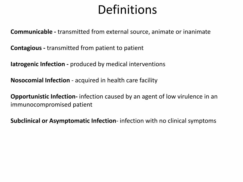
Definitions
Communicable - transmitted from external source, animate or inanimate Contagious - transmitted from patient to patient Iatrogenic Infection - produced by medical interventions Nosocomial Infection - acquired in health care facility Opportunistic Infection- infection caused by an agent of low virulence in an immunocompromised patient Subclinical or Asymptomatic Infection- infection with no clinical symptoms
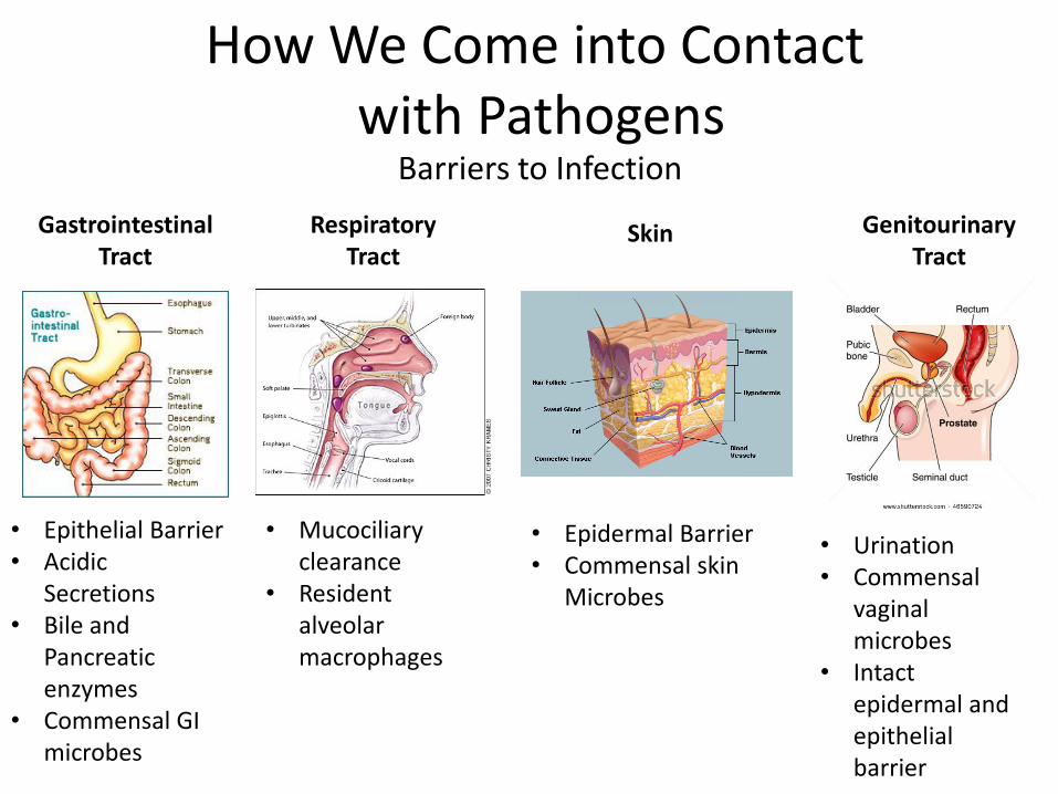
How We Come into Contact with Pathogens
Barriers to Infection
Gastrointestinal Tract
Respiratory Tract
Skin Genitourinary Tract
• Epidermal Barrier • Commensal skin
Microbes
• Epithelial Barrier • Acidic
Secretions • Bile and
Pancreatic enzymes
• Commensal GI microbes
• Mucociliary clearance
• Resident alveolar macrophages
• Urination • Commensal
vaginal microbes
• Intact epidermal and epithelial barrier

Fecal to oral Modes of Transmission
Basis for Failure of Barrier to Infection (GI Tract)
Epithelial Barrier (Attachment and local proliferation of microbes) • Vibrio
cholerae • Giardia
Epithelial Barrier (Attachment and local invasion of microbes) • Shigella • Salmonella • Campylobacter
Acidic Secretions (Acid-resistant cysts and eggs) • Entamoebae • Cryptosporidium
Commensal GI Microbes (Broad Spectrum Antiobiotic use) • Clostridium
difficile
Bile and Pancreatic Enzymes (Resistant Microbial external coats) • Hepatitis A • Rotavirus • Norovirus
Causes Diarrheal Illness

Person to person by respiratory droplets Modes of Transmission
Basis for Failure of Barrier to Infection (Respiratory Tract)
Mucociliary Clearance (Attachment and local proliferation of microbes)
• Influenza virus • Cold Virus
Mucociliary Clearance (Ciliary paralysis by toxins)
• Haemophilus influenzae • Mycoplasma
pneumoniae • Bordetella pertussis
Resident alveolar macrophages (Resistance to killing by phagocytes)
• Mycobacterium
tuberculosis
Causes Respiratory Illness

Modes of Transmission
Basis for Failure of Barrier to Infection (Skin)
Epidermal barrier (Mechanical defects, punctures, burns, ulcers) • Staphylococcus aureus • Candida albicans • Pseudomonas
aeuginosa
Epidermal barrier (Needle sticks) • HIV, • hepatitis
viruses
Epidermal barrier (Arthropod and animal bites) • Yellow fever • Plague • Lyme disease • Malaria • Rabies
Epidermal barrier (Direct infection/local invasion) • Hookworm • Strongyloides
Causes skin and soft tissue infections, and systemic illness Blood borne

Modes of Transmission
Basis for Failure of Barrier to Infection (GenitourinaryTract)
Urination (Obstruction, attachment and local proliferation) • Escherichia
coli
Commensal vaginal microbes (Antibiotic use) • Candida
albicans
Epithelial barrier (Microbial attachment and local proliferation) • Neissieria
gonohorrea
Epithelial barrier (Direct infection local invasion) • Herpes
viruses • Syphilis
Causes localized urogenital infections
Sexual/Body Fluids/Contact with Commensal Organisms
Epithelial barrier (Local trauma) • HPV • HIV

How do Organisms Cause Disease? (Which Microbe goes Where, and Why?)

How do Organisms Cause Disease?
Tropism – The specificity of a microbe for a host, host tissue, or cell type Determined by ligand and receptor binding for the microbe and the host
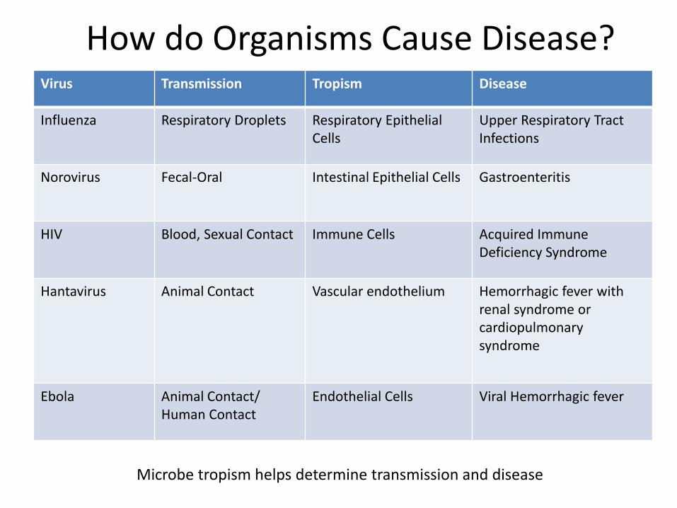
Virus Transmission Tropism Disease
Influenza Respiratory Droplets Respiratory Epithelial Cells
Upper Respiratory Tract Infections
Norovirus Fecal-Oral Intestinal Epithelial Cells Gastroenteritis
HIV Blood, Sexual Contact Immune Cells Acquired Immune Deficiency Syndrome
Hantavirus Animal Contact Vascular endothelium Hemorrhagic fever with renal syndrome or cardiopulmonary syndrome
Ebola Animal Contact/ Human Contact
Endothelial Cells Viral Hemorrhagic fever
How do Organisms Cause Disease?
Microbe tropism helps determine transmission and disease

Viruses HIV (AIDS) Ebola (Viral hemorrhagic fever) Hepatitis A,B,C (Hepatitis) Influenza (The flu) Adenovirus (Common Cold)
What types of Microbes Exist?
4 Main Classes of Infectious Agents
Bacteria Streptococcus pyogenes (Strep Throat) Neisseria meningitidis (Meningitis) Bordetella pertussis (Whooping Cough) Clostridium perfringens (Gas gangrene) Treponema pallidum (Syphilis)
Fungus Candida albicans (Thrush) Aspergillus fumigatus (Aspergillosis) Dermatophytes (Athlete’s Foot) Coccidioides immitis (Valley Fever) Cryptocoocus neoformans (Meningitis)
Parasites Plasmodium falciparum (Malaria) Trypansoma cruzi (Chagas disease) Ascaris lumbricoides (Intestinal roundworms) Giardia lamblia (Giardiasis) Trichomonas vaginalis (Trichomoniasis)

• The smallest infectious agents (20-300 nm) • Composed of nucleic acid genome surrounded by a protein coat called capsid • May contain DNA or RNA but not both • Some viruses are enveloped – Capsid is surrounded by plasma membrane • Composed of virus specific proteins and nucleic acid
Class #1: Viruses (Structure)
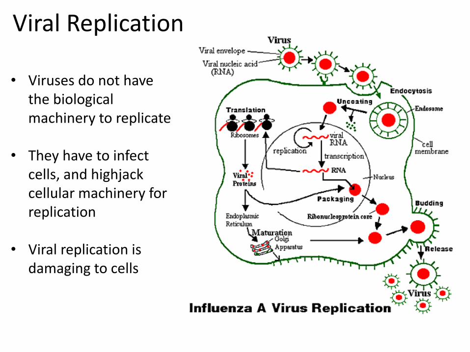
Viral Replication
• Viruses do not have the biological machinery to replicate
• They have to infect cells, and highjack cellular machinery for replication
• Viral replication is damaging to cells
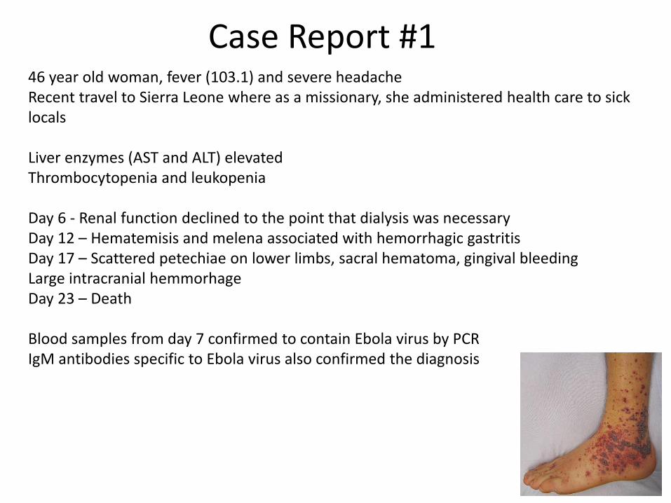
Case Report #1 46 year old woman, fever (103.1) and severe headache Recent travel to Sierra Leone where as a missionary, she administered health care to sick locals Liver enzymes (AST and ALT) elevated Thrombocytopenia and leukopenia Day 6 - Renal function declined to the point that dialysis was necessary Day 12 – Hematemisis and melena associated with hemorrhagic gastritis Day 17 – Scattered petechiae on lower limbs, sacral hematoma, gingival bleeding Large intracranial hemmorhage Day 23 – Death Blood samples from day 7 confirmed to contain Ebola virus by PCR IgM antibodies specific to Ebola virus also confirmed the diagnosis

Ebola Virus Where does the microbe live? What class of microbe
is causing disease?
How did the patient contract the disease?

How does Ebola Virus cause Disease? Viral Hemorrhagic Fever
Ebola virus tropism – Human vascular endothelial cell (blood vessel) infection
Early symptoms – fever, severe headache, muscle pain, weakness, diarrhea, vomiting, unexplained bleeding and bruising, rash Late symptoms – blood in diarrhea in vomitus, internal hemmorhaging, organ failure
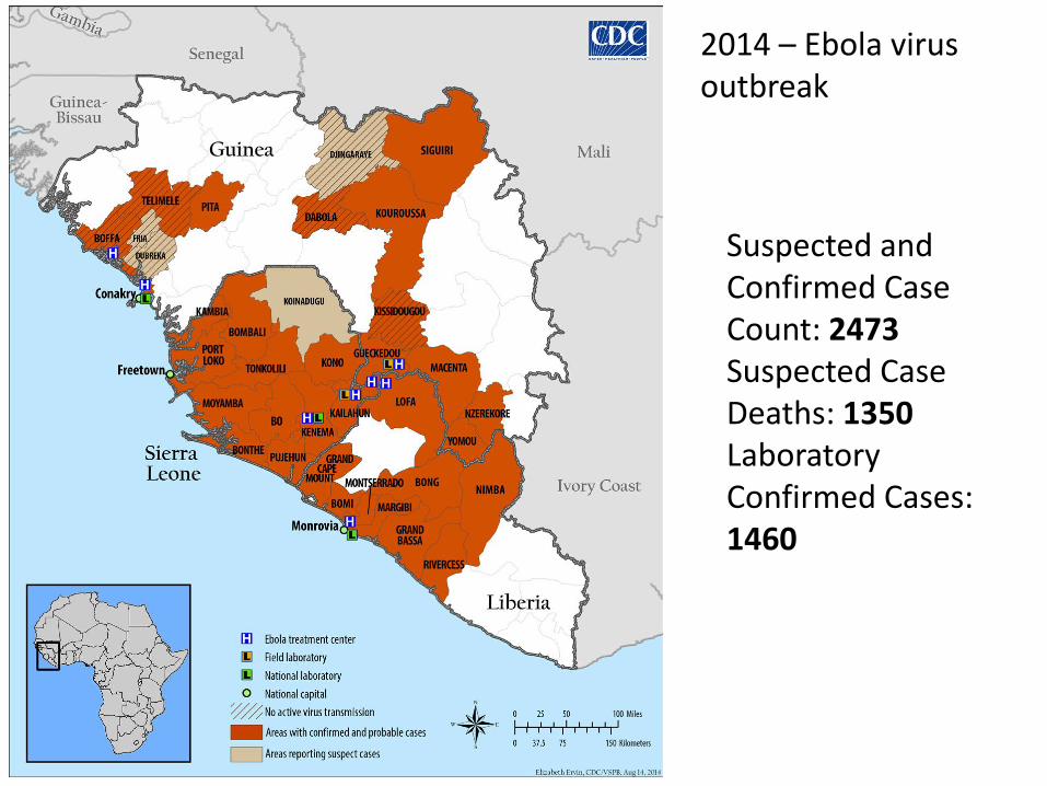
Suspected and Confirmed Case Count: 2473 Suspected Case Deaths: 1350 Laboratory Confirmed Cases: 1460
2014 – Ebola virus outbreak

Class #2: Bacteria Single Celled prokaryotic organism Contain both DNA and RNA Have No Nucleus or any other membrane bound organelles (golgi, mitochondria . . .) Reproduce by binary fission Have bacterial specific nucleic acid, proteins, carbohydrates and lipids

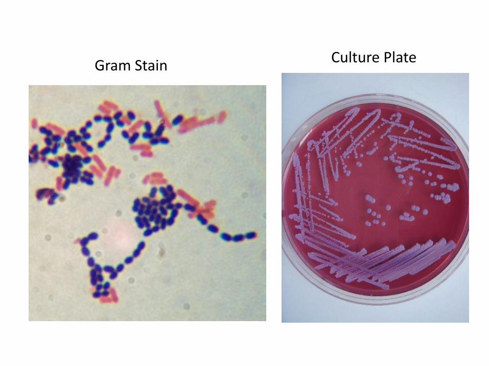
Gram Stain Culture Plate

Some bacteria can cause a wide variety of infections Tropism for certain bacteria is very broad, can infect a wide variety of cell types Many bacteria can produce toxins that can also cause disease

Case Report #2
9 yr old, fever 102, severe throat pain Difficulty swallowing Red swollen tonsils with white patches Lab cultures Streptococcus pyogenes (Group A strep) from a swab of the throat
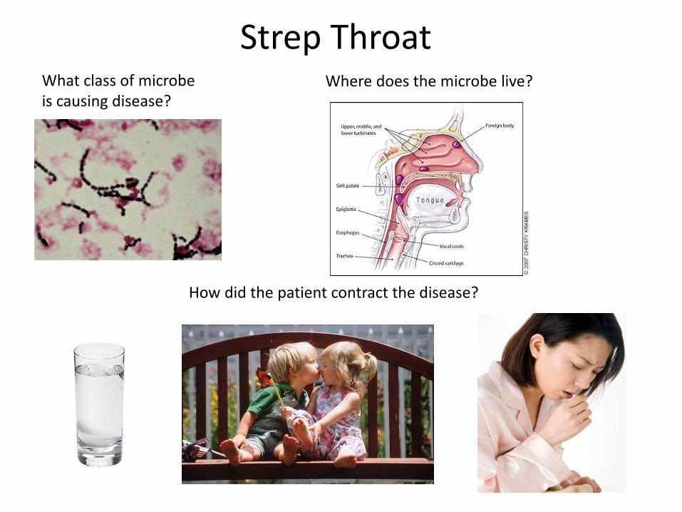
Strep Throat Where does the microbe live? What class of microbe
is causing disease?
How did the patient contract the disease?
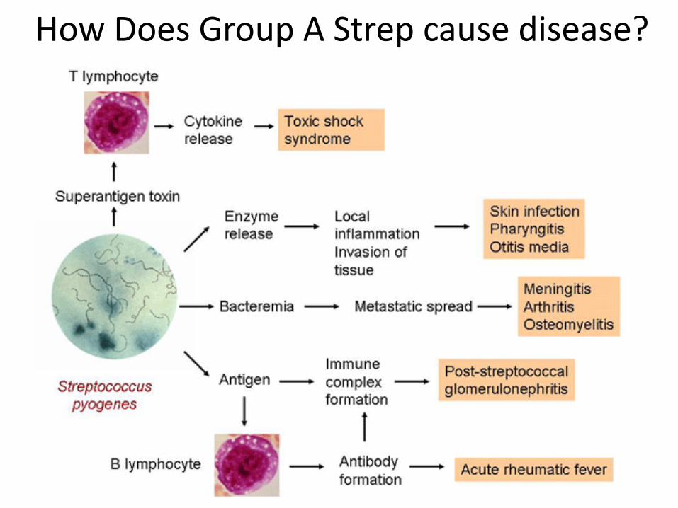
How Does Group A Strep cause disease?

Class #3: Fungus
Eukaryotic organism Contains a cell wall, nucleus and membrane bound organelles Both DNA and RNA Has fungus specific nucleic acid, proteins, carbohydrates and lipids

Molds (multicellular)
Hyphae- tubelike, basic structure
Mycelium- intertwined hyphae
Yeast (single celled)
Asexual reproduction by blastoconidia formation (budding)
Sexual reproduction by production of ascospores or basidiospores.
Two types of Fungus

Yeast Mold A soft, pasty, smooth colony; usually no filamentous (fuzzy) growth can be observed macroscopically
A filamentous fungus: fuzzy, powdery, woolly, velvety, or relatively smooth.


Case Report #3
22 yr old man, itchy, cracking, red feet, especially between the toes Lab cultures Trichophyton (Dermatophyte) from foot culture

Dermatophyte Where does the microbe live?
What class of microbe is causing disease?
How did the patient contract the disease?

Class #4: Parasites Eukaryotic orgainsms Contains a cell membrane, nucleus and membrane bound organelles Both DNA and RNA Has parasite specific nucleic acid, proteins, carbohydrates and lipids

Single Celled Parasites

Large Complex Multicellular parasites Well Defined organs and tissues (GI Tract, Genital Tract)

Parasites have complex life cycles Infective Stage: Cyst or egg that can survive a harsh environment Diagnostic Stage: Trophozoite or worm that causes disease

Case Report #4 23 year old man, fever (103.1) , headaches, body aches Recent travel to the Ivory Coast for vacation Microscopic Blood Smear analysis revealed Plasmodium falciparum
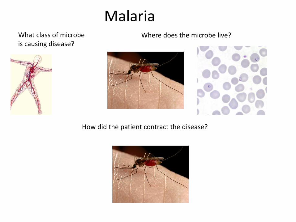
Malaria Where does the microbe live? What class of microbe
is causing disease?
How did the patient contract the disease?
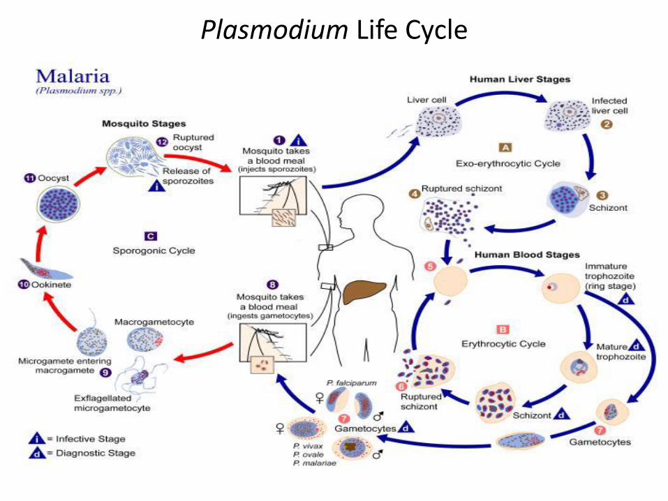
Plasmodium Life Cycle
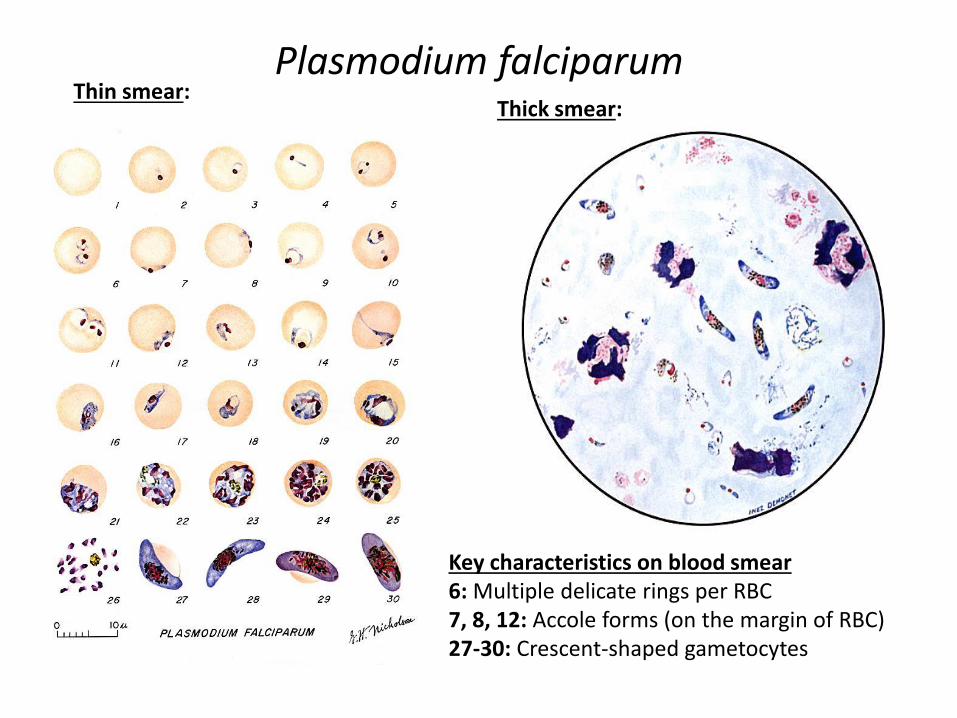
Plasmodium falciparum Thin smear:
Thick smear:
Key characteristics on blood smear 6: Multiple delicate rings per RBC 7, 8, 12: Accole forms (on the margin of RBC) 27-30: Crescent-shaped gametocytes
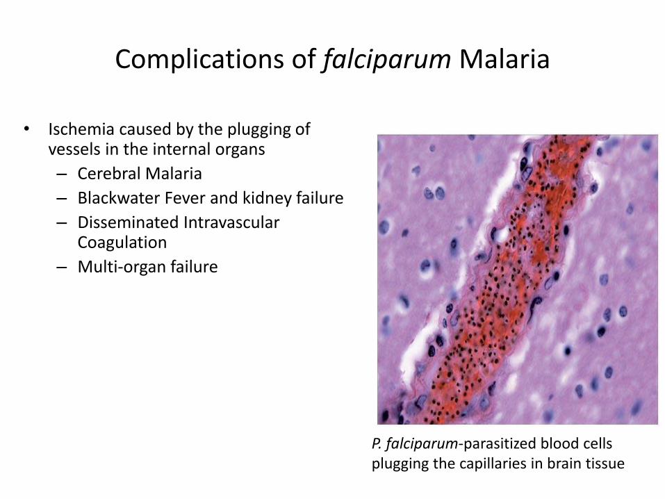
Complications of falciparum Malaria
• Ischemia caused by the plugging of vessels in the internal organs
– Cerebral Malaria
– Blackwater Fever and kidney failure
– Disseminated Intravascular Coagulation
– Multi-organ failure
P. falciparum-parasitized blood cells plugging the capillaries in brain tissue


53
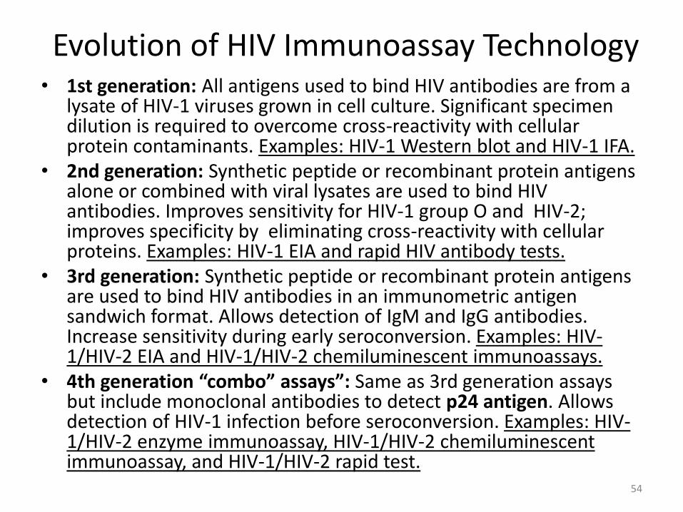
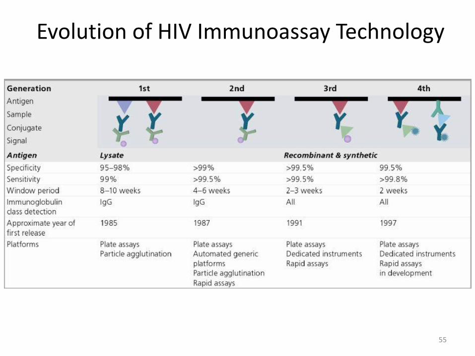
Evolution of HIV Immunoassay Technology • 1st generation: All antigens used to bind HIV antibodies are from a
lysate of HIV-1 viruses grown in cell culture. Significant specimen dilution is required to overcome cross-reactivity with cellular protein contaminants. Examples: HIV-1 Western blot and HIV-1 IFA.
• 2nd generation: Synthetic peptide or recombinant protein antigens alone or combined with viral lysates are used to bind HIV antibodies. Improves sensitivity for HIV-1 group O and HIV-2; improves specificity by eliminating cross-reactivity with cellular proteins. Examples: HIV-1 EIA and rapid HIV antibody tests.
• 3rd generation: Synthetic peptide or recombinant protein antigens are used to bind HIV antibodies in an immunometric antigen sandwich format. Allows detection of IgM and IgG antibodies. Increase sensitivity during early seroconversion. Examples: HIV-1/HIV-2 EIA and HIV-1/HIV-2 chemiluminescent immunoassays.
• 4th generation “combo” assays”: Same as 3rd generation assays but include monoclonal antibodies to detect p24 antigen. Allows detection of HIV-1 infection before seroconversion. Examples: HIV-1/HIV-2 enzyme immunoassay, HIV-1/HIV-2 chemiluminescent immunoassay, and HIV-1/HIV-2 rapid test.
54
Evolution of HIV Immunoassay Technology
55

Structural genes • Gag is p55 from which three core proteins (p15, p17 and p24) are
formed
• Env gene codes for envelope proteins gp160, gp120 and gp41
• Pol codes for p66 and p51 subunits of reverse transcriptase and p31 an endonuclease

Old HIV Diagnostic Algorithm
1. Screen
immunoassay (EIA/CIA)
rapid tests
2. Confirm
Western Blot (98%)
IFA
APTIMA qualitative NAAT

Laboratory diagnosis of HIV infection
ELISA Testing
• first serological test developed to detect HIV infection
• antibodies detected include those directed against p24, gp120, gp160 and gp41, detected first in infection and appear in most individuals
• used for screening only, false positives do occur (recent acute illness, allergies)
• highly sensitive, not specific

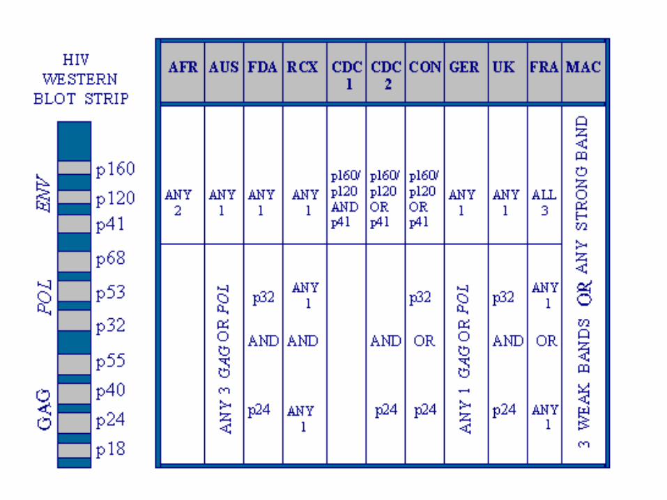
Laboratory diagnosis of HIV infection Western Blot Testing • most popular confirmatory test • antibodies to p24 and p55 appear earliest but decrease or
become undetectable • antibodies to gp31, gp41, gp120, and gp160 appear later but
are present throughout all stages of the disease Western Blot Testing = interpretation of result • no bands, negative • in order to be interpreted as positive a minimum of 3 bands
directed against the following antigens must be present : p24, p31, gp41 or gp120/160
• CDC criteria require 2 bands of the following : p24, gp41 or gp120/160
Laboratory diagnosis of HIV infection Western Blot Testing = interpretation of result
• indeterminate results are those samples that produce bands but not enough to be positive, may be due to the following:
1. prior blood transfusions, even with non-HIV-1 infected blood
2. prior or current infection with syphilis
3. prior or current infection with malaria
4. autoimmune diseases
5. infection with other human retroviruses
6. second or subsequent pregnancies in women
*** run an alternate HIV confirmatory assay

CDC criteria require 2 bands of the following : p24, gp41 or gp120/160


HIV-1 / HIV-2 Ag/Ab Immunoassay


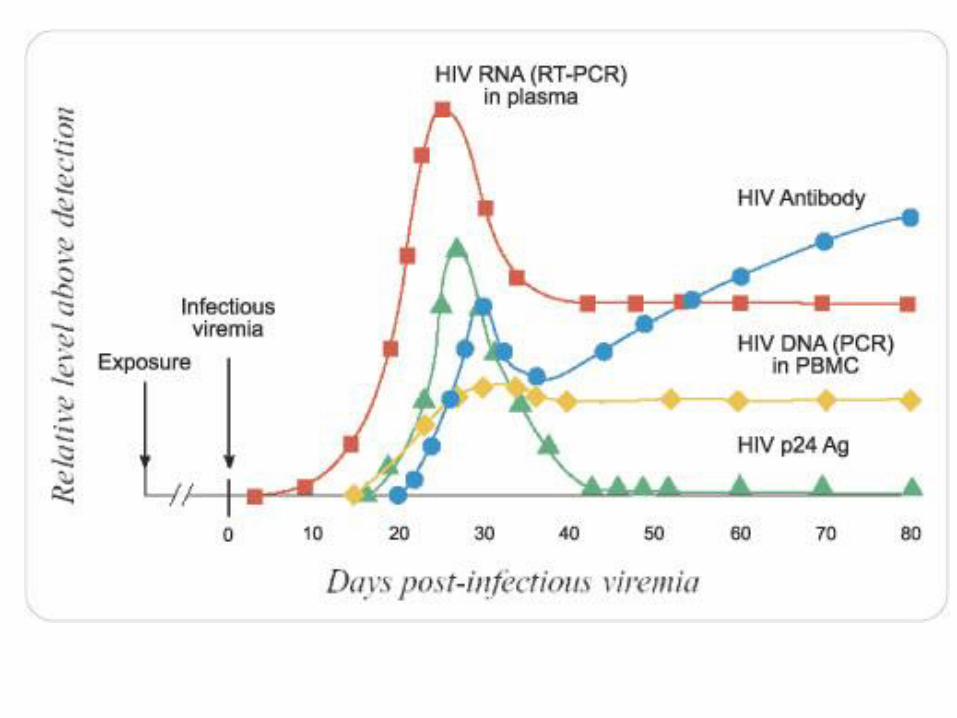
Laboratory diagnosis of HIV infection Viral Load Tests
• viral load or viral burden is the quantity of HIV-RNA that is in the blood
• measures the amount of HIV-RNA in one milliliter of blood

HIV RNA Test • The COBAS® AmpliPrep/COBAS® TaqMan® HIV-1 Test
(Roche) uses reverse transcription and PCR amplification primers that define sequences within the highly conserved regions of the HIV-1 gag gene and of the HIV-1 LTR region.
• Reportable range: 20–10,000,000 copies/ml; LOD = 20
70