Ubiquitin-conjugating enzyme Ubc13 controls … enzyme Ubc13 controls breast cancer metastasis...
6
Ubiquitin-conjugating enzyme Ubc13 controls breast cancer metastasis through a TAK1-p38 MAP kinase cascade Xuefeng Wu a,b,c , Weizhou Zhang a,b,c,d , Joan Font-Burgada a,b,c , Trenis Palmer b , Alexander S. Hamil e , Subhra K. Biswas f , Michael Poidinger f,g , Nicholas Borcherding d , Qing Xie d , Lesley G. Ellies c , Nikki K. Lytle b,h , Li-Wha Wu a,b,c,i , Raymond G. Fox b,h , Jing Yang b , Steven F. Dowdy e , Tannishtha Reya b,h , and Michael Karin a,b,c,1 a Laboratory of Gene Regulation and Signal Transduction and Departments of b Pharmacology, c Pathology, and e Cellular and Molecular Medicine, School of Medicine, University of California, San Diego, La Jolla, CA 92093; d Department of Pathology, Carver College of Medicine, University of Iowa, Iowa City, IA 52242; f Singapore Immunology Network, Agency for Science, Technology and Research (A*STAR), Singapore 138648; g Department of Biological Sciences, National University of Singapore, Singapore 117543; h Sanford Consortium for Regenerative Medicine, La Jolla, CA 92093; and i Institute of Molecular Medicine, College of Medicine, National Cheng Kung University, Tainan 70101, Taiwan Contributed by Michael Karin, July 29, 2014 (sent for review June 6, 2014) Metastatic spread is the leading cause of cancer mortality. Breast cancer (BCa) metastatic recurrence can happen years after removal of the primary tumor. Here we show that Ubc13, an E2 enzyme that catalyzes K63-linked protein polyubiquitination, is largely dispens- able for primary mammary tumor growth but is required for metastatic spread and lung colonization by BCa cells. Loss of Ubc13 inhibited BCa growth and survival only at metastatic sites. Ubc13 was dispensable for transforming growth factor β (TGFβ)-induced SMAD activation but was required for activation of non-SMAD sig- naling via TGFβ-activating kinase 1 (TAK1) and p38, whose activity controls expression of numerous metastasis promoting genes. p38 activation restored metastatic activity to Ubc13-deficient cells, and its pharmacological inhibition attenuated BCa metastasis in mice, sug- gesting it is a therapeutic option for metastatic BCa. ubiquitination-mediated signaling | pre-clinical studies B reast cancer (BCa) is the leading invasive cancer among women worldwide. BCa-related mortality is usually caused by distant metastases rather than primary tumors (1, 2). The spread of cancer cells from primary tumors to distant organs, termed metastasis, is a multistep process in which cancer cells must (i ) invade through the extracellular matrix (ECM), (ii ) disseminate into the bloodstream, (iii ) survive in the circulation, and (iv) extravasate and successfully colonize distant sites (3). Conventional therapeutic strategies have limited success in preventing and treating metastatic cancer, and BCa metastases can recur many years after removal of the primary tumor. This phenomenon could be due to the complex nature of metastasis itself, and, more realistically, the limitation of current treatments that are effective against primary BCa, i.e., surgical removal and localized radiotherapy, but do little to prevent metastatic re- currence. Even chemotherapy is not very effective against met- astatic tumors (4). Unfortunately, the pharmaceutical industry has been reluctant to conduct metastasis prevention trials on patients with early stage cancer using survival and reduction of metastatic load as end points, because such studies are lengthy and require a large number of patients with otherwise relatively good survival prospects (4). Consequently, the development of agents that prevent metastasis from occurring and trigger regression of established metastatic lesions is an urgent unmet need. It was reported that expression of the ubiquitin conjugating enzyme (E2) Ubc13 is up-regulated in metastatic BCa (5). Ubc13, which heterodimerizes with Uev1a, catalyzes formation of lysine 63 (K63)-linked polyubiquitin chains, which control protein–protein interactions involved in DNA damage repair and protein kinase activation (6, 7). In certain immune cells, Ubc13 is required for IκB kinase (IKK)–nuclear factor κB (NF-κB) activation, but a more ubiquitous role for Ubc13 was observed in the activation of MAPK signaling (8–11). We found that Ubc13 is required for activation of mitogen-activated protein kinase kinase kinase 1 (MEKK1), transforming growth factor β (TGFβ)-activating kinase 1 (TAK1), and downstream MAPK cascades on CD40 engagement in B cells (10). Impor- tantly, MEKK1 and TAK1 are also required for BCa metastasis (12, 13). Of the numerous signaling pathways affecting BCa metastasis, the TGFβ pathway has some of the strongest effects, and it promotes metastasis by inducing migration, intravasation, and epithelial-mesenchymal transition (EMT) of carcinoma cells (14). TGFβ signaling is mediated through SMAD-dependent and -independent (non-SMAD) pathways (15, 16). Non-SMAD TGFβ signaling is positively regulated by multiple molecules including TAK1 (17), tumor necrosis factor receptor-associated factor 6 (TRAF6) (18), and TRAF4 (19). The p38 MAPK also participates in different steps of metastasis, including ECM in- vasion by primary cancer cells, migration across the surrounding tissue, entry into the circulation, and colonization of distant sites (20). p38 inhibitors are not toxic and were found effective in prevention and attenuation of inflammatory pain in humans (21, 22). Here we show that a Ubc13-controled TAK1–p38 cascade controls BCa metastatic dissemination and that a p38 inhibitor can cause regression of established metastases. Significance We demonstrate that ubiquitin-conjugating enzyme Ubc13, whose expression is elevated in primary and metastatic breast cancer (BCa), promotes metastatic spread of BCa cells by con- trolling their lung-colonizing ability while having little effect on primary tumor growth. Mechanistically, Ubc13 is required for TGFβ-induced non-SMAD signaling via TAK1 and p38, a pathway that is first activated in the primary tumor. An Ubc13- and p38-dependent metastatic gene signature was identified, explaining how p38 may control metastasis and providing a measure for monitoring the effectiveness of pharmacologic p38 inhibition, which inhibits the growth of established meta- static lesions. We suggest that p38 inhibition should be consid- ered as a potential treatment for metastatic BCa. Author contributions: X.W. designed research; X.W., A.S.H., S.K.B., M.P., and N.K.L. per- formed research; W.Z., J.F.-B., T.P., L.G.E., L.-W.W., R.G.F., J.Y., and S.F.D. contributed new reagents/analytic tools; X.W., W.Z., N.B., Q.X., N.K.L., T.R., and M.K. analyzed data; and X.W. and M.K. wrote the paper. The authors declare no conflict of interest. Data deposition: The data reported in this paper have been deposited in the Gene Ex- pression Omnibus (GEO) database, www.ncbi.nlm.nih.gov/geo (accession no. GSE55649). 1 To whom correspondence should be addressed. Email: [email protected]. This article contains supporting information online at www.pnas.org/lookup/suppl/doi:10. 1073/pnas.1414358111/-/DCSupplemental. 13870–13875 | PNAS | September 23, 2014 | vol. 111 | no. 38 www.pnas.org/cgi/doi/10.1073/pnas.1414358111
Transcript of Ubiquitin-conjugating enzyme Ubc13 controls … enzyme Ubc13 controls breast cancer metastasis...
Ubiquitin-conjugating enzyme Ubc13 controls breastcancer metastasis through a TAK1-p38 MAPkinase cascadeXuefeng Wua,b,c, Weizhou Zhanga,b,c,d, Joan Font-Burgadaa,b,c, Trenis Palmerb, Alexander S. Hamile, Subhra K. Biswasf,Michael Poidingerf,g, Nicholas Borcherdingd, Qing Xied, Lesley G. Elliesc, Nikki K. Lytleb,h, Li-Wha Wua,b,c,i,Raymond G. Foxb,h, Jing Yangb, Steven F. Dowdye, Tannishtha Reyab,h, and Michael Karina,b,c,1
aLaboratory of Gene Regulation and Signal Transduction and Departments of bPharmacology, cPathology, and eCellular and Molecular Medicine, Schoolof Medicine, University of California, San Diego, La Jolla, CA 92093; dDepartment of Pathology, Carver College of Medicine, University of Iowa, Iowa City,IA 52242; fSingapore Immunology Network, Agency for Science, Technology and Research (A*STAR), Singapore 138648; gDepartment of Biological Sciences,National University of Singapore, Singapore 117543; hSanford Consortium for Regenerative Medicine, La Jolla, CA 92093; and iInstitute of MolecularMedicine, College of Medicine, National Cheng Kung University, Tainan 70101, Taiwan
Contributed by Michael Karin, July 29, 2014 (sent for review June 6, 2014)
Metastatic spread is the leading cause of cancer mortality. Breastcancer (BCa) metastatic recurrence can happen years after removalof the primary tumor. Here we show that Ubc13, an E2 enzyme thatcatalyzes K63-linked protein polyubiquitination, is largely dispens-able for primary mammary tumor growth but is required formetastatic spread and lung colonization by BCa cells. Loss of Ubc13inhibited BCa growth and survival only at metastatic sites. Ubc13was dispensable for transforming growth factor β (TGFβ)-inducedSMAD activation but was required for activation of non-SMAD sig-naling via TGFβ-activating kinase 1 (TAK1) and p38, whose activitycontrols expression of numerous metastasis promoting genes. p38activation restored metastatic activity to Ubc13-deficient cells, and itspharmacological inhibition attenuated BCa metastasis in mice, sug-gesting it is a therapeutic option for metastatic BCa.
ubiquitination-mediated signaling | pre-clinical studies
Breast cancer (BCa) is the leading invasive cancer amongwomen worldwide. BCa-related mortality is usually caused
by distant metastases rather than primary tumors (1, 2). Thespread of cancer cells from primary tumors to distant organs,termed metastasis, is a multistep process in which cancer cellsmust (i) invade through the extracellular matrix (ECM), (ii)disseminate into the bloodstream, (iii) survive in the circulation,and (iv) extravasate and successfully colonize distant sites (3).Conventional therapeutic strategies have limited success inpreventing and treating metastatic cancer, and BCa metastasescan recur many years after removal of the primary tumor. Thisphenomenon could be due to the complex nature of metastasisitself, and, more realistically, the limitation of current treatmentsthat are effective against primary BCa, i.e., surgical removal andlocalized radiotherapy, but do little to prevent metastatic re-currence. Even chemotherapy is not very effective against met-astatic tumors (4). Unfortunately, the pharmaceutical industryhas been reluctant to conduct metastasis prevention trials onpatients with early stage cancer using survival and reduction ofmetastatic load as end points, because such studies are lengthy andrequire a large number of patients with otherwise relatively goodsurvival prospects (4). Consequently, the development of agentsthat prevent metastasis from occurring and trigger regression ofestablished metastatic lesions is an urgent unmet need.It was reported that expression of the ubiquitin conjugating
enzyme (E2) Ubc13 is up-regulated in metastatic BCa (5).Ubc13, which heterodimerizes with Uev1a, catalyzes formationof lysine 63 (K63)-linked polyubiquitin chains, which controlprotein–protein interactions involved in DNA damage repairand protein kinase activation (6, 7). In certain immune cells,Ubc13 is required for IκB kinase (IKK)–nuclear factor κB(NF-κB) activation, but a more ubiquitous role for Ubc13 was
observed in the activation of MAPK signaling (8–11). We foundthat Ubc13 is required for activation of mitogen-activatedprotein kinase kinase kinase 1 (MEKK1), transforming growthfactor β (TGFβ)-activating kinase 1 (TAK1), and downstreamMAPK cascades on CD40 engagement in B cells (10). Impor-tantly, MEKK1 and TAK1 are also required for BCa metastasis(12, 13). Of the numerous signaling pathways affecting BCametastasis, the TGFβ pathway has some of the strongest effects,and it promotes metastasis by inducing migration, intravasation,and epithelial-mesenchymal transition (EMT) of carcinoma cells(14). TGFβ signaling is mediated through SMAD-dependentand -independent (non-SMAD) pathways (15, 16). Non-SMADTGFβ signaling is positively regulated by multiple moleculesincluding TAK1 (17), tumor necrosis factor receptor-associatedfactor 6 (TRAF6) (18), and TRAF4 (19). The p38 MAPK alsoparticipates in different steps of metastasis, including ECM in-vasion by primary cancer cells, migration across the surroundingtissue, entry into the circulation, and colonization of distant sites(20). p38 inhibitors are not toxic and were found effective inprevention and attenuation of inflammatory pain in humans (21,22). Here we show that a Ubc13-controled TAK1–p38 cascadecontrols BCa metastatic dissemination and that a p38 inhibitorcan cause regression of established metastases.
Significance
We demonstrate that ubiquitin-conjugating enzyme Ubc13,whose expression is elevated in primary and metastatic breastcancer (BCa), promotes metastatic spread of BCa cells by con-trolling their lung-colonizing ability while having little effecton primary tumor growth. Mechanistically, Ubc13 is requiredfor TGFβ-induced non-SMAD signaling via TAK1 and p38,a pathway that is first activated in the primary tumor. An Ubc13-and p38-dependent metastatic gene signature was identified,explaining how p38 may control metastasis and providinga measure for monitoring the effectiveness of pharmacologicp38 inhibition, which inhibits the growth of established meta-static lesions. We suggest that p38 inhibition should be consid-ered as a potential treatment for metastatic BCa.
Author contributions: X.W. designed research; X.W., A.S.H., S.K.B., M.P., and N.K.L. per-formed research; W.Z., J.F.-B., T.P., L.G.E., L.-W.W., R.G.F., J.Y., and S.F.D. contributed newreagents/analytic tools; X.W., W.Z., N.B., Q.X., N.K.L., T.R., and M.K. analyzed data; andX.W. and M.K. wrote the paper.
The authors declare no conflict of interest.
Data deposition: The data reported in this paper have been deposited in the Gene Ex-pression Omnibus (GEO) database, www.ncbi.nlm.nih.gov/geo (accession no. GSE55649).1To whom correspondence should be addressed. Email: [email protected].
This article contains supporting information online at www.pnas.org/lookup/suppl/doi:10.1073/pnas.1414358111/-/DCSupplemental.
13870–13875 | PNAS | September 23, 2014 | vol. 111 | no. 38 www.pnas.org/cgi/doi/10.1073/pnas.1414358111

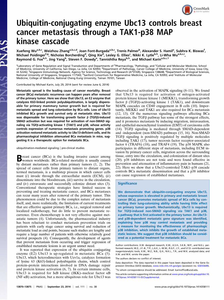
ResultsUbc13 Controls BCa Lung Metastasis. Ubc13 is up-regulated in met-astatic BCa (5). Using the Oncomine platform, we confirmed thatUbc13 is up-regulated in various tumor tissues, including breast,pancreas, colon, prostate, and lymphoma (Fig. S1A). A search ofThe Cancer Genome Atlas (TCGA) datasets showed that Ubc13(UBE2N) and its targets vascular cell adhesion molecule 1(VCAM-1) and intercellular adhesion molecule 1 (ICAM-1) (seebelow) are up-regulated in human tumor and metastasis specimenscompared with normal tissue (Fig. S1B). Up-regulation wasenriched in basal and Her2+ BCa subtypes (Fig. S1B), which areknown to be more metastatic. We used a human BCa cell line, LM2or 4175, which grows in mice and preferentially metastasizes to lung(23), to study the role of Ubc13 in metastatic spread. Ubc13 ex-pression in LM2 cells was silenced by lentiviral shRNA delivery, andresulting shUbc13 and control LM2 cells (shControl) were injectedinto the #4 mammary fat pads of NOD/SCID recipient mice. After8 wk, mice were killed, and their lung sections were H&E stained todetect metastases. Lung sections from LM2-shControl cells injectedinto mice showed large areas containing densely packed cancer cells(Fig. 1A), indicating successful lung colonization. In sharp contrast,LM2-shUbc13 cells formed much smaller areas containing cancercells (Fig. 1A). LM2-shControl cells gave rise to >10-fold more lungmetastases than LM2-shUbc13 cells, based on lung areas occupiedby cancer cells or quantitative RT-PCR (qRT-PCR) analysis ofhuman-specific β2-microglobulin (B2M) normalized to mouseβ-actin mRNA (Fig. 1B). Immunoblot analysis of cancer cells iso-lated from xenografts confirmed that Ubc13 expression was stablysilenced (Fig. 1C). Importantly, Ubc13 silencing had no detectableeffect on primary tumor growth and mass (Fig. 1 D and E). We alsoinoculated LM2 cells via the tail vein to rule out any effects on theprimary tumor and found that Ubc13 silencing dramatically re-duced lung metastasis, determined by bioluminescence measure-ment and surface nodule numbers (Fig. 1 F–H).To further confirm the requirement of Ubc13 for metastasis, we
carried out similar experiments using the mouse metastatic
mammary cancer 4T1 cell line. Again, Ubc13 silencing had noeffect on primary tumor growth but greatly reduced the ability of4T1 cells to metastasize to lung in Balb/C mice (Fig. S2A). We alsoused another metastatic mouse mammary cancer cell line, MT2,derived from MMTV-cNeu/ErbB2–induced tumors in Friendvirus B-type (FVB) mice (24). When transplanted into the #2mammary gland, shControl-MT2 cells formed lung metastasesin 60% of transplanted mice, but shUbc13-MT2 cells did not me-tastasize (Fig. S2B). However, Ubc13 silencing in MT2 cells didpartially affect primary tumor growth (Fig. S2B). We also crossedUbc13F/F mice (8) with MMTV-cNeu mice (25) (both in the FVBbackground). The resulting MMTV-cNeu;Ubc13F/F mice formedspontaneous mammary tumors starting at 6 mo of age. We iso-lated cells from these tumors and infected them with Adeno-Creto delete Ubc13 ex vivo orAdenoGFP as a control (Fig. S3A). TheUbc13-depleted ErbB2 tumor cells failed to form lung metastasesafter orthotopic transplantation or tail vein injection into FVBmice (Fig. S3A). Last, we reconstituted Ubc13-silenced cells witheither WT or a catalytically inactive mutant (C87A) form ofUbc13 (Fig. S3B) and transplanted the resulting cells into mice.Importantly, WT-rescued cells formed lung metastases, whereascells reconstituted with Ubc13(C87A) did not (Fig. S3B). Thus,Ubc13’s catalytic activity is required for BCa metastatic spread.
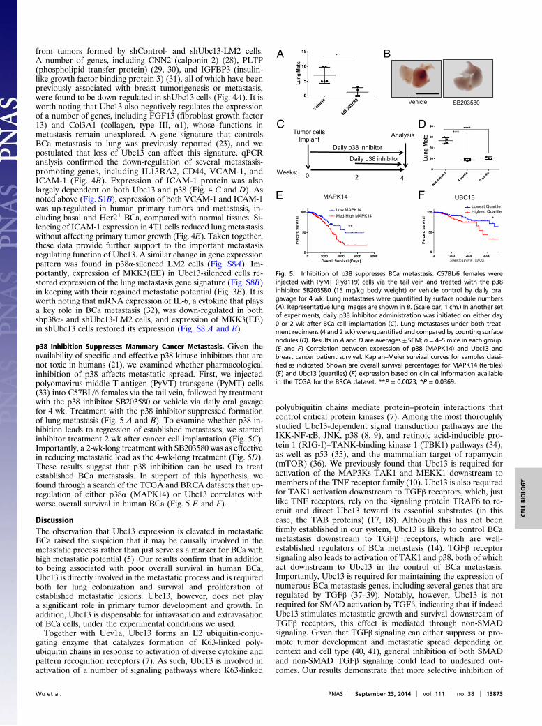
Ubc13 Controls BCa Lung Colonization. To address how Ubc13controls metastasis, we used an inducible shRNA lentivirus thatallows gene silencing on doxycycline (Dox) treatment while label-ing transduced cells with red fluorescent protein (RFP) (26).In LM2 cells transduced with this construct, Dox led to stable
shControl 0
5
10
15 **
shUbc13 Lung
met
s(%
are
a)
0123456
Tum
or W
eigh
t (g
)
Ubc13
Tubulin
IKK
A B C
D E F G
shControl shUbc13
ShControl ShUbc13
0
5
10
15
20
25
Lung
Pho
ton
Flux
(107
) *
H
05
1015202530
Nod
ule
Num
ber
***
0
0.2
0.4
0.6
B2M
Exp
res.
**
shControlshUbc13
shControlshUbc13
shControlshUbc13
shControlshUbc13
Mouse #
Fig. 1. Ubc13 controls BCa metastatic spread. (A–E) Role of Ubc13 in lungmetastasis of orthotopically transplanted BCa cells. (A) H&E staining of lungsections frommice orthotopically transplantedwith shControl- and shUbc13-LM2cells. [Scale bars, 800 μm (top two inlets) and 100 μm (bottom).] (B) Lung met-astatic load was determined by calculating the percentage of the lung surfaceoccupied by cancer cells (Left) or by qRT-PCR of human-specific β2-microglobulin(B2M) mRNA normalized to mouse β-actin mRNA (Right). (C) Ubc13 proteinexpression in cancer cells isolated from primary shControl- and shUbc13-LM2orthotopic tumors (mouse numbers indicated above the lanes). (D) Tumorgrowth curve (the whole 8-wk time course) and (E) weights (end point, i.e., 8 wkafter injection) of primary tumors formed by shControl- and shUbc13-LM2 cells.(F–H) Requirement of Ubc13 for lung colonization by tail vein injected BCa cells.(F) BLI (bioluminescence) images of NOD/SCID mice injected with luciferase-labeled shControl- and shUbc13-LM2 cells via the tail vein. (G) Quantification oflung photon flux. (H) Quantification of lung surface nodules. Data are averages ±SEM; n = 5 mice. ■, shControl; □, shUbc13. *P < 0.05, **P < 0.01, ***P < 0.001.
15 KDa
+ Dox0 21
- Dox3 4 5 0 21 3 4 5
Ubc13Days
50 KDa Tubulin
BF RFPC ED- +
FSC
RFP
A B
Dox:
p10-
shC
trlp1
0-sh
Ubc
13
F
shControl shUbc13
Ki67
Cleaved Caspase 3
N.S.
Fig. 2. Ubc13 is required for lung colonization by BCa cells. (A) Dynamic,doxycycline (Dox)-regulated, Ubc13 silencing in p10-shUbc13–infected LM2cells. (B) Analysis of RFP expression by p10-shCtrl or p10-shUbc13–infected LM2cells cultured in the absence (−) or presence (+) of Dox for 4 d. (C) BLI mea-surement of mice injected with Dox-treated p10-Ctrl or p10-shUbc13 LM2 cells,given Dox in water for the first week and switiched to regular water for thefollowing 3 wk. (D) BLI measurement of mice injected with p10-shCtrl or p10-shUbc13 LM2 cells that were not treated with Dox. Mice were given regularwater for the first week and switiched to Dox-containing water for the fol-lowing 3 wk. Data in C and D are averages ± SEM; n = 3 mice. (E) Represen-tative bright field (BF) and RFP images of lungs from mice transplanted withp10-shCtrl (Upper) or p10-shUbc13 (Lower) LM2 cells and treated as in D. (Scalebar, 1 cm.) (F) Ki67 and cleaved caspase 3 staining of lung lesions in mice thatwere i.v. inoculated with shControl- or shUbc13-LM2 cells (4 wk after injection).Five independent high-power fields (HPFs) were quantitated, and the resultsare shown on the right as averages ± SEM. (Scale bar, 100 μm.)
Wu et al. PNAS | September 23, 2014 | vol. 111 | no. 38 | 13871
CELL
BIOLO
GY

inhibition of Ubc13 expression within 3 d, and Dox withdrawalrestored Ubc13 expression as early as day 3 with full expression onday 5 (Fig. 2A). Flow cytometry and immunofluorescence (IF)microscopy confirmed that both p10-shCtrl– and p10-shUbc13–transduced cells were uniformly RFP positive after Dox addition(Fig. 2B and Fig. S4). To address whether Ubc13 is required forentry of BCa cells into the lung, p10-shControl– and shUbc13-transduced LM2 cells were cultured with Dox for 4 d, and tail veininjected into NOD/SCID mice that were kept on Dox-containingwater for 1 wk and switched to regular drinking water for 3 wk.Lung metastasis was monitored weekly by a bioluminescence assay.Curiously, no differences in lung metastasis were observed betweenthe two groups (Fig. 2C), indicating that Ubc13 activity is not re-quired for lung seeding, a process that was probably completedwithin the first 24 h. We also injected p10-shControl and shUbc13LM2 cells into NOD/SCID mice that were kept on regular waterfor 1 wk, allowing the cells to enter the lung and colonize it. Themice were then given Dox-containing water to silence Ubc13 ex-pression. Whereas p10-shControl cells formed detectable lungmetastases as early as 2 wk after injection, p10-shUbc13 cells didnot form detectable metastases in Dox-treated mice (Fig. 2D).Microscopic analysis under bright field (BF) and red fluorescence(RFP) confirmed that shUbc13-LM2 cells formed much fewer andsmaller lung nodules than shControl-LM2 cells (Fig. 2E). To fur-ther study how Ubc13 affects metastatic growth, we performedtumorsphere formation assays on control and shUbc13 cells andfound that Ubc13 silencing had no effect on these properties (Fig.S5 A and B). These results are consistent with the finding thatUbc13 is generally dispensable for primary tumor growth. Loss ofUbc13 in BCa cells also did not affect their proliferation as evidentby carboxyfluorescein succinimidyl ester labeling (Fig. S5C). Im-portantly, loss of Ubc13 also did not affect LM2 cell intravasationor extravasation quantified by qPCR (Fig. S5D). Through real-timein vivo imaging, we observed no difference in frequencies of cir-culating tumor cells between shControl and shUbc13 LM2 trans-planted mice (Fig. S5 E and F, Table S1, and Movie S1). Wetherefore reasoned that Ubc13 could specifically control metastaticBCa growth properties. Indeed, shUbc13 BCa cells residing insmall lung lesions were less proliferative than shControl cells inlung lesions and were more likely to show caspase 3 activation (Fig.2F). In keeping with Ubc13 being dispensable for primary tumorgrowth, we did not observe a difference in proliferation and
apoptosis of BCa cells within primary tumors formed byshControl- or shUbc13-LM2 cells (Fig. S6).
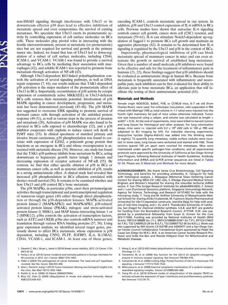
Ubc13 Controls BCa Metastasis Through TAK1 and p38 MAPK. Ubc13is involved in both NF-κB and MAPK activation, but thedependence of either response on Ubc13 activity is cell typespecific (8, 9). To better understand the role of Ubc13 in sig-naling within BCa cells, we stimulated LM2 cells with TNF.Although Ubc13 silencing had no effect on IκBα degradationand resynthesis, it inhibited p38α phosphorylation (Fig. 3A).However, Ubc13 silencing had no significant effect on JNK ac-tivation. Because TGFβ signaling is more relevant to the controlof BCa metastasis than TNF (16), we examined the role ofUbc13 in TGFβ-induced SMAD and non-SMAD signaling inLM2 cells. Although Ubc13 silencing had no effect on SMADphosphorylation, it inhibited TGFβ-induced p38α phosphoryla-tion (Fig. 3B). TNF receptor family members signal to p38 viathe MAPK kinase kinases (MAP3K) MEKK1 and TAK1 (10).We found that TGFβ-induced TAK1 phosphorylation was sub-stantially reduced on Ubc13 silencing (Fig. 3C). Silencing ofTAK1 or p38α in BCa cells led to dramatically reduced lungmetastasis (Fig. S7 A and B). Compared with shControl-LM2cells, shUbc13-LM2 cells exhibited lower p38 phosphorylation(i.e., activation) in both lung lesions and primary tumors (Fig.S7C). Expression of constitutively active MKK3, which acts be-tween TAK1 and p38, so-called MKK3(EE) (27), in Ubc13-silenced4T1 cells fully restored their metastatic potential while having noeffect on primary tumor growth, which was not influenced by theabsence of Ubc13 (Fig. 3 D and E). In conclusion, Ubc13 controlsBCa metastasis through TAK1, MKK3 (or MKK6), and p38α.
A Metastatic Gene Signature That Is Controlled by Ubc13 and p38. Togain an insight to the genes whose expression depends on Ubc13activity, we performed a gene array analysis on cells isolated
Fig. 3. Ubc13 controls BCa metastasis through p38 MAPK. shControl- orshUbc13-LM2 cells were incubated with TNF (20 ng/mL) for the indicatedtimes and assayed for IκBα degradation, p38 phosphorylation, and JNK ac-tivation by immunoblotting or in vitro kinase assay at the indicated times(A); or treated with TGFβ1 (10 ng/mL) and analyzed for p38 and SMAD (B) orTAK1 (C) phosphorylation by immunoblotting. (D) Flag-tagged MKK3(EE)was introduced into shUbc13-4T1 cells, and its expression was analyzed byimmunoblotting. (E) The indicated derivatives of 4T1 cells were orthotopi-cally (second right mammary gland) transplanted into Balb/C mice. Shownare tumor growth curves (Top), tumor weights (Middle), and lung nodulenumbers (Bottom) at 4 wk. Results are averages ± SEM, n = 5 mice.
37 kDa p38
Actin50 kDa
100 kDaICAM-1
ICAM-1
Non-stained Control
4T1 shICAM-14T1 Scrambled
D
B
E
100 kDa
ShControl ShUbc13
15 kDa
37 kDa
ICAM-1
p38
Ubc13
0
0.001
0.002
0.003
0.004
ICAM-1
**
AshControl shUbc13
C0
0.05
0.1
0.15
CD44
***
0
0.005
0.01
0.015
0.02
IL13RA2
***
0
0.0005
0.001
0.0015
0.002
VCAM-1
**
Rel
ativ
e m
RN
A
shControlshUbc13
Mouse #
Fig. 4. Ubc13- and p38-dependent metastasis gene signature. (A) Purifiedepithelial cells from shControl- and shUbc13-LM2 cells derived xenograftswere subjected to transcriptomic analysis. The figures show differentiallyexpressed genes (DEGs) in a heatmap with up-regulated and down-regu-lated genes in red and green, respectively. Data were z-score normalized byrow. (B) Expression of indicated genes was confirmed by qRT-PCR. Results areaverages ± SEM, n = 4 each. (C) ICAM-1 protein amounts in primary tumorsgenerated by shControl- and shUbc13-LM2 tumor cells indicated by animalnumbers. (D) ICAM-1 protein amounts in p38α-silenced LM2 cells. (E) FACSstaining of ICAM-1 in 4T1 cells infected with scrambled or ICAM-1 shRNAs(Left), which were inoculated into the second right mammary fat pad ofBalb/C mice. Four weeks later, primary tumors were weighted (Center), andlung metastases were quantified by surface nodule numbers (Right). **P <0.05 ***P < 0.001. Data are averages ± SEM; n = 5 mice each group.
13872 | www.pnas.org/cgi/doi/10.1073/pnas.1414358111 Wu et al.
from tumors formed by shControl- and shUbc13-LM2 cells.A number of genes, including CNN2 (calponin 2) (28), PLTP(phospholipid transfer protein) (29, 30), and IGFBP3 (insulin-like growth factor binding protein 3) (31), all of which have beenpreviously associated with breast tumorigenesis or metastasis,were found to be down-regulated in shUbc13 cells (Fig. 4A). It isworth noting that Ubc13 also negatively regulates the expressionof a number of genes, including FGF13 (fibroblast growth factor13) and Col3A1 (collagen, type III, α1), whose functions inmetastasis remain unexplored. A gene signature that controlsBCa metastasis to lung was previously reported (23), and wepostulated that loss of Ubc13 can affect this signature. qPCRanalysis confirmed the down-regulation of several metastasis-promoting genes, including IL13RA2, CD44, VCAM-1, andICAM-1 (Fig. 4B). Expression of ICAM-1 protein was alsolargely dependent on both Ubc13 and p38 (Fig. 4 C and D). Asnoted above (Fig. S1B), expression of both VCAM-1 and ICAM-1was up-regulated in human primary tumors and metastasis, in-cluding basal and Her2+ BCa, compared with normal tissues. Si-lencing of ICAM-1 expression in 4T1 cells reduced lung metastasiswithout affecting primary tumor growth (Fig. 4E). Taken together,these data provide further support to the important metastasisregulating function of Ubc13. A similar change in gene expressionpattern was found in p38α-silenced LM2 cells (Fig. S8A). Im-portantly, expression of MKK3(EE) in Ubc13-silenced cells re-stored expression of the lung metastasis gene signature (Fig. S8B)in keeping with their regained metastatic potential (Fig. 3E). It isworth noting that mRNA expression of IL-6, a cytokine that playsa key role in BCa metastasis (32), was down-regulated in bothshp38α- and shUbc13-LM2 cells, and expression of MKK3(EE)in shUbc13 cells restored its expression (Fig. S8 A and B).
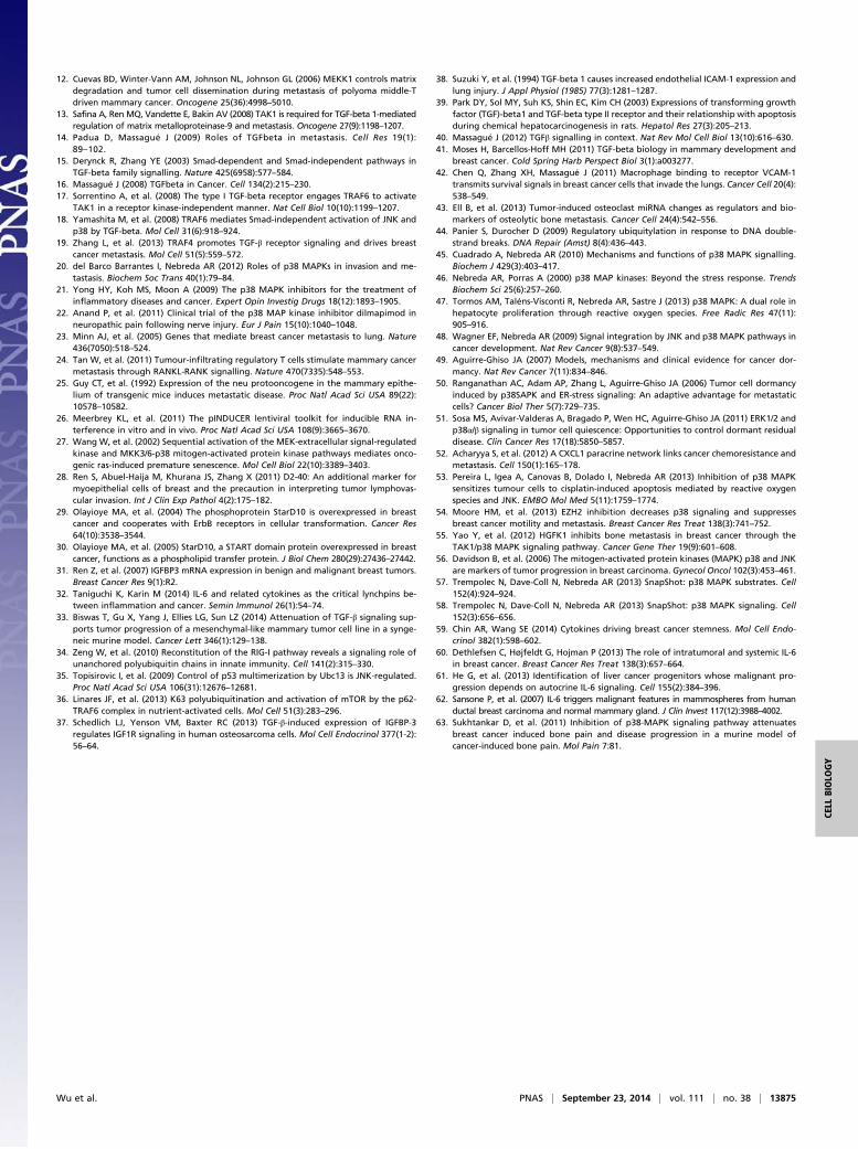
p38 Inhibition Suppresses Mammary Cancer Metastasis. Given theavailability of specific and effective p38 kinase inhibitors that arenot toxic in humans (21), we examined whether pharmacologicalinhibition of p38 affects metastatic spread. First, we injectedpolyomavirus middle T antigen (PyVT) transgene (PyMT) cells(33) into C57BL/6 females via the tail vein, followed by treatmentwith the p38 inhibitor SB203580 or vehicle via daily oral gavagefor 4 wk. Treatment with the p38 inhibitor suppressed formationof lung metastasis (Fig. 5 A and B). To examine whether p38 in-hibition leads to regression of established metastases, we startedinhibitor treatment 2 wk after cancer cell implantation (Fig. 5C).Importantly, a 2-wk-long treatment with SB203580 was as effectivein reducing metastatic load as the 4-wk-long treatment (Fig. 5D).These results suggest that p38 inhibition can be used to treatestablished BCa metastasis. In support of this hypothesis, wefound through a search of the TCGA and BRCA datasets that up-regulation of either p38α (MAPK14) or Ubc13 correlates withworse overall survival in human BCa (Fig. 5 E and F).
DiscussionThe observation that Ubc13 expression is elevated in metastaticBCa raised the suspicion that it may be causally involved in themetastatic process rather than just serve as a marker for BCa withhigh metastatic potential (5). Our results confirm that in additionto being associated with poor overall survival in human BCa,Ubc13 is directly involved in the metastatic process and is requiredboth for lung colonization and survival and proliferation ofestablished metastatic lesions. Ubc13, however, does not playa significant role in primary tumor development and growth. Inaddition, Ubc13 is dispensable for intravasation and extravasationof BCa cells, under the experimental conditions we used.Together with Uev1a, Ubc13 forms an E2 ubiquitin-conju-
gating enzyme that catalyzes formation of K63-linked poly-ubiquitin chains in response to activation of diverse cytokine andpattern recognition receptors (7). As such, Ubc13 is involved inactivation of a number of signaling pathways where K63-linked
polyubiquitin chains mediate protein–protein interactions thatcontrol critical protein kinases (7). Among the most thoroughlystudied Ubc13-dependent signal transduction pathways are theIKK-NF-κB, JNK, p38 (8, 9), and retinoic acid-inducible pro-tein 1 (RIG-I)–TANK-binding kinase 1 (TBK1) pathways (34),as well as p53 (35), and the mammalian target of rapamycin(mTOR) (36). We previously found that Ubc13 is required foractivation of the MAP3Ks TAK1 and MEKK1 downstream tomembers of the TNF receptor family (10). Ubc13 is also requiredfor TAK1 activation downstream to TGFβ receptors, which, justlike TNF receptors, rely on the signaling protein TRAF6 to re-cruit and direct Ubc13 toward its essential substrates (in thiscase, the TAB proteins) (17, 18). Although this has not beenfirmly established in our system, Ubc13 is likely to control BCametastasis downstream to TGFβ receptors, which are well-established regulators of BCa metastasis (14). TGFβ receptorsignaling also leads to activation of TAK1 and p38, both of whichact downstream to Ubc13 in the control of BCa metastasis.Importantly, Ubc13 is required for maintaining the expression ofnumerous BCa metastasis genes, including several genes that areregulated by TGFβ (37–39). Notably, however, Ubc13 is notrequired for SMAD activation by TGFβ, indicating that if indeedUbc13 stimulates metastatic growth and survival downstream ofTGFβ receptors, this effect is mediated through non-SMADsignaling. Given that TGFβ signaling can either suppress or pro-mote tumor development and metastatic spread depending oncontext and cell type (40, 41), general inhibition of both SMADand non-SMAD TGFβ signaling could lead to undesired out-comes. Our results demonstrate that more selective inhibition of
Vehicle SB203580
BA
D
Weeks: 4
Tumor cells Implant
0 2
Daily p38 inhibitor
Daily p38 inhibitor
AnalysisC
**
MAPK14
*
UBC13Lowest Quartile Highest Quartile Low MAPK14
Med-High MAPK14
E F
Fig. 5. Inhibition of p38 suppresses BCa metastasis. C57BL/6 females wereinjected with PyMT (Py8119) cells via the tail vein and treated with the p38inhibitor SB203580 (15 mg/kg body weight) or vehicle control by daily oralgavage for 4 wk. Lung metastases were quantified by surface nodule numbers(A). Representative lung images are shown in B. (Scale bar, 1 cm.) In another setof experiments, daily p38 inhibitor administration was initiated on either day0 or 2 wk after BCa cell implantation (C). Lung metastases under both treat-ment regimens (4 and 2 wk) were quantified and compared by counting surfacenodules (D). Results in A and D are averages ± SEM; n = 4–5 mice in each group.(E and F) Correlation between expression of p38 (MAPK14) and Ubc13 andbreast cancer patient survival. Kaplan–Meier survival curves for samples classi-fied as indicated. Shown are overall survival percentages for MAPK14 (tertiles)(E) and Ubc13 (quartiles) (F) expression based on clinical information availablein the TCGA for the BRCA dataset. **P = 0.0023, *P = 0.0369.
Wu et al. PNAS | September 23, 2014 | vol. 111 | no. 38 | 13873
CELL
BIOLO
GY
non-SMAD signaling through interference with Ubc13 or itsdownstream effector p38 does lead to effective inhibition ofmetastatic spread and even compromises the survival of existingmetastases. We speculate that Ubc13 exerts its prometastatic ac-tivity by controlling expression of cell surface molecules on BCacells. These molecules play pivotal roles in interacting with thehostile microenvironment, present at metastatic (or premetastatic)sites but are not required for survival and growth at the primarytumor site. Indeed, we found that loss of Ubc13 led to down-reg-ulation of a subset of cell surface molecules, including CD44,ICAM-1, and VCAM-1. VCAM-1 was found to provide a survivaladvantage to BCa cells by mediating their association with mac-rophages (42), and soluble ICAM-1 was reported to promote bonemetastasis through activation of NF-κB (43).Although Ubc13-dependent K63-linked polyubiquitination con-
trols the activation of several signaling pathways, as well as DNArepair responses (7, 44), our results indicate that TAK1-dependentp38 activation is the major mediator of the prometastatic effect ofUbc13 in BCa. Importantly, reconstitution of p38 activity by ectopicexpression of constitutively active MKK3(EE) in Ubc13-deficientBCa cells restores their metastatic potential. Involvement of p38MAPK signaling in cancer development, progression, and metas-tasis has been demonstrated previously (45–48). The p38 MAPKwas suggested to overcome ERK signaling to promote survival ofdormant cancer cells through activation of the unfolded proteinresponse (49–51), as well as various steps in the process of invasionand metastasis (20). Activation of p38 MAPK was also found to beelevated in BCa cells exposed to chemotherapy (52), and a p38inhibitor cooperates with cisplatin to induce cancer cell death inPyMT mice (53). In clinical specimens of matched primary andinvasive breast carcinomas, p38 phosphorylation was found to cor-relate with expression of EZH2, a polycomb group protein thatfunctions as an oncogene in BCa and whose overexpression is as-sociated with metastatic disease (54). However, one study has foundthat the TAK1–p38 pathway inhibits bone metastasis by BCa, actingdownstream to hepatocyte growth factor kringle 1 domain anddecreasing expression of receptor activator of NF-κB (55). Bycontrast, we find that either specific ablation of p38 in BCa ormammary cancer cells, as well as systemic inhibition of p38, resultsin a strong antimetastatic effect. A clinical study had revealed thatincreased p38 phosphorylation in BCa effusions correlated withshorter overall survival (56). It remains to be examined whether andhow Ubc13 and p38 control BCa bone metastasis.The p38 MAPKs, in particular p38α, exert their protumorigenic
activities through transcriptional and posttranscriptional regulationof numerous target genes (48) either through direct phosphoryla-tion or through the p38-dependent kinases: MAPK-activatedprotein kinase-2 (MAPKAPK2) and MAPKAPK3, p38-related/activated protein kinase (PRAK), mitogen- and stress-activatedprotein kinase-1( MSK1), and MAP kinase-interacting kinase 1 or2 (MNK1/2). p38α controls the activation of transcription factors,such as ATF2 and CREB; p38α also controls mRNA turnover andtranslation through various RNA binding proteins (57, 58). Usinggene expression analysis, we identified several target genes, pre-viously shown to affect BCa metastasis, whose expression is p38dependent, including CNN2, PLTP, IGFBP3, IL-6, IL13RA2,CD44, VCAM-1, and ICAM-1. At least one of these genes,
encoding ICAM-1, controls metastatic spread in our system. Inaddition, p38 and Ubc13 control expression of IL-6 mRNA in BCacells. Previous studies have shown that autocrine IL-6 signalingcontrols cancer cell growth, cancer stem cell (CSC) renewal, andmetastasis (59–61). IL-6 can stimulate Notch3-dependent up-reg-ulation of Jagged-1 to promote BCa cell growth and maintain anaggressive phenotype (62). It remains to be determined how IL-6signaling is regulated by the Ubc13 and p38 in the context of BCa.Importantly, pharmacological inhibition of p38 can block
metastatic spread of mammary cancer in mice and can even at-tenuate the growth or survival of established lung metastases.Given that a number of small molecule p38 inhibitors were foundto be effective and safe for the treatment of inflammatory pain inhumans (21, 22), these findings suggest that p38 inhibitors shouldbe evaluated as antimetastatic drugs in human BCa. Because bonemetastasis is frequently associated with inflammatory and neuro-pathic pain, such inhibitors can be first evaluated for their ability toalleviate pain in bone metastatic BCa, an application that will fa-cilitate the testing of their antimetastatic potential (63).
Materials and MethodsFemale virgin NOD/SCID, Balb/C, FVB, or C57BL/6 mice, 6–7 wk old (fromCharles River), were used. For orthotopic inoculation, cells suspended in PBSmixed with Matrigel (100 μL total volume in 1:1 ratio) were injected into thefourth right mammary fat pad of mice unless otherwise indicated. Tumorsize was measured using a caliper, and volume was calculated as length ×width2 × 0.52. At the end of experiments, mice were killed to harvest tumorsand lung tissues for histological and other analyses. For lung colonizationassays, mice were i.v. injected with 0.2 × 10⁶ LM2 cells in 100 μL PBS andsubjected to BLI imaging by IVIS. For inducible silencing experiments,doxycycline hyclate (Sigma-Aldrich) was added into the drinking water(2 mg/mL). To quantify lung metastasis, visible surface nodules were coun-ted. In some cases, lung tissues were sectioned and stained with H&E. Threesections spaced 100 μm apart were counted for metastases. Mice weremaintained under specific pathogen-free conditions, and all experimentalprotocols were approved by the University of California at San Diego AnimalCare Program, following National Institutes of Health Guidelines. Antibodyinformation and shRNA and Q-PCR primer sequences are listed in TablesS2–S4. Please see SI Materials and Methods for more details.
ACKNOWLEDGMENTS. We thank Santa Cruz Biotechnology, Cell SignalingTechnology, and GeneTex for providing antibodies, K. Taniguchi for helpwith histological analysis, J. Massague (Memorial Sloan Kettering CancerCenter) for sharing MDA-231 LM2 cells, S. Elledge (Harvard Medical School,Brigham and Women’s Hospital) for sharing the inducible lentiviral shRNAvector, P. Sun (The Scripps Research Institute) for pBabeMKK3(EE), F. Zolezziand J. Lum [Functional Genomics platform, Singapore Immunology Network,Agency for Science, Technology and Research (A*STAR)] for help with themicroarray study, Y. Yang and M. Kelliher (University of Massachusetts Med-ical School) for sharing shUbc13 plasmids, M. Fujimuro (Kyoto PharmaceuticalUniversity) for Ubc13 expression constructs, Jeevisha Bajaj for help with anal-ysis of real time imaging, and J. Xu and E. Theodorakis (University of Califor-nia, San Diego) for chemical inhibitor synthesis. S.K.B. and M.P. are gratefulfor funding from the Biomedical Research Council, A*STAR. X.W. was sup-ported by a postdoctoral fellowship from Susan G. Komen for the Cure(KG111506). Funding was provided by National Institutes of Health (NIH)Grants 1R01CA168689 (to J.Y.), 3R01CA168689-02W1 (to T.P.), DP1CA174422(to T.R.), T32 CA009523-29 (to R.G.F.), T32 GM007752-36 (to N.K.L.). Work wasalso supported by NIH Grants CA163798 and AI043477 and a San Diego Can-cer Center Council Collaborative Translational Grant sponsored by Pedal theCause San Diego (to M.K.). M.K. is an American Cancer Society Research Pro-fessor and holds the Ben and Wanda Hildyard Chair for Mitochondrial andMetabolic Diseases.
1. Desantis C, Ma J, Bryan L, Jemal A (2014) Breast cancer statistics, 2013. CA Cancer J Clin64(1):52–62.
2. Ferlay J, et al. (2013) Cancer incidence and mortality patterns in Europe: Estimates for40 countries in 2012. Eur J Cancer 49(6):1374–1403.
3. Fidler IJ (2003) The pathogenesis of cancer metastasis: The ‘seed and soil’ hypothesisrevisited. Nat Rev Cancer 3(6):453–458.
4. Wan L, Pantel K, Kang Y (2013) Tumor metastasis: Moving new biological insights intothe clinic. Nat Med 19(11):1450–1464.
5. Pestlin G, et al. (2006) US Patent 20060257950A.6. Bhoj VG, Chen ZJ (2009) Ubiquitylation in innate and adaptive immunity. Nature
458(7237):430–437.
7. Wang G, et al. (2012) K63-linked ubiquitination in kinase activation and cancer. FrontOncology 2:1–13.
8. Yamamoto M, et al. (2006) Key function for the Ubc13 E2 ubiquitin-conjugatingenzyme in immune receptor signaling. Nat Immunol 7(9):962–970.
9. Yamamoto M, et al. (2006) Cutting Edge: Pivotal function of Ubc13 in thymocyte TCRsignaling. J Immunol 177(11):7520–7524.
10. Matsuzawa A, et al. (2008) Essential cytoplasmic translocation of a cytokine receptor-assembled signaling complex. Science 321(5889):663–668.
11. Tseng PH, et al. (2010) Different modes of ubiquitination of the adaptor TRAF3 se-lectively activate the expression of type I interferons and proinflammatory cytokines.Nat Immunol 11(1):70–75.
13874 | www.pnas.org/cgi/doi/10.1073/pnas.1414358111 Wu et al.
12. Cuevas BD, Winter-Vann AM, Johnson NL, Johnson GL (2006) MEKK1 controls matrixdegradation and tumor cell dissemination during metastasis of polyoma middle-Tdriven mammary cancer. Oncogene 25(36):4998–5010.
13. Safina A, Ren MQ, Vandette E, Bakin AV (2008) TAK1 is required for TGF-beta 1-mediatedregulation of matrix metalloproteinase-9 and metastasis. Oncogene 27(9):1198–1207.
14. Padua D, Massagué J (2009) Roles of TGFbeta in metastasis. Cell Res 19(1):89–102.
15. Derynck R, Zhang YE (2003) Smad-dependent and Smad-independent pathways inTGF-beta family signalling. Nature 425(6958):577–584.
16. Massagué J (2008) TGFbeta in Cancer. Cell 134(2):215–230.17. Sorrentino A, et al. (2008) The type I TGF-beta receptor engages TRAF6 to activate
TAK1 in a receptor kinase-independent manner. Nat Cell Biol 10(10):1199–1207.18. Yamashita M, et al. (2008) TRAF6 mediates Smad-independent activation of JNK and
p38 by TGF-beta. Mol Cell 31(6):918–924.19. Zhang L, et al. (2013) TRAF4 promotes TGF-β receptor signaling and drives breast
cancer metastasis. Mol Cell 51(5):559–572.20. del Barco Barrantes I, Nebreda AR (2012) Roles of p38 MAPKs in invasion and me-
tastasis. Biochem Soc Trans 40(1):79–84.21. Yong HY, Koh MS, Moon A (2009) The p38 MAPK inhibitors for the treatment of
inflammatory diseases and cancer. Expert Opin Investig Drugs 18(12):1893–1905.22. Anand P, et al. (2011) Clinical trial of the p38 MAP kinase inhibitor dilmapimod in
neuropathic pain following nerve injury. Eur J Pain 15(10):1040–1048.23. Minn AJ, et al. (2005) Genes that mediate breast cancer metastasis to lung. Nature
436(7050):518–524.24. Tan W, et al. (2011) Tumour-infiltrating regulatory T cells stimulate mammary cancer
metastasis through RANKL-RANK signalling. Nature 470(7335):548–553.25. Guy CT, et al. (1992) Expression of the neu protooncogene in the mammary epithe-
lium of transgenic mice induces metastatic disease. Proc Natl Acad Sci USA 89(22):10578–10582.
26. Meerbrey KL, et al. (2011) The pINDUCER lentiviral toolkit for inducible RNA in-terference in vitro and in vivo. Proc Natl Acad Sci USA 108(9):3665–3670.
27. Wang W, et al. (2002) Sequential activation of the MEK-extracellular signal-regulatedkinase and MKK3/6-p38 mitogen-activated protein kinase pathways mediates onco-genic ras-induced premature senescence. Mol Cell Biol 22(10):3389–3403.
28. Ren S, Abuel-Haija M, Khurana JS, Zhang X (2011) D2-40: An additional marker formyoepithelial cells of breast and the precaution in interpreting tumor lymphovas-cular invasion. Int J Clin Exp Pathol 4(2):175–182.
29. Olayioye MA, et al. (2004) The phosphoprotein StarD10 is overexpressed in breastcancer and cooperates with ErbB receptors in cellular transformation. Cancer Res64(10):3538–3544.
30. Olayioye MA, et al. (2005) StarD10, a START domain protein overexpressed in breastcancer, functions as a phospholipid transfer protein. J Biol Chem 280(29):27436–27442.
31. Ren Z, et al. (2007) IGFBP3 mRNA expression in benign and malignant breast tumors.Breast Cancer Res 9(1):R2.
32. Taniguchi K, Karin M (2014) IL-6 and related cytokines as the critical lynchpins be-tween inflammation and cancer. Semin Immunol 26(1):54–74.
33. Biswas T, Gu X, Yang J, Ellies LG, Sun LZ (2014) Attenuation of TGF-β signaling sup-ports tumor progression of a mesenchymal-like mammary tumor cell line in a synge-neic murine model. Cancer Lett 346(1):129–138.
34. Zeng W, et al. (2010) Reconstitution of the RIG-I pathway reveals a signaling role ofunanchored polyubiquitin chains in innate immunity. Cell 141(2):315–330.
35. Topisirovic I, et al. (2009) Control of p53 multimerization by Ubc13 is JNK-regulated.Proc Natl Acad Sci USA 106(31):12676–12681.
36. Linares JF, et al. (2013) K63 polyubiquitination and activation of mTOR by the p62-TRAF6 complex in nutrient-activated cells. Mol Cell 51(3):283–296.
37. Schedlich LJ, Yenson VM, Baxter RC (2013) TGF-β-induced expression of IGFBP-3regulates IGF1R signaling in human osteosarcoma cells. Mol Cell Endocrinol 377(1-2):56–64.
38. Suzuki Y, et al. (1994) TGF-beta 1 causes increased endothelial ICAM-1 expression andlung injury. J Appl Physiol (1985) 77(3):1281–1287.
39. Park DY, Sol MY, Suh KS, Shin EC, Kim CH (2003) Expressions of transforming growthfactor (TGF)-beta1 and TGF-beta type II receptor and their relationship with apoptosisduring chemical hepatocarcinogenesis in rats. Hepatol Res 27(3):205–213.
40. Massagué J (2012) TGFβ signalling in context. Nat Rev Mol Cell Biol 13(10):616–630.41. Moses H, Barcellos-Hoff MH (2011) TGF-beta biology in mammary development and
breast cancer. Cold Spring Harb Perspect Biol 3(1):a003277.42. Chen Q, Zhang XH, Massagué J (2011) Macrophage binding to receptor VCAM-1
transmits survival signals in breast cancer cells that invade the lungs. Cancer Cell 20(4):538–549.
43. Ell B, et al. (2013) Tumor-induced osteoclast miRNA changes as regulators and bio-markers of osteolytic bone metastasis. Cancer Cell 24(4):542–556.
44. Panier S, Durocher D (2009) Regulatory ubiquitylation in response to DNA double-strand breaks. DNA Repair (Amst) 8(4):436–443.
45. Cuadrado A, Nebreda AR (2010) Mechanisms and functions of p38 MAPK signalling.Biochem J 429(3):403–417.
46. Nebreda AR, Porras A (2000) p38 MAP kinases: Beyond the stress response. TrendsBiochem Sci 25(6):257–260.
47. Tormos AM, Taléns-Visconti R, Nebreda AR, Sastre J (2013) p38 MAPK: A dual role inhepatocyte proliferation through reactive oxygen species. Free Radic Res 47(11):905–916.
48. Wagner EF, Nebreda AR (2009) Signal integration by JNK and p38 MAPK pathways incancer development. Nat Rev Cancer 9(8):537–549.
49. Aguirre-Ghiso JA (2007) Models, mechanisms and clinical evidence for cancer dor-mancy. Nat Rev Cancer 7(11):834–846.
50. Ranganathan AC, Adam AP, Zhang L, Aguirre-Ghiso JA (2006) Tumor cell dormancyinduced by p38SAPK and ER-stress signaling: An adaptive advantage for metastaticcells? Cancer Biol Ther 5(7):729–735.
51. Sosa MS, Avivar-Valderas A, Bragado P, Wen HC, Aguirre-Ghiso JA (2011) ERK1/2 andp38α/β signaling in tumor cell quiescence: Opportunities to control dormant residualdisease. Clin Cancer Res 17(18):5850–5857.
52. Acharyya S, et al. (2012) A CXCL1 paracrine network links cancer chemoresistance andmetastasis. Cell 150(1):165–178.
53. Pereira L, Igea A, Canovas B, Dolado I, Nebreda AR (2013) Inhibition of p38 MAPKsensitizes tumour cells to cisplatin-induced apoptosis mediated by reactive oxygenspecies and JNK. EMBO Mol Med 5(11):1759–1774.
54. Moore HM, et al. (2013) EZH2 inhibition decreases p38 signaling and suppressesbreast cancer motility and metastasis. Breast Cancer Res Treat 138(3):741–752.
55. Yao Y, et al. (2012) HGFK1 inhibits bone metastasis in breast cancer through theTAK1/p38 MAPK signaling pathway. Cancer Gene Ther 19(9):601–608.
56. Davidson B, et al. (2006) The mitogen-activated protein kinases (MAPK) p38 and JNKare markers of tumor progression in breast carcinoma. Gynecol Oncol 102(3):453–461.
57. Trempolec N, Dave-Coll N, Nebreda AR (2013) SnapShot: p38 MAPK substrates. Cell152(4):924–924.
58. Trempolec N, Dave-Coll N, Nebreda AR (2013) SnapShot: p38 MAPK signaling. Cell152(3):656–656.
59. Chin AR, Wang SE (2014) Cytokines driving breast cancer stemness. Mol Cell Endo-crinol 382(1):598–602.
60. Dethlefsen C, Højfeldt G, Hojman P (2013) The role of intratumoral and systemic IL-6in breast cancer. Breast Cancer Res Treat 138(3):657–664.
61. He G, et al. (2013) Identification of liver cancer progenitors whose malignant pro-gression depends on autocrine IL-6 signaling. Cell 155(2):384–396.
62. Sansone P, et al. (2007) IL-6 triggers malignant features in mammospheres from humanductal breast carcinoma and normal mammary gland. J Clin Invest 117(12):3988–4002.
63. Sukhtankar D, et al. (2011) Inhibition of p38-MAPK signaling pathway attenuatesbreast cancer induced bone pain and disease progression in a murine model ofcancer-induced bone pain. Mol Pain 7:81.
Wu et al. PNAS | September 23, 2014 | vol. 111 | no. 38 | 13875
CELL
BIOLO
GY