Giant mesenteric-cyst-cause-of-abdominal-distension-managed-with-laparotomy-a-case-report

CASE REPORT Open Access
Two-stage surgery for intraperitoneal andretroperitoneal multicentric liposarcomacausing hydronephrosis: a case reportRyohei Murata1* , Tadashi Yoshida2, Nobuhiro Kobayashi1, Yoshito Watanabe1, Shigenori Homma2,Hayato Echizenya1 and Akinobu Taketomi2
Abstract
Background: Liposarcoma is a soft tissue sarcoma of adipocyte origin. Liposarcoma represents 20–30% of adultsoft tissue tumors, which was most frequently seen in the retroperitoneal space in 45% and abdominal space inonly 5% of cases, but the multicentric case is unknown. Herein, we describe a rare case of multicentric, large, intra-abdominal and retroperitoneal liposarcoma, one of which had caused infection and pressing the right uretercausing hydronephrosis, which was resected by two-stage surgery.
Case presentation: The patient was a 46-year-old man who was referred for abdominal bloating and fatigue. Enhancedcomputed tomography showed a 23-cm intra-abdominal tumor and a 14.6-cm left retroperitoneal tumor. The intra-abdominal tumor which compressed the right ureter caused right unilateral hydronephrosis and deteriorated the renalfunction. The intra-abdominal tumor had also formed an intra-abdominal abscess. We performed emergent laparotomyand resected the intra-abdominal tumor. After the recovery of renal function, we resected the residual retroperitonealtumor. Histopathological examination showed both tumors to be myxoid/round cell type liposarcoma. Consideringclinical findings and their location, he was diagnosed with multicentric liposarcoma. He underwent adjuvantchemotherapy and has been alive without any recurrence for 9 months after the operation.
Conclusions: We successfully resected large intra-abdominal and retroperitoneal multicentric myxoid/roundcell liposarcomas. A two-stage surgery was a rational choice as it provides time to confirm the recovery ofrenal function.
Keywords: Intra-abdominal, Liposarcoma, Multicentric, Myxoid, Round cell, Retroperitoneal
BackgroundLiposarcoma represents 20–30% of adult soft tissue tu-mors, which is most frequently seen in the retroperiton-eal space in 45% and in the abdominal space only in 5%of cases, but multicentric cases are unknown [1]. It isoften detected when it is bigger and heavy, because itrarely presents any symptom during the early period,and many patients who sought clinical help had a chiefcomplaint of abdominal distention or mass [2]. Surgicalinterventions in liposarcoma differ case by case, accord-ing to the relationship with adjacent organs. Herein, we
describe a rare case of large, multicentric, intra-abdom-inal and retroperitoneal liposarcoma, one of which hadcaused infection and pressed the right ureter causinghydronephrosis, which was resected by a two-stagesurgery.
Case presentationA 46-year-old man complained of heartburn, abdominaldistention, and anorexia which persisted for over 2weeks. He had no significant personal and family history.Body temperature on admission was 39.2 °C. Abdominalphysical examination revealed large abdominal masseswithout tenderness or rebound. Blood examinationsshowed increased inflammation reaction (white bloodcell count, 29,700/μL; C-reactive protein, 33.30 mg/dL).
* Correspondence: [email protected] of Surgery, Otaru General Hospital, 047-8550, 1-1-1, Wakamatsu,Otaru-shi, Hokkai-do, JapanFull list of author information is available at the end of the article
© The Author(s). 2019 Open Access This article is distributed under the terms of the Creative Commons Attribution 4.0International License (http://creativecommons.org/licenses/by/4.0/), which permits unrestricted use, distribution, andreproduction in any medium, provided you give appropriate credit to the original author(s) and the source, provide a link tothe Creative Commons license, and indicate if changes were made.
Murata et al. Surgical Case Reports (2019) 5:18 https://doi.org/10.1186/s40792-019-0576-y

Renal dysfunction was also recognized (Table 1). Ab-dominal computed tomography revealed two large in-homogeneous masses with a diameter of 230 and 146mm, respectively. The upper part of the intra-abdominaltumor contained liquid and air, which indicated abscessformation (Fig. 1a–c). As the continuity to the gastro-intestinal tract was inexplicit, a mesenchymal tumor was
mostly suspected. The tumor compressed the right ur-eter, which caused right hydronephrosis (Fig. 1b). Onthe contrary, the left-sided tumor was solid, and it wasthought to be derived from the left kidney without lefthydronephrosis. Plane abdominal magnetic resonanceimaging, T2-weighed imaging (T2WI), and fat saturationT2WI showed mainly high intensity with some
Table 1 Laboratory test results on admission. Increase in inflammation reaction and renal dysfunction was recognized
AFP alpha-fetoprotein, Alb albumin, ALP alkaline phosphatase, ALT alanine aminotransferase, APTT activated partial thromboplastin time, ST aspartateaminotransferase, CEA carcinoembryonic antigen, Cl chloride, Cre creatinine, CRP C-reactive protein, CYFRA cytokeratin fragment, Hb hemoglobin, K potassium, LDHlactate dehydrogenase, NA sodium, Neut neutrophil, Plt platelet, PSA prostate-specific antigen, PT-INR prothrombin time-international normalized ratio, SCCsquamous cell carcinoma, T-bil total bilirubin, TP total protein, WBC white blood cell, γGT gamma-glutamyltransferase
a c
b
Fig. 1 An abdominal enhanced computed tomography scan. a An intra-abdominal mass with a diameter of 230 mm (white arrow). The dorsalpart of the intra-abdominal tumor contained liquid and air, which indicated abscess formation. b A retroperitoneal mass under the left kidneywith a diameter of 146 mm (black arrow), intra-abdominal tumor compressed the right ureter, and the right kidney was in hydronephrosis state(white arrowhead). c Both tumors had internal inhomogeneity, and the upper part of the intra-abdominal tumor would contain abscess (whitearrow, intra-abdominal tumor; black arrow, retroperitoneal tumor)
Murata et al. Surgical Case Reports (2019) 5:18 Page 2 of 7

low-intensity area (Fig. 2). There is also high intensity indiffusion-weighted imaging and low apparent diffusioncoefficient values at solid areas of both masses.We thought that the intra-abdominal tumor had
formed abscess and caused bacterial infection. We de-cided to resect the right intra-abdominal tumor first tostabilize the general condition from a systemic inflam-matory state. After the recovery of renal function, theleft retroperitoneal tumor resection was scheduled.In the first surgery, we inserted a ureteral stent to the
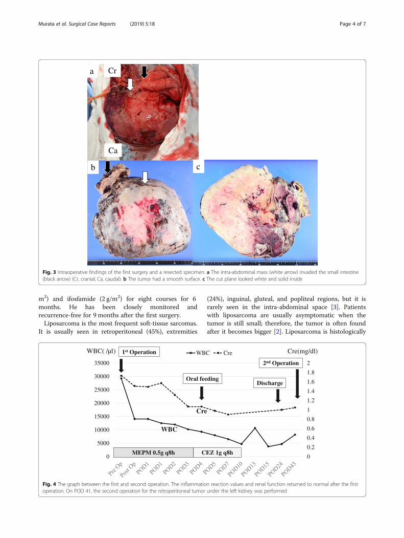
right ureter and successfully performed resection of theright tumor without invasion to the right ureter. How-ever, the tumor appeared to invade the small intestine,and combined resection was performed (Fig. 3a). Theoperation time was 274 min, and the total blood loss was3500 ml. We infused 14 units of red blood cells and 16units of fresh frozen plasma transfusion. The tumor hadsmooth surface, 26 × 23 cm in size, and weighed 3.9 kg(Fig. 3b, c). After the first operation, the inflammationreaction values and renal function returned to normal,and he was discharged on postoperative day (POD) 17.On POD 41, the second surgery was performed to re-move the retroperitoneal tumor under the left kidney(Fig. 4). Via the same incision as the first surgery, wereached the retroperitoneal tumor transperitoneally. Theretroperitoneal tumor was in contact with but not
derived from the left kidney, and complete tumor resec-tion was performed without left nephrectomy. The oper-ation time was 224 min, and the total blood loss was640 ml without any transfusion. Macroscopic findings ofthe retroperitoneal tumor were similar to those of theright intra-abdominal tumor, and it weighed 1.4 kg(Fig. 5). Histopathological examination of theintra-abdominal tumor on the right side revealed varioussizes of spindle-shaped or round-shaped cells in themyxoid parenchyma which were surrounded by fibrouscapsule, with hyperplasia of the capillary or microvascu-lar vessel, collagen fibers, and adipocyte cells (Fig. 6a–c).Alcian blue staining for mucinous component was posi-tive in most areas (Fig. 6d). The tumor cells were immu-nohistochemically positive for S100, MDM2, Rb1, andbcl2, but devoid of CD34, smooth muscle actin, and des-min (Fig. 6e–g). No invasion to the small intestine wasseen, and surgical margin was free (Fig. 6a). The charac-teristics of the retroperitoneal tumor were almost similarto those of the intra-abdominal tumor which suggestedthat both tumors had the same differentiation status.(Fig. 7a–f ). Finally, according to these findings, he wasdiagnosed with multicentric myxoid and round cell lipo-sarcoma. After the second operation, he was dischargedwithout any complications on POD 14 and received ad-juvant triweekly chemotherapy with doxorubicin (30 mg/
a b
c
Fig. 2 Non-contrast abdominal magnetic resonance image. a The solid part of both intra-abdominal (white arrow) and retroperitoneal (blackarrow) masses showed inhomogeneously high intensity in T2-weighted imaging (T2WI). b Both masses also showed almost the same pattern asT2WI in fat saturation T2WI. c Both masses also showed inhomogeneously high intensity in diffusion-weighted imaging
Murata et al. Surgical Case Reports (2019) 5:18 Page 3 of 7
m2) and ifosfamide (2 g/m2) for eight courses for 6months. He has been closely monitored andrecurrence-free for 9 months after the first surgery.Liposarcoma is the most frequent soft-tissue sarcomas.
It is usually seen in retroperitoneal (45%), extremities
(24%), inguinal, gluteal, and popliteal regions, but it israrely seen in the intra-abdominal space [3]. Patientswith liposarcoma are usually asymptomatic when thetumor is still small; therefore, the tumor is often foundafter it becomes bigger [2]. Liposarcoma is histologically
Ca
Cra
b c
Fig. 3 Intraoperative findings of the first surgery and a resected specimen. a The intra-abdominal mass (white arrow) invaded the small intestine(black arrow) (Cr, cranial; Ca, caudal). b The tumor had a smooth surface. c The cut plane looked white and solid inside
0
0.2
0.4
0.6
0.8
1
1.2
1.4
1.6
1.8
2
0
5000
10000
15000
20000
25000
30000
35000
WBC CreWBC( /µl) 1st Operation
Oral feedingDischarge
2nd Operation
Cre(mg/dl)
MEPM 0.5g q8h CEZ 1g q8h
WBC
Cre
Fig. 4 The graph between the first and second operation. The inflammation reaction values and renal function returned to normal after the firstoperation. On POD 41, the second operation for the retroperitoneal tumor under the left kidney was performed
Murata et al. Surgical Case Reports (2019) 5:18 Page 4 of 7

classified into five categories, namely, well-differentiated(45%), differentiated (20%), pleomorphic (10%), roundcell (5%), and myxoid type (rare). Most giant liposarco-mas are usually seen as differentiated tumors [4, 5].Myxoid and round cell tumors share the same character-istic cytogenic abnormalities: the translocation oft(12;16)(q13;p11) leading to the genes’ fusion DDIT3and FUS with generation of a hybrid protein FUS/DDIT3 [6]. Essentially, round cells are frequently foundin myxoid liposarcoma, which is considered to be themarker of poor prognosis when presenting 5% or moreof the mass in localized myxoid liposarcoma [7]. The
prognosis differs depending on these subtypes as follows:the 5-year survival rate is 95% in well-differentiated type,92% in mucinous type, 74% in round-cell type, and 59%in pleomorphic type [5].Liposarcoma is usually a single tumor. When multiple
tumors are seen, one of the main problems is to differ-entiate multicentric lesions from metastases of a singletumor. Multicentric liposarcoma accounts for only 1–2%of all liposarcomas in the USA and Europe [8]. Thereare no diagnostic criteria, but some features of multi-centric liposarcoma are indicated [1]: Primary liposarco-mas commonly occur in areas such as the thigh,popliteal fossa, retroperitoneum, peritoneal cavity (in-cluding the mesentery and greater omentum), upper ex-tremities, and thoracic serosa [2]. There is nooccurrence in sites where metastases are usually found,such as the lungs, liver, and skeletal system [3]. Histo-pathological type of the tumors is well differentiated ormyxoid patterns, which rarely metastasize [4]. There is along time interval between the occurrence of the firstand subsequent tumors [9]. In most cases, it is difficultto differentiate the multicentric liposarcoma from meta-static liposarcoma and there are no criteria. In our case,both liposarcomas had myxoid/round cell types in thecommon sites and were considered multicentric.The treatment for liposarcoma has not been established,
but en bloc resection is recommended not only for treat-ment but for pathological diagnosis [5]. Our case devel-oped renal dysfunction due to compression of the rightureter by the intra-abdominal tumor. If we tried to re-move both tumors simultaneously, and if the retroperiton-eal tumor was derived from the left kidney, composite
Fig. 5 The specimen of the second surgery. The retroperitoneal tumorlooked flat and smooth similar to the intra-abdominal tumor
a b c
e f g
d
Fig. 6 Histopathological examination of the intra-abdominal tumor. a–c Hematoxylin and eosin-stained sample showed various sizes of spindle-shaped or round-shaped cells in the myxoid parenchyma, surrounded by fibrous capsule, with hyperplasia of the capillary or microvascular vessel,collagen fibers, and adipocyte cells. a The small intestine serosa shown above the tumor surface was intact (arrowhead), and no tumor invasionto the small intestine was observed. d. Alcian blue staining for the mucinous component was positive. e–g Immunostaining with S100 andMDM2 was positive and negative with CD34
Murata et al. Surgical Case Reports (2019) 5:18 Page 5 of 7

resection with the kidney was unsafe, as it might affectkidney function. Thus, we decided to first resect theintra-abdominal tumor which caused abdominal abscess.In this case, multicentric tumors were composed of
multiple compartments. Therefore, we thought that hisprognosis would be poor and he should undergo chemo-therapy in the adjuvant settings. The effectiveness of ad-juvant chemotherapy remains unproven, and properregimen has not been established.Tumor size (> 20 cm), histological subtypes, and tumor
dissemination are considered important prognostic fac-tors [10]. In this case, both tumors were myxoid/roundcell types and found in two different compartments. Thepatient was thought to be at risk for recurrence and re-ceived adjuvant chemotherapy, but radiation therapywas not initiated because of the wide range of tumorbeds. Myxoid/round cell liposarcoma is a chemosensitivesoft tissue tumor, and adjuvant chemotherapy withdoxorubicin is recommended [11]. A meta-analysisshowed that the effect of the doxorubicin-based chemo-therapy improved relapse-free survival, but not overallsurvival [12]. In this case, he underwent triweeklychemotherapy with doxorubicin (30 mg/m2) and ifosfa-mide (2 g/m2) for five cycles, and there has been no re-currence for 9 months. In our case of multicentricmyxoid/round cell liposarcoma, en bloc surgical resec-tion and adjuvant chemotherapy were effective.
ConclusionWe successfully resected large, intra-abdominal, andretroperitoneal multicentric myxoid/round cell liposar-comas. A two-stage surgery was a rational choice as itprovides time to confirm the recovery of kidneyfunction.
AbbreviationsCT: Computed tomography; T2WI: T2-weighed imaging
AcknowledgementsWe would like to thank Editage (www.editage.jp) for the English languageediting, and Kiyoshi Kasai for histopathological examination.
FundingThis report did not receive any specific grant from funding agencies in thepublic, commercial, or not-for-profit sectors.
Authors’ contributionsRM described and designed the study. TY and HE supervised the writing ofthe manuscript. Other co-authors collected data and discussed the contentof the manuscript. All authors read and approved the final manuscript.
Ethics approval and consent to participateNot applicable.
Consent for publicationInformed consent was obtained from the patient and his family for thepublication of this case report.
Competing interestsThe authors declare that they have no competing interests.
Publisher’s NoteSpringer Nature remains neutral with regard to jurisdictional claims inpublished maps and institutional affiliations.
Author details1Department of Surgery, Otaru General Hospital, 047-8550, 1-1-1, Wakamatsu,Otaru-shi, Hokkai-do, Japan. 2Department of Gastroenterological Surgery I,Hokkaido University Hospital, 060-8648, Nishi 5 chome, Kita 14 jyo, Kita-ku,Sapporo-shi, Japan.
Received: 22 October 2018 Accepted: 24 January 2019
References1. Roviello F. Giant liposarcoma of the mesentery. Giant liposarcoma of the
mesentery. Report of a case. Ann Ital Chir. 2007;78:443–5.
a b c
e f g
d
Fig. 7 Histopathological examination of the retroperitoneal tumor specimen. a, b Hematoxylin and eosin-stained sample showed almost thesame character with the intra-abdominal tumor, suggesting they had the same differentiation status. c. Alcian blue staining for the mucinouscomponent was positive. d–f Immunostaining with S100 and MDM2 was positive and negative with CD34
Murata et al. Surgical Case Reports (2019) 5:18 Page 6 of 7

2. Jaques DP, Coit DG, Hajdu SI, Brennan MF. Management of primary andrecurrent soft-tissue sarcoma of the retroperitoneum. Ann Surg. 1990;212:51–9.
3. McKinley SK, Abreu N, Patalas E, Chang A. Large retroperitoneal liposarcomadiagnosed upon radiological evaluation of mild right-sided inguinal hernia.Case Rep Radiol. 2013;2013:187957.
4. Eltweri AM, Gravante G, Read-Jones SL, Rai S, Bowrey DJ, Haynes IG. A caseof recurrent mesocolon myxoid liposarcoma and review of the literature.Case Rep Oncol Med. 2013;2013:692754.
5. Sharma M, Mannan R, Bhasin TS, Manjari M, Punj R. Giant inflammatoryvariant of well differentiated liposarcoma: a case report of a rare entity. JClin Diagn Res. 2013;7:1720–1.
6. Göransson M, Andersson MK, Forni C, Ståhlberg A, Andersson C, Olofsson A,Mantovani R, Aman P. The myxoid liposarcoma FUS-DDIT3 fusiononcoprotein deregulates NF-kappaB target genes by interaction withNFKBIZ. Oncogene. 2009;28:270–8.
7. Fernández-Aceñero MJ, López-Criado P, López-Franco M, Meizoso T, CalvoC. Multicentric myxoid liposarcoma: report of two cases. World J SurgOncol. 2007;5:139.
8. Pack GT, Pierson JC. Liposarcoma: a study of 105 cases. Surgery. 1954;36:687–712.
9. Seenu V, Kriplani AK, Shukla NK, Raina V, Thakur K, Kapur BM. Multicentricliposarcoma: report of two cases. Surg Today. 1995;25:447–50.
10. Duman K, Girgin M, Artasc G. A case report: giant intra-abdominalliposarcoma presenting acute renal failure. Ann Med Surg (Lond). 2016;12:90–3.
11. Grimer R, Judson I, Peake D, Seddon B. Guidelines for the management ofsoft tissue sarcomas. Sarcoma. 2010;2010:506182.
12. O’Connor JM, Chacón M, Petracci FE, Chacón RD. Adjuvant chemotherapyin soft tissue sarcoma (STS): a meta-analysis of published data. J Clin Oncol.2008;26:10526.
Murata et al. Surgical Case Reports (2019) 5:18 Page 7 of 7