Two-photon laser-scanning microscopy for single and ...
16
1 Two-photon laser-scanning microscopy for single and repetitive imaging of dorsal and lateral spinal white matter in vivo Fabien Nadrigny 1,2,* , Karim Le Meur 1,3,* , Heinz Steffens 1,4,5,* , Eike D. Schomburg 1,4 , Sam Safavi-Abbasi 6,7 & Payam Dibaj 1 1 Max-Planck-Institute for Experimental Medicine, Göttingen, Germany; 2 U862, Institut François Magendie, Bordeaux, France; 3 Department of Pharmacy, Ludwig-Maximilians- University, Munich, Germany ; 4 Institute of Physiology, University of Göttingen, Germany; 5 Department of NanoBiophotonics, Max-Planck-Institute for Biophysical Chemistry, Göttingen, Germany; 6 Department of Neurosurgery, University of Oklahoma Health Sciences Center, Oklahoma City, USA; 7 Division of Neurological Surgery, St. Joseph`s Hospital and Medical Center, Phoenix, USA *: equal contribution Corresponding author: Payam Dibaj, MD, Max-Planck-Institute for Experimental Medicine, Göttingen, Germany, D-37075 Göttingen, Germany, e-mail: [email protected] Short title: In vivo imaging of spinal white matter Pages: 13; Figures: 4; Abstract: 99 words; total (Introduction, Methods, Results and Discussion): 1755 words. Abbreviations: 2P-LSM, two-photon laser-scanning microscopy; ECFP/EGFP/EYFP, enhanced cyan/green/yellow fluorescent protein.
Transcript of Two-photon laser-scanning microscopy for single and ...

1
Two-photon laser-scanning microscopy for single and repetitive imaging of dorsal and
lateral spinal white matter in vivo
Fabien Nadrigny1,2,*, Karim Le Meur1,3,*, Heinz Steffens1,4,5,*, Eike D. Schomburg1,4, Sam
Safavi-Abbasi6,7 & Payam Dibaj1
1Max-Planck-Institute for Experimental Medicine, Göttingen, Germany; 2U862, Institut
François Magendie, Bordeaux, France; 3Department of Pharmacy, Ludwig-Maximilians-
University, Munich, Germany ; 4Institute of Physiology, University of Göttingen, Germany;
5Department of NanoBiophotonics, Max-Planck-Institute for Biophysical Chemistry,
Göttingen, Germany; 6Department of Neurosurgery, University of Oklahoma Health Sciences
Center, Oklahoma City, USA; 7Division of Neurological Surgery, St. Joseph`s Hospital and
Medical Center, Phoenix, USA
*: equal contribution
Corresponding author: Payam Dibaj, MD, Max-Planck-Institute for Experimental Medicine,
Göttingen, Germany, D-37075 Göttingen, Germany, e-mail: [email protected]
Short title: In vivo imaging of spinal white matter
Pages: 13; Figures: 4; Abstract: 99 words; total (Introduction, Methods, Results and
Discussion): 1755 words.
Abbreviations: 2P-LSM, two-photon laser-scanning microscopy; ECFP/EGFP/EYFP,
enhanced cyan/green/yellow fluorescent protein.
Zdenka.Stadnikova
Pre-press

2
Abstract
We developed appropriate surgical procedures for single and repetitive multi-photon imaging
of spinal cord in vivo. By intravenous anesthesia, artificial ventilation and laminectomy, acute
experiments were performed in the dorsal and lateral white matter. By volatile anesthesia
and minimal-invasive surgery, chronic repetitive imaging up to 8 months were performed in
the dorsal column through the window between two adjacent spines. Transgenic mouse
technology enabled simultaneous imaging of labeled axons, astrocytes and microglia.
Repetitive imaging showed positional shifts of microglia over time. These techniques serve
for investigations of cellular dynamics and cell-cell interactions in intact and pathologically
changed spinal tissue.
Keywords: two-photon laser-scanning microscopy, in vivo imaging, spinal cord, white
matter, glia.

3
Introduction
Two-photon laser-scanning microscopy (2P-LSM) is an appropriate tool to image nervous
tissue in vivo (Davalos et al. 2005; Kerschensteiner et al. 2005; Kobat et al. 2009; Dibaj et al.
2010a). Time-lapse imaging is enabled by some advantages of this technique: recording of
deep stacks with high z-resolution within a relatively short time course and negligible
bleaching effects on simultaneously excited fluorescent proteins (FPs). The tool serves for
the study of cellular dynamics and interactions in intact and pathologically changed nervous
tissue (Davalos et al. 2005; Dibaj et al. 2010a; Dibaj et al. 2010b; Dibaj et al. 2011a; Nikic et
al. 2011; Dibaj et al. 2012; Nishida et al. 2014; Matsumura et al. 2015). Thereby, white
matter is the appropriate CNS region for studying axon-glia interactions (Nave 2010). In
contrast to the grey matter, accessibility of white matter for in vivo 2P-LSM is limited in the
brain. Spinal white matter with its superficial location provides an appropriate access. Here,
we demonstrate techniques for in vivo imaging of dorsal and lateral spinal white matter in a
single session. Repetitive 2P-LSM imaging of the spinal dorsal column has been performed
after laminectomy without (Dray et al. 2009; Di Maio et al. 2011; Davalos and Akassoglou
2012) or with implantation of a chamber (Farrar et al. 2012; Figley et al. 2013) or of a glass
window (Fenrich et al. 2012; Fenrich et al. 2013). Here, we demonstrate a technique for
repetitive imaging of the dorsal column by minimal-invasive surgery without the need of
laminectomy or implantatory procedures.

4
Methods
Ethics statement
The experiments were performed according to the ethical guidelines of the national animal
protection law and were authorized by the ethical committee of the State of Lower Saxony.
Mice
Adult triple-transgenic TgN(GFAP-ECFP)xTgH(CX3CR1-EGFP)xTgN(THY1-EYFP) mice (2-
5 months of age at the beginning of the experiments) were used for the experiments.
TgH(CX3CR1-EGFP)xTgN(THY1-EYFP) mice were obtained by crossbreeding homozygous
CX3CR1-EGFP mice, in which the expression of the green fluorescent protein EGFP in
monocytes, macrophages and microglia is achieved by placement of the EGFP reporter
gene into the Cx3cr1 locus encoding the chemokine receptor CX3CR1, with transgenic
THY1-EYFP mice expressing the yellow fluorescent protein EYFP in projection neurons
(Dibaj et al. 2010a). TgH(CX3CR1-EGFP)xTgN(THY1-EYFP) mice were next crossbred with
GFAP-ECFP mice expressing the cyan fluorescent protein ECFP in astrocytes (Dibaj et al.,
2011b). TgH(CX3CR1-EGFP), TgN(THY1-EYFP) and TgN(GFAP-ECFP) mice were of
C57Bl6 background for more than 10 generations.
Anesthesia and surgery
Anesthesia in acute single experiments
Anesthesia was initiated by 80 mg/kg pentobarbital injected i. p. The mouse was placed on a
support heated by a flat pad (Figure 1). After cannulation of the jugular vein by a catheter
with an outer diameter of 0.61 mm (Figure 2), anesthesia was continued intravenously with
40-60 mg methohexital-sodium per kg and h. A tracheotomy was performed and a tube
inserted for artificial ventilation (Figure 2). Active respiratory movements were abolished by
paralysis with pancuronium (800 µg per kg supplemented i. p. every hour) and artificial
ventilation was performed with a gas mixture of CO2 (2.5 %), O2 (47.5 %), and N2 (50 %) at
120 strokes/min (120-160 µl/stroke depending on the weight). Rectal body temperature,

5
heart rate (ECG: platinum wires of 0.3 mm diameter inserted into both forelegs and
connected via a differential amplifier with an oscilloscope) and O2 blood saturation
(MouseOx®, Starr Life Sciences, Oakmont, USA) were continuously monitored. Appropriate
dose of anesthesia was initially established by the absence of corneal and pinna reflexes,
later after paralysis, by falling body temperature and heart rate.
Anesthesia in chronic repetitive experiments
After initial anesthesia in a box filled with a mixture of gas (1:1 O2:N2O; 1000 ml/min)
enriched with 5% isoflurane, the mouse was placed on the heated support and anesthesia
was continued via a mask. Gas flow was reduced to 200 ml/min O2 and 150 ml/min N2O
which was enriched with 2% isoflurane. Appropriate dose of anesthesia was established by
the absence of corneal and pinna reflexes as well as by falling respiratory rate. Rectal body
temperature was continuously monitored.
Spinal preparation for single experiments
After incision of the skin and removal of tendons and muscle tissue from the lumbar dorsal
spinal processes and spinal arcs, a laminectomy was performed from vertebrae L1 to L4 or
L5 to expose the spinal cord segments L2 to L4 as well as to expose the dorsal roots L3 to
L5. For imaging purposes of the lateral spinal cord with dorsal root ganglia and ventral roots
(Dibaj et al. 2011a; Dibaj et al. 2011b; Dibaj et al. 2012), spinal arcs of the corresponding
vertebral bodies were removed on one side. After paralysis, dura mater was removed if
required for drug application (Dibaj et al. 2010a; Dibaj et al. 2012).
Spinal preparation for repetitive experiments
After removal of tendons and muscle tissue in the cleft between vertebral arcs L1 and L2,
imaging was performed through the cleaned intact dura mater. After imaging, the wound was
sutured, and 1 µg buprenorphine was given twice daily s. c. for two days. Without the need of

6
removal of spinal arcs by this procedure no signs of disability were observed between the
experiments.
Mounting the vertebral column
For imaging sessions nearly free of motion artefact the vertebral column was rigidly fixed with
two custom-made clamps (Figure 1 and 3a). For imaging of the lateral spinal cord two other
custom-made clamps were used to tilt the Mouse up to 90° (Figure 1c,d).
Two-photon laser-scanning microscopy and image acquisition
For 2P-LSM we built a custom-made microscope with four channels for simultaneous
recording of different FPs. Three dichroic mirrors split the fluorescence signal between a
Zeiss W Plan Apochromat objective (20x, NA 1.0, water immersion; Carl Zeiss GmbH, Jena,
Germany) and four Photo Multiplier Tubes (Hamamatsu, Japan). Modified ScanImage
software (Pologruto et al. 2003) was used to drive the microscope. Photographic and
epifluorescence overviews of the spinal cord were acquired using a 5x objective (NA 0.15;
Zeiss, Jena, Germany) and a CCD camera (Figure 3b,c). High resolution in vivo imaging
was performed using the custom-made microscope equipped with a fs-pulsed titanium-
sapphire laser (Chameleon Ultra II; Coherent, Glasgow, UK). For simultaneous excitation of
all FPs, the laser wavelength was set to 925 nm (Bestvater et al. 2002; Spiess et al. 2005).
Parallel, uniformly spaced planes were recorded, digitized and processed to obtain z-stacks
of images. The total acquisition time for a stack of 20 to 30 images and 40 to 60 µm depth
was approximately 2 min. In acute experiments, several stacks were taken continuously to
record time-lapse series (Dibaj et al. 2010a; Dibaj et al. 2011a).

7
Results and Discussion
In vivo imaging of the spinal cord
Due to high penetration depth and low photo-toxicity 2P-LSM is the method of choice for in
vivo imaging. We developed a method for stable in vivo imaging of superficial regions within
the white matter of the spinal cord. Figure 3 exemplarily shows a dorsal and a lateral aspect
of exposed spinal cords. Anesthesia and spinal preparation with respect to acute single and
chronic repetitive imaging experiments are described in detail in “Methods”.
Position permanence of simultaneously recorded spinal axons, astrocytes and
microglia
To image axons and glia in CNS white matter, we performed in vivo 2P-LSM of the spinal
cord of multiple-transgenic mice expressing ECFP, EGFP and EYFP in astrocytes, microglia
and projection neurons, respectively. Long-term repetitive imaging of the same region in the
dorsal spinal cord over more than 200 days revealed different spatial stability over time for
axons, astrocytes and microglia (Figure 4). Axonal structures as well as blood vessels show
stable spatial stability during the complete investigation period and, therefore, served as
landmarks for repetitive imaging of the same region. Astrocytes show a relatively high
positional stability with a “positional shift half time” (time by which half of the imaged cells
changed their position) of around 130 days, whereas microglial cells often change their
position. Thereby, the “positional shift half time” of perivascular microglia (around 36 days) is
distinctively higher than that of parenchymal microglia (around 1.6 days).
A major problem during in vivo imaging of the spinal cord is the drift of the imaged area
mainly due to motion artifacts resulting from the neighborhood of the spine to the lungs.
Anesthesia by i. p. injection of ketamin, xylazine and acepromazine together with suspending
the whole mouse in the air has been suggested to overcome respiratory movements
(Davalos and Akassoglou 2012). In the presented chronic repetitive experiments using
volatile anesthesia, also used by Farrar et al. (Farrar et al. 2012; Farrar et al. 2015),
8
suspending of the mouse was not necessary thus facilitating permanent contact to the
heated support. The method of imaging the spinal cord through the window between two
adjacent spines distinctly minimizes not only the trauma during surgery when compared with
exposure by laminectomy but also the rate of artificial movements. By the presented minimal-
invasive surgical protocol, without any implantatory procedures, we were able to perform
longitudinal studies over more than 200 days with up to ten separate imaging sessions in the
same mouse without any observations of motor or other behavioral deficits.
For the performance of acute single experiments we recommend i. v. anesthesia
together with intubation and artificial respiration. The animals can hereby be paralyzed which
lead to a distinct reduction of active contraction-induced movements. Moreover, the
respiration trigger might be used as imaging trigger leading to further reduction of motion
artefacts. To our knowledge, we are the only group being able to image lateral white matter
of the spinal cord in vivo (Dibaj et al. 2011a; Dibaj et al. 2011b; Dibaj et al. 2012). In this
approach, for surgical and imaging purposes, motion artifacts have to be maximally reduced
by paralysis and artificial respiration. In contrast to fasciculus gracilis of the dorsal column
with its ascending sensory fibers, descending motor fibers can only be imaged in vivo in the
lateral white matter. This is of particular interest for imaging studies of murine disease
models predominantly affecting motor areas of the central nervous system, for instance,
SOD1-model of amyotrophic lateral sclerosis (Dibaj et al. 2011a; Dibaj et al. 2011b; Dibaj et
al. 2012).
The demonstrated techniques for in vivo imaging of dorsal and lateral spinal white
matter in a single session as well as for repetitive imaging of the dorsal column by minimal-
invasive surgery without the need of laminectomy or implantatory procedures serve for
preclinical studies of healthy and pathologically changed spinal cord. Using murine models of
neurological disorders, imaging studies in vivo, especially those with longitudinal scheme,
may help to identify effective therapeutic agents.

9
Figure 1 Equipment for fixation of the mouse during surgery and imaging. (a) Profile of the
heatable mouse support with rails to fit in the cross table of the microscope and rails to fix the
vertebral column clamps. The stainless steel plate is isolated by cork to heat up only the
steel plate via the heat pad. (b-d) Mouse support, covered with water proof pad, and clamps
for fixation of the vertebral column. (b) Modified Narishige clamps for the vertebral column,
used for the dorsal aspect of the spinal cord. The jaws are tilted to leave enough room for the
Microscope lens (see Figure 3a). (c,d) Two other custom-made clamps for the vertebral
column, used for the lateral aspect of the spinal cord. (c) Position of the jaws during fixation
of the vertebral column. (d) Position of the jaws after tilting the vertebral column for imaging
lateral spinal cord.

10
Figure 2 Infusion anesthesia and artificial ventilation. (a) Ventral aspect of the mouse neck,
with central vein catheter and tracheal tube. (b) Diagram of respiration gas flow during
artificial respiration.
11
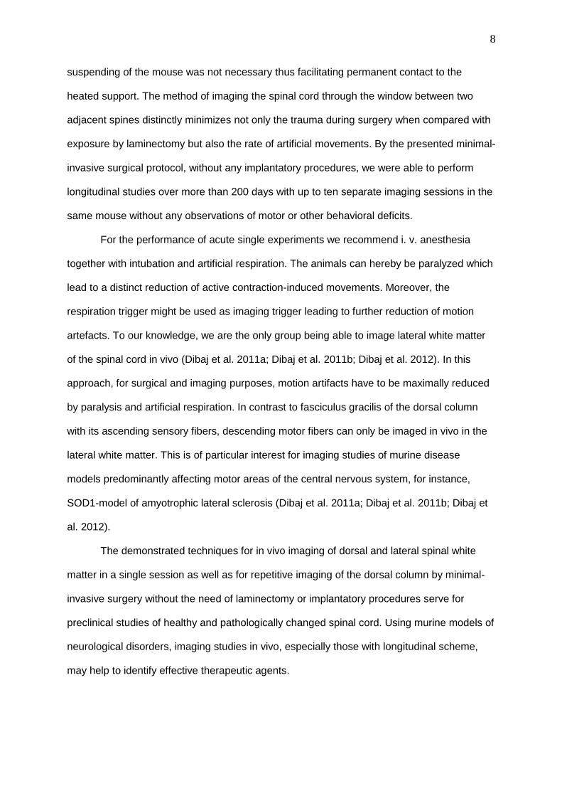
Figure 3 Mounted vertebral column with spinal cord ready for microscopy. (a) Vertebral
column fixed by clamps; objective lens in position. (b) Dorsal aspect of the spinal cord; view
through the cleft between lumbar spines 1 and 2; dura intact. (c) Lateral aspect of the spinal
cord; spinal arcs L1 to L3 dorsally and on the left side completely removed; dorsal roots L3
and L4 shifted dorsally. Arrows show rostrally. DCV: dorsal central vein, DGL: lumbar dorsal

12
root ganglion, DL: lumbar dorsal root, LSC: lateral spinal cord, VL: lumbar ventral root; the
numbers correspond to the lumbar spinal cord segments.
Figure 4 Long-term repetitive multi-cellular imaging of the dorsal spinal cord. (a-e) Repetitive
2P-LSM images of the same region in the dorsal white matter of the lumbar spinal cord over
150 days in a triple transgenic mouse expressing ECFP in astrocytes, EGFP in microglia and

13
EYFP in axons. Each image is a Maximum Intensity Projection of a 40-µm thick stack. The
central vein is on the left side, the dorsal root entry zone on the right. All images are
arranged such that rostral is to the upper side. Axons are stable structures and are used as
landmarks for repetitive imaging. The ellipse marks an astrocyte which is visible in all
images, whereas a parenchymal microglia in the neighborhood, marked by a square, is only
shortly present in the imaged stack. In contrast, another microglia, marked by an arrow in a-
d, is present over a relatively long period. A perivascular microglia, marked by a circle, is
also present over a relatively long period. Arrows in e mark elongated bi-polarized
astrocytes. The arrowhead marks a node of Ranvier in e. Scale bar, 50 µm. (f) Measuring the
fraction of cells remaining at the same place over time revealed the spatial stability of each
cell type over time. Although astrocytes show a relatively high positional stability (“positional
shift half time” τastro ~ 130 days), parenchymal microglia changed position at a very fast rate
(τmicro ~ 1.6 days). Respective half time of perivascular microglia laid in between (τperivasc. micro
~ 36 days).

14
References
Bestvater F, Spiess E, Stobrawa G, Hacker M, Feurer T, Porwol T, Berchner-Pfannschmidt
U, Wotzlaw C, Acker H: Two-photon fluorescence absorption and emission spectra of
dyes relevant for cell imaging. J Microsc 208: 108-115, 2002.
Davalos D, Akassoglou K: In vivo imaging of the mouse spinal cord using two-photon
microscopy. J Vis Exp 59: e2760, 2012.
Davalos D, Grutzendler J, Yang G, Kim JV, Zuo Y, Jung S, Littman DR, Dustin ML, Gan WB:
ATP mediates rapid microglial response to local brain injury in vivo. Nat Neurosci 8:
752-758, 2005.
Dibaj P, Nadrigny F, Steffens H, Scheller A, Hirrlinger J, Schomburg ED, Neusch C, Kirchhoff
F: NO mediates microglial response to acute spinal cord injury under ATP control in
vivo. Glia 58: 1133-1144, 2010a.
Dibaj P, Steffens H, Nadrigny F, Neusch C, Kirchhoff F, Schomburg ED: Long-lasting post-
mortem activity of spinal microglia in situ in mice. J Neurosci Res 88: 2431-2440,
2010b.
Dibaj P, Steffens H, Zschüntzsch J, Nadrigny F, Schomburg ED, Kirchhoff F, Neusch C: In
Vivo imaging reveals distinct inflammatory activity of CNS microglia versus PNS
macrophages in a mouse model for ALS. PLoS One 6: e17910, 2011a.
Dibaj P, Steffens H, Zschüntzsch J, Kirchhoff F, Schomburg ED, Neusch C: In vivo imaging
reveals rapid morphological reactions of astrocytes towards focal lesions in an ALS
mouse model. Neurosci Lett 497: 148-151, 2011b.
Dibaj P, Zschüntzsch J, Steffens H, Scheffel J, Göricke B, Weishaupt JH, Le Meur K,
Kirchhoff F, Hanisch UK, Schomburg ED, Neusch C: Influence of methylene blue on
microglia-induced inflammation and motor neuron degeneration in the SOD1 (G93A)
model for ALS. PLoS One 8: e43963, 2012.

15
Di Maio A, Skuba A, Himes BT, Bhagat SL, Hyun JK, Tessler A, Bishop D, Son Y: In vivo
imaging of dorsal root regeneration: rapid immobilization and presynaptic
differentiation at the CNS/PNS border. J Neurosci 31: 4569-4582, 2011.
Dray C, Rougon G, Debarbieux F: Quantitative analysis by in vivo imaging of the dynamics of
vascular and axonal networks in injured mouse spinal cord. Proc Natl Acad Sci U S A
106: 9459-9464, 2009.
Farrar MJ, Bernstein IM, Schlafer DH, Cleland TA, Fetcho JR, Schaffer CB: Chronic in vivo
imaging in the mouse spinal cord using an implanted chamber. Nat Methods 9: 297-
302, 2012.
Farrar MJ, Rubin JD, Diago DM, Schaffer CB: Characterization of blood flow in the mouse
dorsal spinal venous system before and after dorsal spinal vein occlusion. J Cereb
Blood Flow Metab 35: 667-675, 2015.
Fenrich KK, Weber P, Hocine M, Zalc M, Rougon G, Debarbieux F: Long-term in vivo
imaging of normal and pathological mouse spinal cord with subcellular resolution
using implanted glass windows. J Physiol 590: 3665-3675, 2012.
Fenrich KK, Weber P, Rougon G, Debarbieux F: Long- and short-term intravital imaging
reveals differential spatiotemporal recruitment and function of myelomonocytic cells
after spinal cord injury. J Physiol 591: 4895-4902, 2013.
Figley SA, Chen Y, Maeda A, Conroy L, McMullen JD, Silver JI, Stapleton S, Vitkin A,
Lindsay P, Burrell K, Zadeh G, Fehlings MG, DaCosta RS. A spinal cord window
chamber model for in vivo longitudinal multimodal optical and acoustic imaging in a
murine model. PLoS One 8: e58081, 2013.
Kerschensteiner M, Schwab ME, Lichtman JW, Misgeld T: In vivo imaging of axonal
degeneration and regeneration in the injured spinal cord. Nat Med 11: 572-577, 2005.
Kobat D, Durst ME, Nishimura N, Wong AW, Schaffer CB, Xu C: Deep tissue multiphoton
microscopy using longer wavelength excitation. Opt Express 17: 13354-13364, 2009.

16
Matsumura S, Taniguchi W, Nishida K, Nakatsuka T, Ito S: In vivo two-photon imaging of
structural dynamics in the spinal dorsal horn in an inflammatory pain model. Eur J
Neurosci 41: 989-997, 2015.
Nave KA: Myelination and support of axonal integrity by glia. Nature 468: 244-252, 2010.
Nikic I, Merkler D, Sorbara C, Brinkoetter M, Kreutzfeldt M, Bareyre FM, Bruck W, Bishop D,
Misgeld T, Kerschensteiner M: Reversible form of axon damage in experimental
autoimmune encephalomyelitis and multiple sclerosis. Nat Med 17: 495-499, 2011.
Nishida K, Matsumura S, Taniguchi W, Uta D, Furue H, Ito S: Three-dimensional distribution
of sensory stimulation-evoked neuronal activity of spinal dorsal horn neurons
analyzed by in vivo calcium imaging. PLoS One 9: e103321, 2014.
Pologruto TA, Sabatini BL, Svoboda K: ScanImage: flexible software for operating laser
scanning microscopes. Biomed Eng Online 2: 13, 2003.
Spiess E, Bestvater F, Heckel-Pompey A, Toth K, Hacker M, Stobrawa G, Feurer T, Wotzlaw
C, Berchner-Pfannschmidt U, Porwol T, Acker H: Two-photon excitation and emission
spectra of the green fluorescent protein variants ECFP, EGFP and EYFP. J Microsc
217: 200-204, 2005.


![Knife-Edge Scanning Microscopy: High-throughput Imaging and …jkwon/publications/files/choe.hpc08... · more advanced schemes such as multi-photon microscopy [3], optical sectioning](https://static.fdocuments.net/doc/165x107/5f787d3f59b36f6e7179727c/knife-edge-scanning-microscopy-high-throughput-imaging-and-jkwonpublicationsfileschoehpc08.jpg)