
TWO-PHOTON EXCITATION FLUORESCENCE MICROSCOPY...
42
? Annu. Rev. Biomed. Eng. 2000. 02:399–429 Copyright c 2000 by Annual Reviews. All rights reserved T WO-PHOTON EXCITATION FLUORESCENCE MICROSCOPY Peter T. C. So 1 , Chen Y. Dong 1 , Barry R. Masters 2 , and Keith M. Berland 3 1 Department of Mechanical Engineering, Massachusetts Institute of Technology, Cambridge, Massachusetts 02139; e-mail: [email protected] 2 Department of Ophthalmology, University of Bern, Bern, Switzerland 3 Department of Physics, Emory University, Atlanta, Georgia 30322 Key Words multiphoton, fluorescence spectroscopy, single molecule, functional imaging, tissue imaging ■ Abstract Two-photon fluorescence microscopy is one of the most important re- cent inventions in biological imaging. This technology enables noninvasive study of biological specimens in three dimensions with submicrometer resolution. Two-photon excitation of fluorophores results from the simultaneous absorption of two photons. This excitation process has a number of unique advantages, such as reduced specimen photodamage and enhanced penetration depth. It also produces higher-contrast im- ages and is a novel method to trigger localized photochemical reactions. Two-photon microscopy continues to find an increasing number of applications in biology and medicine. CONTENTS INTRODUCTION ................................................ 400 HISTORICAL REVIEW OF TWO-PHOTON MICROSCOPY TECHNOLOGY ... 401 BASIC PRINCIPLES OF TWO-PHOTON MICROSCOPY .................. 402 Physical Basis for Two-Photon Excitation ............................. 402 Optical Properties of Two-Photon Microscopy .......................... 403 TWO-PHOTON MICROSCOPY INSTRUMENTATION .................... 404 Two-Photon Laser Sources ........................................ 404 Scanning Fluorescence Microscopy Optics ............................. 405 Fluorescence Detection System ..................................... 406 FLUORESCENT PROBES USED IN TWO-PHOTON MICROSCOPY ......... 406 Extrinsic and Endogenous Two-Photon Fluorophores ..................... 406 Recent Efforts in Two-Photon Probe Development ....................... 408 PHYSIOLOGICAL EFFECTS OF NEAR-IR MICROBEAM ILLUMINATION ... 409 MOLECULAR-LEVEL APPLICATIONS OF TWO-PHOTON MICROSCOPY ... 410 Single-Molecule Detection in Solution ................................ 410 Imaging Single Molecules with Two-Photon Excitation .................... 411 1523-9829/00/0825-0399$14.00 399 Annu. Rev. Biomed. Eng. 2000.2:399-429. Downloaded from arjournals.annualreviews.org by University of Illinois - Urbana Champaign on 02/02/09. For personal use only.
Transcript of TWO-PHOTON EXCITATION FLUORESCENCE MICROSCOPY...
P1: FhN/ftt P2: FhN
July 10, 2000 11:18 Annual Reviews AR106-15
?Annu. Rev. Biomed. Eng. 2000. 02:399–429
Copyright c© 2000 by Annual Reviews. All rights reserved
TWO-PHOTON EXCITATION FLUORESCENCE
MICROSCOPY
Peter T. C. So1, Chen Y. Dong1, Barry R. Masters2,and Keith M. Berland31Department of Mechanical Engineering, Massachusetts Institute of Technology,Cambridge, Massachusetts 02139; e-mail: [email protected] of Ophthalmology, University of Bern, Bern, Switzerland3Department of Physics, Emory University, Atlanta, Georgia 30322
Key Words multiphoton, fluorescence spectroscopy, single molecule, functionalimaging, tissue imaging
■ Abstract Two-photon fluorescence microscopy is one of the most important re-cent inventions in biological imaging. This technology enables noninvasive study ofbiological specimens in three dimensions with submicrometer resolution. Two-photonexcitation of fluorophores results from the simultaneous absorption of two photons.This excitation process has a number of unique advantages, such as reduced specimenphotodamage and enhanced penetration depth. It also produces higher-contrast im-ages and is a novel method to trigger localized photochemical reactions. Two-photonmicroscopy continues to find an increasing number of applications in biology andmedicine.
CONTENTS
INTRODUCTION . . . . . . . . . . . . . . . . . . . . . . . . . . . . . . . . . . . . . . . . . . . . . . . . 400HISTORICAL REVIEW OF TWO-PHOTON MICROSCOPY TECHNOLOGY. . . 401BASIC PRINCIPLES OF TWO-PHOTON MICROSCOPY. . . . . . . . . . . . . . . . . . 402
Physical Basis for Two-Photon Excitation. . . . . . . . . . . . . . . . . . . . . . . . . . . . . 402Optical Properties of Two-Photon Microscopy. . . . . . . . . . . . . . . . . . . . . . . . . . 403
TWO-PHOTON MICROSCOPY INSTRUMENTATION. . . . . . . . . . . . . . . . . . . . 404Two-Photon Laser Sources. . . . . . . . . . . . . . . . . . . . . . . . . . . . . . . . . . . . . . . . 404Scanning Fluorescence Microscopy Optics. . . . . . . . . . . . . . . . . . . . . . . . . . . . . 405Fluorescence Detection System. . . . . . . . . . . . . . . . . . . . . . . . . . . . . . . . . . . . . 406
FLUORESCENT PROBES USED IN TWO-PHOTON MICROSCOPY. . . . . . . . . 406Extrinsic and Endogenous Two-Photon Fluorophores. . . . . . . . . . . . . . . . . . . . . 406Recent Efforts in Two-Photon Probe Development. . . . . . . . . . . . . . . . . . . . . . . 408
PHYSIOLOGICAL EFFECTS OF NEAR-IR MICROBEAM ILLUMINATION . . . 409MOLECULAR-LEVEL APPLICATIONS OF TWO-PHOTON MICROSCOPY. . . 410
Single-Molecule Detection in Solution. . . . . . . . . . . . . . . . . . . . . . . . . . . . . . . . 410Imaging Single Molecules with Two-Photon Excitation. . . . . . . . . . . . . . . . . . . . 411
1523-9829/00/0825-0399$14.00 399
Ann
u. R
ev. B
iom
ed. E
ng. 2
000.
2:39
9-42
9. D
ownl
oade
d fr
om a
rjou
rnal
s.an
nual
revi
ews.
org
by U
nive
rsity
of
Illin
ois
- U
rban
a C
ham
paig
n on
02/
02/0
9. F
or p
erso
nal u
se o
nly.
P1: FhN/ftt P2: FhN
July 10, 2000 11:18 Annual Reviews AR106-15
?400 SO ET AL
Fluorescence Correlation Spectroscopy. . . . . . . . . . . . . . . . . . . . . . . . . . . . . . . 411Photon-Counting Histogram. . . . . . . . . . . . . . . . . . . . . . . . . . . . . . . . . . . . . . . 413
CELLULAR-LEVEL APPLICATIONS OF TWO-PHOTON MICROSCOPY. . . . . 414Two-Photon In Vivo Cellular Imaging—Minimizing Photodamage
and Bleaching. . . . . . . . . . . . . . . . . . . . . . . . . . . . . . . . . . . . . . . . . . . . . . . . 414Multiphoton Imaging of Far-UV Fluorophores. . . . . . . . . . . . . . . . . . . . . . . . . . 414Two-Photon Multiple Color Imaging. . . . . . . . . . . . . . . . . . . . . . . . . . . . . . . . . 415Three-Dimensional Localized Uncaging of Signaling Molecules. . . . . . . . . . . . . 415
TISSUE LEVEL APPLICATIONS OF TWO-PHOTON MICROSCOPY. . . . . . . . . 416Applying Two-Photon Microscopy to Study Tissue Physiology. . . . . . . . . . . . . . 416Applying Two-Photon Excitation for Clinical Diagnosis and Treatment. . . . . . . . 417
NEW DEVELOPMENTS IN TWO-PHOTON INSTRUMENTATION. . . . . . . . . . 418Two-Photon Video Rate Microscopy. . . . . . . . . . . . . . . . . . . . . . . . . . . . . . . . . 418Simultaneous Two-Photon Fluorescence and Reflected-Light
Confocal Microscopy. . . . . . . . . . . . . . . . . . . . . . . . . . . . . . . . . . . . . . . . . . . 419Integrating Fluorescence Spectroscopy into Two-Photon Microscopes. . . . . . . . . 420
CONCLUSION. . . . . . . . . . . . . . . . . . . . . . . . . . . . . . . . . . . . . . . . . . . . . . . . . . 421
INTRODUCTION
The need for better diagnostic tools has triggered a renaissance in optical-micro-scopy instrumentation development. Two-photon fluorescence microscopy (TPM),invented by Denk et al in 1990 (1), is a three-dimensional (3D) imaging technologybased on the nonlinear excitation of fluorophores. TPM is considered a revolution-ary development in biological imaging because of its four unique capabilities.First, TPM greatly reduces photodamage and allows imaging of living specimens.Second, TPM can image turbid specimens with submicrometer resolution down toa depth of a few hundred micrometers. Third, TPM allows high-sensitivity imagingby eliminating the contamination of the fluorescence signal by the excitation light.Fourth, TPM can initiate photochemical reaction within a subfemtoliter volumeinside cells and tissues.
This review covers the historical development of two-photon fluorescence mi-croscopy techniques and the underlying physical principles. The basic instrumenta-tion design for TPM is explained. We describe the two-photon absorption propertiesof a number of commonly used fluorophores and the recent efforts in synthesizingtwo-photon optimized new probes. To effectively apply this new technique for livespecimen imaging, we need to understand two-photon photodamage mechanisms.We discuss a number of biomedical uses of two-photon excitation at the molecular,cellular, and tissue levels. Finally, we survey recent instrumentation developmentefforts that may lead to more novel applications.
The reader is referred to several previous reviews of topics included in thischapter (2–4). The patent from the Cornell group of Webb and coworkers, whichdescribes multiphoton excitation microscopy, is critical reading (5). An intro-duction to confocal microscopy and its applications is provided elsewhere (6, 7).
Ann
u. R
ev. B
iom
ed. E
ng. 2
000.
2:39
9-42
9. D
ownl
oade
d fr
om a
rjou
rnal
s.an
nual
revi
ews.
org
by U
nive
rsity
of
Illin
ois
- U
rban
a C
ham
paig
n on
02/
02/0
9. F
or p
erso
nal u
se o
nly.
P1: FhN/ftt P2: FhN
July 10, 2000 11:18 Annual Reviews AR106-15
?TWO-PHOTON MICROSCOPY 401
Confocal microscopy is reviewed in a new book of reprinted selected historicalpapers and patents (8).
HISTORICAL REVIEW OF TWO-PHOTONMICROSCOPY TECHNOLOGY
The potential for highly intense light to trigger nonlinear processes has long beenrecognized. In particular, multiphoton excitation processes were predicted byMaria Goppert-Mayer in her doctoral dissertation on the theory of two-photonquantum transitions in atoms (9). Experimental work in nonlinear optics may havebegun with the work by Franken and his group in 1961, focusing on second har-monic generation of light (10). They showed that ruby laser light, at wavelengthλ, propagating through a quartz crystal will generate light at the second harmonicfrequency with a wavelength ofλ/2. In 1963, a few weeks after the publicationof the paper by Franken et al, Kaiser & Garret published the first report on two-photon excitation (TPE) of CaF2:Eu2+ fluorescence (11). They later demonstratedthat TPE also can excite the fluorescence of organic dyes. Since then, many ex-amples of TPE processes in molecular spectroscopy have been reported (12, 13).Two-photon spectroscopy has become an important tool to study the electronicstructure of the molecular excited states (14, 15). G¨oppert-Mayer’s theory wasfinally verified 32 years after its formulation. By analogy with the two-photonprocesses, three-photon excitation spectroscopy has also been described. Three-photon absorption processes were first reported by Singh & Bradley (16). Sincethen others have demonstrated three-photon excitation processes (17, 18). Today,the term multiphoton excitation commonly describes two and higher numbers ofphoton excitation processes.
After the development of nonlinear optical spectroscopy, the potential of non-linear optical effects in microscopy was soon recognized. In a conventional lightmicroscope, the source of the contrast is the differences in the absorption co-efficients and the optical density of the specimens. For nonlinear microscopy, aspecimen with a nonlinear optical cross-section will produce higher harmoniclight emission under sufficiently intense illumination. The nonlinear harmonicgeneration is a function of the molecular structure. All materials possess third-order nonlinear susceptibility and higher-order terms; second-order nonlinear sus-ceptibility exists in specimens that have non-centrosymmetric geometry, such asLiNbO3 crystals and some biological specimens. The principle of nonlinear scan-ning microscopy has been simply explained (6). Practical applications of nonlinearmicroscopy started with works of Freund & Kopf (18a); they determined the prop-erties of ferroelectric domains by an analysis of the intensity and the angulardistribution of the second harmonic generated within the crystals. Hellwarth &Christensen developed a second harmonic microscope to study microstructuresin polycrystalline ZnS materials (19). The potential for incorporating nonlinearoptical effects in scanning microscopy has been suggested by an Oxford group
Ann
u. R
ev. B
iom
ed. E
ng. 2
000.
2:39
9-42
9. D
ownl
oade
d fr
om a
rjou
rnal
s.an
nual
revi
ews.
org
by U
nive
rsity
of
Illin
ois
- U
rban
a C
ham
paig
n on
02/
02/0
9. F
or p
erso
nal u
se o
nly.
P1: FhN/ftt P2: FhN
July 10, 2000 11:18 Annual Reviews AR106-15
?402 SO ET AL
(20, 21). They realized that the nonlinear processes are confined to the focal planeof the objective, because the image intensity would depend quadratically on theillumination power. However, the major impact of nonlinear optics in microscopywas not realized until the seminal work of Denk et al (1), who investigated thepotential of imaging two-photon excited fluorescence in a scanning microscopewith ultrafast pulsed lasers. The use of fluorescence techniques allows specific la-beling of biological structures and provides a sensitive means to study biochemicalprocesses such as calcium signaling in cells.
In addition to second- and higher-order harmonic-light generation, microscopesbased on other nonlinear optical effects, such as sum frequency generation, co-herent anti-Stoke Raman scattering, and parametric oscillations, have also beenconsidered and implemented [see recent works (22–24)]. The potential of usingthese newer techniques for biological and medical research still requires furtherevaluation and is not covered in this review.
BASIC PRINCIPLES OF TWO-PHOTON MICROSCOPY
Physical Basis for Two-Photon Excitation
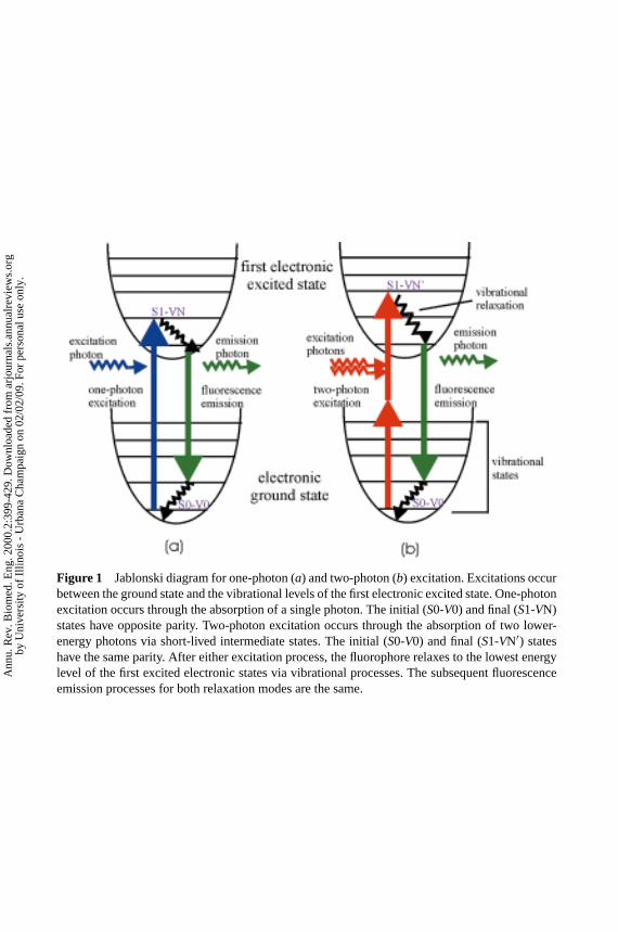
TPE of molecules is a nonlinear process involving the absorption of two photonswhose combined energy is sufficient to induce a molecular transition to an excitedelectronic state. A comparison between one- and two-photon absorption is shownin Figure 1 (see color insert). Conventional one-photon techniques use UV orvisible light to excite fluorescent molecules of interest. Excitation occurs when theabsorbed photon energy matches the energy gap between the ground and excitedstates. The same transition can be excited by a two-photon process in which twoless energetic photons are simultaneously absorbed. Quantum mechanically, asingle photon excites the molecule to a virtual intermediate state, and the moleculeis eventually brought to the final excited state by the absorption of the secondphoton.
The theory of TPE was predicted by G¨oppert-Mayer in 1931 (9). The basicphysics of this phenomenon has also been described elsewhere (25, 26). Fluores-cence excitation is an interaction between the fluorophore and an excitation elec-tromagnetic field. This process is described by a time-dependent Schroedingerequation, in which the Hamiltonian contains an electric dipole interaction term:EEγ · Er , where EEγ is the electric field vector of the photons andEr is the position op-
erator. This equation can be solved by perturbation theory. The first-order solutioncorresponds to the one-photon excitation (OPE), and the multiphoton transitionsare represented by higher order solutions. In the case of TPE, the transition prob-ability between the molecular initial state|i 〉 and the final state| f 〉 is given by
P ∼∣∣∣∣∣∑
m
⟨f∣∣ EEγ · Er
∣∣m⟩⟨m∣∣ EEγ · Er∣∣i ⟩
εγ − εm
∣∣∣∣∣2
(1)
Ann
u. R
ev. B
iom
ed. E
ng. 2
000.
2:39
9-42
9. D
ownl
oade
d fr
om a
rjou
rnal
s.an
nual
revi
ews.
org
by U
nive
rsity
of
Illin
ois
- U
rban
a C
ham
paig
n on
02/
02/0
9. F
or p
erso
nal u
se o
nly.
P1: FhN/FGI P2: FPX/FOK QC: FHN/fgm T1: FhN
July 31, 2000 13:37 Annual Reviews AR106-01
?Figure 1 Jablonski diagram for one-photon (a) and two-photon (b) excitation. Excitations occurbetween the ground state and the vibrational levels of the first electronic excited state. One-photonexcitation occurs through the absorption of a single photon. The initial (S0-V0) and final (S1-VN)states have opposite parity. Two-photon excitation occurs through the absorption of two lower-energy photons via short-lived intermediate states. The initial (S0-V0) and final (S1-VN′) stateshave the same parity. After either excitation process, the fluorophore relaxes to the lowest energylevel of the first excited electronic states via vibrational processes. The subsequent fluorescenceemission processes for both relaxation modes are the same.
Ann
u. R
ev. B
iom
ed. E
ng. 2
000.
2:39
9-42
9. D
ownl
oade
d fr
om a
rjou
rnal
s.an
nual
revi
ews.
org
by U
nive
rsity
of
Illin
ois
- U
rban
a C
ham
paig
n on
02/
02/0
9. F
or p
erso
nal u
se o
nly.
P1: FhN/ftt P2: FhN
July 10, 2000 11:18 Annual Reviews AR106-15
?TWO-PHOTON MICROSCOPY 403
whereεγ is the photonic energy associated with the electric field vectorEEγ , thesummation is over all intermediate statesm, andεm is the energy difference betweenthe statem and the ground state. Note that the dipole operator has odd parity (i.e.absorbing one photon changes the parity of the state), and the one-photon transitionmoment〈 f | EEγ · Er |i 〉 dictates that the initial and final states have opposite parity.The two-photon moment〈 f | EEγ · Er |m〉〈m| EEγ · Er |i 〉 allows transition in which thetwo states have the same parity (25, 26).
Optical Properties of Two-Photon Microscopy
In TPE microscopy, a high numerical aperture objective is used to focus the exci-tation source to a diffraction-limited spot. The characteristic spatial profile at thefocal plane for a circular lens withNA= sin(α), focusing light of wavelengthλ, is
I (u, v) =∣∣∣∣2∫ 1
0J0(vρ)e
− i2 uρ2
ρ dρ
∣∣∣∣2 (2)
whereJ0 is the zeroth-order Bessel function,u = 4k sin2(α/2)z, andv = k sin(α)rare the respective dimensionless axial and radial coordinates normalized to wavenumberk = 2π/λ (27, 28). Because TPE depends on the square of incident photonflux, the point spread function (PSF), which represents the geometry of the exci-tation volume, isI 2(u/2, v/2). Compared with the conventional one-photon PSF,I (u, v), the two-photon result has several major differences leading to distinctadvantages for bioimaging applications.
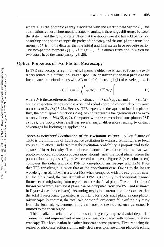
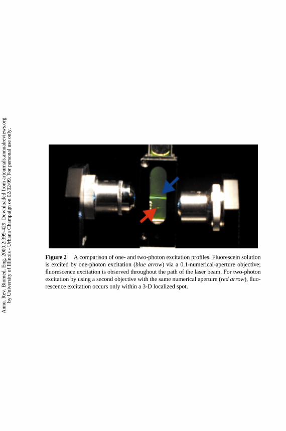
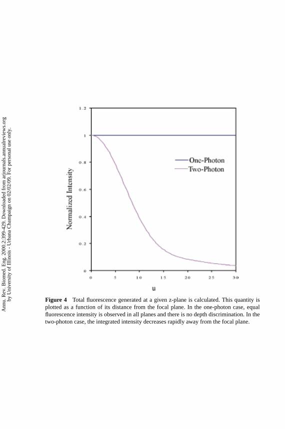
Three-Dimensional Localization of the Excitation Volume A key feature ofTPM is the limitation of fluorescence excitation to within a femtoliter size focalvolume. Equation 1 indicates that the excitation probability is proportional to thesquare of laser intensity. The nonlinear feature of excitation implies that two-photon–induced absorption occurs most strongly near the focal plane, where thephoton flux is highest (Figure 2; see color insert). Figure 3 (see color insert)compares the radial and axial PSF for one-photon microscopy and TPM. Notethat TPE wavelength is twice that of the one-photon case. Owing to the longerwavelength used, TPM has a wider PSF when compared with the one-photon case.On the other hand, the true strength of TPM is its ability to discriminate againstfluorescence originating from regions outside the focal plane. The contribution offluorescence from each axial plane can be computed from the PSF and is shownin Figure 4 (see color insert). Assuming negligible attenuation, one can see thatthe total fluorescence generated is constant for each axial plane in one-photonmicroscopy. In contrast, the total two-photon fluorescence falls off rapidly awayfrom the focal plane, demonstrating that most of the fluorescence generated islimited to the focal region.
This localized excitation volume results in greatly improved axial depth dis-crimination and improvement in image contrast, compared with conventional mi-croscopy. This localization has other important consequences. First, reducing theregion of photointeraction significantly decreases total specimen photobleaching
Ann
u. R
ev. B
iom
ed. E
ng. 2
000.
2:39
9-42
9. D
ownl
oade
d fr
om a
rjou
rnal
s.an
nual
revi
ews.
org
by U
nive
rsity
of
Illin
ois
- U
rban
a C
ham
paig
n on
02/
02/0
9. F
or p
erso
nal u
se o
nly.
P1: FhN/FGI P2: FPX/FOK QC: FHN/fgm T1: FhN
July 31, 2000 13:37 Annual Reviews AR106-01
?Figure 2 A comparison of one- and two-photon excitation profiles. Fluorescein solutionis excited by one-photon excitation (blue arrow) via a 0.1-numerical-aperture objective;fluorescence excitation is observed throughout the path of the laser beam. For two-photonexcitation by using a second objective with the same numerical aperture (red arrow), fluo-rescence excitation occurs only within a 3-D localized spot.
Ann
u. R
ev. B
iom
ed. E
ng. 2
000.
2:39
9-42
9. D
ownl
oade
d fr
om a
rjou
rnal
s.an
nual
revi
ews.
org
by U
nive
rsity
of
Illin
ois
- U
rban
a C
ham
paig
n on
02/
02/0
9. F
or p
erso
nal u
se o
nly.
P1: FhN/FGI P2: FPX/FOK QC: FHN/fgm T1: FhN
July 31, 2000 13:37 Annual Reviews AR106-01
?Figure 3 A comparison of the one- and two-photon point spread functions in the (a)radial and (b) axial directions. In these figures,v andu are normalized optical coordinatesalong radial and axial directions as defined in the main text.
Ann
u. R
ev. B
iom
ed. E
ng. 2
000.
2:39
9-42
9. D
ownl
oade
d fr
om a
rjou
rnal
s.an
nual
revi
ews.
org
by U
nive
rsity
of
Illin
ois
- U
rban
a C
ham
paig
n on
02/
02/0
9. F
or p
erso
nal u
se o
nly.
P1: FhN/FGI P2: FPX/FOK QC: FHN/fgm T1: FhN
July 31, 2000 13:37 Annual Reviews AR106-01
?Figure 4 Total fluorescence generated at a given z-plane is calculated. This quantity isplotted as a function of its distance from the focal plane. In the one-photon case, equalfluorescence intensity is observed in all planes and there is no depth discrimination. In thetwo-photon case, the integrated intensity decreases rapidly away from the focal plane.
Ann
u. R
ev. B
iom
ed. E
ng. 2
000.
2:39
9-42
9. D
ownl
oade
d fr
om a
rjou
rnal
s.an
nual
revi
ews.
org
by U
nive
rsity
of
Illin
ois
- U
rban
a C
ham
paig
n on
02/
02/0
9. F
or p
erso
nal u
se o
nly.
P1: FhN/ftt P2: FhN
July 10, 2000 11:18 Annual Reviews AR106-15
?404 SO ET AL
and photodamage. Second, photoinitiated chemical reaction can be locally trig-gered in 3-D–resolved volumes.
Reduced Attenuation in Biological SpecimensAnother major advantage of TPEis its ability to image thick biological specimens, owing to the reduced scatteringand absorption of near IR light (relative to UV and visible wavelengths) in bi-ological samples. In Rayleigh scattering, the scatterer is much smaller than thewavelength of light, and the scattering cross-section is inversely proportional tothe fourth power of wavelength. When the equivalent wavelengths are used in OPEand TPE, a scattering event in a two-photon transition is over an order of mag-nitude less likely to occur than its one-photon counterpart. This results in deeperpenetration of the excitation source into scattering samples. Rayleigh scattering isonly an approximation of how light propagates in tissues, but the general inverserelationship between scattering and excitation wavelength remains valid. Most tis-sue also has reduced absorption in the near IR, thus TPE can effectively exploitthe tissue “optical window” at 700–1000 nm. Tissue absorbance in this window isorders of magnitude less than the absorption in the UV or blue-green region. Thedeep penetration depth of TPM is a result of both reduced scattering and reducedabsorption.
High Signal-to-Background Ratio Fluorescence DetectionIn standard one-photon microscopy, the excitation wavelength is spectrally close to the fluores-cence emission band. To eliminate the leak-through of the excitation light into thedetection channel, the barrier filter often cuts off a part of the emission band. Theresult is a reduction in microscope sensitivity. For TPE, the excitation wavelengthis much farther removed from the emission band, and highly efficient filters canbe applied to eliminate the excitation with a minimal attenuation of the signal.
TWO-PHOTON MICROSCOPY INSTRUMENTATION
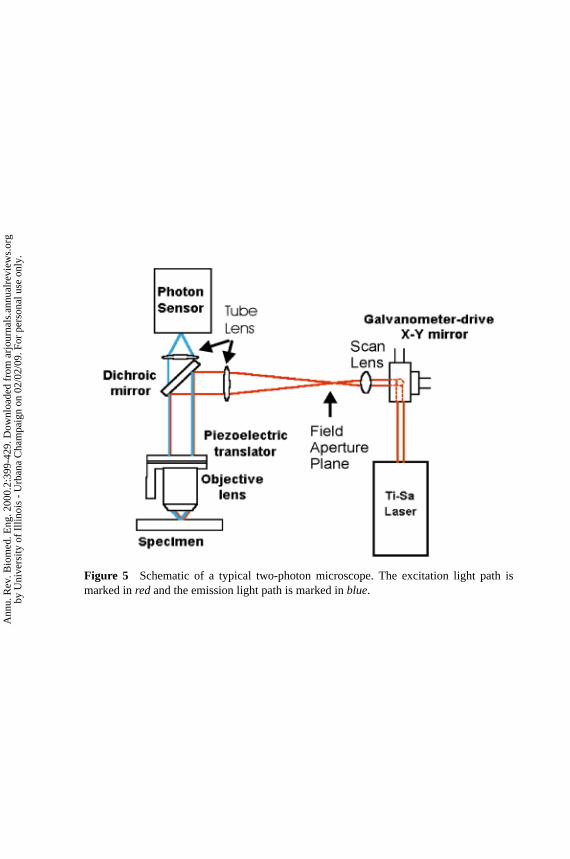
Two-photon microscopes are commercially available; however, one can also beconstructed from components (29) or by modifying an existing confocal micro-scope (1, 30). The basic designs of these systems are very similar, and the crit-ical components are shown in Figure 5 (see color insert). A typical two-photonmicroscope features three basic components: an excitation light source, a high-throughput scanning fluorescence microscope, and a high-sensitivity detectionsystem.
Two-Photon Laser Sources
Because two-photon absorption is a second-order process with a small cross-section on the order of 10−50 cm4 s (defined as 1 GM in honor of G¨oppert-Mayer), high-photon flux needs to be delivered to the sample to generate efficient
Ann
u. R
ev. B
iom
ed. E
ng. 2
000.
2:39
9-42
9. D
ownl
oade
d fr
om a
rjou
rnal
s.an
nual
revi
ews.
org
by U
nive
rsity
of
Illin
ois
- U
rban
a C
ham
paig
n on
02/
02/0
9. F
or p
erso
nal u
se o
nly.
P1: FhN/FGI P2: FPX/FOK QC: FHN/fgm T1: FhN
July 31, 2000 13:37 Annual Reviews AR106-01
?Figure 5 Schematic of a typical two-photon microscope. The excitation light path ismarked inredand the emission light path is marked inblue.
Ann
u. R
ev. B
iom
ed. E
ng. 2
000.
2:39
9-42
9. D
ownl
oade
d fr
om a
rjou
rnal
s.an
nual
revi
ews.
org
by U
nive
rsity
of
Illin
ois
- U
rban
a C
ham
paig
n on
02/
02/0
9. F
or p
erso
nal u
se o
nly.
P1: FhN/ftt P2: FhN
July 10, 2000 11:18 Annual Reviews AR106-15
?TWO-PHOTON MICROSCOPY 405
absorption. This is typically achieved using ultrashort pulsed laser excitation. Ithas been pointed out thatna, the number of photons absorbed per fluorophore perpulse, is given by
na ≈ p20δ
τp f 2p
((NA)2
2hcλ
)2
(3)
whereτp is the pulse duration,δ is the fluorophore’s two-photon absorption atwavelengthλ, p0 is the average laser intensity,f p is the laser’s repetition rate,NA is the numerical aperture of the focusing objective, and ¯h andc are Planck’sconstant and the speed of light, respectively (1). Equation 3 shows that, for thesame average laser power and repetition frequency, the excitation probability isincreased by increasing the NA of the focusing lens and by reducing the pulsewidth of the laser. Increasing NA corresponds to spatially confining the excitationpower to a smaller focal volume.
Femtosecond, picosecond, and continuous-wave (cw) laser sources have beenused for TPM. Currently, the most commonly used laser source for multipho-ton microscopy is femtosecond titanium-sapphire (Ti-Sapphire) systems. Thesepulsed femtosecond systems are capable of generating a 100-fs pulse train at rep-etition rates of∼80 MHz. The tuning range of Ti-Sapphire systems extends from700 to 1000 nm. Other commonly used femtosecond sources are Cr-LiSAF andpulse-compressed Nd-YLF lasers (31). From Equation 3, it is clear that TPE canalso be generated by using picosecond light sources, although at a lower exci-tation efficiency. Commonly available picosecond systems include mode-lockedNd-YAG (∼100 ps), picosecond Ti-Sapphire lasers, and pulsed-dye lasers (∼1 ps).TPE with cw lasers has also been demonstrated. Compared with a standard fem-tosecond light source, a cw light source requires an∼200-fold increase in averagepower to achieve the same excitation rate. This has been accomplished using cwsources such as ArKr laser and Nd-YAG laser (32). The main advantage in usingcw laser sources is the significant reduction in system cost.
Scanning Fluorescence Microscopy Optics
In a typical two-photon microscope, images are generated by raster scanning thex-y mirrors of a galvanometer-driven scanner. After appropriate beam power con-trol and pulse width compensation, the excitation light enters the microscope viaa modified epiluminescence light path. The scan lens is positioned such that thex-y scanner is at its eye point while the field aperture plane is at its focal point.For infinity-corrected objectives, a tube lens is positioned to recollimate the exci-tation light. The scan lens and the tube lens function together as a beam expanderthat overfills the back aperture of the objective lens. A dichroic mirror reflectsthe excitation light to the objective. The dichroic mirrors are short-pass filters,which maximize reflection in the IR and transmission in the blue-green regionof the spectrum. Typically, high-numerical-aperture objectives are used to maxi-mize excitation efficiency. Thex-y galvanometer-driven scanners provide lateral
Ann
u. R
ev. B
iom
ed. E
ng. 2
000.
2:39
9-42
9. D
ownl
oade
d fr
om a
rjou
rnal
s.an
nual
revi
ews.
org
by U
nive
rsity
of
Illin
ois
- U
rban
a C
ham
paig
n on
02/
02/0
9. F
or p
erso
nal u
se o
nly.
P1: FhN/ftt P2: FhN
July 10, 2000 11:18 Annual Reviews AR106-15
?406 SO ET AL
focal-point positioning. An objective positioner translates the focal point axiallyand allows 3-D raster scanning.
Fluorescence Detection System
The fluorescence emission is collected by the imaging objective and transmit-ted through the dichroic mirror along the emission path. An additional barrierfilter is needed to further attenuate the scattered excitation light because of thehigh excitation intensity used. The fluorescence signal is directed to the detectorsystem. Photodetectors that have been used in two-photon microscope systems in-clude photomultiplier tubes (PMTs), avalanche photodiodes, and charge-coupled-device (CCD) cameras. PMTs are the most common implementation becausethey are robust and low cost, have large active areas, and have relatively goodsensitivity.
A major advantage of TPM is that, unlike confocal microscopy, emission pin-holes and descanning optics are not necessary to achieve axial depth discrimination.TPE is already localized to the focal volume, and there is no appreciable off-focalfluorescence to reject. The addition of a pinhole can enhance resolution but at acost of signal loss (33, 34). An important consideration in designing the detec-tion pathway is whether to implement a de-scan lens in the emission path. If thescanned region in the object plane isd, then the de-scan lens is not necessary ifthe detector area is larger than a characteristic area given byd × M , whereMis the overall magnification of the detection path (35). However, for nonuniformdetectors, as some PMTs are, it may be desirable to implement descanning opticsto prevent detection efficiency variation when the fluorescence emission is incidenton different positions of the photocathode surface.
FLUORESCENT PROBES USED IN TWO-PHOTONMICROSCOPY
It is important to examine the nonlinear absorption characteristics of fluorescentmolecules. In general, most chromophores can be excited in two-photon modeat twice their one-photon absorption maximum. However, because one- and two-photon absorption processes have different quantum mechanical selection rules,a fluorophore’s TPE spectrum scaled to half the wavelength is not necessarilyequivalent to its OPE spectrum. Spectroscopic properties of fluorophores undernonlinear excitation need to be better quantified to optimize their use in TPM.Below, we discuss the two-photon absorption properties of some typical extrinsicand intrinsic fluorophores.
Extrinsic and Endogenous Two-Photon Fluorophores
The spectroscopic study of rhodamine under TPE was one of the earliest efforts inthis area (36). With the advent of TPM, spectral characterization of fluorophores
Ann
u. R
ev. B
iom
ed. E
ng. 2
000.
2:39
9-42
9. D
ownl
oade
d fr
om a
rjou
rnal
s.an
nual
revi
ews.
org
by U
nive
rsity
of
Illin
ois
- U
rban
a C
ham
paig
n on
02/
02/0
9. F
or p
erso
nal u
se o
nly.
P1: FhN/ftt P2: FhN
July 10, 2000 11:18 Annual Reviews AR106-15
?TWO-PHOTON MICROSCOPY 407
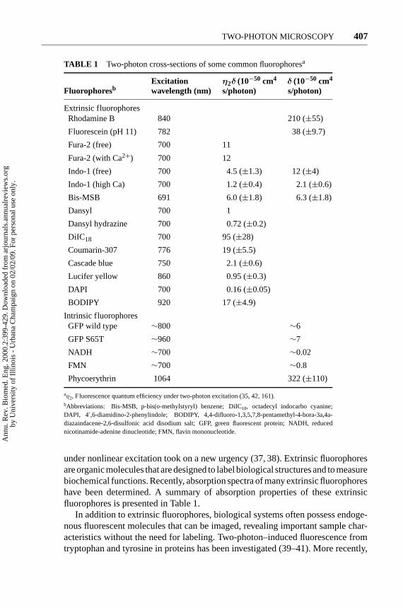
TABLE 1 Two-photon cross-sections of some common fluorophoresa
Excitation η2δ (10−50 cm4 δ (10−50 cm4
Fluorophoresb wavelength (nm) s/photon) s/photon)
Extrinsic fluorophoresRhodamine B 840 210 (±55)
Fluorescein (pH 11) 782 38 (±9.7)
Fura-2 (free) 700 11
Fura-2 (with Ca2+) 700 12
Indo-1 (free) 700 4.5 (±1.3) 12 (±4)
Indo-1 (high Ca) 700 1.2 (±0.4) 2.1 (±0.6)
Bis-MSB 691 6.0 (±1.8) 6.3 (±1.8)
Dansyl 700 1
Dansyl hydrazine 700 0.72 (±0.2)
DiIC18 700 95 (±28)
Coumarin-307 776 19 (±5.5)
Cascade blue 750 2.1 (±0.6)
Lucifer yellow 860 0.95 (±0.3)
DAPI 700 0.16 (±0.05)
BODIPY 920 17 (±4.9)
Intrinsic fluorophoresGFP wild type ∼800 ∼6
GFP S65T ∼960 ∼7
NADH ∼700 ∼0.02
FMN ∼700 ∼0.8
Phycoerythrin 1064 322 (±110)
aη2, Fluorescence quantum efficiency under two-photon excitation (35, 42, 161).bAbbreviations: Bis-MSB, p-bis(o-methylstyryl) benzene; DiIC18, octadecyl indocarbo cyanine;DAPI, 4′,6-diamidino-2-phenylindole; BODIPY, 4,4-difluoro-1,3,5,7,8-pentamethyl-4-bora-3a,4a-diazaindacene-2,6-disulfonic acid disodium salt; GFP, green fluorescent protein; NADH, reducednicotinamide-adenine dinucleotide; FMN, flavin mononucleotide.
under nonlinear excitation took on a new urgency (37, 38). Extrinsic fluorophoresare organic molecules that are designed to label biological structures and to measurebiochemical functions. Recently, absorption spectra of many extrinsic fluorophoreshave been determined. A summary of absorption properties of these extrinsicfluorophores is presented in Table 1.
In addition to extrinsic fluorophores, biological systems often possess endoge-nous fluorescent molecules that can be imaged, revealing important sample char-acteristics without the need for labeling. Two-photon–induced fluorescence fromtryptophan and tyrosine in proteins has been investigated (39–41). More recently,
Ann
u. R
ev. B
iom
ed. E
ng. 2
000.
2:39
9-42
9. D
ownl
oade
d fr
om a
rjou
rnal
s.an
nual
revi
ews.
org
by U
nive
rsity
of
Illin
ois
- U
rban
a C
ham
paig
n on
02/
02/0
9. F
or p
erso
nal u
se o
nly.
P1: FhN/ftt P2: FhN
July 10, 2000 11:18 Annual Reviews AR106-15
?408 SO ET AL
the absorption cross-section of phycoerythrin has been measured and shown to bemuch greater than that of rhodamine 6G at the excitation wavelength of 1064 nm(42). Multiphoton-induced fluorescence of the neurotransmitter serotonin has alsobeen demonstrated (43). The exciting development of green fluorescent protein(GFP) introduces a convenient fluorescent marker for monitoring gene expressionin cells and tissues (44). Two-photon imaging parameters of the GFPs have beenoptimized (45, 46). Nicotinamides [NAD(P)Hs] represent another intrinsic fluo-rophore that can be excited by using a two-photon microscope (41). NAD(P)Hlevels in cells are related to their metabolic rates (47), and NAD(P)H fluorescencehas been used to monitor redox state in cornea (48) and skin (49). The two-photon spectroscopic characteristics of these endogenous probes are also listed inTable 1.
Recent Efforts in Two-Photon Probe Development
One recent development in two-photon probe utilization has been in identifyingdrug molecules that are excitable by two-photon light sources. The ability to moni-tor the drug distribution in a native environment allows a better assessment of drugdelivery efficiency and can be important for developing therapeutic strategies (50).However, monitoring fluorescent drugs under physiological conditions with OPEcan be difficult because of short image penetration depth and high backgroundfluorescence contribution from other naturally occurring fluorescent compounds(51). Therefore, the identification of two-photon excitable drugs has importantclinical and pharmaceutical consequences. In this area, the anticancer drug topote-can has been identified as a good two-photon probe with a two-photon absorptioncross-section of>20 GM at 840-nm excitation. Under physiological conditions,two-photon–induced fluorescence from topotecan has been detected in plasma andin whole blood down to respective concentrations of 0.05µM and 1µM (51). Inthe foreseeable future, it is likely that further efforts in identifying two-photon–excitable pharmaceutical agents for implementation in clinical settings will bemade.
Another important area is the development of extrinsic fluorophores with op-timal two-photon absorption properties. The fluorophores listed in Table 1 haveall been conventional one-photon excitable probes. Because OPE and TPE obeydifferent selection rules, one should not expect these probes necessarily to have op-timized properties for TPE. Recent searches for molecules with high two-photonabsorption cross-sections have led to discovery of molecules with two-photoncross-sections of>1000 GM (52). Optimizing two-photon absorption propertiesin fluorophores has two important consequences. The development of efficientfluorophores can reduce the excitation laser intensity required for imaging, andthus reduce specimen photodamage. Alternatively, with high two-photon cross-sections, significant excitation can be achieved with the more economical cwlasers, thus reducing the cost of two-photon systems that typically use femtosecondTi-Sapphire lasers.
Ann
u. R
ev. B
iom
ed. E
ng. 2
000.
2:39
9-42
9. D
ownl
oade
d fr
om a
rjou
rnal
s.an
nual
revi
ews.
org
by U
nive
rsity
of
Illin
ois
- U
rban
a C
ham
paig
n on
02/
02/0
9. F
or p
erso
nal u
se o
nly.
P1: FhN/ftt P2: FhN
July 10, 2000 11:18 Annual Reviews AR106-15
?TWO-PHOTON MICROSCOPY 409
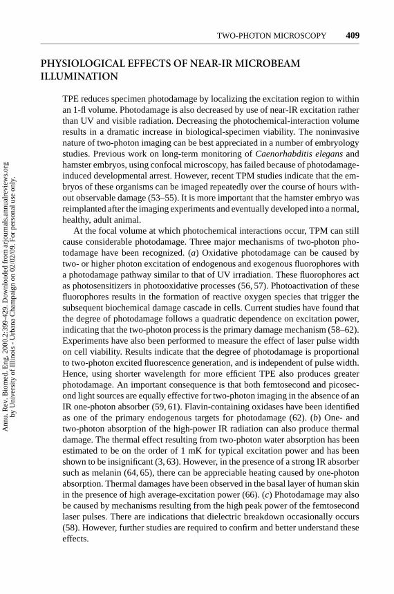
PHYSIOLOGICAL EFFECTS OF NEAR-IR MICROBEAMILLUMINATION
TPE reduces specimen photodamage by localizing the excitation region to withinan 1-fl volume. Photodamage is also decreased by use of near-IR excitation ratherthan UV and visible radiation. Decreasing the photochemical-interaction volumeresults in a dramatic increase in biological-specimen viability. The noninvasivenature of two-photon imaging can be best appreciated in a number of embryologystudies. Previous work on long-term monitoring ofCaenorhabditis elegansandhamster embryos, using confocal microscopy, has failed because of photodamage-induced developmental arrest. However, recent TPM studies indicate that the em-bryos of these organisms can be imaged repeatedly over the course of hours with-out observable damage (53–55). It is more important that the hamster embryo wasreimplanted after the imaging experiments and eventually developed into a normal,healthy, adult animal.
At the focal volume at which photochemical interactions occur, TPM can stillcause considerable photodamage. Three major mechanisms of two-photon pho-todamage have been recognized. (a) Oxidative photodamage can be caused bytwo- or higher photon excitation of endogenous and exogenous fluorophores witha photodamage pathway similar to that of UV irradiation. These fluorophores actas photosensitizers in photooxidative processes (56, 57). Photoactivation of thesefluorophores results in the formation of reactive oxygen species that trigger thesubsequent biochemical damage cascade in cells. Current studies have found thatthe degree of photodamage follows a quadratic dependence on excitation power,indicating that the two-photon process is the primary damage mechanism (58–62).Experiments have also been performed to measure the effect of laser pulse widthon cell viability. Results indicate that the degree of photodamage is proportionalto two-photon excited fluorescence generation, and is independent of pulse width.Hence, using shorter wavelength for more efficient TPE also produces greaterphotodamage. An important consequence is that both femtosecond and picosec-ond light sources are equally effective for two-photon imaging in the absence of anIR one-photon absorber (59, 61). Flavin-containing oxidases have been identifiedas one of the primary endogenous targets for photodamage (62). (b) One- andtwo-photon absorption of the high-power IR radiation can also produce thermaldamage. The thermal effect resulting from two-photon water absorption has beenestimated to be on the order of 1 mK for typical excitation power and has beenshown to be insignificant (3, 63). However, in the presence of a strong IR absorbersuch as melanin (64, 65), there can be appreciable heating caused by one-photonabsorption. Thermal damages have been observed in the basal layer of human skinin the presence of high average-excitation power (66). (c) Photodamage may alsobe caused by mechanisms resulting from the high peak power of the femtosecondlaser pulses. There are indications that dielectric breakdown occasionally occurs(58). However, further studies are required to confirm and better understand theseeffects.
Ann
u. R
ev. B
iom
ed. E
ng. 2
000.
2:39
9-42
9. D
ownl
oade
d fr
om a
rjou
rnal
s.an
nual
revi
ews.
org
by U
nive
rsity
of
Illin
ois
- U
rban
a C
ham
paig
n on
02/
02/0
9. F
or p
erso
nal u
se o
nly.
P1: FhN/ftt P2: FhN
July 10, 2000 11:18 Annual Reviews AR106-15
?410 SO ET AL
MOLECULAR-LEVEL APPLICATIONS OF TWO-PHOTONMICROSCOPY
In recent years, a number of technological advances in light sources, detectors,and optics have led to tremendously improved sensitivity in fluorescence methods.Single-molecule sensitivity is now routinely achieved (67–70). Although most ofthe single-molecule work has used OPE, two-photon methods can offer improve-ment in the signal-to-background ratio (SBR), owing to excitation volume localiza-tion and the wide spectral separation of the emission, excitation, and Raman bands.
It should be noted that, although ultrasensitive applications of two-photon fluo-rescence require careful optimization of instrumentation and experimental setups,they are not fundamentally different from two-photon fluorescence instrumentationdiscussed previously in this review. The following discussion of single-moleculeapplications of two-photon fluorescence has been divided into three parts: fluo-rescence burst detection of single molecules, single-molecule imaging, and fluo-rescence correlation spectroscopy (FCS).
Single-Molecule Detection in Solution
The first demonstration of single-molecule detection by TPE in solution was pre-sented by Mertz et al (71), using rhodamine B molecules in water. They observedphoton bursts from single molecules diffusing through the two-photon volume,with an average SBR of∼10. A number of other reports have since demonstratedefficient fluorescence burst detection of single molecules by TPE in free solution(72, 73), in flow cells (74–76), and in low-temperature solids (77, 78). As antic-ipated, a common finding among most of these reports is the high SBR, largelycaused by the very low background levels associated with TPE. A quantitative studyof the two-photon background, including contributions from two-photon hyper-Rayleigh and hyper-Raman scattering of water, has shown reduced backgroundlevels for TPE vs OPE (79). Care must be taken in determining TPE illuminationconditions, because too much input power can lead to continuum generation in thesolvent, and thus greatly increased background levels (73).
Although the SBR has generally been favorable with TPE, the absolute countrates per molecule are not always comparable with OPE detection (71, 73, 80–82). The reduced magnitude of fluorescence yield is hypothesized to arise fromlong-lived, more highly excited states reached by TPE, increased photobleaching,intersystem crossing, or perhaps some other saturation or multiphoton phenomena.The mechanism is sample dependent, and there are counter examples in whichthe TPE fluorescence intensity is comparable with (enhanced green-fluorescenceprotein) or exceeds (7-amino-A-methylcoumarin) the OPE fluorescence level, atleast under certain illumination conditions (73, 83). The photophysics of a givenmolecule of interest is thus important in determining the relative advantages of one-and two-photon fluorescence detection. Newly designed chromophores with hightwo-photon cross-sections may enhance the advantages of TPE in single-moleculeapplications (52, 84).
Ann
u. R
ev. B
iom
ed. E
ng. 2
000.
2:39
9-42
9. D
ownl
oade
d fr
om a
rjou
rnal
s.an
nual
revi
ews.
org
by U
nive
rsity
of
Illin
ois
- U
rban
a C
ham
paig
n on
02/
02/0
9. F
or p
erso
nal u
se o
nly.
P1: FhN/ftt P2: FhN
July 10, 2000 11:18 Annual Reviews AR106-15
?TWO-PHOTON MICROSCOPY 411
Imaging Single Molecules with Two-Photon Excitation
Spatially resolved applications of ultrasensitive two-photon fluorescence have alsoshown promising results. Sanchez and coworkers first demonstrated two-photonimaging (far-field) of single rhodamine B molecules immobilized on glass surfaceswith an SBR of 30 (80). In addition, emission spectra were acquired for individualmolecules by using a cooled CCD and a spectrograph. Discrete photobleaching ofindividual fluorescence peaks, as well as polarized emission, served as evidence fortrue single-molecule detection. It is interesting that this group found that TPE leadsto faster photobleaching of single rhodamine B molecules than OPE, by a factor of∼2. This number is expected to be sample dependent. A report by Bopp et al, alsousing rhodamine B, demonstrates the ability to determine molecular orientation,as well as to measure laser pulse parameters at the sample by TPE fluorescence(85). Their results also showed reduced fluorescence emission from rhodamine Bfor TPE vs OPE. An interesting variation of TPE imaging methods uses two-photon “wide-field” illumination (∼5-µm-diameter spot); the larger spot size isachieved by underfilling the objective lens. This approach allowed Sonnleitneret al to not only observe single molecules, but track them within the spot overtime, measuring diffusion of single labeled lipid molecules (86). The authors planto test this approach for tracking single molecules on live cell membranes.
Imaging applications of TPE have also been extended to the near-field, usinguncoated fiber tips (81, 87). The study by Kirsch et al makes use of cw TPE.These applications have the exciting potential of ultraresolved imaging with highSBRs and improvedz-resolution as compared with one-photon near-field imaging.To fully realize these advantages requires selection of fluorescent probes that arereasonably stable with TPE, because increased bleaching (as seen with rhodamineprobes) would otherwise detract from the TPE advantages.
Finally, an apertureless variation of near-field imaging was recently introducedwith the promise of extremely highly resolved imaging not limited by a mini-mum aperture size (88). An exciting direction in this field is the use of enhancedradiation (from electromagnetic interaction of the tip and the optical field) local-ized at the tip end to improve resolution and contrast. The quadratic dependenceof TPE on excitation intensity makes it ideal to exploit these locally enhancedfields (89, 90). The first experimental realization of this approach demonstratedextremely high-resolution images (∼20 nm) of photosynthetic membrane frag-ments andJ-aggregates of pseudoisocyanine dye (91). Fluorescence quenching bythe near-field probe is a problem for this method, which, nonetheless, has veryexciting possibilities.
Fluorescence Correlation Spectroscopy
FCS, first introduced by Magde et al (92) and Thompson (93), is proving to bea powerful method for studying a large variety of experimental systems. Appli-cations include measurement of diffusion, chemical reactions, molecular interac-tions, number concentration, hydrodynamic flow, and photophysical parameters
Ann
u. R
ev. B
iom
ed. E
ng. 2
000.
2:39
9-42
9. D
ownl
oade
d fr
om a
rjou
rnal
s.an
nual
revi
ews.
org
by U
nive
rsity
of
Illin
ois
- U
rban
a C
ham
paig
n on
02/
02/0
9. F
or p
erso
nal u
se o
nly.
P1: FhN/ftt P2: FhN
July 10, 2000 11:18 Annual Reviews AR106-15
?412 SO ET AL
such as triplet state lifetimes. FCS experiments are performed by recording spon-taneous equilibrium fluctuations in fluorescence intensity (i.e. fluorescence bursts)from a small (<1-fl) open volume. Information about various experimental pa-rameters is extracted through temporal analysis of these fluctuations by calculatingthe autocorrelation (or cross correlation) of the fluorescence signal, defined as
G(τ ) = 〈δF(t)× δF(t + τ)〉〈F(t)〉2 (4)
HereF(t) is the time-dependent fluorescence signal,δF(t) is the time-dependentdeviation from the average fluorescence intensity, and the angle brackets representthe time average. Because this review is focused on TPM, we here cover onlytwo-photon FCS measurements, and the reader is referred to the above referencesfor a more general discussion and review of the FCS literature.
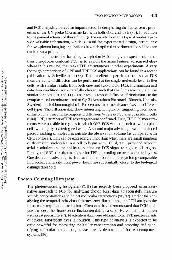
TPE was first applied in FCS measurements by Berland et al. The optical sec-tioning of TPE was introduced as an alternative to confocal detection for FCS in3-D systems (94). This report demonstrated the capability to measure translationaldiffusion coefficients of small (7- and 15-nm radius) fluorescent latex beads insolution and, for the first time using FCS, in the cytoplasm of live cells (Figure 6).The low background levels associated with TPE and reduced autofluorescenceachieved by using 960-nm excitation were of critical importance for the cellularmeasurements. A subsequent report by these authors introduced the capability tomeasure molecular concentration and thus detect protein association/dissociationreactions and kinetics in solution (95). As mentioned above, FCS can also be apowerful tool in studying the photophysical characteristics of fluorescence probes,
Figure 6 Result of a fluorescence correlation spectroscopic measurement of the diffusioncoefficient of 7-nm-radius latex spheres in the cytoplasm of CRL 1503 mouse embryonicfibroblast cells. The data are acquired 30 min after microinjection. The diffusion rate(8×10⁻⁸ cm2/s) is found to be∼fivefold slower than in water(3× 10⁻⁷ cm2/s). This figureis adapted from 94.
Ann
u. R
ev. B
iom
ed. E
ng. 2
000.
2:39
9-42
9. D
ownl
oade
d fr
om a
rjou
rnal
s.an
nual
revi
ews.
org
by U
nive
rsity
of
Illin
ois
- U
rban
a C
ham
paig
n on
02/
02/0
9. F
or p
erso
nal u
se o
nly.
P1: FhN/ftt P2: FhN
July 10, 2000 11:18 Annual Reviews AR106-15
?TWO-PHOTON MICROSCOPY 413
and FCS analysis provided an important tool in deciphering the fluorescence prop-erties of the UV probe Coumarin-120 with both OPE and TPE (73). In additionto the general interest of these findings, the results from this type of analysis pro-vide valuable information, which is useful for experimental design, particularlyfor two-photon imaging applications in which optimal experimental conditions arenot known a priori.
The main motivation for using two-photon FCS in a given experiment, ratherthan one-photon confocal FCS, is to exploit the same features (discussed else-where in this review) that make TPE advantageous in other experiments. A verythorough comparison of OPE and TPE FCS applications can be found in a recentpublication by Schwille et al (83). This excellent paper demonstrates that FCSmeasurements of diffusion can be performed at the single-molecule level in livecells, with similar results from both one- and two-photon FCS. Illumination anddetection conditions were carefully chosen, such that the fluorescence yield wassimilar for both OPE and TPE. Their results resolve diffusion of rhodamines in thecytoplasm and membranes, and of Cy-3 (Amersham Pharmacia Biotech, Uppsala,Sweden)-labeled immunoglobulin E receptors in the membrane of several differentcell types. The diffusion data show interesting complexity, suggesting anomalousdiffusion or at least multicomponent diffusion. Whereas FCS was possible in cellsusing OPE, a number of TPE advantages were confirmed. First, TPE FCS measure-ments were possible in regions in which OPE FCS was not, such as within plantcells with highly scattering cell walls. A second major advantage was the reducedphotobleaching of molecules outside the observation volume (as compared withOPE confocal). This can be exceedingly important when there are small numbersof fluorescent molecules in a cell to begin with. Third, TPE provided superioraxial resolution and the ability to confine the FCS signal to a given cell region.Finally, the SBR can also be higher for TPE, depending on probes and cell types.One distinct disadvantage is that, for illumination conditions yielding comparablefluorescence intensity, TPE power levels are substantially closer to the biologicaldamage threshold.
Photon-Counting Histogram
The photon-counting histogram (PCH) has recently been proposed as an alter-native approach to FCS for analyzing photon burst data, to accurately measuresample concentrations and detect molecular interactions (96, 97). Rather than an-alyzing the temporal behavior of fluorescence fluctuations, the PCH analyzes thefluctuation amplitude distributions. Chen et al have demonstrated that PCH anal-ysis can describe fluorescence fluctuation data as a super-Poissonian distributionwith great precision (97). Fluctuation data were obtained from TPE measurementsof several fluorescent dyes in solution. This type of analysis is expected to bequite powerful for measuring molecular concentration and detecting and quan-tifying molecular interactions, as was already demonstrated for two-componentsystems (96).
Ann
u. R
ev. B
iom
ed. E
ng. 2
000.
2:39
9-42
9. D
ownl
oade
d fr
om a
rjou
rnal
s.an
nual
revi
ews.
org
by U
nive
rsity
of
Illin
ois
- U
rban
a C
ham
paig
n on
02/
02/0
9. F
or p
erso
nal u
se o
nly.
P1: FhN/ftt P2: FhN
July 10, 2000 11:18 Annual Reviews AR106-15
?414 SO ET AL
CELLULAR-LEVEL APPLICATIONS OF TWO-PHOTONMICROSCOPY
The ability to simultaneously monitor cellular biochemical activity and structureat the subcellular level is vital to understand many important processes. The needfor functional imaging dictates the use of minimally invasive technology suchas TPM. The ability of TPE to initiate photochemial reaction in a subfemtolitervolume further opens up new windows of opportunities for 3-D, localized uncagingand photobleaching experiments.
Two-Photon In Vivo Cellular Imaging—MinimizingPhotodamage and Bleaching
Although higher-resolution methods such as electron microscopy can produce im-ages with finer details, optical microscopy is unique because it reveals dynamicprocesses such as signaling, intracellular transport, and cell migration. Some cel-lular processes have been successfully studied with white light video microscopy(98). However, many functional studies require fluorescence. Fluorescence imag-ing of live cells is difficult, especially when 3-D information is required. Beforethe invention of TPM, fluorescence confocal microscopy was the only option. Theuse of high-intensity UV or blue-green radiation in the confocal system results insignificant photodamage and compromises the validity of the research.
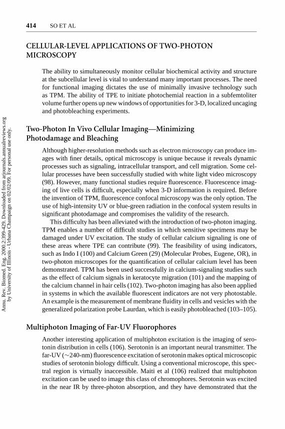
This difficulty has been alleviated with the introduction of two-photon imaging.TPM enables a number of difficult studies in which sensitive specimens may bedamaged under UV excitation. The study of cellular calcium signaling is one ofthese areas where TPE can contribute (99). The feasibility of using indicators,such as Indo I (100) and Calcium Green (29) (Molecular Probes, Eugene, OR), intwo-photon microscopes for the quantification of cellular calcium level has beendemonstrated. TPM has been used successfully in calcium-signaling studies suchas the effect of calcium signals in keratocyte migration (101) and the mapping ofthe calcium channel in hair cells (102). Two-photon imaging has also been appliedin systems in which the available fluorescent indicators are not very photostable.An example is the measurement of membrane fluidity in cells and vesicles with thegeneralized polarization probe Laurdan, which is easily photobleached (103–105).
Multiphoton Imaging of Far-UV Fluorophores
Another interesting application of multiphoton excitation is the imaging of sero-tonin distribution in cells (106). Serotonin is an important neural transmitter. Thefar-UV (∼240-nm) fluorescence excitation of serotonin makes optical microscopicstudies of serotonin biology difficult. Using a conventional microscope, this spec-tral region is virtually inaccessible. Maiti et al (106) realized that multiphotonexcitation can be used to image this class of chromophores. Serotonin was excitedin the near IR by three-photon absorption, and they have demonstrated that the
Ann
u. R
ev. B
iom
ed. E
ng. 2
000.
2:39
9-42
9. D
ownl
oade
d fr
om a
rjou
rnal
s.an
nual
revi
ews.
org
by U
nive
rsity
of
Illin
ois
- U
rban
a C
ham
paig
n on
02/
02/0
9. F
or p
erso
nal u
se o
nly.
P1: FhN/ftt P2: FhN
July 10, 2000 11:18 Annual Reviews AR106-15
?TWO-PHOTON MICROSCOPY 415
distribution of serotonin in intracellular granules can be mapped. Equally importantis the subsequent discovery by Shear et al (43) that serotonin may be further ex-cited by a six-photon process in which the molecule is converted by the absorptionof four photons to a two-photon–excitable by-product. Serotonin distribution canthen be imaged based on the blue-green fluorescence of this by-product (43). Thetwo basic principles described here are important. First, multiphoton excitationcan be used to access new far-UV chromophores. Second, multiphoton chemicalprocesses may be used to create new fluorophores based on endogenous cellularmolecules.
Two-Photon Multiple Color Imaging
The possibility of using TPM to simultaneously excite different color fluorophoresfor multiple label imaging has been explored (29, 107) (Figure 7; see color insert).Xu et al imaged rat basophilic leukemia cells simultaneously labeled with pyrenelysophosphatidylcholine, a UV-emitting plasma membrane probe; DAPI, a blue-emitting nucleic probe; Bodipy sphingomyelin, a green-emitting Golgi label; andrhodamine 123, a red-emitting mitochondria probe. This study demonstrates thesimultaneous imaging of four cellular structural components and the potential forstudies in which the interaction of various cellular organelles can be monitoredover time in 3-D.
Three-Dimensional Localized Uncaging of SignalingMolecules
An important property of TPM is its ability to initiate localized chemical reac-tions such as the uncaging of signaling molecules. Denk led the development ofthis novel technique by mapping the distribution of nicotin acetylcholine recep-tors in a muscle cell line (BC3H1) (108). The membrane potential of a chosencell was monitored by the whole-cell patch clamp technique. Caged carbamoyl-choline was placed in the medium. Upon two-photon uncaging, a 3-D localizedburst of carbamoylcholine was released at the focal point of the laser light. Thewhole-cell current measured by the patch clamp is a function of the relative spa-tial distance between the carbamoylcholine burst and the proximal acetylocholinereceptors. In the presence of many close-by receptors, a larger current will be ob-served and vice versa. By scanning the uncaging beam throughout the cell volume,the receptor distribution can be determined. Similar techniques have been subse-quently applied to map glutamate receptors on hippocampal pyramidal neurons(109).
In addition to receptor mapping, localized uncaging is also valuable for thestudy of intracellular-signaling pathways. The ability to generate active signalingmolecules localized in 3-D with submicrometer resolution allows investigators tomonitor intracellular signal propagation. Localized uncaging of fluorescein hasbeen demonstrated by Denk and coworkers (1). However, the uncaging of moreimportant cellular messengers, such as calcium, has been difficult because of the
Ann
u. R
ev. B
iom
ed. E
ng. 2
000.
2:39
9-42
9. D
ownl
oade
d fr
om a
rjou
rnal
s.an
nual
revi
ews.
org
by U
nive
rsity
of
Illin
ois
- U
rban
a C
ham
paig
n on
02/
02/0
9. F
or p
erso
nal u
se o
nly.

P1: FhN/FGI P2: FPX/FOK QC: FHN/fgm T1: FhN
July 31, 2000 13:37 Annual Reviews AR106-01
?Figure 7 Two-photon multiple color imaging of bovine pulmonary artery endothelialcells labeled with DAPI (nucleus), BODIPYO-FL phallacidin (actin), and MitoTrackeroRed CMXRos (mitochondria) (Molecular Probes, Eugene, OR). All three probes wereexcited at 780 nm, and images were acquired by three independent detection channels.
Ann
u. R
ev. B
iom
ed. E
ng. 2
000.
2:39
9-42
9. D
ownl
oade
d fr
om a
rjou
rnal
s.an
nual
revi
ews.
org
by U
nive
rsity
of
Illin
ois
- U
rban
a C
ham
paig
n on
02/
02/0
9. F
or p
erso
nal u
se o
nly.

P1: FhN/ftt P2: FhN
July 10, 2000 11:18 Annual Reviews AR106-15
?416 SO ET AL
lack of an efficient two-photon photolabile cage. This situation is improving asnew cage groups targeted for two-photon applications are being developed (110).This new technology has been used in a number of pilot studies (111–116).
Two-photon photobleaching recovery is a similar technique that can be appliedto measure intracellular transport, viscosity, and diffusion (117). Similar to con-ventional photobleaching-recovery studies, molecules labeled with fluorophores ofinterest can be photobleached within a 3-D localized volume. The recovery of lossfluorescence is correlated with fresh molecules diffusing back into the excitationvolume and provides information on the intracellular environment.
TISSUE LEVEL APPLICATIONS OF TWO-PHOTONMICROSCOPY
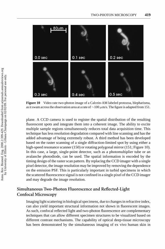
A recent comparison study has convincingly demonstrated that TPM is a superiormethod in the imaging of thick, highly scattering specimens (33).
Applying Two-Photon Microscopy to StudyTissue Physiology
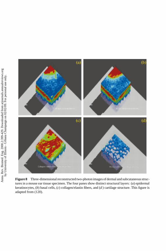
The potential of applying TPM to study tissue physiology has been recognizedsince its inception (1). Two-photon tissue imaging has been successfully appliedto study the physiology of many tissue types, including the corneal structure ofrabbit eyes (48, 66), the light-induced calcium signals in salamander retina (118),the human and mouse dermal and subcutaneous structures (49, 119, 120) (Figure 8;see color insert), the toxin effect on human intestinal mucosa (121), and themetabolic processes of pancreatic islets (122, 123). Today, TPM is particularlywidely used in two areas—neurobiology (124) and embryology. In neurobiologystudies, TPM has been applied to study the neuron structure and function in in-tact brain slices (125), the role of calcium signaling in dendritic spine function(126–134), neuronal plasticity and the associating cellular morphological changes(135), and hemodynamics in rat neocortex (136). In embryology studies, two-photon imaging has been used to examine calcium passage during sperm-eggfusion (137), the origin of bilateral axis in sea urchin embryos (138), cell fusionevents inC. eleganshypodermis (53, 54), and hamster embryo development (55).It is expected that two-photon imaging will be applied to an increasing number oftissue systems as better commercial instruments become available.
It is important to examine the technological limitations of applying two-photonimaging in tissues. (a) It should be recognized that the imaging depth of differentspecimens could be drastically different. For example, in the cornea of the eye,a unique optically transparent organ, an autofluorescence image can be obtainedfrom depths beyond a millimeter, whereas, inside a highly scattering specimensuch as human skin, the contrast of autofluorescence images is significantly de-graded at∼200–300µm. The maximum imaging depth depends on the scatteering
Ann
u. R
ev. B
iom
ed. E
ng. 2
000.
2:39
9-42
9. D
ownl
oade
d fr
om a
rjou
rnal
s.an
nual
revi
ews.
org
by U
nive
rsity
of
Illin
ois
- U
rban
a C
ham
paig
n on
02/
02/0
9. F
or p
erso
nal u
se o
nly.
P1: FhN/FGI P2: FPX/FOK QC: FHN/fgm T1: FhN
July 31, 2000 13:37 Annual Reviews AR106-01
?Figure 8 Three-dimensional reconstructed two-photon images of dermal and subcutaneous struc-tures in a mouse ear tissue specimen. The four panes show distinct structural layers: (a) epidermalkeratinocytes, (b) basal cells, (c) collagen/elastin fibers, and (d ) cartilage structure. This figure isadapted from (120).
Ann
u. R
ev. B
iom
ed. E
ng. 2
000.
2:39
9-42
9. D
ownl
oade
d fr
om a
rjou
rnal
s.an
nual
revi
ews.
org
by U
nive
rsity
of
Illin
ois
- U
rban
a C
ham
paig
n on
02/
02/0
9. F
or p
erso
nal u
se o
nly.
P1: FhN/ftt P2: FhN
July 10, 2000 11:18 Annual Reviews AR106-15
?TWO-PHOTON MICROSCOPY 417
and absorption coefficients of the tissue, the efficiency of the fluorophore, and thethroughput of the microscope optics. (b) The two-photon PSF is well characterizedin thin specimens. In a number of tissue models, two-photon PSF degradation ap-pears to be minimal up to a depth of 100µm (33). However, the aberration of PSFin real tissue specimens has not been satisfactorily measured. A number of projectsare now underway to better understand the physics of light propagation in turbidmedium and to quantify resolution in both tissue phantoms and real tissues. (c) Flu-orescence labeling of deep tissue structure is a major technical challenge. Commonfluorescent probes are designed for labeling cultured cells. Diffusional delivery ofthese probes is not ideal in tissues. Probe distributions tend to vary greatly as afunction of depth. Fluorophores could also be delivered by microinjection, but thismethod suffers from its invasive nature and its limited ability to label multiplecells. Tissue structure may also be imaged with endogenous fluorophores such asNAD(P)H and flavoproteins (47). Unfortunately, these fluorophores suffer fromlow quantum yield, and their fluorescence emissions in the blue-green spectralregion are strongly attenuated by the tissue. Molecular biology methods allow thelabeling of specific tissue structures with fluorescent probes such as GFP, but thismethod can be applied only to specimens with well-controlled genetic makeup.The lack of universal and effective methods for fluorescent labeling of living tissuesis a major obstacle in two-photon tissue imaging.
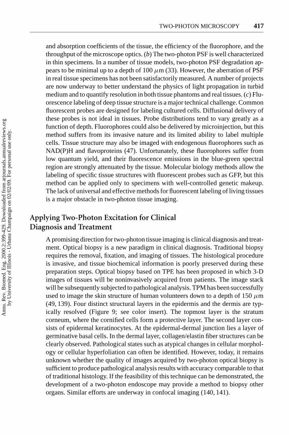
Applying Two-Photon Excitation for ClinicalDiagnosis and Treatment
A promising direction for two-photon tissue imaging is clinical diagnosis and treat-ment. Optical biopsy is a new paradigm in clinical diagnosis. Traditional biopsyrequires the removal, fixation, and imaging of tissues. The histological procedureis invasive, and tissue biochemical information is poorly preserved during thesepreparation steps. Optical biopsy based on TPE has been proposed in which 3-Dimages of tissues will be noninvasively acquired from patients. The image stackwill be subsequently subjected to pathological analysis. TPM has been successfullyused to image the skin structure of human volunteers down to a depth of 150µm(49, 139). Four distinct structural layers in the epidermis and the dermis are typ-ically resolved (Figure 9; see color insert). The topmost layer is the stratumcorneum, where the cornified cells form a protective layer. The second layer con-sists of epidermal keratinocytes. At the epidermal-dermal junction lies a layer ofgerminative basal cells. In the dermal layer, collagen/elastin fiber structures can beclearly observed. Pathological states such as atypical changes in cellular morphol-ogy or cellular hyperfoliation can often be identified. However, today, it remainsunknown whether the quality of images acquired by two-photon optical biopsy issufficient to produce pathological analysis results with accuracy comparable to thatof traditional histology. If the feasibility of this technique can be demonstrated, thedevelopment of a two-photon endoscope may provide a method to biopsy otherorgans. Similar efforts are underway in confocal imaging (140, 141).
Ann
u. R
ev. B
iom
ed. E
ng. 2
000.
2:39
9-42
9. D
ownl
oade
d fr
om a
rjou
rnal
s.an
nual
revi
ews.
org
by U
nive
rsity
of
Illin
ois
- U
rban
a C
ham
paig
n on
02/
02/0
9. F
or p
erso
nal u
se o
nly.

P1: FhN/FGI P2: FPX/FOK QC: FHN/fgm T1: FhN
July 31, 2000 13:37 Annual Reviews AR106-01
?Figure 9 Three-dimensional reconstructed two-photon images of in vivo human skin. Thestrata corneum and the basal layers are clearly visible. The figure is adapted from (159).
Ann
u. R
ev. B
iom
ed. E
ng. 2
000.
2:39
9-42
9. D
ownl
oade
d fr
om a
rjou
rnal
s.an
nual
revi
ews.
org
by U
nive
rsity
of
Illin
ois
- U
rban
a C
ham
paig
n on
02/
02/0
9. F
or p
erso
nal u
se o
nly.
P1: FhN/ftt P2: FhN
July 10, 2000 11:18 Annual Reviews AR106-15
?418 SO ET AL
In addition to diagnosis, TPE may also find applications in clinical treatmentbased on photodynamic therapy. Photodynamic therapy allows the destructionof specific tissue, such as a tumor, by preferentially loading the tissue with aphotosensitizer. Photosensitizer-loaded tissue is subsequently destroyed by laserillumination. Unfortunately, while the photosensitizer uptake in tumorous tissue ishigher, there is often non-negligible uptake in normal tissues. Peripheral damage tohealthy tissues is a common occurrence. The potential of using two-photon imagingto first localize the tumor and then of applying TPE to initiate photodynamic actionat the selected site is a very attractive option. Preliminary work in this area hasbeen reported (142–144).
NEW DEVELOPMENTS IN TWO-PHOTONINSTRUMENTATION
Although the standard TPM, as described previously, functions very well as ageneral purpose instrument, a number of exciting new applications demand newinstrumentation capabilities. This section describes the frontiers of two-photoninstrumentation research and their biomedical potentials.
Two-Photon Video Rate Microscopy
A major limitation of standard two-photon microscopes is their speed, which is∼0.5 Hz, and the typical time required to obtain a high-resolution 3-D image stackis∼10 min. This is clearly unsuitable for applications such as clinical biopsy, inwhich efficiency is crucial. Furthermore, this slow imaging speed is also incompat-ible with intravital imaging of patients or animals, in which the presence of motionartifacts is a major concern. Finally, the study of many cellular processes, such ascalcium signaling or neuronal communication, requires imaging with millisecondtime resolution. This problem is addressed by the development of a number ofvideo rate two-photon microscopes.
The first video rate two-photon imaging system is based on the line-scanningapproach. Image acquisition time is reduced by covering the image plane with aline instead of a point (145, 146). The line focus is typically achieved by usinga cylindrical element in the excitation beam path. The resulting fluorescent lineimage is acquired with a spatially resolved detector such as a CCD camera. Themain drawback associated with line scanning is the inevitable degradation of theimage PSF, especially in the axial direction. A second approach, which has beentermed “multiphoton multifocal microscopy” (147, 148), is analogous to Nipkowdisk-based confocal systems (149). This elegant approach is based on a customfabricated lenslet array, in place of the scan lens, that focuses the incident laserinto multiple focal spots at the field aperture plane. The lenslet lenses are arrangedin patterns similar to the traditional Nipkow disk designs. Upon the rotation of thelenslet lens, the projection of the lenslet will uniformly cover the field aperture
Ann
u. R
ev. B
iom
ed. E
ng. 2
000.
2:39
9-42
9. D
ownl
oade
d fr
om a
rjou
rnal
s.an
nual
revi
ews.
org
by U
nive
rsity
of
Illin
ois
- U
rban
a C
ham
paig
n on
02/
02/0
9. F
or p
erso
nal u
se o
nly.
P1: FhN/ftt P2: FhN
July 10, 2000 11:18 Annual Reviews AR106-15
?TWO-PHOTON MICROSCOPY 419
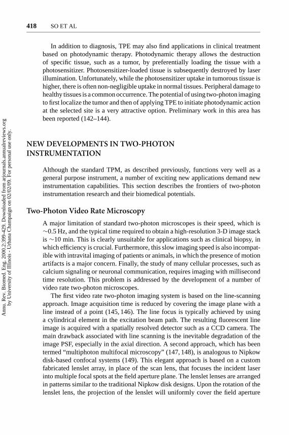
Figure 10 Video rate two-photon image of a Calcein-AM labeled protozoa, blepharisma,as it swam across the observation area at a rate of∼100µm/s. The figure is adapted from 151.
plane. A CCD camera is used to register the spatial distribution of the resultingfluorescent spots and integrate them into a coherent image. The ability to excitemultiple sample regions simultaneously reduces total data acquisition time. Thistechnique has less resolution degradation compared with line scanning and has theadded advantage of being extremely robust. A third method has been developedbased on the raster scanning of a single diffraction-limited spot by using either ahigh-speed resonance scanner (150) or rotating polygonal mirror (151; Figure 10).In this case, a large, single-point detector, such as a photomultiplier tube or anavalanche photodiode, can be used. The spatial information is encoded by thetiming design of the raster scan pattern. By replacing the CCD imager with a singlepixel detector, the image resolution may be improved by removing the dependenceon the emission PSF. This is particularly important in turbid specimens in whichthe scattered fluorescence signal is not confined in a single pixel of the CCD imagerand may degrade the image resolution.
Simultaneous Two-Photon Fluorescence and Reflected-LightConfocal Microscopy
Imaging light scattering in biological specimens, due to changes in refractive index,can also yield important structural information not shown in fluorescent images.As such, confocal reflected light and two-photon fluorescence are complementarytechniques that can allow different specimen structures to be visualized based ondifferent contrast mechanisms. The capability of optical deep-tissue microscopyhas been demonstrated by the simultaneous imaging of ex vivo human skin in
Ann
u. R
ev. B
iom
ed. E
ng. 2
000.
2:39
9-42
9. D
ownl
oade
d fr
om a
rjou
rnal
s.an
nual
revi
ews.
org
by U
nive
rsity
of
Illin
ois
- U
rban
a C
ham
paig
n on
02/
02/0
9. F
or p
erso
nal u
se o
nly.
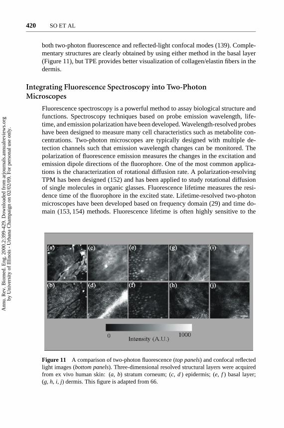
P1: FhN/ftt P2: FhN
July 10, 2000 11:18 Annual Reviews AR106-15
?420 SO ET AL
both two-photon fluorescence and reflected-light confocal modes (139). Comple-mentary structures are clearly obtained by using either method in the basal layer(Figure 11), but TPE provides better visualization of collagen/elastin fibers in thedermis.
Integrating Fluorescence Spectroscopy into Two-PhotonMicroscopes
Fluorescence spectroscopy is a powerful method to assay biological structure andfunctions. Spectroscopy techniques based on probe emission wavelength, life-time, and emission polarization have been developed. Wavelength-resolved probeshave been designed to measure many cell characteristics such as metabolite con-centrations. Two-photon microscopes are typically designed with multiple de-tection channels such that emission wavelength changes can be monitored. Thepolarization of fluorescence emission measures the changes in the excitation andemission dipole directions of the fluorophore. One of the most common applica-tions is the characterization of rotational diffusion rate. A polarization-resolvingTPM has been designed (152) and has been applied to study rotational diffusionof single molecules in organic glasses. Fluorescence lifetime measures the resi-dence time of the fluorophore in the excited state. Lifetime-resolved two-photonmicroscopes have been developed based on frequency domain (29) and time do-main (153, 154) methods. Fluorescence lifetime is often highly sensitive to the
Figure 11 A comparison of two-photon fluorescence (top panels) and confocal reflectedlight images (bottom panels). Three-dimensional resolved structural layers were acquiredfrom ex vivo human skin: (a, b) stratum corneum; (c, d ) epidermis; (e, f ) basal layer;(g, h, i, j) dermis. This figure is adapted from 66.
Ann
u. R
ev. B
iom
ed. E
ng. 2
000.
2:39
9-42
9. D
ownl
oade
d fr
om a
rjou
rnal
s.an
nual
revi
ews.
org
by U
nive
rsity
of
Illin
ois
- U
rban
a C
ham
paig
n on
02/
02/0
9. F
or p
erso
nal u
se o
nly.
P1: FhN/ftt P2: FhN
July 10, 2000 11:18 Annual Reviews AR106-15
?TWO-PHOTON MICROSCOPY 421
fluorophore biochemical microenvironment. Lifetime-sensitive functional probeshave been identified for common applications such as Ca2+ or pH measurements,providing a complementary approach to wavelength-resolved measurement. It ismore important that there are interesting processes that can be best measured bylifetime-resolved imaging. Examples include the determination of tissue oxygena-tion based on lifetime changes on oxygen quenching (155, 156). Another exam-ple includes the use of lifetime resolved measurement to quantitatively monitorthe proteolytic process of antigenic materials in macrophage vacuoles (157, 158)(Figure 12; see color insert).
CONCLUSION
Within a decade after the invention of TPM, this technology has found applicationsin many diverse areas, ranging from the study of single molecules to tissue imag-ing. It is important that TPM is also starting to attract attention from the clinicaland industrial communities. The development of a two-photon endoscope will bea critical step toward realizing noninvasive optical biopsy. As a complement totraditional pathology, the ability for a two-photon endoscope to rapidly and non-invasively acquire tissue-structural images may find use in areas such as surgicalmargin determination. TPM is also being applied by the pharmaceutical industryfor high-throughput drug screening in which TPE or three-photon excitation oftryptophan or tryosine allows label-free study of protein-drug interactions. Highlysensitive two-photon FCS and PCH may be applied for further quantitative anal-ysis. Given the importance of studying biological systems in vivo, we can projectthat the use of TPM in academic research will continue to increase. As two-photontechnology reaches maturity, it is also promising that high-impact clinical andindustrial applications may be identified.
ACKNOWLEDGMENTS
P. T. C. So acknowledges kind support from the National Science Foundation(MCB-9604382), National Institute of Health (R29GM56486-01), and Ameri-can Cancer Society (RPG-98-058-01-CCE). C. Y. Dong acknowledges fellowshipsupport from NIH 5F32CA75736-02.
Visit the Annual Reviews home page at www.AnnualReviews.org
LITERATURE CITED
1. Denk W, Strickler JH, Webb WW. 1990.Two-photon laser scanning fluorescence mi-croscopy.Science248:73–76
2. Williams RM, Piston DW, Webb WW. 1994.Two-photon molecular excitation provides
intrinsic 3-dimensional resolution for laser-based microscopy and microphotochem-istry. FASEB J.8:804–13
3. Denk WJ, Piston DW, Webb WW. 1995.Two-photon molecular excitation laser-
Ann
u. R
ev. B
iom
ed. E
ng. 2
000.
2:39
9-42
9. D
ownl
oade
d fr
om a
rjou
rnal
s.an
nual
revi
ews.
org
by U
nive
rsity
of
Illin
ois
- U
rban
a C
ham
paig
n on
02/
02/0
9. F
or p
erso
nal u
se o
nly.
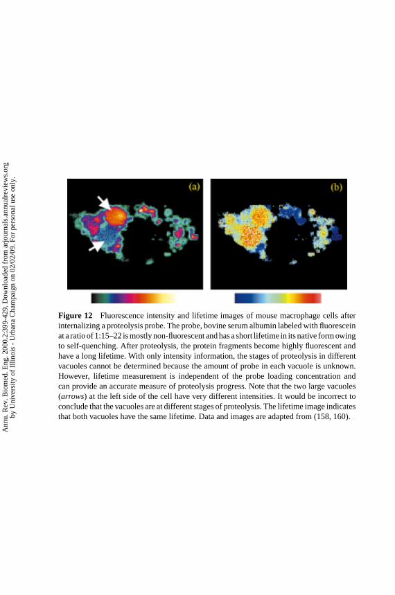
P1: FhN/FGI P2: FPX/FOK QC: FHN/fgm T1: FhN
July 31, 2000 13:37 Annual Reviews AR106-01
?Figure 12 Fluorescence intensity and lifetime images of mouse macrophage cells afterinternalizing a proteolysis probe. The probe, bovine serum albumin labeled with fluoresceinat a ratio of 1:15–22 is mostly non-fluorescent and has a short lifetime in its native form owingto self-quenching. After proteolysis, the protein fragments become highly fluorescent andhave a long lifetime. With only intensity information, the stages of proteolysis in differentvacuoles cannot be determined because the amount of probe in each vacuole is unknown.However, lifetime measurement is independent of the probe loading concentration andcan provide an accurate measure of proteolysis progress. Note that the two large vacuoles(arrows) at the left side of the cell have very different intensities. It would be incorrect toconclude that the vacuoles are at different stages of proteolysis. The lifetime image indicatesthat both vacuoles have the same lifetime. Data and images are adapted from (158, 160).
Ann
u. R
ev. B
iom
ed. E
ng. 2
000.
2:39
9-42
9. D
ownl
oade
d fr
om a
rjou
rnal
s.an
nual
revi
ews.
org
by U
nive
rsity
of
Illin
ois
- U
rban
a C
ham
paig
n on
02/
02/0
9. F
or p
erso
nal u
se o
nly.
P1: FhN/ftt P2: FhN
July 10, 2000 11:18 Annual Reviews AR106-15
?422 SO ET AL
scanning microscopy. InHandbook of Bio-logical Confocal Microscopy, ed. JB Paw-ley, pp. 445–58. New York: Plenum
4. Hell SW. 1996. Nonlinear optical mi-croscopy.Bioimaging4:121–23
5. Denk WJ, Strickler JP, Webb WW. 1991.US Patent No. 5034613
6. Wilson T, Sheppard CJR. 1984.Theory andPractice of Scanning Optical Microscopy.New York: Academic. 213 pp.
7. Pawley JB, ed. 1995.Handbook of Confo-cal Microscopy. New York: Plenum. 632pp.
8. Masters BR. 1996.Selected Papers onConfocal Microscopy. Bellingham, WA:SPIE. 702 pp.
9. Goppert-Mayer M. 1931. Uber elemen-tarakte mit zwei quantensprungen.Ann.Phys. (Leipzig)5:273–94
10. Franken PA, Hill AE, Peters CW, Weinre-ich G. 1961. Generation of optical harmon-ics.Phys. Rev. Lett.7:118–19
11. Kaiser W, Garrett CGB. 1961. Two-photonexcitation in CaF2:Eu2+. Phys. Rev. Lett.7:229–31
12. McClain WM. 1971. Excited state sym-metry assignment through polarized two-photon absorption studies in fluids.J.Chem. Phys.55:2789
13. Friedrich DM, McClain WM. 1980. Two-photon molecular electronic spectroscopy.Annu. Rev. Phys. Chem.31:559–77
14. Friedrich DM. 1982. Two-photon molec-ular spectroscopy.J. Chem. Educ.59:472
15. Birge RR. 1986. Two-photon spectroscopyof protein-bound fluorophores.Acc. Chem.Res.19:138–46
16. Singh S, Bradley LT. 1964. Three-photonabsorption in naphthalene crystals by laserexcitation.Phys. Rev. Lett.12:162–64
17. Rentzepis PM, Mitschele CJ, Saxman AC.1970. Measurement of ultrashort laserpulses by three-photon fluorescence.Appl.Phys. Lett.7:229–31
18. Gryczynski I, Szmacinski H, Lakowicz JR.1995. On the possibility of calcium imag-
ing using Indo-1 with three-photon excita-tion. Photochem. Photobiol.62:804–8
18a. Freund I, Kopf L. 1970. Long Range Orderin NH4Cl. Phys. Rev. Lett.24:1017–21
19. Hellwarth R, Christensen P. 1974. Non-linear optical microscopic examination ofstructures in polycrystaline ZnSe.Opt.Commun.12:318–22
20. Sheppard CJR, Kompfner R, Gannaway J,Walsh D. 1977.The scanning harmonic op-tical microscope. Presented at IEEE/OSAConf. Laser Eng. Appl., Washington, DC
21. Gannaway JN, Sheppard CJR. 1978. Sec-ond harmonic imaging in the scanning op-tical microscope.Opt. Quantum Electron.10:435–39
22. Barad Y, Eisenberg H, Horowitz M, Sil-berberg Y. 1997. Nonlinear scanning lasermicroscopy by third harmonic generation.Appl. Phys. Lett.70:922–24
23. Squier JA, Muller M, Brakenhoff GJ, Wil-son KR. 1998. Third harmonic generationmicroscopy.Opt. Exp.3:315–24
24. Campagnola PJ, Wei M-D, Lewis A, LoewLM. 1999. High-resolution nonlinear op-tical imaging of live cells by second har-monic generation.Biophys. J.77:3341–49
25. Baym G. 1974. Lectures on Quan-tum Mechanics. Menlo Park, CA: Ben-jamin/Cummins. 594 pp.
26. Callis PR. 1997. The theory of two-photoninduced fluorescence anisotropy. InNon-linear and Two-Photon-Induced Fluores-cence, ed. J Lakowicz, pp. 1–42. New York:Plenum
27. Sheppard CJR, Gu M. 1990. Image for-mation in two-photon fluorescence micro-scope.Optik86:104–6
28. Gu M, Sheppard CJR. 1995. Compari-son of three-dimensional imaging prop-erties between two-photon and single-photon fluorescence microscopy.J. Mi-crosc.177:128–37
29. So PTC, French T, Yu WM, Berland KM,Dong CY, Gratton E. 1995. Time-resolvedfluorescence microscopy using two-photonexcitation.Bioimaging3:49–63
Ann
u. R
ev. B
iom
ed. E
ng. 2
000.
2:39
9-42
9. D
ownl
oade
d fr
om a
rjou
rnal
s.an
nual
revi
ews.
org
by U
nive
rsity
of
Illin
ois
- U
rban
a C
ham
paig
n on
02/
02/0
9. F
or p
erso
nal u
se o
nly.
P1: FhN/ftt P2: FhN
July 10, 2000 11:18 Annual Reviews AR106-15
?TWO-PHOTON MICROSCOPY 423
30. Diaspro A, Corosu M, Ramoino P, RobelloM. 1999. Adapting a compact confocal mi-croscope system to a two-photon excita-tion fluorescence imaging architecture.Mi-crosc. Res. Tech.47:196–205
31. Wokosin DL, Centonze VE, White J, Arm-strong D, Robertson G, Ferguson AI.1996. All-solid-state ultrafast lasers facil-itate multiphoton excitation fluorescenceimaging.IEEE J. Sel. Top. Quantum Elec-tron. 2:1051–65
32. Hell SW, Booth M, Wilms S. 1998. Two-photon near- and far-field fluorescence mi-croscopy with continuous-wave excitation.Opt. Lett.23:1238–40
33. Centonze VE, White JG. 1998. Multi-photon excitation provides optical sec-tions from deeper within scattering spec-imens than confocal imaging.Biophys. J.75:2015–24
34. Gauderon R, Lukins RB, Sheppard CJR.1999. Effects of a confocal pinhole in two-photon microscopy.Microsc. Res. Tech.47:210–14
35. Xu C, Webb WW. 1997. Multiphotonexcitation of molecular fluorophores andnonlinear laser microscopy. InNonlinearand Two-Photon-Induced Fluorescence,ed. JR Lakowicz, pp. 471–540. New York:Plenum
36. Hermann JP, Ducuing J. 1972. Dispersionof the two-photon cross section in rho-damine dyes.Opt. Commun.6:101–5
37. Xu C, Guild J, Webb WW, Denk W. 1995.Determination of absolute two-photon ex-citation cross sections by in situ second-order autocorrelation.Opt. Lett.20:2372–74
38. Albota MA, Xu C, Webb WW. 1998. Two-photon fluorescence excitation cross sec-tions of biomolecular probes from 690 to960 nm.Appl. Opt.37:7352–56
39. Lakowicz JR, Gryczynski I. 1992. Trypto-phan fluorescence intensity and anisotropydecays of human serum albumin resultingfrom one-photon and two-photon excita-tion. Biophys. Chem.45:1–6
40. Lakowicz JR, Kierdaszuk B, Callis P,Malak H, Gryczynski I. 1995. Fluores-cence anisotropy of tyrosine using one-and two-photon excitation.Biophys. Chem.56:263–71
41. Kierdaszuk B, Malak H, Gryczynski I, Cal-lis P, Lakowicz JR. 1996. Fluorescence ofreduced nicotinamides using one- and two-photon excitation.Biophys. Chem.62:1–13
42. Chen ZP, Kaplan DL, Yang K, KumarJ, Marx KA, Tripathy SK. 1997. Two-photon-induced fluorescence from the phy-coerythrin protein.Appl. Opt.36:1655–59
43. Shear JB, Xu C, Webb WW. 1997.Multiphoton-excited visible emission byserotonin solutions.Photochem. Photo-biol. 65:931–36
44. Chalfie M, Tu Y, Euskirchen G, Ward WW,Prasher DC. 1994. Green fluorescent pro-tein as a marker for gene expression.Sci-ence263:802–5
45. Niswender KD, Blackman SM, Rohde L,Magnuson MA, Piston DW. 1995. Quanti-tative imaging of green fluorescent proteinin cultured cells: comparison of microscop-ic techniques, use in fusion proteins anddetection limits.J. Microsc.180:109–16
46. Potter SM, Wang CM, Garrity PA, FraserSE. 1996. Intravital imaging of green flu-orescent protein using two-photon laser-scanning microscopy.Gene173:25–31
47. Masters BR, Chance B. 1999. Redox con-focal imaging: intrinsic fluorescent probesof cellular metabolism. InFluorescent andLuminescent Probes for Biological Activ-ity, ed. WT Mason, pp. 361–74. London:Academic
48. Piston DW, Masters BR, Webb WW. 1995.Three-dimensionally resolved NAD(P)Hcellular metabolic redox imaging of thein situ cornea with two-photon excita-tion laser scanning microscopy.J. Microsc.178:20–27
49. Masters BR, So PTC, Gratton E. 1997.Multiphoton excitation fluorescence mi-croscopy and spectroscopy of in vivo hu-man skin.Biophys. J.72:2405–12
Ann
u. R
ev. B
iom
ed. E
ng. 2
000.
2:39
9-42
9. D
ownl
oade
d fr
om a
rjou
rnal
s.an
nual
revi
ews.
org
by U
nive
rsity
of
Illin
ois
- U
rban
a C
ham
paig
n on
02/
02/0
9. F
or p
erso
nal u
se o
nly.
P1: FhN/ftt P2: FhN
July 10, 2000 11:18 Annual Reviews AR106-15
?424 SO ET AL
50. Wang X, Krebs LJ, Al-Nuri M, PudavarHE, Ghosal S, et al. 1999. A chemi-cally labeled cytotoxic agent: two-photonfluorophore for optical tracking of cellu-lar pathway in chemotherapy.Proc. Natl.Acad. Sci. USA96:11081–84
51. Gryczynski I, Gryczynski Z, Lakowicz JR,Yang D, Burke TG. 1999. Fluorescencespectral properties of the anticancer drugtopotecan by steady-state and frequencydomain fluorometry with one-photon andmulti-photon excitation.Photochem. Pho-tobiol. 69:421–28
52. Albota M, Beljonne D, Bredas J-L, EhrlichJE, Fu J-Y, et al. 1998. Design of organicmolecules with large two-photon absorp-tion cross sections.Science281:1653–56
53. Mohler WA, Simske JS, Williams-MassonEM, Hardin JD, White JG. 1998. Dynam-ics and ultrastructure of developmental cellfusions in the Caenorhabditis elegans hy-podermis.Curr. Biol. 8:1087–90
54. Mohler WA, White JG. 1998. Stereo-4-Dreconstruction and animation from liv-ing fluorescent specimens.BioTechniques24:1006–12
55. Squirrell JM, Wokosin DL, White JG,Bavister BD. 1999. Long-term two-photonfluorescence imaging of mammalian em-bryos without compromising viability.Nat.Biotechnol.17:763–67
56. Keyse SM, Tyrrell RM. 1990. Induction ofthe heme oxygenase gene in human skin fi-broblasts by hydrogen peroxide and UVA(365 nm) radiation: evidence for the in-volvement of the hydroxyl radical.Car-cinogenesis11:787–91
57. Tyrrell RM, Keyse SM. 1990. New trendsin photobiology: the interaction of UVA ra-diation with cultured cells.J. Photochem.Photobiol. B4:349–61
58. Konig K, So PTC, Mantulin WW,Tromberg BJ, Gratton E. 1996. Two-photon excited lifetime imaging of autoflu-orescence in cells during UVA and NIRphotostress.J. Microsc.183:197–204
59. Konig K, Becker TW, Fischer P, Riemann
I, Halbhuber K-J. 1999. Pulse-length de-pendence of cellular response to intensenear-infrared laser pulses in multiphotonmicroscopes.Opt. Lett.24:113–15
60. Sako Y, Sekihata A, Yanagisawa Y, Ya-mamoto M, Shimada Y, et al. 1997. Com-parison of two-photon excitation laserscanning microscopy with UV-confocallaser scanning microscopy in three-dimensional calcium imaging using thefluorescence indicator Indo-1.J. Microsc.185:9–20
61. Koester HJ, Baur D, Uhl R, Hell SW.1999. Ca2+ fluorescence imaging withpico- and femtosecond two-photon excita-tion: signal and photodamage.Biophys. J.77:2226–36
62. Hockberger PE, Skimina TA, CentonzeVE, Lavin C, Chu S, et al. 1999. Activa-tion of flavin-containing oxidases underlieslight-induced production of H2O2 in mam-malian cells.Proc. Natl. Acad. Sci. USA96:6255–60
63. Schonle A, Hell SW. 1998. Heating by ab-sorption in the focus of an objective lens.Opt. Lett.23:325–27
64. Jacques SL, McAuliffe DJ, Blank IH, Par-rish JA. 1987. Controlled removal of hu-man stratum corneum by pulsed laser.J.Invest. Dermatol.88:88–93
65. Pustovalov VK. 1995. Initiation of ex-plosive boiling and optical breakdown asa result of the action of laser pulseson melanosome in pigmented biotissues.Kvantovaya Elektron.22:1091–94
66. Buehler C, Kim KH, Dong CY, MastersBR, So PTC. 1999. Innovations in two-photon deep tissue microscopy.IEEE Eng.Med. Biol. Mag.18:23–30
67. Nie SM, Zare RN. 1997. Optical detectionof single molecules.Annu. Rev. Biophys.Biomol. Struct.26:567–96
68. Ambrose WP, Goodwin PM, Jett JH, Or-den AV, Werner JH, Keller RA. 1999. Sin-gle molecule fluorescence spectroscopy atambient temperature.Chem. Rev.99:2929–56
Ann
u. R
ev. B
iom
ed. E
ng. 2
000.
2:39
9-42
9. D
ownl
oade
d fr
om a
rjou
rnal
s.an
nual
revi
ews.
org
by U
nive
rsity
of
Illin
ois
- U
rban
a C
ham
paig
n on
02/
02/0
9. F
or p
erso
nal u
se o
nly.
P1: FhN/ftt P2: FhN
July 10, 2000 11:18 Annual Reviews AR106-15
?TWO-PHOTON MICROSCOPY 425
69. Weiss S. 1999. Fluorescence spectroscopyof single biomolecules.Science283:1676–83
70. Xie XS, Lu HP. 1999. Single-molecule en-zymology.J. Biol. Chem.274:15967–70
71. Mertz J, Xu C, Webb WW. 1995. Single-molecule detection by two-photon-excitedfluorescence.Opt. Lett.20:2532–34
72. Overway KS, Duhachek SD, LoeffelmannK, Zugel SA, Lytle FE. 1996. Blank-freetwo-photon excited fluorescence detection.Appl. Spectrosc.50:1335–37
73. Brand L, Eggeling C, Seidel CAM. 1997.Single-molecule detection of coumarin-120.Nucleosides Nucleotides16:551–56
74. Song JM, Inoue T, Kawazumi H, Ogawa T.1998. Single molecule detection by lasertwo-photon excited fluorescence in a cap-illary flowing cell. Anal. Sci.14:913–16
75. Hanninen PE, Soini JT, Soini E. 1999.Photon-burst analysis in two-photon fluo-rescence excitation flow cytometry.Cytom-etry36:183–88
76. Van Orden A, Cai H, Goodwin PM,Keller RA. 1999. Efficient detection ofsingle DNA fragments in flowing samplestreams by two-photon fluorescence exci-tation.Anal. Chem.71:2108–16
77. Plakhotnik T, Walser D, Pirotta M, RennA, Wild UP. 1996. Nonlinear spectroscopyon a single quantum system: two-photonabsorption of a single molecule.Science271:1703–5
78. Walser D, Plakhotnik T, Renn A, Wild UP.1997. One- and two-photon spectroscopyon single molecules of diphenyloctate-traene.Chem. Phys. Lett.270:16–22
79. Xu C, Shear JB, Webb WW. 1997.Hyper-Rayleigh and hyper-Raman scat-tering background of liquid water intwo-photon excited fluorescence detection.Anal. Chem.69:1285–87
80. Sanchez EJ, Novotny L, Holtom GR,Xie XS. 1997. Room-temperature fluores-cence imaging and spectroscopy of sin-gle molecules by two-photon excitation.J.Phys. Chem. A101:7019–23
81. Lewis MK, Wolanin P, Gafni A, SteelDG. 1998. Near-field scanning optical mi-croscopy of single molecules by fem-tosecond two-photon excitation.Opt. Lett.23:1111–13
82. Schwille P, Korlach J, Webb WW.1999. Fluorescence correlation spectro-scopy with single-molecule sensitivity oncell and model membranes.Cytometry36:176–82
83. Schwille P, Haupts U, Maiti S, Webb WW.1999. Molecular dynamics in living cellsobserved by fluorescence correlation spec-troscopy with one- and two-photon excita-tion. Biophys. J.77:2251–65
84. Bhawalkar JD, He GS, Park C-K, Zhao CF,Ruland G, Prasad PN. 1996. Efficient, two-photon pumped green upconverted cavitylasing in a new dye.Opt. Commun.124:33–37
85. Bopp MA, Jia Y, Haran G, Morlino EA,Hochstrasser RM. 1998. Single-moleculespectroscopy with 27 fs pulses: time-resolved experiments and direct imagingof orientational distributions.Appl. Phys.Lett.73:7–9
86. Sonnleitner M, Schutz GJ, Schmidt T.1999. Imaging individual molecules bytwo-photon excitation.Chem. Phys. Lett.300:221–26
87. Kirsch AK, Subramaniam V, Striker G,Schnetter C, Arndt-Jovin DJ, Jovin TM.1998. Continuous wave two-photon scan-ning near-field optical microscopy.Bio-phys. J.75:1513–21
88. Zenhausern F, O’Boyle MP, Wickramas-inghe HK. 1994. Apertureless near-fieldoptical microscope.Appl. Phys. Lett.65:1623–25
89. Novotny L, Sanchez EJ, Xie XS. 1998.Near-field optical imaging using metaltips illuminated by higher-order Hermite-Gaussian beams.Ultramicroscopy71:21–29
90. Kawata Y, Xu C, Denk W. 1999. Feasibil-ity of molecular-resolution fluorescencenear-field microscopy using multi-photon
Ann
u. R
ev. B
iom
ed. E
ng. 2
000.
2:39
9-42
9. D
ownl
oade
d fr
om a
rjou
rnal
s.an
nual
revi
ews.
org
by U
nive
rsity
of
Illin
ois
- U
rban
a C
ham
paig
n on
02/
02/0
9. F
or p
erso
nal u
se o
nly.
P1: FhN/ftt P2: FhN
July 10, 2000 11:18 Annual Reviews AR106-15
?426 SO ET AL
absorption and field enhancement near asharp tip.J. Appl. Phys.85:1294–301
91. Sanchez EJ, Novotny L, Xie XS. 1999.Near-field fluorescence microscopy basedon two-photon excitation with metal tips.Phys. Rev. Lett.82:4014–17
92. Magde D, Elson E, Webb WW. 1972.Thermodynamic fluctuations in a reactingsystem: measurement by fluorescencecorrelation spectroscopy.Phys. Rev. Lett.29:705–8
93. Thompson NL. 1991. Fluorescence cor-relation spectroscopy. InTopics in Fluo-rescence Spectroscopy, ed. JR Lakowicz,pp. 337–78. New York: Plenum
94. Berland KM, So PTC, Gratton E.1995. Two-photon fluorescence correla-tion spectroscopy: method and applica-tion to the intracellular environment.Bio-phys. J.68:694–701
95. Berland KM, So PTC, Chen Y, Man-tulin WW, Gratton E. 1996. Scanningtwo-photon fluctuation correlation spec-troscopy: particle counting measurementsfor detection of molecular aggregation.Biophys. J.71:410–20
96. Chen Y, Muller JD, Berland KM, GrattonE. 1999. Fluorescence fluctuation spec-troscopy.Methods Companion MethodsEnzymol.19:234–52
97. Chen Y, Muller JD, So PTC, GrattonE. 1999. The photon counting histogramin fluorescence fluctuation spectroscopy.Biophys. J.77:553–67
98. Inoue S, Spring KR. 1997.Video Mi-croscopy: The Fundamentals. New York:Plenum. 741 pp.
99. Koutalos Y. 1999. Intracellular spread-ing of second messengers.J. Physiol.519(3):629
100. Szmacinski H, Gryczynski I, Lakow-icz JR. 1993. Calcium-dependent fluores-cence lifetimes of Indo-1 for one- andtwo-photon excitation of fluorescence.Photochem. Photobiol.58:341–45
101. Brust-Mascher I, Webb WW. 1998. Cal-cium waves induced by large voltage
pulses in fish keratocytes.Biophys. J.75:1669–78
102. Denk W, Holt JR, Shepherd GM, CoreyDP. 1995. Calcium imaging of singlestereocilia in hair cells: localization oftransduction channels at both ends of tiplinks. Neuron15:1311–21
103. Yu WM, So PTC, French T, Gratton E.1996. Fluorescence generalized polariza-tion of cell membranes: a two-photonscanning microscopy approach.Biophys.J. 70:626–36
104. Parasassi T, Gratton E, Zajicek H, LeviM, Yu W. 1999. Detecting membranelipid microdomains by two-photon flu-orescence microscopy.IEEE Eng. Med.Biol. Mag.18:92–99
105. Bagatolli LA, Gratton E. 1999. Two-photon fluorescence microscopy observa-tion of shape changes at the phase tran-sition in phospholipid giant unilamellarvesicles.Biophys. J.77:2090–101
106. Maiti S, Shear JB, Williams RM, ZipfelWR, Webb WW. 1997. Measuring sero-tonin distribution in live cells with three-photon excitation.Science275:530–32
107. Xu C, Zipfel W, Shear JB, WilliamsRM, Webb WW. 1996. Multiphotonfluorescence excitation: new spectralwindows for biological nonlinear mi-croscopy.Proc. Natl. Acad. Sci. USA93:10763–68
108. Denk W. 1994. Two-photon scanningphotochemical microscopy: mappingligand-gated ion channel distributions.Proc. Natl. Acad. Sci. USA91:6629–33
109. Pettit DL, Wang SS, Gee KR, Au-gustine GJ. 1997. Chemical two-photonuncaging: a novel approach to mappingglutamate receptors.Neuron19:465–71
110. Furuta T, Wang SS, Dantzker JL, DoreTM, Bybee WJ, et al. 1999. Brominated7-hydroxycoumarin-4-ylmethyls: photo-labile protecting groups with biologi-cally useful cross-sections for two pho-ton photolysis.Proc. Natl. Acad. Sci. USA96:1193–200
Ann
u. R
ev. B
iom
ed. E
ng. 2
000.
2:39
9-42
9. D
ownl
oade
d fr
om a
rjou
rnal
s.an
nual
revi
ews.
org
by U
nive
rsity
of
Illin
ois
- U
rban
a C
ham
paig
n on
02/
02/0
9. F
or p
erso
nal u
se o
nly.
P1: FhN/ftt P2: FhN
July 10, 2000 11:18 Annual Reviews AR106-15
?TWO-PHOTON MICROSCOPY 427
111. Jaconi M, Pyle J, Bortolon R, Ou J,Clapham D. 1997. Calcium release andinflux colocalize to the endoplasmic retic-ulum.Curr. Biol. 7:599–602
112. Lipp P, Niggli E. 1998. Fundamentalcalcium release events revealed by two-photon excitation photolysis of caged cal-cium in guinea pig cardiac myocytes.J.Physiol.508:801–9
113. Brown EB, Webb WW. 1998. Two-photonactivation of caged calcium with submi-cron, submillisecond resolution.MethodsEnzymol.291:356–80
114. Soeller C, Cannell MB. 1999. Two-photon microscopy: imaging in scatter-ing samples and three-dimensionally re-solved flash photolysis.Microsc. Res.Tech.47:182–95
115. DelPrincipe F, Egger M, Ellis-DaviesGC, Niggli E. 1999. Two-photon andUV-laser flash photolysis of the Ca2+cage, dimethoxynitrophenyl-EGTA-4.Cell Calcium25:85–91
116. Brown EB, Shear JB, Adams SR, TsienRY, Webb WW. 1999. Photolysis of cagedcalcium in femtoliter volumes using two-photon excitation.Biophys. J.76:489–99
117. Brown EB, Wu ES, Zipfel W, Webb WW.1999. Measurement of molecular diffu-sion in solution by multiphoton fluores-cence photobleaching recovery.Biophys.J. 77:2837–49
118. Denk W, Detwiler PB. 1999. Opticalrecording of light-evoked calcium sig-nals in the functionally intact retina.Proc.Natl. Acad. Sci. USA96:7035–40
119. Masters BR, So PTC, Gratton E.1998. Optical biopsy of in vivo hu-man skin: multi-photon excitation micro-scopy.Lasers Med. Sci.13:196–203
120. So PTC, Kim H, Kochevar IE. 1998. Two-photon deep tissue ex vivo imaging ofmouse dermal and subcutaneous struc-tures.Opt. Exp.3:339–50
121. Riegler M, Castagliuolo I, So PTC, LotzM, Wang C, et al. 1999. Effects of sub-
stance P on human colonic mucosa invitro. Am. J. Physiol.276:G1473–83
122. Bennett BD, Jetton TL, Ying G, Mag-nuson MA, Piston DW. 1996. Quan-titative subcellular imaging of glucosemetabolism within intact pancreaticislets.J. Biol. Chem.271:3647–51
123. Piston DW, Knobel SM, Postic C, SheltonKD, Magnuson MA. 1999. Adenovirus-mediated knockout of a conditional glu-cokinase gene in isolated pancreatic isletsreveals an essential role for proximalmetabolic coupling events in glucose-stimulated insulin secretion.J. Biol.Chem.274:1000–4
124. Fetcho JR, O’Malley DM. 1997. Imagingneuronal networks in behaving animals.Curr. Opin. Neurobiol.7:832–38
125. Denk W, Delaney KR, Gelperin A, Kle-infeld D, Strowbridge BW, et al. 1994.Anatomical and functional imaging ofneurons using 2-photon laser scanningmicroscopy. J. Neurosci. Methods54:151–62
126. Yuste R, Denk W. 1995. Dendritic spinesas basic functional units of neuronal inte-gration.Nature375:682–84
127. Yuste R, Majewska A, Cash SS, DenkW. 1999. Mechanisms of calcium influxinto hippocampal spines: heterogeneityamong spines, coincidence detection byNMDA receptors, and optical quantalanalysis.J. Neurosci.19:1976–87
128. Denk W, Sugimori M, Llinas R. 1995.Two types of calcium response limited tosingle spines in cerebellar Purkinje cells.Proc. Natl. Acad. Sci. USA92:8279–82
129. Svoboda K, Denk W, Kleinfeld D, TankDW. 1997. In vivo dendritic calcium dy-namics in neocortical pyramidal neurons.Nature385:161–65
130. Svoboda K, Helmchen F, Denk W, TankDW. 1999. Spread of dendritic excitationin layer 2/3 pyramidal neurons in rat bar-rel cortex in vivo.Nat. Neurosci.2:65–73
131. Helmchen F, Svoboda K, Denk W, TankDW. 1999. In vivo dendritic calcium
Ann
u. R
ev. B
iom
ed. E
ng. 2
000.
2:39
9-42
9. D
ownl
oade
d fr
om a
rjou
rnal
s.an
nual
revi
ews.
org
by U
nive
rsity
of
Illin
ois
- U
rban
a C
ham
paig
n on
02/
02/0
9. F
or p
erso
nal u
se o
nly.
P1: FhN/ftt P2: FhN
July 10, 2000 11:18 Annual Reviews AR106-15
?428 SO ET AL
dynamics in deep-layer cortical pyrami-dal neurons.Nat. Neurosci.2:989–96
132. Shi SH, Hayashi Y, Petralia RS, ZamanSH, Wenthold RJ, et al. 1999. Rapid spinedelivery and redistribution of AMPA re-ceptors after synaptic NMDA receptor ac-tivation.Science284:1811–16
133. Mainen ZF, Malinow R, Svoboda K.1999. Synaptic calcium transients in sin-gle spines indicate that NMDA receptorsare not saturated.Nature399:151–55
134. Maletic-Savatic M, Malinow R, SvobodaK. 1999. Rapid dendritic morphogenesisin CA1 hippocampal dendrites induced bysynaptic activity.Science283:1923–27
135. Engert F, Bonhoeffer T. 1999. Dendriticspine changes associated with hippocam-pal long-term synaptic plasticity.Nature399:66–70
136. Kleinfeld D, Mitra PP, Helmchen F,Denk W. 1998. Fluctuations and stimulus-induced changes in blood flow observedin individual capillaries in layers 2through 4 of rat neocortex.Proc. Natl.Acad. Sci. USA95:15741–46
137. Jones KT, Soeller C, Cannell MB. 1998.The passage of Ca2+ and fluorescent mar-kers between the sperm and egg afterfusion in the mouse.Development125:4627–35
138. Summers RG, Piston DW, Harris KM,Morrill JB. 1996. The orientation offirst cleavage in the sea urchin embryo,Lytechinus variegatus, does not specifythe axes of bilateral symmetry.Dev. Biol.175:177–83
139. Kim KH, So PTC, Kochevar IE, MastersBR, Gratton E. 1998. Two-photon fluroes-cence and confocal reflected light imag-ing of thick tissue structures.SPIE Proc.3260:46–57
140. Dabbs T, Glass M. 1992. Fiber-optic con-focal microscopy: FOCON.Appl. Opt.31:3030–35
141. Giniunas L, Justkaitis R, Shatalin SV.1993. Endoscope with optical sectioningcapability.Appl. Opt.32:2888–90
142. Bhawalkar JD, Kumar ND, Zhao CF,Prasad PN. 1997. Two-photon photody-namic therapy.J. Clin. Laser Med. Surg.15:201–4
143. Bodaness RS, Heller DF, KrasinskiJ, King DS. 1986. The two-photonlaser-induced fluorescence of the tumor-localizing photosensitizer hematopor-phyrin derivative: Resonance-enhanced750 nm two-photon excitation into thenear-UV Soret band.J. Biol. Chem.261:12098–101
144. Fisher WG, Partridge WP Jr, Dees C,Wachter EA. 1997. Simultaneous two-photon activation of type-I photodynamictherapy agents.Photochem. Photobiol.66:141–55
145. Guild JB, Webb WW. 1995. Line scan-ning microscopy with two-photon fluo-rescence excitation.Biophys. J.68:A290
146. Brakenhoff GJ, Squier J, Norris T, BlitonAC, Wade WH, Athey B. 1996. Real-timetwo-photon confocal microscopy using afemtosecond, amplified Ti:Sapphire sys-tem.J. Microsc.181:253–59
147. Bewersdorf J, Pick R, Hell SW. 1998.Mulitfocal multiphoton microscopy.Opt.Lett.23:655–57
148. Buist AH, Muller M, Squire J, BrakenhoffGJ. 1998. Real time two-photon absorp-tion microscopy using multi point excita-tion. J. Microsc.192:217–26
149. Petran M, Hadravsky M, Egger MD,Galambos R. 1958. Tandem scanning re-flected light microscope.J. Opt. Soc. Am.58:661–64
150. Fan GY, Fujisaki H, Miyawaki A, TsayRK, Tsien RY, Ellisman MH. 1999.Video-rate scanning two-photon excita-tion fluorescence microscopy and ratioimaging with cameleons.Biophys. J.76:2412–20
151. Kim KH, Buehler C, So PTC. 1999. High-speed, two-photon scanning microscope.Appl. Opt.38:6004–9
152. Farrer RA, Previte MJR, Olson CE,Peyser LA, Fourkas JT, So PTC. 1999.
Ann
u. R
ev. B
iom
ed. E
ng. 2
000.
2:39
9-42
9. D
ownl
oade
d fr
om a
rjou
rnal
s.an
nual
revi
ews.
org
by U
nive
rsity
of
Illin
ois
- U
rban
a C
ham
paig
n on
02/
02/0
9. F
or p
erso
nal u
se o
nly.
P1: FhN/ftt P2: FhN
July 10, 2000 11:18 Annual Reviews AR106-15
?TWO-PHOTON MICROSCOPY 429
Single-molecule detection with a two-photon fluorescence microscope withfast-scanning capabilities and polariza-tion sensitivity.Opt. Lett.24:1832–34
153. Straub M, Hell SW. 1998. Fluorescencelifetime three-dimensional microscopywith picosecond precision using a mul-tifocal multiphoton microscope.Appl.Phys. Lett.73:1769–71
154. Sytsma J, Vroom JM, de Grauw CJ, Ger-ritsen HC. 1998. Time-gated fluorescencelifetime imaging and microvolume spec-troscopy using two-photon excitation.J.Microsc.191:39–51
155. Helmlinger G, Yuan F, Dellian M, JainRK. 1997. Interstitial pH and pO2 gradi-ents in solid tumors in vivo: high-resolu-tion measurements reveal a lack of corre-lation.Nat. Med.3:177–82
156. Roh HD, Boucher Y, Kalnicki S, Buchs-baum R, Bloomer WD, Jain RK. 1991.Interstitial hypertension in carcinoma ofuterine cervix in patients: possible cor-relation with tumor oxygenation and ra-diation response.Cancer Res.51:6695–98
157. Weaver DJ Jr, Cherukuri A, Carrero J,Coelho-Sampaio T, Durack G, Voss EWJr. 1996. Macrophage mediated process-ing of an exogenous antigenic fluorescentprobe: time-dependent elucidation of theprocessing pathway.Biol. Cell. 87:95–104
158. French T, So PTC, Weaver DJ Jr, Coelho-Sampaio T, Gratton E, et al. 1997. Two-photon fluorescence lifetime imaging mi-croscopy of macrophage-mediated anti-gen processing.J. Microsc.185:339–53
159. Masters BR, So PTC. 1999. Multi-photonexcitation microscopy and confocal mi-croscopy imaging of in vivo human skin:a comparison.Microsc. Microanal. 5:282–89
160. So PTC, Konig K, Berland K, DongCY, French T, et al. 1998. New time-resolved techniques in two-photon mi-croscopy.Cell Mol. Biol.44:771–93
161. Xu C, Webb WW. 1996. Measurementof two-photon excitation cross sectionsof molecular fluorophores with data from690 to 1050 nm.J. Opt. Soc. Am. B13:481–91
Ann
u. R
ev. B
iom
ed. E
ng. 2
000.
2:39
9-42
9. D
ownl
oade
d fr
om a
rjou
rnal
s.an
nual
revi
ews.
org
by U
nive
rsity
of
Illin
ois
- U
rban
a C
ham
paig
n on
02/
02/0
9. F
or p
erso
nal u
se o
nly.
Annual Review of Bimedical Engineering Volume 2, 2000
CONTENTS
PIERRE M. GALLETTI: A Personal Reflection, Robert M. Nerem 1
PHYSICOCHEMICAL FOUNDATIONS AND STRUCTURAL DESIGN OF HYDROGELS IN MEDICINE AND BIOLOGY, N. A. Peppas, Y. Huang, M. Torres-Lugo, J. H. Ward, J. Zhang 9
BIOENGINEERING MODELS OF CELL SIGNALING, Anand R. Asthagiri, Douglas A. Lauffenburger 31
FUNDAMENTALS OF IMPACT BIOMECHANICS: Part I - Biomechanics of the Head, Neck, and Thorax, Albert I. King 55INJURY AND REPAIR OF LIGAMENTS AND TENDONS, Savio L.-Y. Woo, Richard E. Debski, Jennifer Zeminski, Steven D. Abramowitch, Serena S. Chan Saw, MS, James A. Fenwick 83
ELECTROPHYSIOLOGICAL MODELING OF CARDIAC VENTRICULAR FUNCTION: From Cell to Organ, R. L. Winslow, D. F. Scollan, A. Holmes, C. K. Yung, J. Zhang, M. S. Jafri 119
CRYOSURGERY, Boris Rubinsky 157CELL MECHANICS: Mechanical Response, Cell Adhesion, and Molecular Deformation, Cheng Zhu, Gang Bao, Ning Wang 189
MICROENGINEERING OF CELLULAR INTERACTIONS, Albert Folch, Mehmet Toner 227
QUANTITATIVE MEASUREMENT AND PREDICTION OF BIOPHYSICAL RESPONSE DURING FREEZING IN TISSUES, John C. Bischof 257
MICROFABRICATED MICRONEEDLES FOR GENE AND DRUG DELIVERY, Devin V. McAllister, Mark G. Allen, Mark R. Prausnitz 289CURRENT METHODS IN MEDICAL IMAGE SEGMENTATION, Dzung L. Pham, Chenyang Xu, Jerry L. Prince 315
ANTIBODY ENGINEERING, Jennifer Maynard, George Georgiou 339
NEW CURRENTS IN ELECTRICAL STIMULATION OF EXCITABLE TISSUES, Peter J. Basser, Bradley J. Roth 377
TWO-PHOTON EXCITATION FLUORESCENCE MICROSCOPY, Peter T. C. So, Chen Y. Dong, Barry R. Masters, Keith M. Berland 399IMAGING THREE-DIMENSIONAL CARDIAC FUNCTION, W. G. O'Dell, A. D. McCulloch 431
THREE-DIMENSIONAL ULTRASOUND IMAGING, Aaron Fenster, Donal B. Downey 457
BIOPHYSICAL INJURY MECHANISMS IN ELECTRICAL SHOCK TRAUMA, Raphael C. Lee, Dajun Zhang, Jurgen Hannig 477
WAVELETS IN TEMPORAL AND SPATIAL PROCESSING OF BIOMEDICAL IMAGES, Andrew F. Laine 511
Ann
u. R
ev. B
iom
ed. E
ng. 2
000.
2:39
9-42
9. D
ownl
oade
d fr
om a
rjou
rnal
s.an
nual
revi
ews.
org
by U
nive
rsity
of
Illin
ois
- U
rban
a C
ham
paig
n on
02/
02/0
9. F
or p
erso
nal u
se o
nly.
MICRODEVICES IN MEDICINE, Dennis L. Polla, Arthur G. Erdman, William P. Robbins, David T. Markus, Jorge Diaz-Diaz, Raed Rizq, Yunwoo Nam, Hui Tao Brickner, Amy Wang, Peter Krulevitch 551NEUROENGINEERING MODELS OF BRAIN DISEASE, Leif H. Finkel 577
EXTRACORPOREAL TISSUE ENGINEERED LIVER-ASSIST DEVICES, Emmanouhl S. Tzanakakis, Donavon J. Hess, Timothy D. Sielaff, Wei-Shou Hu 607
MAGNETIC RESONANCE STUDIES OF BRAIN FUNCTION AND NEUROCHEMISTRY, Kâmil Ugurbil, Gregor Adriany, Peter Andersen, Wei Chen, Rolf Gruetter, Xiaoping Hu, Hellmut Merkle, Dae-Shik Kim, Seong-Gi Kim, John Strupp, Xiao Hong Zhu, Seiji Ogawa 633
INTERVENTIONAL AND INTRAOPERATIVE MAGNETIC RESONANCE IMAGING, J. Kettenbach, D. F. Kacher, S. K. Koskinen, Stuart G. Silverman, A. Nabavi, Dave Gering, Clare M. C. Tempany, R. B. Schwartz, R. Kikinis, P. M. Black, F. A. Jolesz 661CARTILAGE TISSUE REMODELING IN RESPONSE TO MECHANICAL FORCES, Alan J. Grodzinsky, Marc E. Levenston, Moonsoo Jin, Eliot H. Frank 691
IN VIVO NEAR-INFRARED SPECTROSCOPY, Peter Rolfe 715
Ann
u. R
ev. B
iom
ed. E
ng. 2
000.
2:39
9-42
9. D
ownl
oade
d fr
om a
rjou
rnal
s.an
nual
revi
ews.
org
by U
nive
rsity
of
Illin
ois
- U
rban
a C
ham
paig
n on
02/
02/0
9. F
or p
erso
nal u
se o
nly.