Two Motifs Target Batten Disease Protein CLN3 to Lysosomes in ...
11
Molecular Biology of the Cell Vol. 15, 1313–1323, March 2004 Two Motifs Target Batten Disease Protein CLN3 to Lysosomes in Transfected Nonneuronal and Neuronal Cells Aija Kytta ¨la ¨,* Gudrun Ihrke,* Jouni Vesa, † Michael J. Schell, † and J. Paul Luzio* ‡ *Cambridge Institute for Medical Research and Department of Clinical Biochemistry, University of Cambridge, Cambridge CB2 2XY, United Kingdom; † David Geffen School of Medicine at University of California, Los Angeles, California 90095-7088; and † Department of Pharmacology, University of Cambridge, Cambridge CB2 1QJ, United Kingdom Submitted February 28, 2003; Revised October 20, 2003; Accepted October 23, 2003 Monitoring Editor: Guido Guidotti Batten disease is a neurodegenerative disorder resulting from mutations in CLN3, a polytopic membrane protein, whose predominant intracellular destination in nonneuronal cells is the lysosome. The topology of CLN3 protein, its lysosomal targeting mechanism, and the development of Batten disease are poorly understood. We provide experimental evidence that both the N and C termini and one large loop domain of CLN3 face the cytoplasm. We have identified two lysosomal targeting motifs that mediate the sorting of CLN3 in transfected nonneuronal and neuronal cells: an unconventional motif in the long C-terminal cytosolic tail consisting of a methionine and a glycine separated by nine amino acids [M(X) 9 G], and a more conventional dileucine motif, located in the large cytosolic loop domain and preceded by an acidic patch. Each motif on its own was sufficient to mediate lysosomal targeting, but optimal efficiency required both. Interestingly, in primary neurons, CLN3 was prominently seen both in lysosomes in the cell body and in endosomes, containing early endosomal antigen-1 along neuronal processes. Because there are few lysosomes in axons and peripheral parts of dendrites, the presence of CLN3 in endosomes of neurons may be functionally important. Endosomal association of the protein was independent of the two lysosomal targeting motifs. INTRODUCTION Batten disease or juvenile-onset neuronal ceroid lipofuscino- sis is the most common disorder in a group of severe neu- rodegenerative diseases, the NCLs, affecting people world- wide, and being as frequent as 1 in 12,500 births in some parts of northern Europe and the United States. The main manifestations of the NCLs are almost entirely restricted to the central nervous system with patients suffering from vi- sual failures, epilepsy, phychomotor deterioration, and pre- mature death (Santavuori, 1988). Various forms of NCLs have been shown to result from mutations in different genes, CLN1–CLN8, encoding proteins that are mostly localized in lysosomes but also in other organelles, including the endo- plasmic reticulum (ER) (reviewed by Weimer et al., 2002). Recently, an interaction of three NCL proteins (CLN5 with CLN2 and CLN3, respectively) was described, linking for the first time different NCL proteins at the molecular level (Vesa et al., 2002). The major Batten disease causing mutation is a 1.02-kb deletion in the CLN3 gene that is found in 85% of the disease chromosomes (The International Batten Disease Consor- tium, 1995). Additionally, 31 other Batten disease mutations have been described (http://www.ucl.ac.uk/ncl/CLN3.html). The CLN3 gene encodes a 438 amino acid protein, predicted to contain several transmembrane domains (TMDs), but no N- terminal signal sequence (The International Batten Disease Consortium, 1995; Janes et al., 1996; Mao et al., 2003). The low abundance of CLN3 protein in mammalian cells has contrib- uted to difficulties in establishing its intracellular distribution. However, the consensus view based on various studies to localize the endogenous and exogenously expressed protein is that CLN3 is most likely a lysosomal/endosomal protein traf- ficking through the ER and Golgi (Ja ¨rvela ¨ et al., 1998; Kida et al., 1999; Haskell et al., 2000; reviewed in Pearce, 2000). A poten- tially important additional site of localization has been ob- served in neurons where CLN3 protein was found in the synaptic region (Ja ¨rvela ¨ et al., 1999; Haskell et al., 2000; Luiro et al., 2001). Consistent with a lysosomal location of CLN3 protein there is compelling evidence that it is functionally important in controlling the acidic pH in lysosomes. This evidence comes from studies of fibroblasts from Batten disease patients, which show elevated lysosomal pH (Holopainen et al., 2001), and of Btn1p, the yeast homologue of CLN3 protein, which localizes to the vacuole, the yeast equivalent of the lysosome. Yeast lacking Btn1p have abnormal vacuolar pH in the early phases of growth, a defect that can be reversed by complementation with either the yeast wild-type gene or human CLN3 (Pearce et al., 1999; Chattopadhyay et al., 2000). Alternative or additional Article published online ahead of print. Mol. Biol. Cell 10.1091/ mbc.E03– 02– 0120. Article and publication date are available at www.molbiolcell.org/cgi/doi/10.1091/mbc.E03– 02– 0120. ‡ Corresponding author. E-mail address: [email protected]. Abbreviations used: CI-M6PR, cation-independent mannose 6-phosphate receptor; EEA1, early endosomal antigen-1; GST, glutathione S-transferase; lgp, lysosomal membrane glycopro- tein; NCL, neuronal ceroid lipofuscinosis; NRK, normal rat kid- ney; PDI, protein disulfide isomerase; PBS, phosphate-buffered saline; PFA, paraformaldehyde; TMD, transmembrane domain; wt, wild type. © 2004 by The American Society for Cell Biology 1313
Transcript of Two Motifs Target Batten Disease Protein CLN3 to Lysosomes in ...
Molecular Biology of the CellVol. 15, 1313–1323, March 2004
Two Motifs Target Batten Disease Protein CLN3 toLysosomes in Transfected Nonneuronal and NeuronalCellsAija Kyttala,* Gudrun Ihrke,* Jouni Vesa,† Michael J. Schell,† andJ. Paul Luzio*‡
*Cambridge Institute for Medical Research and Department of Clinical Biochemistry, University ofCambridge, Cambridge CB2 2XY, United Kingdom; †David Geffen School of Medicine at Universityof California, Los Angeles, California 90095-7088; and †Department of Pharmacology, University ofCambridge, Cambridge CB2 1QJ, United Kingdom
Submitted February 28, 2003; Revised October 20, 2003; Accepted October 23, 2003Monitoring Editor: Guido Guidotti
Batten disease is a neurodegenerative disorder resulting from mutations in CLN3, a polytopic membrane protein, whosepredominant intracellular destination in nonneuronal cells is the lysosome. The topology of CLN3 protein, its lysosomaltargeting mechanism, and the development of Batten disease are poorly understood. We provide experimental evidencethat both the N and C termini and one large loop domain of CLN3 face the cytoplasm. We have identified two lysosomaltargeting motifs that mediate the sorting of CLN3 in transfected nonneuronal and neuronal cells: an unconventional motifin the long C-terminal cytosolic tail consisting of a methionine and a glycine separated by nine amino acids [M(X)9G], anda more conventional dileucine motif, located in the large cytosolic loop domain and preceded by an acidic patch. Eachmotif on its own was sufficient to mediate lysosomal targeting, but optimal efficiency required both. Interestingly, inprimary neurons, CLN3 was prominently seen both in lysosomes in the cell body and in endosomes, containing earlyendosomal antigen-1 along neuronal processes. Because there are few lysosomes in axons and peripheral parts ofdendrites, the presence of CLN3 in endosomes of neurons may be functionally important. Endosomal association of theprotein was independent of the two lysosomal targeting motifs.
INTRODUCTION
Batten disease or juvenile-onset neuronal ceroid lipofuscino-sis is the most common disorder in a group of severe neu-rodegenerative diseases, the NCLs, affecting people world-wide, and being as frequent as 1 in 12,500 births in someparts of northern Europe and the United States. The mainmanifestations of the NCLs are almost entirely restricted tothe central nervous system with patients suffering from vi-sual failures, epilepsy, phychomotor deterioration, and pre-mature death (Santavuori, 1988). Various forms of NCLshave been shown to result from mutations in different genes,CLN1–CLN8, encoding proteins that are mostly localized inlysosomes but also in other organelles, including the endo-plasmic reticulum (ER) (reviewed by Weimer et al., 2002).Recently, an interaction of three NCL proteins (CLN5 withCLN2 and CLN3, respectively) was described, linking forthe first time different NCL proteins at the molecular level(Vesa et al., 2002).
The major Batten disease causing mutation is a 1.02-kbdeletion in the CLN3 gene that is found in 85% of the diseasechromosomes (The International Batten Disease Consor-tium, 1995). Additionally, 31 other Batten disease mutationshave been described (http://www.ucl.ac.uk/ncl/CLN3.html).The CLN3 gene encodes a 438 amino acid protein, predicted tocontain several transmembrane domains (TMDs), but no N-terminal signal sequence (The International Batten DiseaseConsortium, 1995; Janes et al., 1996; Mao et al., 2003). The lowabundance of CLN3 protein in mammalian cells has contrib-uted to difficulties in establishing its intracellular distribution.However, the consensus view based on various studies tolocalize the endogenous and exogenously expressed protein isthat CLN3 is most likely a lysosomal/endosomal protein traf-ficking through the ER and Golgi (Jarvela et al., 1998; Kida et al.,1999; Haskell et al., 2000; reviewed in Pearce, 2000). A poten-tially important additional site of localization has been ob-served in neurons where CLN3 protein was found in thesynaptic region (Jarvela et al., 1999; Haskell et al., 2000; Luiro etal., 2001). Consistent with a lysosomal location of CLN3 proteinthere is compelling evidence that it is functionally important incontrolling the acidic pH in lysosomes. This evidence comesfrom studies of fibroblasts from Batten disease patients, whichshow elevated lysosomal pH (Holopainen et al., 2001), and ofBtn1p, the yeast homologue of CLN3 protein, which localizesto the vacuole, the yeast equivalent of the lysosome. Yeastlacking Btn1p have abnormal vacuolar pH in the early phasesof growth, a defect that can be reversed by complementationwith either the yeast wild-type gene or human CLN3 (Pearce etal., 1999; Chattopadhyay et al., 2000). Alternative or additional
Article published online ahead of print. Mol. Biol. Cell 10.1091/mbc.E03–02–0120. Article and publication date are available atwww.molbiolcell.org/cgi/doi/10.1091/mbc.E03–02–0120.
‡ Corresponding author. E-mail address: [email protected] used: CI-M6PR, cation-independent mannose6-phosphate receptor; EEA1, early endosomal antigen-1; GST,glutathione S-transferase; lgp, lysosomal membrane glycopro-tein; NCL, neuronal ceroid lipofuscinosis; NRK, normal rat kid-ney; PDI, protein disulfide isomerase; PBS, phosphate-bufferedsaline; PFA, paraformaldehyde; TMD, transmembrane domain;wt, wild type.
© 2004 by The American Society for Cell Biology 1313
functions of CLN3 protein have been suggested, including themaintenance of biophysical membrane properties (Das et al.,2001), control of apoptosis (Puranam et al., 1999; Persaud-Sawin et al., 2002) and control of protein trafficking (Chatto-padhyay et al., 2003).
In this study, we have examined the intracellular traffick-ing of the polytopic CLN3 protein to lysosomes. Little ispresently known about this except that it occurs indepen-dently of its glycosylation status (Kida et al., 1999). To locatetargeting motifs in CLN3, we have experimentally deter-mined the major cytosolic domains of the protein. Using thisinformation, we have generated cDNA constructs encodingchimeric proteins with isolated cytosolic domains of CLN3attached to a reporter molecule or encoding full-lengthCLN3 with mutations in putative targeting motifs. We haveidentified two targeting motifs that mediate trafficking ofCLN3 to lysosomes both in transfected neuronal and non-neuronal cells. CLN3 protein expressed in neurons wasfound not only in lysosomes but also in early endosomesindependently of the presence or absence of the lysosomaltargeting motifs. This suggests that intracellular traffickingof CLN3 includes sorting in early endosomes and empha-sizes the possible functional importance of CLN3 in endo-somal compartments, especially in neurons.
MATERIALS AND METHODS
cDNA ConstructsHuman CLN3 cDNA (Jarvela et al., 1998) was cloned into pCMV5 (Anderssonet al., 1989) for transient expression experiments in HeLa cells and hippocam-pal neuronal cells or into � pMEP4 (Girotti and Banting, 1996) for stabletransfection of normal rat kidney (NRK) cells. Chimeric CD8-CLN3 constructswere generated in CD8-pBluescript (Ihrke et al., 2000) by replacing the cyto-solic tail of CD8 with either the cytosolic loop (amino acids 237–271) or thecytosolic tail (amino acids 398–438) of CLN3 produced by polymerase chainreaction, and subcloned into � pMEP4. Two constructs mimicking naturallyoccurring juvenile-onset neuronal ceroid lipofuscinosis mutations (Munroe etal., 1997) were created by introducing two stop codons (tgatga) into theCLN3-pCMV5 construct after the gag sequence encoding E399 in the CLN3protein, or by deleting g1272 in a long CLN3p-CMV5 construct containingnoncoding 3� sequence of CLN3 (cloned from a human liver cDNA library;BD Biosciences Clontech, Palo Alto, CA). All point mutations, insertions,deletions, and addition of the FLAG-tag after S401, were generated in theconstructs in pCMV5 or pBluescript, by using a QuikChange site-directed invitro mutagenesis kit according to manufacturer’s protocols (Stratagene, Am-sterdam, The Netherlands). All constructs were verified by sequencing.
Cell Culture and TransfectionNRK and HeLa cells were grown on coverslips and transfected using Fu-GENE 6 transfection reagent as described previously (Ihrke et al., 2000).Transient expression of CLN3-pCMV5 constructs in HeLa cells was analyzed48 h after transfection. Stable NRK cell lines expressing � pMEP4 constructswere generated as described previously (Ihrke et al., 2000). Expression of the� pMEP4 constructs was induced by adding 2–5 �M CdCl2 to culture media16–24 h before experiments. Lysosomal degradation of the lumenal CD8portion of chimeric proteins was inhibited by adding 21 �M leupeptin (SigmaChemical, Poole, Dorset, United Kingdom) at the start of induction (Ihrke etal., 2000).
Hippocampal cultures were prepared from neonatal rats (age 6–24 h), asdescribed previously (Schell et al., 2001). Briefly, �24 hippocampi were dis-sociated in papain, dispersed by trituration through pipets, plated onto 72poly-d-lysine–coated coverslips (22 � 22 mm), and incubated overnight inNeurobasal medium (Invitrogen, Paisley, United Kingdom) supplementedwith B27 (Invitrogen), and 20% horse serum. The next morning, medium wasreplaced with serum-free medium plus B27 and cells were maintained in thismedium for up to 4 wk. In most experiments, cells were transfected after 7 din culture by using a modified calcium phosphate method (Schell et al., 2001)and then fixed for immunocytochemistry 3–5 d later.
Immunofluorescence Labeling, Antibodies, and MicroscopyFor immunofluorescence labeling, cells were fixed either with methanol at–20°C or 4% paraformaldehyde (PFA) in phosphate buffered saline (PBS), pH7.4. PFA-fixed cells were permeabilized by keeping 0.2% saponin (SigmaChemical) in all solutions during labeling, or by 5-min incubation in 0.1%Triton X-100-PBS before adding antibodies [for hippocampal neurons and for
labeling with antiprotein disulfide isomerase [PDI] and anti-early endosomalantigen-1 [EEA1] antibodies]. Recombinant CLN3 was detected either withrabbit anti-peptide antibody m385 against the amino acids 242–258 of mouseCLN3 (Luiro et al., 2001), h385 against amino acids 242–258 of human CLN3(Jarvela et al., 1998), or a new affinity-purified rabbit antibody 33 aff againstamino acids 1–33 of human CLN3 protein. Briefly, the sequence encodingamino acids 1–99 of the human CLN3 protein was cloned into pGEX4T-3(Amersham Biosciences UK, Little Chalfont, Buckinghamshire, United King-dom), expressed as a fusion protein with glutathione S-transferase (GST) inEscherichia coli, purified, and injected into two rabbits (3326 and 3327). Anti-serum 3327 was affinity purified by removing the anti-GST antibodies on aGST-coupled cyanogen bromide column followed by binding to GST-CLN3(aa 1–33) on a nitrocellulose membrane, and elution with 200 mM glycine,0.1% gelatin, pH 2. Because antibody h385 was raised against a peptidecontaining LI(254-3), it did not recognize proteins carrying mutations in thismotif and therefore, antibodies m385, 33aff, or 3326 were used for detection ofthese mutated proteins.
CD8-CLN3 chimeric proteins were detected with a rat monoclonal antibody(mAb) to human CD8� (a gift from Dr. G. Hale, University of Oxford, Oxford,United Kingdom). Lysosomes were detected with mouse monoclonal anti-bodies against rat lgp120 (GM10; Reaves et al., 1996) and human lamp-1(H4A3; Developmental Studies Hybridoma Bank, Department of BiologicalSciences, University of Iowa, Iowa City, IA) in NRK and HeLa cells, respec-tively. Mouse monoclonal anti-PDI antibody (ID3; a gift from Dr. David Vaux,Sir William Dunn School of Pathology, University of Oxford, Oxford, UnitedKingdom) and anti-EEA1 antibody (BD Transduction Laboratories, San Di-ego, CA) were used for detection of the ER and early endosomes, respectively.Mouse monoclonal anti-synapsin antibody (Chemicon, Hampshire, UnitedKingdom) was used as a presynaptic and neuronal marker in hippocampalcell cultures. Secondary antibodies were from Jackson ImmunoResearch Lab-oratories (West Grove, PA) or Molecular Probes Europe (Lerden, The Neth-erlands). The labeled coverslips were mounted using ProLong Antifade (Mo-lecular Probes Europe) and visualized using either an Axioplan microscopeequipped with a charge-coupled device camera (Figures 2, 7, and 9; CarlZeiss, Jena, Germany) or an MRC 1024 confocal microscope (Bio-Rad, Her-cules, CA). Adobe Photoshop software was used for image processing.
Determination of Cytosolic Domains of CLN3 Protein bySelective PermeabilizationFor selective permeabilization, we modified the protocol of Davies and Ioan-nou (2000). Cells were incubated at 4°C for 45 min in sucrose buffer (1%bovine serum albumin, 0.3 M sucrose, 0.1 M KCl, 2.5 mM MgCl2, 1 mMEDTA, 10 mM HEPES, pH 7.4) containing 0, 5, 10, 15, or 20 �g/ml freshlymade digitonin (Sigma Chemical). NRK cells were then incubated with pri-mary antibodies against CLN3 protein (h385or 33aff) and against the lumenaldomain of lgp120 (GM10). As a control for selective permeabilization, cellswere incubated with rabbit antibody 1001, raised against the cytosolic tail ofthe cation-independent mannose 6-phosphate receptor (CI-M6PR) (Reaves etal., 1996) and with GM10. The cells were washed once with cold PBS, fixedwith methanol, blocked for 30 min in 1% bovine serum albumin-PBS, andlabeled with secondary antibodies. For full permeabilization, the cells weretreated as described above, but with 50–100 �g/ml digitonin in sucrosebuffer. In selective permeabilization experiments with HeLa cells, FLAG-tagged CLN3 protein was detected by monoclonal mouse anti-FLAG anti-body (clone M2; Sigma Chemical). The lumenal domain of lysosomal mem-brane protein lamp-1 was detected with fluorescein isothiocyanate-conjugated anti-CD107 antibody (BD Biosciences, Heidelberg, Germany).
Coimmunoprecipitation AssayCoimmunoprecipitations of CLN3 and CLN5 proteins were performed asdescribed previously (Vesa et al., 2002). Briefly, COS-1 cells were cotransfectedwith either wt or the SWE mutant of CLN5 (G253Stop) as well as withwild-type (wt), loop mutant (LI 253-4AA), tail mutant (M409A�G419A), ordouble mutant (LI253-4AA�M409A�G419A) of CLN3, all cloned in pCMV5.Two days posttransfection cells were lysed and supernatants were immuno-precipitated with CLN5-specific antibody CLN5/N (Vesa et al., 2002). Immu-nocomplexes were analyzed by Western blotting by using CLN3-specificantiserum 3326.
RESULTS
CLN3 Protein Is in Lysosomes of TransfectedNonneuronal CellsWe first determined the steady-state localization of humanCLN3 protein in transfected NRK and HeLa cells by immu-nofluorescence microscopy. In neither of these cell typeswere any of our antibodies to human CLN3 protein able todetect endogenous CLN3 protein by using this technique. Intransfected cells transiently expressing CLN3, there was ex-
A. Kyttala et al.
Molecular Biology of the Cell1314
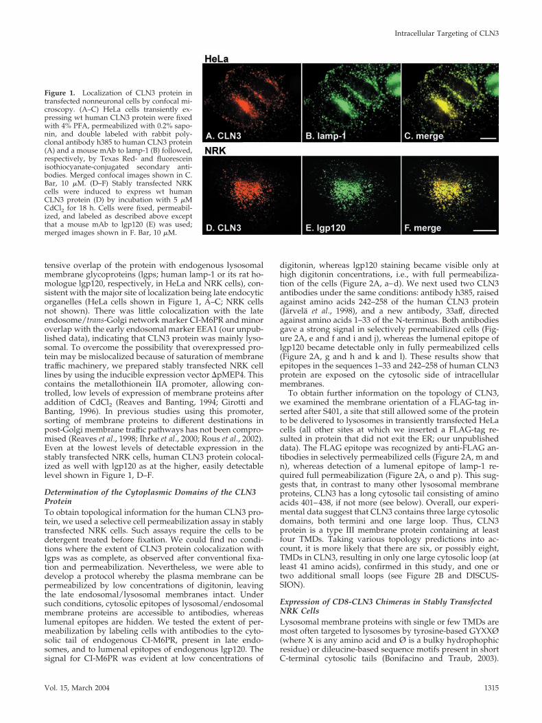
tensive overlap of the protein with endogenous lysosomalmembrane glycoproteins (lgps; human lamp-1 or its rat ho-mologue lgp120, respectively, in HeLa and NRK cells), con-sistent with the major site of localization being late endocyticorganelles (HeLa cells shown in Figure 1, A–C; NRK cellsnot shown). There was little colocalization with the lateendosome/trans-Golgi network marker CI-M6PR and minoroverlap with the early endosomal marker EEA1 (our unpub-lished data), indicating that CLN3 protein was mainly lyso-somal. To overcome the possibility that overexpressed pro-tein may be mislocalized because of saturation of membranetraffic machinery, we prepared stably transfected NRK celllines by using the inducible expression vector �pMEP4. Thiscontains the metallothionein IIA promoter, allowing con-trolled, low levels of expression of membrane proteins afteraddition of CdCl2 (Reaves and Banting, 1994; Girotti andBanting, 1996). In previous studies using this promoter,sorting of membrane proteins to different destinations inpost-Golgi membrane traffic pathways has not been compro-mised (Reaves et al., 1998; Ihrke et al., 2000; Rous et al., 2002).Even at the lowest levels of detectable expression in thestably transfected NRK cells, human CLN3 protein colocal-ized as well with lgp120 as at the higher, easily detectablelevel shown in Figure 1, D–F.
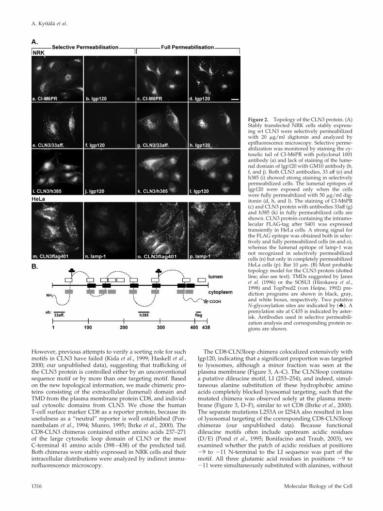
Determination of the Cytoplasmic Domains of the CLN3ProteinTo obtain topological information for the human CLN3 pro-tein, we used a selective cell permeabilization assay in stablytransfected NRK cells. Such assays require the cells to bedetergent treated before fixation. We could find no condi-tions where the extent of CLN3 protein colocalization withlgps was as complete, as observed after conventional fixa-tion and permeabilization. Nevertheless, we were able todevelop a protocol whereby the plasma membrane can bepermeabilized by low concentrations of digitonin, leavingthe late endosomal/lysosomal membranes intact. Undersuch conditions, cytosolic epitopes of lysosomal/endosomalmembrane proteins are accessible to antibodies, whereaslumenal epitopes are hidden. We tested the extent of per-meabilization by labeling cells with antibodies to the cyto-solic tail of endogenous CI-M6PR, present in late endo-somes, and to lumenal epitopes of endogenous lgp120. Thesignal for CI-M6PR was evident at low concentrations of
digitonin, whereas lgp120 staining became visible only athigh digitonin concentrations, i.e., with full permeabiliza-tion of the cells (Figure 2A, a–d). We next used two CLN3antibodies under the same conditions: antibody h385, raisedagainst amino acids 242–258 of the human CLN3 protein(Jarvela et al., 1998), and a new antibody, 33aff, directedagainst amino acids 1–33 of the N-terminus. Both antibodiesgave a strong signal in selectively permeabilized cells (Fig-ure 2A, e and f and i and j), whereas the lumenal epitope oflgp120 became detectable only in fully permeabilized cells(Figure 2A, g and h and k and l). These results show thatepitopes in the sequences 1–33 and 242–258 of human CLN3protein are exposed on the cytosolic side of intracellularmembranes.
To obtain further information on the topology of CLN3,we examined the membrane orientation of a FLAG-tag in-serted after S401, a site that still allowed some of the proteinto be delivered to lysosomes in transiently transfected HeLacells (all other sites at which we inserted a FLAG-tag re-sulted in protein that did not exit the ER; our unpublisheddata). The FLAG epitope was recognized by anti-FLAG an-tibodies in selectively permeabilized cells (Figure 2A, m andn), whereas detection of a lumenal epitope of lamp-1 re-quired full permeabilization (Figure 2A, o and p). This sug-gests that, in contrast to many other lysosomal membraneproteins, CLN3 has a long cytosolic tail consisting of aminoacids 401–438, if not more (see below). Overall, our experi-mental data suggest that CLN3 contains three large cytosolicdomains, both termini and one large loop. Thus, CLN3protein is a type III membrane protein containing at leastfour TMDs. Taking various topology predictions into ac-count, it is more likely that there are six, or possibly eight,TMDs in CLN3, resulting in only one large cytosolic loop (atleast 41 amino acids), confirmed in this study, and one ortwo additional small loops (see Figure 2B and DISCUS-SION).
Expression of CD8-CLN3 Chimeras in Stably TransfectedNRK CellsLysosomal membrane proteins with single or few TMDs aremost often targeted to lysosomes by tyrosine-based GYXXØ(where X is any amino acid and Ø is a bulky hydrophophicresidue) or dileucine-based sequence motifs present in shortC-terminal cytosolic tails (Bonifacino and Traub, 2003).
Figure 1. Localization of CLN3 protein intransfected nonneuronal cells by confocal mi-croscopy. (A–C) HeLa cells transiently ex-pressing wt human CLN3 protein were fixedwith 4% PFA, permeabilized with 0.2% sapo-nin, and double labeled with rabbit poly-clonal antibody h385 to human CLN3 protein(A) and a mouse mAb to lamp-1 (B) followed,respectively, by Texas Red- and fluoresceinisothiocyanate-conjugated secondary anti-bodies. Merged confocal images shown in C.Bar, 10 �M. (D–F) Stably transfected NRKcells were induced to express wt humanCLN3 protein (D) by incubation with 5 �MCdCl2 for 18 h. Cells were fixed, permeabil-ized, and labeled as described above exceptthat a mouse mAb to lgp120 (E) was used;merged images shown in F. Bar, 10 �M.
Intracellular Targeting of CLN3
Vol. 15, March 2004 1315
However, previous attempts to verify a sorting role for suchmotifs in CLN3 have failed (Kida et al., 1999; Haskell et al.,2000; our unpublished data), suggesting that trafficking ofthe CLN3 protein is controlled either by an unconventionalsequence motif or by more than one targeting motif. Basedon the new topological information, we made chimeric pro-teins consisting of the extracellular (lumenal) domain andTMD from the plasma membrane protein CD8, and individ-ual cytosolic domains from CLN3. We chose the humanT-cell surface marker CD8 as a reporter protein, because itsusefulness as a “neutral” reporter is well established (Pon-nambalam et al., 1994; Munro, 1995; Ihrke et al., 2000). TheCD8-CLN3 chimeras contained either amino acids 237–271of the large cytosolic loop domain of CLN3 or the mostC-terminal 41 amino acids (398–438) of the predicted tail.Both chimeras were stably expressed in NRK cells and theirintracellular distributions were analyzed by indirect immu-nofluorescence microscopy.
The CD8-CLN3loop chimera colocalized extensively withlgp120, indicating that a significant proportion was targetedto lysosomes, although a minor fraction was seen at theplasma membrane (Figure 3, A–C). The CLN3loop containsa putative dileucine motif, LI (253–254), and indeed, simul-taneous alanine substitution of these hydrophobic aminoacids completely blocked lysosomal targeting, such that themutated chimera was observed solely at the plasma mem-brane (Figure 3, D–F), similar to wt CD8 (Ihrke et al., 2000).The separate mutations L253A or I254A also resulted in lossof lysosomal targeting of the corresponding CD8-CLN3loopchimeras (our unpublished data). Because functionaldileucine motifs often include upstream acidic residues(D/E) (Pond et al., 1995; Bonifacino and Traub, 2003), weexamined whether the patch of acidic residues at positions�9 to �11 N-terminal to the LI sequence was part of themotif. All three glutamic acid residues in positions �9 to�11 were simultaneously substituted with alanines, without
Figure 2. Topology of the CLN3 protein. (A)Stably transfected NRK cells stably express-ing wt CLN3 were selectively permeabilizedwith 20 �g/ml digitonin and analyzed byepifluorescence microscopy. Selective perme-abilization was monitored by staining the cy-tosolic tail of CI-M6PR with polyclonal 1001antibody (a) and lack of staining of the lume-nal domain of lgp120 with GM10 antibody (b,f, and j). Both CLN3 antibodies, 33 aff (e) andh385 (i) showed strong staining in selectivelypermeabilized cells. The lumenal epitopes oflgp120 were exposed only when the cellswere fully permeabilized with 50 �g/ml dig-itonin (d, h, and l). The staining of CI-M6PR(c) and CLN3 protein with antibodies 33aff (g)and h385 (k) in fully permeabilized cells areshown. CLN3 protein containing the intramo-lecular FLAG-tag after S401 was expressedtransiently in HeLa cells. A strong signal forthe FLAG epitope was obtained both in selec-tively and fully permeabilized cells (m and o),whereas the lumenal epitope of lamp-1 wasnot recognized in selectively permeabilizedcells (n) but only in completely permeabilizedHeLa cells (p). Bar 10 �m. (B) Most probabletopology model for the CLN3 protein (dottedline; also see text). TMDs suggested by Janeset al. (1996) or the SOSUI (Hirokawa et al.,1998) and TopPred2 (von Heijne, 1992) pre-diction programs are shown in black, gray,and white boxes, respectively. Two putativeN-glycosylation sites are indicated by (�). Aprenylation site at C435 is indicated by aster-isk. Antibodies used in selective permeabili-zation analysis and corresponding protein re-gions are shown.
A. Kyttala et al.
Molecular Biology of the Cell1316
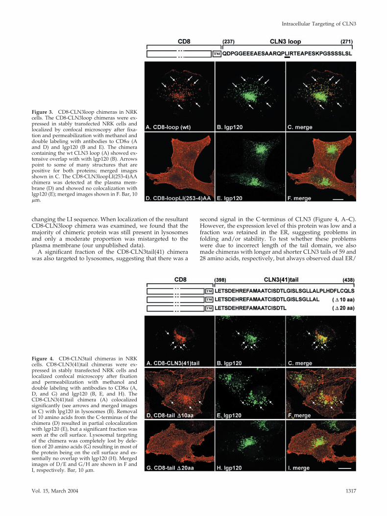
changing the LI sequence. When localization of the resultantCD8-CLN3loop chimera was examined, we found that themajority of chimeric protein was still present in lysosomesand only a moderate proportion was mistargeted to theplasma membrane (our unpublished data).
A significant fraction of the CD8-CLN3tail(41) chimerawas also targeted to lysosomes, suggesting that there was a
second signal in the C-terminus of CLN3 (Figure 4, A–C).However, the expression level of this protein was low and afraction was retained in the ER, suggesting problems infolding and/or stability. To test whether these problemswere due to incorrect length of the tail domain, we alsomade chimeras with longer and shorter CLN3 tails of 59 and28 amino acids, respectively, but always observed dual ER/
Figure 3. CD8-CLN3loop chimeras in NRKcells. The CD8-CLN3loop chimeras were ex-pressed in stably transfected NRK cells andlocalized by confocal microscopy after fixa-tion and permeabilization with methanol anddouble labeling with antibodies to CD8� (Aand D) and lgp120 (B and E). The chimeracontaining the wt CLN3 loop (A) showed ex-tensive overlap with with lgp120 (B). Arrowspoint to some of many structures that arepositive for both proteins; merged imagesshown in C. The CD8-CLN3loopLI(253-4)AAchimera was detected at the plasma mem-brane (D) and showed no colocalization withlgp120 (E); merged images shown in F. Bar, 10�m.
Figure 4. CD8-CLN3tail chimeras in NRKcells. CD8-CLN3(41)tail chimeras were ex-pressed in stably transfected NRK cells andlocalized confocal microscopy after fixationand permeabilization with methanol anddouble labeling with antibodies to CD8� (A,D, and G) and lgp120 (B, E, and H). TheCD8-CLN3(41)tail chimera (A) colocalizedsignificantly (see arrows and merged imagesin C) with lpg120 in lysosomes (B). Removalof 10 amino acids from the C-terminus of thechimera (D) resulted in partial colocalizationwith lgp120 (E), but a significant fraction wasseen at the cell surface. Lysosomal targetingof the chimera was completely lost by dele-tion of 20 amino acids (G) resulting in most ofthe protein being on the cell surface and es-sentially no overlap with lgp120 (H). Mergedimages of D/E and G/H are shown in F andI, respectively. Bar, 10 �m.
Intracellular Targeting of CLN3
Vol. 15, March 2004 1317
lysosomal localization (our unpublished data). To narrowdown the location of lysosomal targeting information withinthe 41 amino acid long tail, we made C-terminal deletions of10 or 20 amino acids. Whereas deletion of 10 amino acidsresulted in an intermediate phenotype with increasedplasma membrane localization of the CD8-CLN3 tail chi-mera (Figure 4D), targeting to lysosomes was completelyabolished by deletion of 20 amino acids (Figure 4G). Thus, itwas likely that targeting information would be locatedwithin the carboxy-terminal 20 amino acids.
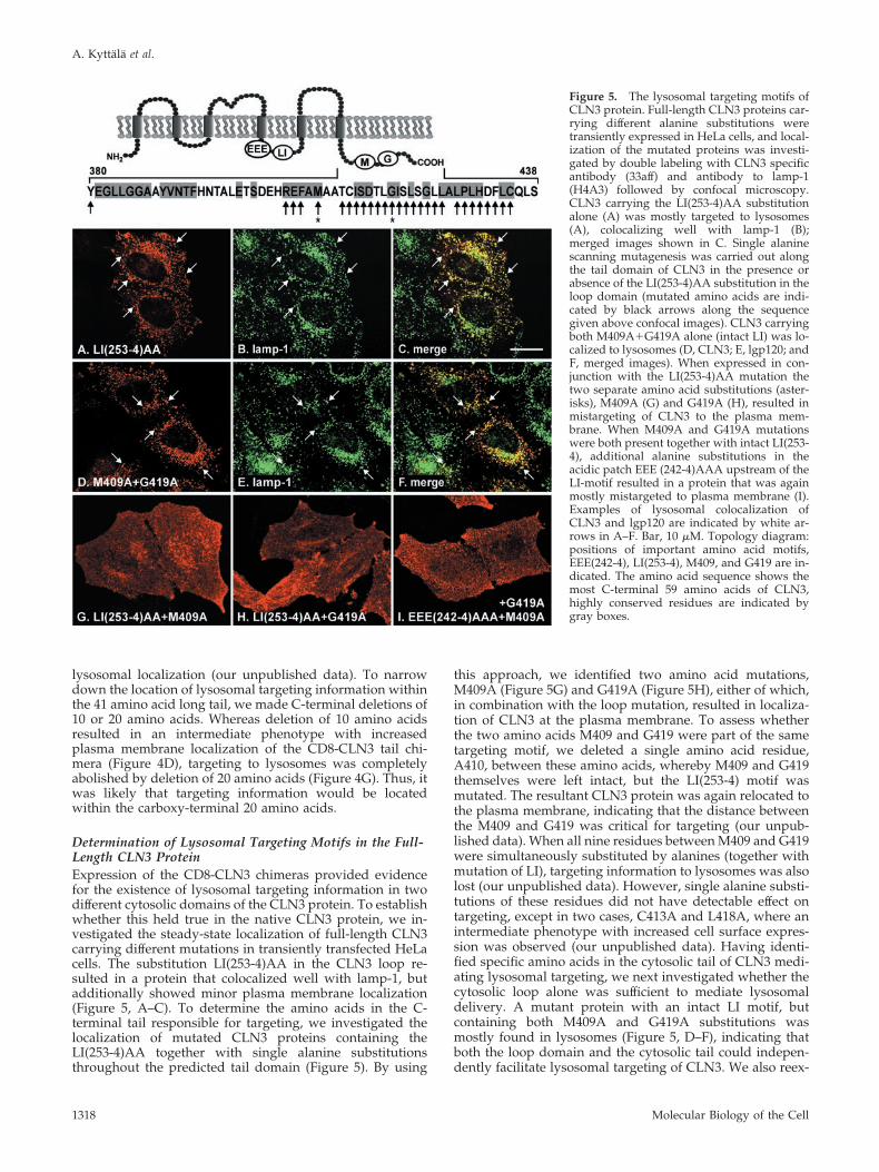
Determination of Lysosomal Targeting Motifs in the Full-Length CLN3 ProteinExpression of the CD8-CLN3 chimeras provided evidencefor the existence of lysosomal targeting information in twodifferent cytosolic domains of the CLN3 protein. To establishwhether this held true in the native CLN3 protein, we in-vestigated the steady-state localization of full-length CLN3carrying different mutations in transiently transfected HeLacells. The substitution LI(253-4)AA in the CLN3 loop re-sulted in a protein that colocalized well with lamp-1, butadditionally showed minor plasma membrane localization(Figure 5, A–C). To determine the amino acids in the C-terminal tail responsible for targeting, we investigated thelocalization of mutated CLN3 proteins containing theLI(253-4)AA together with single alanine substitutionsthroughout the predicted tail domain (Figure 5). By using
this approach, we identified two amino acid mutations,M409A (Figure 5G) and G419A (Figure 5H), either of which,in combination with the loop mutation, resulted in localiza-tion of CLN3 at the plasma membrane. To assess whetherthe two amino acids M409 and G419 were part of the sametargeting motif, we deleted a single amino acid residue,A410, between these amino acids, whereby M409 and G419themselves were left intact, but the LI(253-4) motif wasmutated. The resultant CLN3 protein was again relocated tothe plasma membrane, indicating that the distance betweenthe M409 and G419 was critical for targeting (our unpub-lished data). When all nine residues between M409 and G419were simultaneously substituted by alanines (together withmutation of LI), targeting information to lysosomes was alsolost (our unpublished data). However, single alanine substi-tutions of these residues did not have detectable effect ontargeting, except in two cases, C413A and L418A, where anintermediate phenotype with increased cell surface expres-sion was observed (our unpublished data). Having identi-fied specific amino acids in the cytosolic tail of CLN3 medi-ating lysosomal targeting, we next investigated whether thecytosolic loop alone was sufficient to mediate lysosomaldelivery. A mutant protein with an intact LI motif, butcontaining both M409A and G419A substitutions wasmostly found in lysosomes (Figure 5, D–F), indicating thatboth the loop domain and the cytosolic tail could indepen-dently facilitate lysosomal targeting of CLN3. We also reex-
Figure 5. The lysosomal targeting motifs ofCLN3 protein. Full-length CLN3 proteins car-rying different alanine substitutions weretransiently expressed in HeLa cells, and local-ization of the mutated proteins was investi-gated by double labeling with CLN3 specificantibody (33aff) and antibody to lamp-1(H4A3) followed by confocal microscopy.CLN3 carrying the LI(253-4)AA substitutionalone (A) was mostly targeted to lysosomes(A), colocalizing well with lamp-1 (B);merged images shown in C. Single alaninescanning mutagenesis was carried out alongthe tail domain of CLN3 in the presence orabsence of the LI(253-4)AA substitution in theloop domain (mutated amino acids are indi-cated by black arrows along the sequencegiven above confocal images). CLN3 carryingboth M409A�G419A alone (intact LI) was lo-calized to lysosomes (D, CLN3; E, lgp120; andF, merged images). When expressed in con-junction with the LI(253-4)AA mutation thetwo separate amino acid substitutions (aster-isks), M409A (G) and G419A (H), resulted inmistargeting of CLN3 to the plasma mem-brane. When M409A and G419A mutationswere both present together with intact LI(253-4), additional alanine substitutions in theacidic patch EEE (242-4)AAA upstream of theLI-motif resulted in a protein that was againmostly mistargeted to plasma membrane (I).Examples of lysosomal colocalization ofCLN3 and lgp120 are indicated by white ar-rows in A–F. Bar, 10 �M. Topology diagram:positions of important amino acid motifs,EEE(242-4), LI(253-4), M409, and G419 are in-dicated. The amino acid sequence shows themost C-terminal 59 amino acids of CLN3,highly conserved residues are indicated bygray boxes.
A. Kyttala et al.
Molecular Biology of the Cell1318
amined the putative role of the acidic sequence N-terminalto the LI-motif by using CLN3 constructs with a mutated tailmotif (M409A�G419A). In this context, replacement of glu-tamic acids –9 to –11 with alanines [EEE(242-4)AAA] in thepresence of an intact LI(253-4) motif led to accumulation ofCLN3 at the plasma membrane with little lysosomal local-ization (Figure 5I). This finding suggests that the dileucinetargeting motif in the loop domain of CLN3 includes theupstream acid patch.
The recently described interaction of CLN5, another NCLprotein of lysosomal membranes, with CLN3 (Vesa et al.,
2002) raised the question of whether this interaction is im-portant for correct lysosomal targeting of CLN3. We there-fore coexpressed CLN5, either wild-type or a C-terminallytruncated protein (CLN5 SWE mutation; Vesa et al., 2002),transiently in COS-1 cells together with CLN3 carrying mu-tations in the targeting motifs. Immunoprecipitation with aCLN5 specific antibody, followed by immunoblotting withCLN3-specific antibody 3326 revealed that the two NCLproteins were able to retain their physical interaction evenwhen both lysosomal targeting motifs in CLN3 were mu-tated simultaneously (Figure 6). This result indicates that thetargeting motifs of CLN3 act independently of an interactionwith CLN5 protein, presumably by direct binding to thelysosomal sorting machinery.
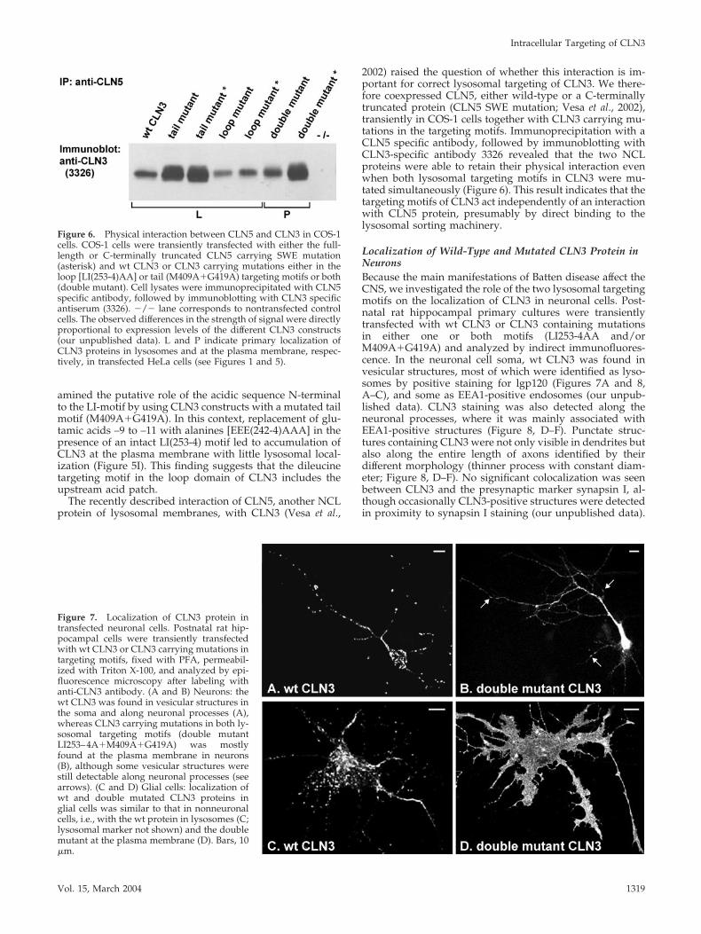
Localization of Wild-Type and Mutated CLN3 Protein inNeuronsBecause the main manifestations of Batten disease affect theCNS, we investigated the role of the two lysosomal targetingmotifs on the localization of CLN3 in neuronal cells. Post-natal rat hippocampal primary cultures were transientlytransfected with wt CLN3 or CLN3 containing mutationsin either one or both motifs (LI253-4AA and/orM409A�G419A) and analyzed by indirect immunofluores-cence. In the neuronal cell soma, wt CLN3 was found invesicular structures, most of which were identified as lyso-somes by positive staining for lgp120 (Figures 7A and 8,A–C), and some as EEA1-positive endosomes (our unpub-lished data). CLN3 staining was also detected along theneuronal processes, where it was mainly associated withEEA1-positive structures (Figure 8, D–F). Punctate struc-tures containing CLN3 were not only visible in dendrites butalso along the entire length of axons identified by theirdifferent morphology (thinner process with constant diam-eter; Figure 8, D–F). No significant colocalization was seenbetween CLN3 and the presynaptic marker synapsin I, al-though occasionally CLN3-positive structures were detectedin proximity to synapsin I staining (our unpublished data).
Figure 6. Physical interaction between CLN5 and CLN3 in COS-1cells. COS-1 cells were transiently transfected with either the full-length or C-terminally truncated CLN5 carrying SWE mutation(asterisk) and wt CLN3 or CLN3 carrying mutations either in theloop [LI(253-4)AA] or tail (M409A�G419A) targeting motifs or both(double mutant). Cell lysates were immunoprecipitated with CLN5specific antibody, followed by immunoblotting with CLN3 specificantiserum (3326). �/� lane corresponds to nontransfected controlcells. The observed differences in the strength of signal were directlyproportional to expression levels of the different CLN3 constructs(our unpublished data). L and P indicate primary localization ofCLN3 proteins in lysosomes and at the plasma membrane, respec-tively, in transfected HeLa cells (see Figures 1 and 5).
Figure 7. Localization of CLN3 protein intransfected neuronal cells. Postnatal rat hip-pocampal cells were transiently transfectedwith wt CLN3 or CLN3 carrying mutations intargeting motifs, fixed with PFA, permeabil-ized with Triton X-100, and analyzed by epi-fluorescence microscopy after labeling withanti-CLN3 antibody. (A and B) Neurons: thewt CLN3 was found in vesicular structures inthe soma and along neuronal processes (A),whereas CLN3 carrying mutations in both ly-sosomal targeting motifs (double mutantLI253–4A�M409A�G419A) was mostlyfound at the plasma membrane in neurons(B), although some vesicular structures werestill detectable along neuronal processes (seearrows). (C and D) Glial cells: localization ofwt and double mutated CLN3 proteins inglial cells was similar to that in nonneuronalcells, i.e., with the wt protein in lysosomes (C;lysosomal marker not shown) and the doublemutant at the plasma membrane (D). Bars, 10�m.
Intracellular Targeting of CLN3
Vol. 15, March 2004 1319
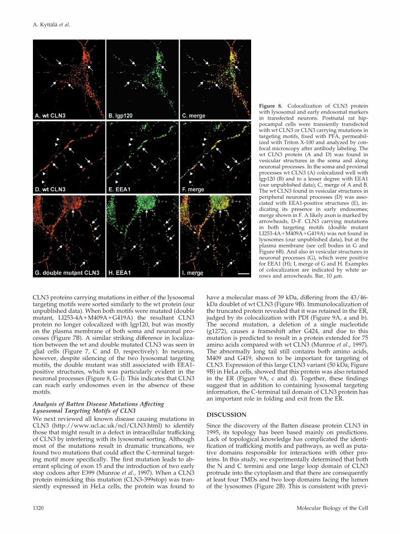
CLN3 proteins carrying mutations in either of the lysosomaltargeting motifs were sorted similarly to the wt protein (ourunpublished data). When both motifs were mutated (doublemutant, LI253-4A�M409A�G419A) the resultant CLN3protein no longer colocalized with lgp120, but was mostlyon the plasma membrane of both soma and neuronal pro-cesses (Figure 7B). A similar striking difference in localiza-tion between the wt and double mutated CLN3 was seen inglial cells (Figure 7, C and D, respectively). In neurons,however, despite silencing of the two lysosomal targetingmotifs, the double mutant was still associated with EEA1-positive structures, which was particularly evident in theneuronal processes (Figure 8, G–I). This indicates that CLN3can reach early endosomes even in the absence of thesemotifs.
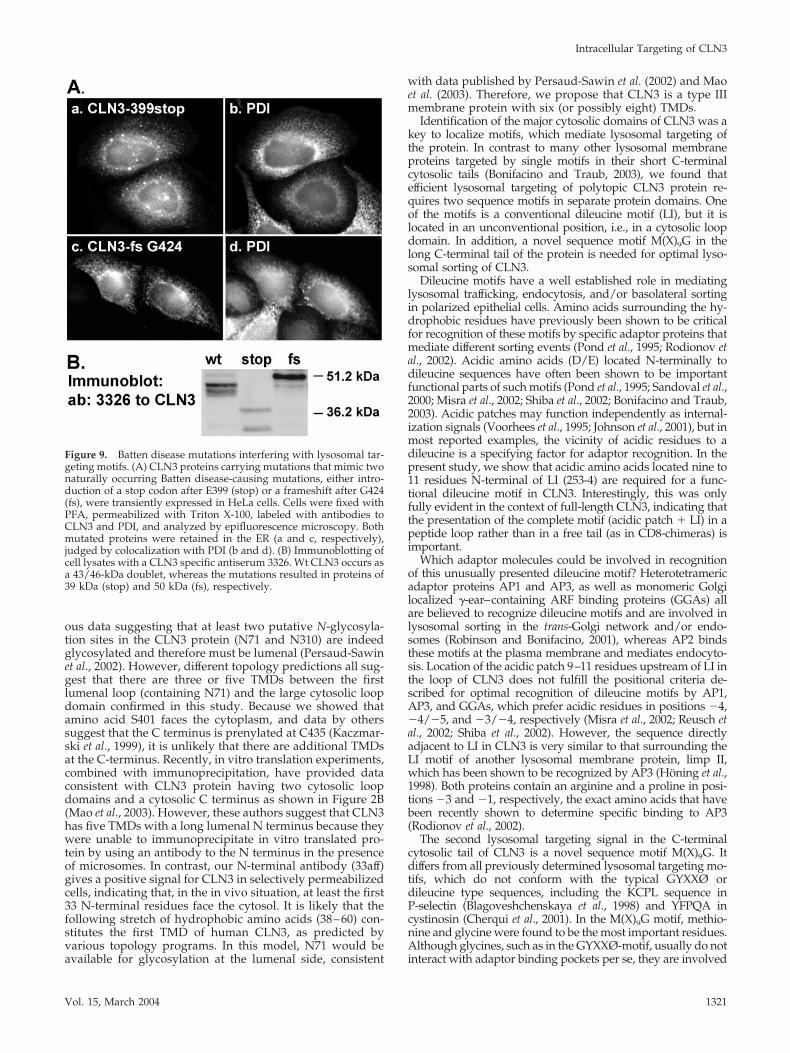
Analysis of Batten Disease Mutations AffectingLysosomal Targeting Motifs of CLN3We next reviewed all known disease causing mutations inCLN3 (http://www.ucl.ac.uk/ncl/CLN3.html) to identifythose that might result in a defect in intracellular traffickingof CLN3 by interfering with its lysosomal sorting. Althoughmost of the mutations result in dramatic truncations, wefound two mutations that could affect the C-terminal target-ing motif more specifically. The first mutation leads to ab-errant splicing of exon 15 and the introduction of two earlystop codons after E399 (Munroe et al., 1997). When a CLN3protein mimicking this mutation (CLN3-399stop) was tran-siently expressed in HeLa cells, the protein was found to
have a molecular mass of 39 kDa, differing from the 43/46-kDa doublet of wt CLN3 (Figure 9B). Immunolocalization ofthe truncated protein revealed that it was retained in the ER,judged by its colocalization with PDI (Figure 9A, a and b).The second mutation, a deletion of a single nucleotide(g1272), causes a frameshift after G424, and due to thismutation is predicted to result in a protein extended for 75amino acids compared with wt CLN3 (Munroe et al., 1997).The abnormally long tail still contains both amino acids,M409 and G419, shown to be important for targeting ofCLN3. Expression of this large CLN3 variant (50 kDa; Figure9B) in HeLa cells, showed that this protein was also retainedin the ER (Figure 9A, c and d). Together, these findingssuggest that in addition to containing lysosomal targetinginformation, the C-terminal tail domain of CLN3 protein hasan important role in folding and exit from the ER.
DISCUSSION
Since the discovery of the Batten disease protein CLN3 in1995, its topology has been based mainly on predictions.Lack of topological knowledge has complicated the identi-fication of trafficking motifs and pathways, as well as puta-tive domains responsible for interactions with other pro-teins. In this study, we experimentally determined that boththe N and C termini and one large loop domain of CLN3protrude into the cytoplasm and that there are consequentlyat least four TMDs and two loop domains facing the lumenof the lysosomes (Figure 2B). This is consistent with previ-
Figure 8. Colocalization of CLN3 proteinwith lysosomal and early endosomal markersin transfected neurons. Postnatal rat hip-pocampal cells were transiently transfectedwith wt CLN3 or CLN3 carrying mutations intargeting motifs, fixed with PFA, permeabil-ized with Triton X-100 and analyzed by con-focal microscopy after antibody labeling. Thewt CLN3 protein (A and D) was found invesicular structures in the soma and alongneuronal processes. In the soma and proximalprocesses wt CLN3 (A) colocalized well withlgp120 (B) and to a lesser degree with EEA1(our unpublished data); C, merge of A and B.The wt CLN3 found in vesicular structures inperipheral neuronal processes (D) was asso-ciated with EEA1-positive structures (E), in-dicating its presence in early endosomes;merge shown in F. A likely axon is marked byarrowheads, D–F. CLN3 carrying mutationsin both targeting motifs (double mutantLI253-4A�M409A�G419A) was not found inlysosomes (our unpublished data), but at theplasma membrane (see cell bodies in G andFigure 6B). And also in vesicular structures inneuronal processes (G), which were positivefor EEA1 (H); I, merge of G and H. Examplesof colocalization are indicated by white ar-rows and arrowheads. Bar, 10 �m.
A. Kyttala et al.
Molecular Biology of the Cell1320
ous data suggesting that at least two putative N-glycosyla-tion sites in the CLN3 protein (N71 and N310) are indeedglycosylated and therefore must be lumenal (Persaud-Sawinet al., 2002). However, different topology predictions all sug-gest that there are three or five TMDs between the firstlumenal loop (containing N71) and the large cytosolic loopdomain confirmed in this study. Because we showed thatamino acid S401 faces the cytoplasm, and data by otherssuggest that the C terminus is prenylated at C435 (Kaczmar-ski et al., 1999), it is unlikely that there are additional TMDsat the C-terminus. Recently, in vitro translation experiments,combined with immunoprecipitation, have provided dataconsistent with CLN3 protein having two cytosolic loopdomains and a cytosolic C terminus as shown in Figure 2B(Mao et al., 2003). However, these authors suggest that CLN3has five TMDs with a long lumenal N terminus because theywere unable to immunoprecipitate in vitro translated pro-tein by using an antibody to the N terminus in the presenceof microsomes. In contrast, our N-terminal antibody (33aff)gives a positive signal for CLN3 in selectively permeabilizedcells, indicating that, in the in vivo situation, at least the first33 N-terminal residues face the cytosol. It is likely that thefollowing stretch of hydrophobic amino acids (38–60) con-stitutes the first TMD of human CLN3, as predicted byvarious topology programs. In this model, N71 would beavailable for glycosylation at the lumenal side, consistent
with data published by Persaud-Sawin et al. (2002) and Maoet al. (2003). Therefore, we propose that CLN3 is a type IIImembrane protein with six (or possibly eight) TMDs.
Identification of the major cytosolic domains of CLN3 was akey to localize motifs, which mediate lysosomal targeting ofthe protein. In contrast to many other lysosomal membraneproteins targeted by single motifs in their short C-terminalcytosolic tails (Bonifacino and Traub, 2003), we found thatefficient lysosomal targeting of polytopic CLN3 protein re-quires two sequence motifs in separate protein domains. Oneof the motifs is a conventional dileucine motif (LI), but it islocated in an unconventional position, i.e., in a cytosolic loopdomain. In addition, a novel sequence motif M(X)9G in thelong C-terminal tail of the protein is needed for optimal lyso-somal sorting of CLN3.
Dileucine motifs have a well established role in mediatinglysosomal trafficking, endocytosis, and/or basolateral sortingin polarized epithelial cells. Amino acids surrounding the hy-drophobic residues have previously been shown to be criticalfor recognition of these motifs by specific adaptor proteins thatmediate different sorting events (Pond et al., 1995; Rodionov etal., 2002). Acidic amino acids (D/E) located N-terminally todileucine sequences have often been shown to be importantfunctional parts of such motifs (Pond et al., 1995; Sandoval et al.,2000; Misra et al., 2002; Shiba et al., 2002; Bonifacino and Traub,2003). Acidic patches may function independently as internal-ization signals (Voorhees et al., 1995; Johnson et al., 2001), but inmost reported examples, the vicinity of acidic residues to adileucine is a specifying factor for adaptor recognition. In thepresent study, we show that acidic amino acids located nine to11 residues N-terminal of LI (253-4) are required for a func-tional dileucine motif in CLN3. Interestingly, this was onlyfully evident in the context of full-length CLN3, indicating thatthe presentation of the complete motif (acidic patch � LI) in apeptide loop rather than in a free tail (as in CD8-chimeras) isimportant.
Which adaptor molecules could be involved in recognitionof this unusually presented dileucine motif? Heterotetramericadaptor proteins AP1 and AP3, as well as monomeric Golgilocalized �-ear–containing ARF binding proteins (GGAs) allare believed to recognize dileucine motifs and are involved inlysosomal sorting in the trans-Golgi network and/or endo-somes (Robinson and Bonifacino, 2001), whereas AP2 bindsthese motifs at the plasma membrane and mediates endocyto-sis. Location of the acidic patch 9–11 residues upstream of LI inthe loop of CLN3 does not fulfill the positional criteria de-scribed for optimal recognition of dileucine motifs by AP1,AP3, and GGAs, which prefer acidic residues in positions �4,�4/�5, and �3/�4, respectively (Misra et al., 2002; Reusch etal., 2002; Shiba et al., 2002). However, the sequence directlyadjacent to LI in CLN3 is very similar to that surrounding theLI motif of another lysosomal membrane protein, limp II,which has been shown to be recognized by AP3 (Honing et al.,1998). Both proteins contain an arginine and a proline in posi-tions �3 and �1, respectively, the exact amino acids that havebeen recently shown to determine specific binding to AP3(Rodionov et al., 2002).
The second lysosomal targeting signal in the C-terminalcytosolic tail of CLN3 is a novel sequence motif M(X)9G. Itdiffers from all previously determined lysosomal targeting mo-tifs, which do not conform with the typical GYXXØ ordileucine type sequences, including the KCPL sequence inP-selectin (Blagoveshchenskaya et al., 1998) and YFPQA incystinosin (Cherqui et al., 2001). In the M(X)9G motif, methio-nine and glycine were found to be the most important residues.Although glycines, such as in the GYXXØ-motif, usually do notinteract with adaptor binding pockets per se, they are involved
Figure 9. Batten disease mutations interfering with lysosomal tar-geting motifs. (A) CLN3 proteins carrying mutations that mimic twonaturally occurring Batten disease-causing mutations, either intro-duction of a stop codon after E399 (stop) or a frameshift after G424(fs), were transiently expressed in HeLa cells. Cells were fixed withPFA, permeabilized with Triton X-100, labeled with antibodies toCLN3 and PDI, and analyzed by epifluorescence microscopy. Bothmutated proteins were retained in the ER (a and c, respectively),judged by colocalization with PDI (b and d). (B) Immunoblotting ofcell lysates with a CLN3 specific antiserum 3326. Wt CLN3 occurs asa 43/46-kDa doublet, whereas the mutations resulted in proteins of39 kDa (stop) and 50 kDa (fs), respectively.
Intracellular Targeting of CLN3
Vol. 15, March 2004 1321
in presentation of the signal (Owen et al., 2001). Therefore, theglycine at position 419 in the M(X)9G motif of CLN3 may fulfilla similar role. Because changes both in the number or thecomposition of residues between M409 and G419 also alteredlocalization of CLN3, it is possible that this motif is part of alarger three-dimensional determinant, rather than being a lin-ear sequence motif. The C-terminal tail structure of CLN3could be stabilized by interaction of hydrophobic amino acidsand/or lipid anchors with the cytosolic leaflet of the membraneand thereby be important for the presentation of the targetingmotif. One possible attachment site is C435, which has beenshown to be prenylated in vitro (Kaczmarski et al., 1999). How-ever, our present data are consistent with a previous reportindicating that substitution of C435 with alanine had no de-tectable effect on steady state localization of CLN3 (Haskell etal., 2000). Thus, additional factors may contribute to the pre-sentation of the M(X)9G motif in CLN3.
There is currently no information about the trafficking ma-chinery that might interact with unusual targeting motifs. Itwas recently reported that the sequence DVPM, found in thesorting receptor sorLA, was the minimal sequence requirementof the form � XX� (where � is an acidic amino acid) forbinding to GGA1 (Jacobsen et al., 2002). A similar sequenceEFAM409 in CLN3 overlaps with the M(X)9G motif. Becausesubstitution of E406 by alanine had no detectable effect onlysosomal targeting of CLN3, it is unlikely that M(X)9G motif isa � XX�-type signal recognized by GGA1. Our experimentsalso showed that interaction with another NCL protein, CLN5,did not depend on the presence of the lysosomal targetingmotifs of CLN3, suggesting that CLN5 binding is not a prereq-uisite for lysosomal targeting of CLN3.
An unresolved question is why the most severe symptomsof Batten disease manifest in the brain. Thus, expression ofCLN3 in neurons has been investigated extensively with thehope of uncovering any putative differences with nonneuronalcells. Consistent with previous studies, we found that CLN3was present in vesicular structures both in soma and in neu-ronal processes (including axons) of cultured rat hippocampalneurons (Jarvela et al., 1999; Haskell et al., 2000; Luiro et al.,2001). Because lysosomes and late endosomes are predomi-nantly located in the cell soma and proximal regions of den-drites (Parton et al., 1992), it is likely that the presence of CLN3in distal parts of neuronal processes represents localizationsother than lysosomes/late endosomes. Previously, CLN3 ex-pressed in neuronal processes has been colocalized with pre-synaptic markers and therefore suggested to have a role insynaptic transmission (Jarvela et al., 1999; Haskell et al., 2000).However, fractionation analysis of mouse brain later revealedthat CLN3 resides in a compartment different from synapticvesicles (Luiro et al., 2001). We also frequently observed CLN3close to a presynaptic marker (synapsin I), but saw very littleprecise overlap. In contrast, we found that CLN3 was associ-ated prominently with EEA1 along neuronal processes dem-onstrating a novel localization of CLN3 in early endosomes. Ithas been previously shown that early endosomes are concen-trated at presynaptic terminals and varicosities in axons ofhippocampal neurons (Parton et al., 1992). The presence ofCLN3 in early endosomes was much less evident in nonneu-ronal cells (HeLa), suggesting this localization could be espe-cially important in neurons. The same lysosomal targetingmotifs of CLN3 operated both in neuronal and nonneuronalcells; however, localization of CLN3 in early endosomes oc-curred independently of lysosomal targeting. Therefore, it ispossible that CLN3 contains additional targeting information,which is only recognized in specialized cells (such as neurons),and/or which regulates the rate of trafficking at the level ofendosomes (Johnson et al., 2001).
Endosomes present in neuronal processes are dynamic andheterogeneous structures (Sulzer and Holtzman, 1989; Cooneyet al., 2002). Among these organelles, multivesicular bodieshave been suggested to be the major structures mediatingtransport of endocytic and autophagic material from the nerveterminals to the lysosomes in the cell body (Parton et al., 1992;Hollenbeck, 1993; Nixon and Cataldo, 1995). It is tempting tohypothesize that the function of CLN3 is to regulate the phys-iological properties of such organelles (including their degra-dative characteristics). This would be consistent with previousreports demonstrating altered intravesicular pH when CLN3was absent or functionally inactive (Golabek et al., 2000; Hol-opainen et al., 2001). The function of many lysosomal proteinsis known to be regulated by the proteolytic activation of theirprecursors in acidic environments. For example, gradual pro-teolytic processing of cathepsin D in acidic compartments isrequired to yield an active enzyme, which has been suggestedto have an important role in neuronal development and/orhomeostasis (Tyynela et al., 2000). Interestingly, altered levelsof CLN3 expression have been reported to affect processing ofcathepsin D as well as amyloid-� protein precursor (Golabek etal., 2000). The highly specialized endosomal–lysosomal systemmay render neurons especially sensitive to impaired (or accel-erated) processing of certain proteins and lipids and therefore,lead to the accumulation of lipofuscin typical for all NCLdisorders.
ACKNOWLEDGMENTS
We thank Sally Gray for technical assistance and Dr. Karin Romish for advicein determination of topology. The work was supported by grants from theWellcome Trust (United Kingdom) to A.K. (Traveling Research Fellowship061077), to J.P.L. and G.I. (057263); The Medical Research Council (UnitedKingdom) to J.P.L.; and The Academy of Finland (48047) and the MaudKuistila Foundation (Finland) to A.K. M.J.S. was supported by the RoyalSociety (United Kingdom). The Cambridge Institute for Medical Research is inreceipt of a strategic award from the Wellcome Trust.
REFERENCES
Andersson, S., Davis, D.L., Dahlback, H., Jornvall, H., and Russell, D.W.(1989). Cloning, structure, and expression of the mitochondrial cytochromeP-450 sterol 26-hydroxylase, a bile acid biosynthetic enzyme. J. Biol. Chem.264, 8222–8229.
Blagoveshchenskaya, A.D., Norcott, J.P., and Cutler, D.F. (1998). Lysosomaltargeting of P-selectin is mediated by a novel sequence within its cytoplasmic tail.J. Biol. Chem. 273, 2729–2737.
Bonifacino, J.S., and Traub, L.M. (2003). Signals for sorting of transmembraneproteins to endosomes and lysosomes. Annu. Rev. Biochem. 72, 395–447.
Chattopadhyay, S., Muzaffar, N.E., Sherman, F., and Pearce, D.A. (2000). Theyeast model for Batten disease: mutations in btn1, btn2, and hsp30 alter pHhomeostasis. J. Bacteriol. 182, 6418–6423.
Chattopadhyay, S., Roberts, P.M., and Pearce, D.A. (2003). The yeast model forBatten disease: a role for Btn2p in the trafficking of the Golgi-associated vesiculartargeting protein, Yif1p. Biochem. Biophys. Res. Commun. 302, 534–538.
Cherqui, S., Kalatzis, V., Trugnan, G., and Antignac, C. (2001). The targeting ofcystinosin to the lysosomal membrane requires a tyrosine-based signal and anovel sorting motif. J. Biol. Chem. 276, 13314–13321.
Cooney, J.R., Hurlburt, J.L., Selig, D.K., Harris, K.M., and Fiala, J.C. (2002).Endosomal compartments serve multiple hippocampal dendritic spines from awidespread rather than a local store of recycling membrane. J. Neurosci. 22,2215–2224.
Das, A.M., von Harlem, R., Feist, M., Lucke, T., and Kohlschutter, A. (2001).Altered levels of high-energy phosphate compounds in fibroblasts from differentforms of neuronal ceroid lipofuscinoses: further evidence for mitochondrialinvolvement. Eur. J. Paediatr. Neurol. 5, 143–146.
Davies, J.P., and Ioannou, Y.A. (2000). Topological analysis of Niemann-Pick C1protein reveals that the membrane orientation of the putative sterol-sensingdomain is identical to those of 3-hydroxy-3-methylglutaryl-CoA reductase andsterol regulatory element binding protein cleavage-activating protein. J. Biol.Chem. 275, 24367–24374.
A. Kyttala et al.
Molecular Biology of the Cell1322
Girotti, M., and Banting, G. (1996). TGN38-green fluorescent protein hybridproteins expressed in stably transfected eukaryotic cells provide a tool for thereal-time, in vivo study of membrane traffic pathways and suggest a possible rolefor rat TGN38. J. Cell Sci. 109, 2915–2926.
Golabek, A.A., Kida, E., Walus, M., Kaczmarski, W., Michalewski, M., andWisniewski, K.E. (2000). CLN3 protein regulates lysosomal pH and alters intra-cellular processing of Alzheimer’s amyloid-beta protein precursor and cathepsinD in human cells. Mol. Genet. Metab. 70, 203–213.
Haskell, R.E., Carr, C.J., Pearce, D.A., Bennett, M.J., and Davidson, B.L. (2000).Batten disease: evaluation of CLN3 mutations on protein localization and func-tion. Hum. Mol. Genet. 9, 735–744.
Hirokawa, T., Boon-Chieng, S., and Mitaku, S. (1998). SOSUI: classification andsecondary structure prediction system for membrane proteins. Bioinformatics 14,378–379.
Hollenbeck, P.J. (1993). Products of endocytosis and autophagy are retrievedfrom axons by regulated retrograde organelle transport. J. Cell Biol. 121, 305–315.
Holopainen, J.M., Saarikoski, J., Kinnunen, P.K., and Jarvela, I.I. (2001). Elevatedlysosomal pH in neuronal ceroid lipofuscinoses (NCLs). Eur. J. Biochem. 268,5851–5856.
Honing, S., Sandoval, I.V., and von Figura, K. (1998). A di-leucine-based motif inthe cytoplasmic tail of LIMP-II and tyrosinase mediates selective binding of AP-3.EMBO J. 17, 1304–1314.
Ihrke, G., Gray, S.R., and Luzio, J.P. (2000). Endolyn is a mucin-like type Imembrane protein targeted to lysosomes by its cytoplasmic tail. Biochem. J. 345,287–296.
Jacobsen, L., Madsen, P., Nielsen, M.S., Geraerts, W.P.M., Gliemann, J., Smit, A.B.,and Petersen, C.M. (2002). The sorLA cytoplasmic domain interacts with GGA1and -2 and defines minimum requirements for GGA binding. FEBS Lett. 511,155–158.
Janes, R.W., Munroe, P.B., Mitchison, H.A., Gardiner, R.M., Mole, S.E., andWallace, B.A. (1996). A model for Batten disease protein CLN 3, functionalimplications from homology and mutations. FEBS Lett. 399, 75–77.
Johnson, A.O., Lampson, M.A., and McGraw, T.E. (2001). A di-leucine sequenceand a cluster of acidic amino acids are required for dynamic retention in theendosomal recycling compartment of fibroblasts. Mol. Biol. Cell 12, 367–381.
Jarvela, I., Lehtovirta, M., Tikkanen, R., Kyttala, A., and Jalanko, A. (1999).Defective intracellular transport of CLN3 is the molecular basis of Batten disease(JNCL). Hum. Mol. Genet. 8, 1091–1098.
Jarvela, I., Sainio, M., Rantamaki, T., Olkkonen, V.M., Carpen, O., Peltonen, L.,and Jalanko, A. (1998). Biosynthesis and intracellular targeting of the CLN3protein defective in Batten disease. Hum. Mol. Genet. 7, 85–90.
Kaczmarski, W., Wisniewski, K.E., Golabek, A., Kaczmarski, A., Kida, E., andMichalewski, M. (1999). Studies of membrane association of CLN3 protein. Mol.Genet. Metab. 66, 261–264.
Kida, E., Kaczmarski, W., Golabek, A.A., Kaczmarski, A., Michalewski, M., andWisniewski, K.E. (1999). Analysis of intracellular distribution and trafficking ofthe CLN3 protein in fusion with the green fluorescent protein in vitro. Mol.Genet. Metab. 66, 265–271.
Luiro, K., Kopra, O., Lehtovirta, M., and Jalanko, A. (2001). CLN3 protein istargeted to neuronal synapses but excluded from synaptic vesicles: new clues toBatten disease. Hum. Mol. Genet. 10, 2123–2131.
Mao, Q., Foster, B.J., Xia, H., and Davidson, B.L. (2003). Membrane topology ofCLN3, the protein underlying Batten disease. FEBS Lett. 27143, 1–7.
Misra, S., Puertollano, R., Kato, Y., Bonifacino, J.S., and Hurley, J.H. (2002).Structural basis for acidic-cluster-dileucine sorting-signal recognition by VHSdomains. Nature 415, 933–937.
Munro, S. (1995). An investigation of the role of transmembrane domains inGolgi protein retention. EMBO J. 14, 4695–4704.
Munroe, P.B., et al. (1997). Spectrum of mutations in the Batten disease gene,CLN3. Am. J. Hum. Genet. 61, 310–316.
Nixon, R.A., and Cataldo, A.M. (1995). The endosomal-lysosomal system ofneurons: new roles. Trends Neurosci. 18, 489–496.
Owen, D.J., Setiadi, H., Evans, P.R., McEver, R.P., and Green, S.A. (2001). A thirdspecificity-determining site in �2 adaptin for sequences upstream of Yxx� sortingmotifs. Traffic 2, 105–110.
Parton, R.G., Simons, K., and Dotti, C.G. (1992). Axonal and dendritic endocyticpathways in cultured neurons. J. Cell Biol. 119, 123–137.
Pearce, D.A. (2000). Localization and processing of CLN3, the protein associatedto Batten disease: where is it and what does it do ? J. Neurosci. Res. 59, 19–23.
Pearce, D.A., Ferea, T., Nosel, S.A., Das, B., and Sherman, F. (1999). Action ofBTN1, the yeast orthologue of the gene mutated in Batten disease. Nat. Genet. 22,55–58.
Persaud-Sawin, D.A., VanDongen, A., and Boustany, R.M. (2002). Motifs withinthe CLN3 protein: modulation of cell growth rates and apoptosis. Hum. Mol.Genet. 11, 2129–2142.
Ponnambalam, S., Rabouille, C., Luzio, J.P., Nilsson, T., and Warren, G. (1994).The TGN38 glycoprotein contains two non-overlapping signals that mediatelocalization to the trans-Golgi network. J. Cell Biol. 125, 253–268.
Pond, L., Kuhn, L.A., Teyton, L., Schutze, M.P., Tainer, J.A., Jackson, M.R., andPeterson, P.A. (1995). A role for acidic residues in di-leucine motif-based target-ing to the endocytic pathway. J. Biol. Chem. 270, 19989–19997.
Puranam, K.L., Guo, W.-X., Qian, W.-H., Nikbakht, K., and Boustany, R.M.(1999). CLN3 defines a novel antiapoptotic pathway operative in neurodegen-eration and mediated by ceramide. Mol. Genet. Metab. 66, 294–308.
Reaves, B., and Banting, G. (1994). Overexpression of TGN38/41 leads to mislo-calisation of �-adaptin. FEBS Lett. 351, 448–456.
Reaves, B.J., Banting, G., and Luzio, J.P. (1998). Lumenal and trans-membranedomains play a role in sorting type I membrane proteins on endocytic pathways.Mol. Biol. Cell 9, 1107–1122.
Reaves, B.J., Bright, N.A., Mullock, B.M., and Luzio, J.P. (1996). The effect ofwortmannin on the localisation of lysosomal type I integral membrane glycop-roteins suggests a role for phosphoinositide 3-kinase activity in regulating mem-brane traffic late in the endocytic pathway. J. Cell Sci. 109, 749–762.
Reusch, U., Bernhard, O., Koszinowski, U., and Schu, P. (2002). AP-1A andAP-3A lysosomal sorting functions. Traffic 3, 752–761.
Robinson, M.S., and Bonifacino, J.S. (2001). Adaptor-related proteins. Curr. Opin.Cell Biol..13, 444–453.
Rodionov, D.G., Honing, S., Silye, A., Kongsvik, T.L., Von Figura, K., and Bakke,O. (2002). Structural requirements for interactions between leucine-sorting sig-nals and clathrin-associated adaptor protein complex AP3. J. Biol. Chem. 277,47436–47443.
Rous, B.A., Reaves, B.J., Ihrke, G., Briggs, J.A.G., Gray, S.R., Stephens, D.J.,Banting, G., and Luzio, J.P. (2002). The role of the adaptor complex AP-3 intargeting wild type and mutated CD63 to lysosomes. Mol. Biol. Cell 13, 1071–1082.
Sandoval, I.V., Martinez-Arca, S., Valdueza, J., Palacios, S., and Holman, G.D.(2000). Distinct reading of different structural determinants modulates thedileucine-mediated transport steps of the lysosomal membrane protein LIMPIIand the insulin-sensitive glucose transporter GLUT4. J. Biol. Chem. 275, 39874–39885.
Santavuori, P. (1988). Neuronal ceroid-lipofuscinoses in childhood. Brain Dev. 10,80–83.
Schell, M.J., Erneux, C., and Irvine, R.F. (2001). Inositol 1, 4, 5-trisphosphate3-kinase A associates with F-actin and dendritic spines via its N terminus. J. Biol.Chem. 276, 37537–37546.
Shiba, T., et al. (2002). Structural basis for recognition of acidic-cluster dileucinesequence by GGA1. Nature 415, 937–941.
Sulzer, D., and Holtzman, E. (1989). Acidification and endosome-like compart-ments in the presynaptic terminals of frog retinal photoreceptors. J. Neurocytol.18, 529–540.
The International Batten Disease Consortium. (1995). Isolation of a novel geneunderlying Batten disease, CLN3. Cell 82, 949–957.
Tyynela, J., Sohar, I., Sleat, D.E., Gin, R.M., Donnelly, R.J., Baumann, M., Haltia,M., and Lobel, P. (2000). A mutation in the ovine cathepsin D gene causes acongenital lysosomal storage disease with profound neurodegeneration. EMBO J.19, 2786–2792.
Vesa, J., Chin, M.H., Oelgeschlager, K., Isosomppi, J., DellAngelica, E.C., Jalanko,A., and Peltonen, L. (2002). Neuronal ceroid lipofuscinoses are connected atmolecular level: interaction of CLN5 protein with CLN2 and CLN3. Mol. Biol.Cell 13, 2410–2420.
von Heijne, G. (1992). Membrane protein structure prediction, hydrophobicityanalysis and the positive-inside rule. J. Mol. Biol. 225, 487–494.
Voorhees, P., Deignan, E., van Donselaar, E., Humphrey, J., Marks, M.S., Peters,P.J., and Bonifacino, J.S. (1995). An acidic sequence within the cytoplasmic do-main of furin functions as a determinant of trans-Golgi network localization andinternalization from the cell surface. EMBO J. 14, 4961–4975.
Weimer, J.M., Kriscenski-Perry, E., Elshatory, Y., and Pearce, D.A. (2002). Theneuronal ceroid lipofuscinoses: mutations in different proteins result in similardisease. Neuromol. Med. 1, 111–124.
Intracellular Targeting of CLN3
Vol. 15, March 2004 1323