Two dimensional electrophoresis, Sample preparation Session I 台大生技教改暑期課程.
75
Two dimensional Two dimensional electrophoresis, Sample electrophoresis, Sample preparation preparation Session I Session I 台台台台台台台台台台
-
Upload
esmond-whitehead -
Category
Documents
-
view
251 -
download
4
Transcript of Two dimensional electrophoresis, Sample preparation Session I 台大生技教改暑期課程.

Two dimensional Two dimensional electrophoresis, Sample electrophoresis, Sample
preparationpreparation
Session ISession I
台大生技教改暑期課程

Only “Proteomics” is the large-scale screening of the Only “Proteomics” is the large-scale screening of the proteins of a cell, organism or biological fluid, a process proteins of a cell, organism or biological fluid, a process which requires which requires stringentlystringently controlled steps of controlled steps of sample sample preparation, 2-D electrophoresis, image detection and preparation, 2-D electrophoresis, image detection and analysis, spot identification, and database searches.analysis, spot identification, and database searches.
The core technology of proteomics is 2-DEThe core technology of proteomics is 2-DE
At present, there is no other technique that is capable of At present, there is no other technique that is capable of simultaneously resolving thousands of proteins in one simultaneously resolving thousands of proteins in one separation procedure. (sited in 2000)separation procedure. (sited in 2000)
Two dimensional electrophoresisTwo dimensional electrophoresis

Traditional IEF procedure:Traditional IEF procedure:
IEF in run in thin polyacrylamide gel rods in glass or plastIEF in run in thin polyacrylamide gel rods in glass or plastic tubes. ic tubes.
Gel rods containing: 1. urea, 2. detergent, 3. reductant, aGel rods containing: 1. urea, 2. detergent, 3. reductant, and 4. carrier ampholytes (form pH gradient).nd 4. carrier ampholytes (form pH gradient).
Problem: 1. tedious. 2. not reproducible.Problem: 1. tedious. 2. not reproducible.
Evolution of 2-DE methodologyEvolution of 2-DE methodology
In the pastIn the past

SDS-PAGE Gel size:SDS-PAGE Gel size:
This “O’Farrell” techniques has been used for 20 years This “O’Farrell” techniques has been used for 20 years without major modification.without major modification.
20 x 20 cm have become a standard for 2-DE. 20 x 20 cm have become a standard for 2-DE.
Assumption: 100 bands can be resolved by 20 cm long Assumption: 100 bands can be resolved by 20 cm long 1-DE. 1-DE.
Therefore, 20 x 20 cm gel can resolved 100 x 100 = Therefore, 20 x 20 cm gel can resolved 100 x 100 = 10,000 proteins, in theory. 10,000 proteins, in theory.
Evolution of 2-DE methodologyEvolution of 2-DE methodology
100100
100100
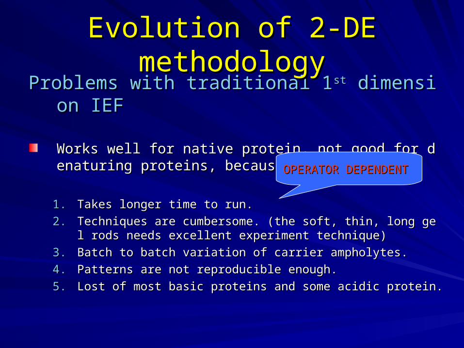
Problems with traditional 1Problems with traditional 1stst dimension IEF dimension IEF
Works well for native protein, not good for denaturing pWorks well for native protein, not good for denaturing proteins, because:roteins, because:
1.1. Takes longer time to run.Takes longer time to run.
2.2. Techniques are cumbersome. (the soft, thin, long gel rods neTechniques are cumbersome. (the soft, thin, long gel rods needs excellent experiment technique)eds excellent experiment technique)
3.3. Batch to batch variation of carrier ampholytes.Batch to batch variation of carrier ampholytes.
4.4. Patterns are not reproducible enough.Patterns are not reproducible enough.
5.5. Lost of most basic proteins and some acidic protein.Lost of most basic proteins and some acidic protein.
Evolution of 2-DE methodologyEvolution of 2-DE methodology
OPERATOR DEPENDENTOPERATOR DEPENDENT

Resolution for IEF: Immobilized pH gradients.Resolution for IEF: Immobilized pH gradients.
Developed by Bjellqvist (Developed by Bjellqvist (1982, Biochem. Biophys Methods, vol 6, p3171982, Biochem. Biophys Methods, vol 6, p317))PH gradient are prepared by co-polymerizing acrylamide PH gradient are prepared by co-polymerizing acrylamide monomers with acrylamide derivatives containing carboxylmonomers with acrylamide derivatives containing carboxylic and tertiary amino groups.ic and tertiary amino groups.
1.1. The pH gradient is fixed, not affected by sample composition.The pH gradient is fixed, not affected by sample composition.2.2. Reproducible data are presented.Reproducible data are presented.3.3. Modified by Angelika Gorg by using thin film to support the thin poModified by Angelika Gorg by using thin film to support the thin po
lyacrylamide IEF gel, named Strips. (lyacrylamide IEF gel, named Strips. (1988, Electrophoresis, vol 9, p 5311988, Electrophoresis, vol 9, p 531))
Evolution of 2-DE methodologyEvolution of 2-DE methodology

II. Immobilized pH gradient, IPGII. Immobilized pH gradient, IPG
First developed by Righetti ,(1990). First developed by Righetti ,(1990).
Immobilized pH gradient generated by buffering Immobilized pH gradient generated by buffering acrylamide derivatives (Immobilines)acrylamide derivatives (Immobilines)
Immobilines are weak acid or weak base.Immobilines are weak acid or weak base.
General structureGeneral structure
CH2 CCN N
R = amino or carboxylic groups
H
H
O
CH2 CCN N
H
R
O
Acrylamide

Schematic drawing of a IPGSchematic drawing of a IPG

Run 2-DE, a quick overviewRun 2-DE, a quick overview

Run 2-DE, step by stepRun 2-DE, step by step
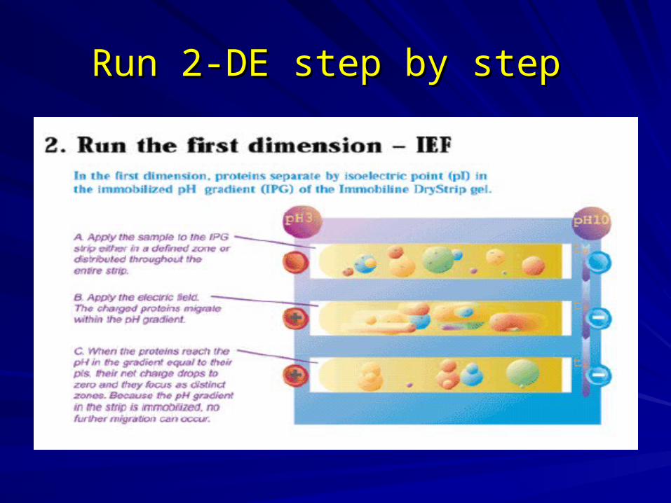
Run 2-DE step by stepRun 2-DE step by step
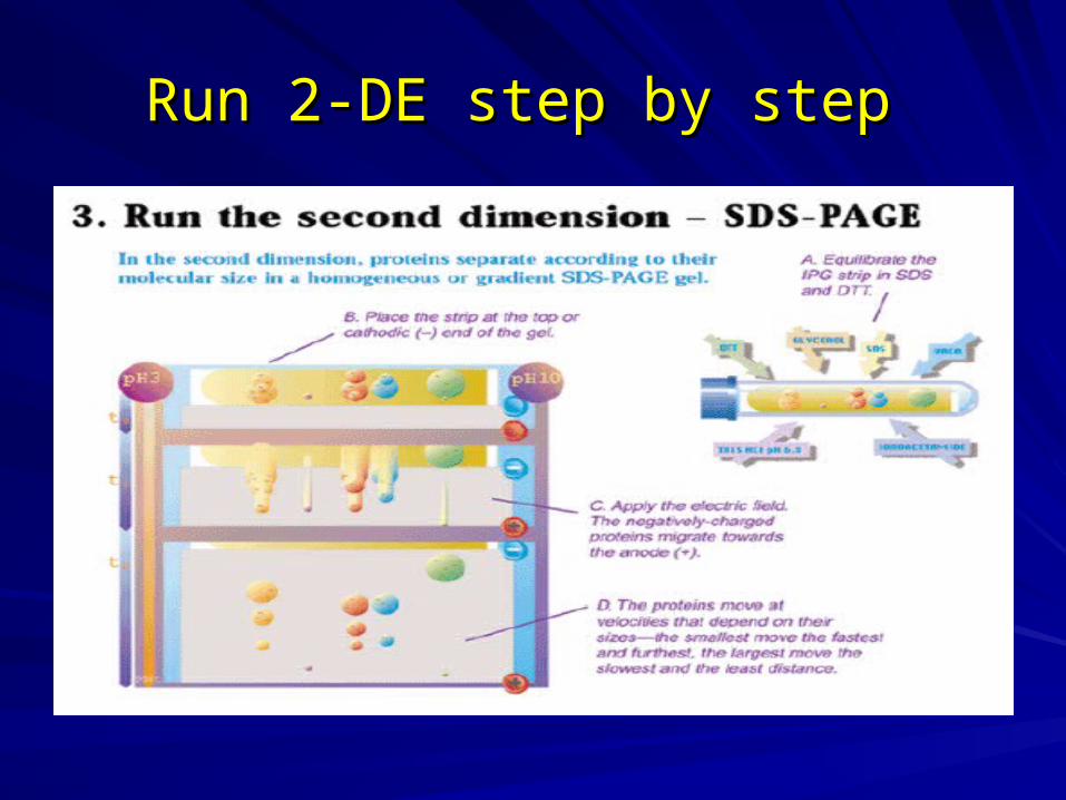
Run 2-DE step by stepRun 2-DE step by step

Run 2-DE step by stepRun 2-DE step by step
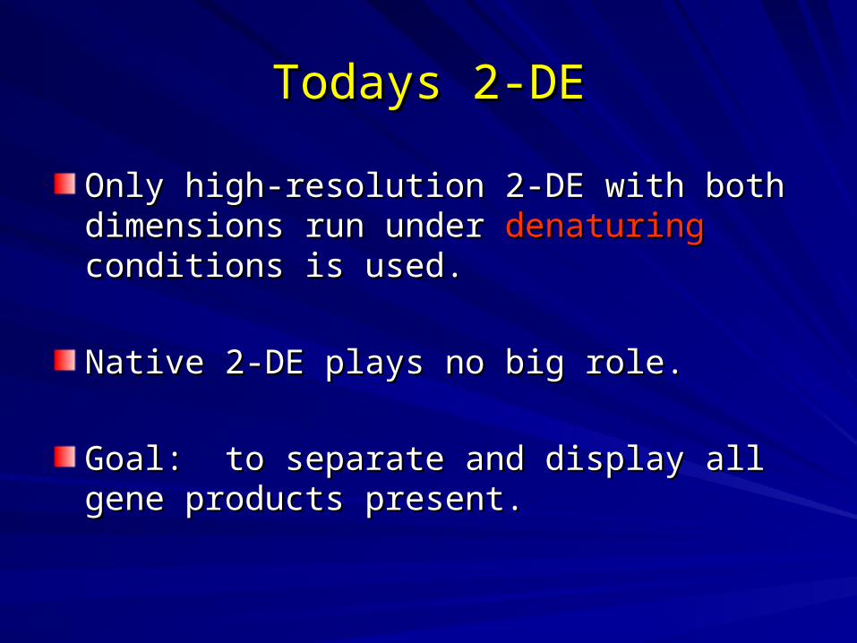
Only high-resolution 2-DE with both dimensions Only high-resolution 2-DE with both dimensions run under run under denaturingdenaturing conditions is used. conditions is used.
Native 2-DE plays no big role.Native 2-DE plays no big role.
Goal: to separate and display all gene products Goal: to separate and display all gene products present.present.
Todays 2-DETodays 2-DE

Challenges for 2-DEChallenges for 2-DE
1. Spot number:1. Spot number:
– 10,000-150,000 gene products in a cell.10,000-150,000 gene products in a cell.
– PTM makes it difficult to predict real number.PTM makes it difficult to predict real number.
– Sensitivity and dynamic range of 2-DE must be adqueSensitivity and dynamic range of 2-DE must be adquete.te.
– It’s imposssible to display all proteins in one singel gelIt’s imposssible to display all proteins in one singel gels.s.

Challenges for 2-DEChallenges for 2-DE
2. Isoelectric point spectrum:2. Isoelectric point spectrum:
– pI of proteins: range from pH 3-13. (by in vitro translatpI of proteins: range from pH 3-13. (by in vitro translated ORF)ed ORF)
– PTM would not alter the pI outside this range.PTM would not alter the pI outside this range.
– pH gradient from 3-13 dose not exist.pH gradient from 3-13 dose not exist.
– For proteins which pI > 11.5, they need to be handed For proteins which pI > 11.5, they need to be handed separately. separately.

Challenges for 2-DEChallenges for 2-DE
3. molecular weights:3. molecular weights:
– Small proteins or peptides can be analysed by modifyiSmall proteins or peptides can be analysed by modifying the gel and buffer condition of SDS-PAGE.ng the gel and buffer condition of SDS-PAGE.
– Protein > 250 kDa do not enter 2Protein > 250 kDa do not enter 2ndnd SDS-PAGE properl SDS-PAGE properly.y.
– 1-DE (SDS-PAGE) can be run in a lane at the side of 1-DE (SDS-PAGE) can be run in a lane at the side of 2-DE. 2-DE.

Challenges for 2-DEChallenges for 2-DE
4. hydrophobic proteins:4. hydrophobic proteins:
–Some very hydrophobic proteins do not go in Some very hydrophobic proteins do not go in solution.solution.
–Some hydrophobic proteins are lost during Some hydrophobic proteins are lost during sample preparation and IEF.sample preparation and IEF.
–More chemical developments are required. More chemical developments are required.

Challenges for 2-DEChallenges for 2-DE
5. Sensitivity of detection:5. Sensitivity of detection:
– Low copy number proteins are very difficult Low copy number proteins are very difficult to detect, even employing most sensitive to detect, even employing most sensitive staining methods.staining methods.
– Sensitivity of staining methods:Sensitivity of staining methods:1.1. Silver stainingSilver staining2.2. Fluorescent stainingFluorescent staining3.3. Dye binding staining (CBR)Dye binding staining (CBR)

Challenges for 2-DEChallenges for 2-DE
6. Loading capacity:6. Loading capacity:
– For detection of low abundant proteins, For detection of low abundant proteins, more sample needs to be loaded.more sample needs to be loaded.
– A wide dynamic range of the SDS-PAGE is A wide dynamic range of the SDS-PAGE is required to prevent merging of highly required to prevent merging of highly abundant protein.abundant protein.
– Loading capacity: IEF > SDS-PAGE.Loading capacity: IEF > SDS-PAGE.
Challenges for 2-DEChallenges for 2-DE
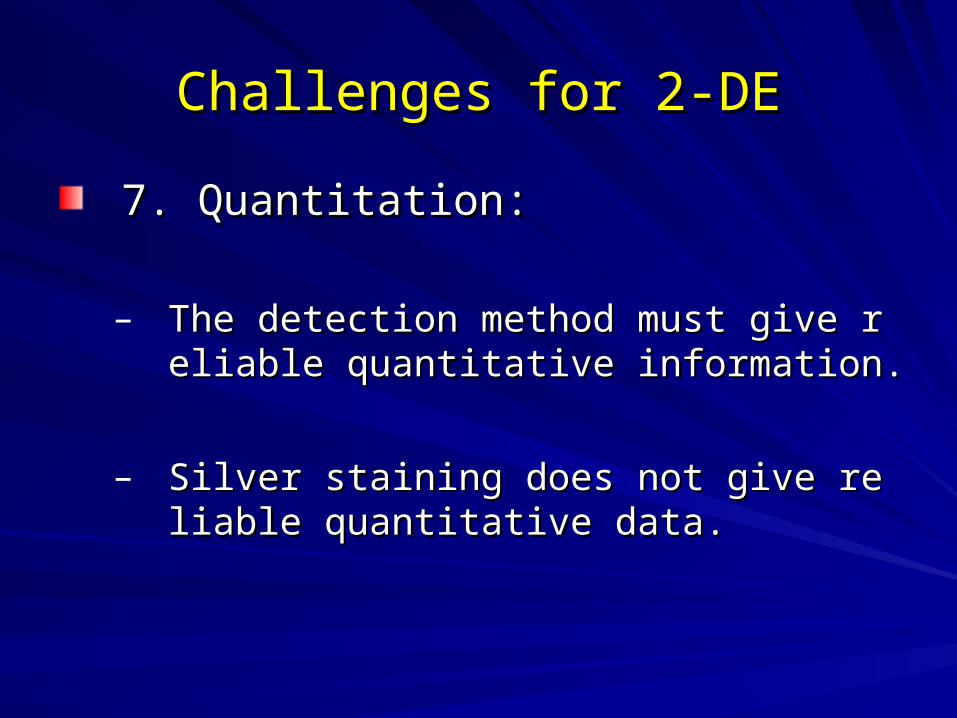
7. Quantitation:7. Quantitation:
– The detection method must give reliable quThe detection method must give reliable quantitative information.antitative information.
– Silver staining does not give reliable quantitSilver staining does not give reliable quantitative data.ative data.

Challenges for 2-DEChallenges for 2-DE
8. Reproducibility:8. Reproducibility:
– Highest importance in 2-DE experiment.Highest importance in 2-DE experiment.
– Immobilized pH gradient strip have Immobilized pH gradient strip have improved a lot for 1improved a lot for 1stst dimension consistency dimension consistency
– Variation most comes from sample Variation most comes from sample preparation.preparation.

A good-looking spot pattern – A good-looking spot pattern – streak and smear free – is not a streak and smear free – is not a
guarantee for best 2-DE protocol.guarantee for best 2-DE protocol.

Sample preparationSample preparation

Some important concepts for Some important concepts for sample preparationsample preparation
1.1. A good sample preparation is the key to good rA good sample preparation is the key to good result.esult.
2.2. The protein composition of the cell lysate or tissThe protein composition of the cell lysate or tissue must be reflected in the patterns of 2-DE.ue must be reflected in the patterns of 2-DE.
3.3. Avoid protein contamination from environment.Avoid protein contamination from environment.4.4. Co-analytical modification (CAM) must be avoidCo-analytical modification (CAM) must be avoid
ed. (pre-purification sometimes leads to CAM)ed. (pre-purification sometimes leads to CAM)5.5. Highly selective procedure for tissue analysis Highly selective procedure for tissue analysis
(Laser capture micro dissection, LCM)(Laser capture micro dissection, LCM)

Some important concepts for Some important concepts for sample preparationsample preparation
6.6. Treatment of sample must be kept to a minimuTreatment of sample must be kept to a minimum to avoid sample loss.m to avoid sample loss.
7.7. Keep sample as cold as possible.Keep sample as cold as possible.
8.8. Shorten processing time as short as possible.Shorten processing time as short as possible.
9.9. Removal of saltsRemoval of salts
10.10. Minimized the unwanted processing, eg proteoMinimized the unwanted processing, eg proteolytic degradation, chemical modification. lytic degradation, chemical modification.

Frequently applied treatmentsFrequently applied treatments
1.1. Cell washingCell washing
2.2. Cell disruptionCell disruption
3.3. Removal of contaminantRemoval of contaminant
4.4. MicrodialysisMicrodialysis
5.5. Electrophretic desaltingElectrophretic desalting
6.6. Precipitation methodsPrecipitation methods
7.7. For very hydrophobic proteinFor very hydrophobic protein

1. Cell washing1. Cell washing
To remove contaminant material.To remove contaminant material.Frequent used bufferFrequent used buffer
– PBS: phosphate buffer saline, sodium chloride, 145 PBS: phosphate buffer saline, sodium chloride, 145 mM (0.85%) in phosphate buffer, 150 mM pH7.2mM (0.85%) in phosphate buffer, 150 mM pH7.2
– Tris buffer sucrose (10mM Tris, 250 mM sucrose, pTris buffer sucrose (10mM Tris, 250 mM sucrose, pH 7,2)H 7,2)
Enough osmoticum to avoid cell lysis Enough osmoticum to avoid cell lysis

2. Cell disruption2. Cell disruption
– Gentle lysis method Gentle lysis method 1.1. Osmotic lysis (cultured cells)Osmotic lysis (cultured cells)
– Suspend cells in hypoosmotic solution.Suspend cells in hypoosmotic solution.
2.2. Repeated freezing and thawing (bacteria)Repeated freezing and thawing (bacteria)– Freeze using liquid nitrogenFreeze using liquid nitrogen
3.3. Detergent lysis (yeast and fungi)Detergent lysis (yeast and fungi)– Lysis buffer (containing urea and detergent)Lysis buffer (containing urea and detergent)– SDS (have to be removed before IEF)SDS (have to be removed before IEF)
4.4. Enzymatic lysis (plant, bacteria, fungi)Enzymatic lysis (plant, bacteria, fungi)– Lysomzyme (bacteria)Lysomzyme (bacteria)– Cellulose and pectinase (plant)Cellulose and pectinase (plant)– Lyticase (yeast)Lyticase (yeast)

2. Cell disruption (continued)2. Cell disruption (continued)
– Vigorous lysis method Vigorous lysis method 1.1. Sonication probe (cell suspension)Sonication probe (cell suspension)
– Avoid overheat, cool on ice between burst.Avoid overheat, cool on ice between burst.
2.2. French pressure (microorganism with cell wall)French pressure (microorganism with cell wall)– Cells are lysed by shear force.Cells are lysed by shear force.
3.3. Mortar and pestle (solid tissue, microorganism)Mortar and pestle (solid tissue, microorganism)– Grind solid tissue to fine powder with liquid nitrogen.Grind solid tissue to fine powder with liquid nitrogen.
4.4. Sample grinding kit (for small amount of sample)Sample grinding kit (for small amount of sample)– For precious sample.For precious sample.
5.5. Glass bead (cell suspension, microorganism)Glass bead (cell suspension, microorganism)– Using abrasive vortexed bead to break cell walls.Using abrasive vortexed bead to break cell walls.

2. Cell disruption (continued)2. Cell disruption (continued)
– Key variable for successful extraction from cruKey variable for successful extraction from crude materialde material
1.1. The method of cell lysisThe method of cell lysis
2.2. The control of pHThe control of pH
3.3. The control of temperatureThe control of temperature
4.4. Avoidance of proteolytic degradationAvoidance of proteolytic degradation

3. Removal of contaminants3. Removal of contaminants
– Major type of contaminants:Major type of contaminants:
1.1. DNA/RNADNA/RNA
2.2. LipidsLipids
3.3. polysaccharidespolysaccharides
4.4. Solid materialSolid material
5.5. SaltSalt

DNA/RNA contaminantDNA/RNA contaminant
– DNA/RNA can be stained by silver staining.DNA/RNA can be stained by silver staining.– They cause horizontal streaking at the acidic part of They cause horizontal streaking at the acidic part of
the gel.the gel.– They precipitate with the proteins when sample appThey precipitate with the proteins when sample app
lying at basic end of IEF gellying at basic end of IEF gel– How to remove:How to remove:
1. precipitation of proteins1. precipitation of proteins
2. DNase/RNase treatment2. DNase/RNase treatment
3. sonication (mechanical breakage)3. sonication (mechanical breakage)
4. DNA/RNA extraction method (phenol/chroloform)4. DNA/RNA extraction method (phenol/chroloform)

Removal of other contaminantsRemoval of other contaminants
– Removal of lipids:Removal of lipids:>2% detergent>2% detergentPrecipitationPrecipitation
– Removal of polysaccharides:Removal of polysaccharides:Enzymatic procedureEnzymatic procedurePrecipitationPrecipitation
– Removal of solid materialRemoval of solid materialCentrifugationCentrifugation
– Removal of saltsRemoval of saltsMicrodialysisMicrodialysisPrecipitationPrecipitation

4. Microdialysis4. Microdialysis
– Specially design for small Specially design for small volume samplesvolume samples
– Membrane cut-off is about Membrane cut-off is about 8000 Da8000 Da
– Drawbacks:Drawbacks:1. Time consuming (som1. Time consuming (some protease might be active protease might be active and digest proteins durie and digest proteins during the dialysis)ng the dialysis)
2. Some proteins precipit2. Some proteins precipitation after dialsis.ation after dialsis.

5. Electrophoretic desalting5. Electrophoretic desalting
– There are some case where the sample muThere are some case where the sample must not be dialysed. (halobacteria lysate)st not be dialysed. (halobacteria lysate)
– Some proteins will gel if desalted. (Bovine viSome proteins will gel if desalted. (Bovine vitreous proteins)treous proteins)
Solution for above: low voltage (100V) for 5 houSolution for above: low voltage (100V) for 5 hours before IEF running. rs before IEF running. (A. Gorg, 1995)(A. Gorg, 1995)

6. Precipitation methods.6. Precipitation methods.
– The reasons for applying protein The reasons for applying protein precipitation procedure:precipitation procedure:
1.1. Concentrate low concentrated protein Concentrate low concentrated protein samples.samples.
2.2. Removal of several disturbing material at the Removal of several disturbing material at the same time.same time.
3.3. Inhibition of protease activity.Inhibition of protease activity.
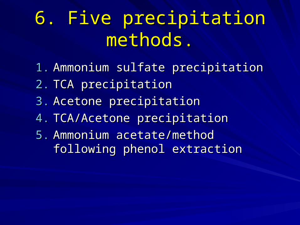
6. Five precipitation methods.6. Five precipitation methods.
1.1. Ammonium sulfate precipitationAmmonium sulfate precipitation
2.2. TCA precipitationTCA precipitation
3.3. Acetone precipitationAcetone precipitation
4.4. TCA/Acetone precipitationTCA/Acetone precipitation
5.5. Ammonium acetate/method following Ammonium acetate/method following phenol extractionphenol extraction

Ammonium sulfate precipitationAmmonium sulfate precipitation
– Proteins tend to aggregate in high concentrProteins tend to aggregate in high concentration of salt (salting out)ation of salt (salting out)
Add Ammonium sulfate slowly into solution anAdd Ammonium sulfate slowly into solution and stir for 10-30 minsd stir for 10-30 mins
Harvest protein by centrifugation.Harvest protein by centrifugation.
– LimitationLimitationSome proteins are soluble at high salt conc.Some proteins are soluble at high salt conc.
Ammonium sulfate seriously affect IEF.Ammonium sulfate seriously affect IEF.

TCA precipitationTCA precipitation
– Trichloroacetic acid is a very affective proteiTrichloroacetic acid is a very affective protein precipitant.n precipitant.
Add TCA to extract to final conc.10-20%.Add TCA to extract to final conc.10-20%.Add 10-20% TCA directly to tissue or cells.Add 10-20% TCA directly to tissue or cells.Harvest protein by centrifugation.Harvest protein by centrifugation.Wash access TCA by ethanol or acetone.Wash access TCA by ethanol or acetone.
– LimitationLimitationSometimes the pellet is hard to redissolve.Sometimes the pellet is hard to redissolve.TCA must remove complete. (affecting IEF)TCA must remove complete. (affecting IEF)Some degradation or modification of protein oSome degradation or modification of protein occursccurs

Acetone precipitationAcetone precipitation
– The most common organic solvent used to precipitaThe most common organic solvent used to precipitated proteins, lipid and detergent remain in solution.ted proteins, lipid and detergent remain in solution.
Add at least 3 vol. of ice-cold acetone into extract.Add at least 3 vol. of ice-cold acetone into extract.
Stand on ice for at least 2 hours.Stand on ice for at least 2 hours.
Harvest protein by centrifugation.Harvest protein by centrifugation.
Remove access acetone by air drying.Remove access acetone by air drying.
– LimitationLimitationSometimes the pellet is hard to redissolve.Sometimes the pellet is hard to redissolve.
Some proteins would not precipitate.Some proteins would not precipitate.
DNA/RNA and glycan also precipitate.DNA/RNA and glycan also precipitate.

Example, Acetone precipitationExample, Acetone precipitation
With Acetone precipitation Crude extract by lysis buffer

TCA/acetone precipitationTCA/acetone precipitation
– The method is more active than TCA or acetone aloThe method is more active than TCA or acetone alone. ne. Most commonly used in 2-DE.Most commonly used in 2-DE.
Suspension samples in 10% TCA/Acetone with 0.07% Suspension samples in 10% TCA/Acetone with 0.07% 2-mercaptoethanol or 20mM DTT.2-mercaptoethanol or 20mM DTT.Stand on -20C for at least 45mins.Stand on -20C for at least 45mins.Harvest protein by centrifugation.Harvest protein by centrifugation.Wash the pellet by acetone with0.07% 2-mercaptoethaWash the pellet by acetone with0.07% 2-mercaptoethanol or 20mM DTT.nol or 20mM DTT.Remove access acetone by air dry.Remove access acetone by air dry.
– LimitationLimitationSometimes the pellet is hard to redissolve.Sometimes the pellet is hard to redissolve.TCA must remove complete. (affecting IEF)TCA must remove complete. (affecting IEF)Some degradation or modification of protein occursSome degradation or modification of protein occurs

Precipitation with ammonium Precipitation with ammonium acetate in methanol following acetate in methanol following
phenol extractionphenol extraction– The method is more suitable for plant sample with The method is more suitable for plant sample with
high level of interfering substancehigh level of interfering substanceProteins are extracted into buffer saturated phenol.Proteins are extracted into buffer saturated phenol.
Precipitated by ammonium acetate/methanol.Precipitated by ammonium acetate/methanol.
Harvest protein by centrifugation.Harvest protein by centrifugation.
Wash with ammonium acetate/methanol followed by Wash with ammonium acetate/methanol followed by acetone.acetone.
– LimitationLimitationComplicated.Complicated.
Time consuming.Time consuming.

C D
A B
823 spots 757 spots
969 spots 899 spots
Acetone Isopropanol
TCA TCA/Acetone

7. For very hydrophobic proteins7. For very hydrophobic proteins
Membrane proteins do not easily go into solutioMembrane proteins do not easily go into solution. A lot of optimization work is required. n. A lot of optimization work is required.
1.1. Thiourea procedureThiourea procedure
2.2. SDS procedureSDS procedure
3.3. New zwitterionic detergent and sulfobetainsNew zwitterionic detergent and sulfobetains

Thiourea procedureThiourea procedure
7M urea + 2M thiourea (Rabilloud, 1998)7M urea + 2M thiourea (Rabilloud, 1998)
Pros: Increase spot number considerably.Pros: Increase spot number considerably.
Cons: Causing artifact spots.Cons: Causing artifact spots.
Causing vertical streaking at acidiCausing vertical streaking at acidic area.c area.

Example, thiourea procedureExample, thiourea procedure
Lysis buffer, 8M urea Lysis buffer, 7M urea+ 2M thiourea

SDS procedureSDS procedure
– For emergency case.For emergency case.– Up to 2% SDS can be used.Up to 2% SDS can be used.– Have to dilute SDS samples at least 20 fold Have to dilute SDS samples at least 20 fold
with urea an a non or zwitterionic detergent with urea an a non or zwitterionic detergent containing solutions.containing solutions.
– The major reasons for using SDS:The major reasons for using SDS:1.1. Formation of oligomers can be preventedFormation of oligomers can be prevented2.2. Dissolved tough cell walls samples (with boilinDissolved tough cell walls samples (with boilin
g)g)3.3. Dissolved very hydrophobic proteinsDissolved very hydrophobic proteins
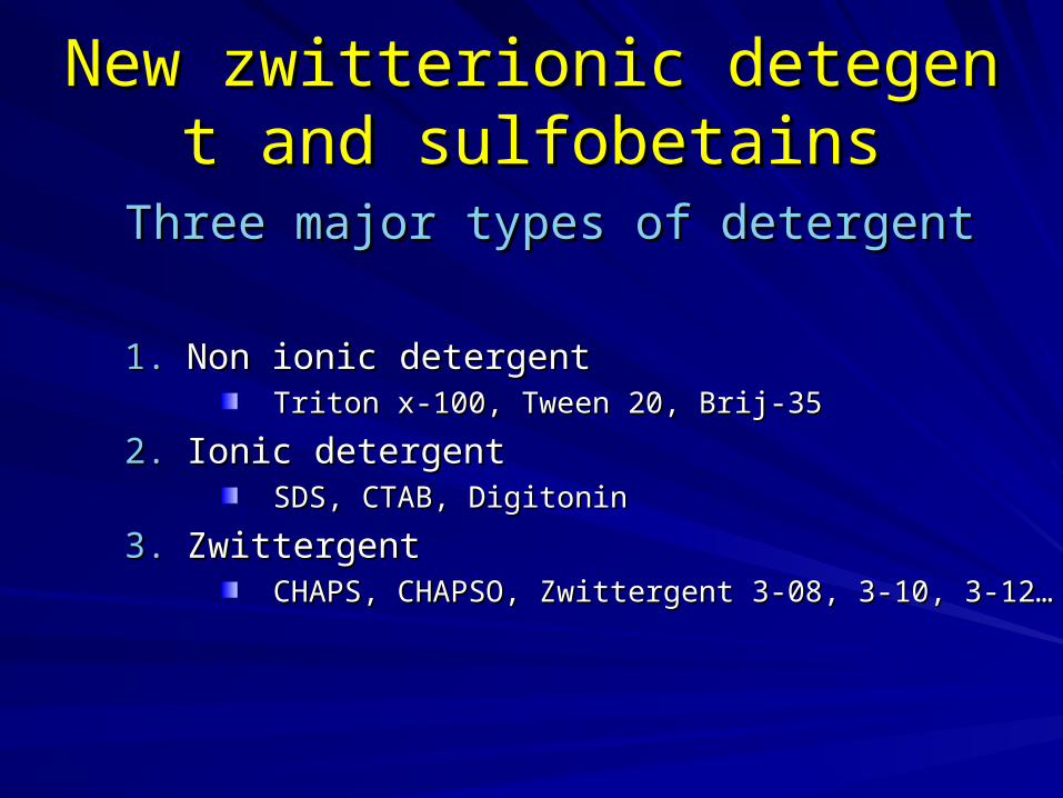
New zwitterionic detegent and sNew zwitterionic detegent and sulfobetainsulfobetains
Three major types of detergentThree major types of detergent
1.1. Non ionic detergentNon ionic detergentTriton x-100, Tween 20, Brij-35Triton x-100, Tween 20, Brij-35
2.2. Ionic detergentIonic detergentSDS, CTAB, DigitoninSDS, CTAB, Digitonin
3.3. ZwittergentZwittergentCHAPS, CHAPSO, Zwittergent 3-08, 3-10, 3-12… CHAPS, CHAPSO, Zwittergent 3-08, 3-10, 3-12…

Now, we are ready to dissolve protein sNow, we are ready to dissolve protein samples in IEF lysis bufferamples in IEF lysis buffer
What is the composition in IEF lysis buffer?

2-DE are in denaturing condition2-DE are in denaturing condition
Three components must present in 2-DE denaturing conditiThree components must present in 2-DE denaturing condition (namely, in IEF lysis buffer)on (namely, in IEF lysis buffer)
1.1. Urea (often > 7M)Urea (often > 7M)
2.2. Reductant (DTT used most widely)Reductant (DTT used most widely)
3.3. Non-ionic or zwitterionic detergent Non-ionic or zwitterionic detergent
4.4. DyeDye

why not using native conditionwhy not using native condition
1.1. Under native condition, a great part of proteins Under native condition, a great part of proteins
exists in several conformations. This leads to exists in several conformations. This leads to more complex 2-DE patterns.more complex 2-DE patterns.
2.2. Native protein complexes sometimes too big to Native protein complexes sometimes too big to enter the gel.enter the gel.
3.3. Reduction of protein-protein interactions.Reduction of protein-protein interactions.
4.4. For match the theoretical pI and MW, all proteiFor match the theoretical pI and MW, all proteins should not have 3D structure or quanternarns should not have 3D structure or quanternary structure. y structure.

Composition of standard lysis buffer (foComposition of standard lysis buffer (for IEF)r IEF)
1.1. 9M urea9M urea
2.2. 4% CHAPS4% CHAPS
3.3. 1% DTT1% DTT
4.4. 0.8% carrier ampholyte0.8% carrier ampholyte
5.5. 0.02% bromophenol blue.0.02% bromophenol blue.
1
2
3
5

Functions of denaturant (Urea)Functions of denaturant (Urea)
1.1. To convert proteins into single confTo convert proteins into single conformation by canceling 2ormation by canceling 2ndnd and 3 and 3rdrd st structure.ructure.
2.2. To keep hydrophobic proteins into To keep hydrophobic proteins into solution.solution.
3.3. To avoid protein-protein interaction.To avoid protein-protein interaction.
4.4. Thio urea: for very hydrophobic proThio urea: for very hydrophobic proteins only.teins only.
Thiourea

Beware when using ureaBeware when using urea
1.1. The purity of urea is very criticalThe purity of urea is very critical
2.2. Isocyanate impurities and heating will cause carIsocyanate impurities and heating will cause carbamylation of the proteins.bamylation of the proteins.
3.3. It does not seem to make a difference what graIt does not seem to make a difference what grade of urea is used because, urea + heat + protede of urea is used because, urea + heat + protein = carbamylation. in = carbamylation.

Carbamylation of proteinsCarbamylation of proteins
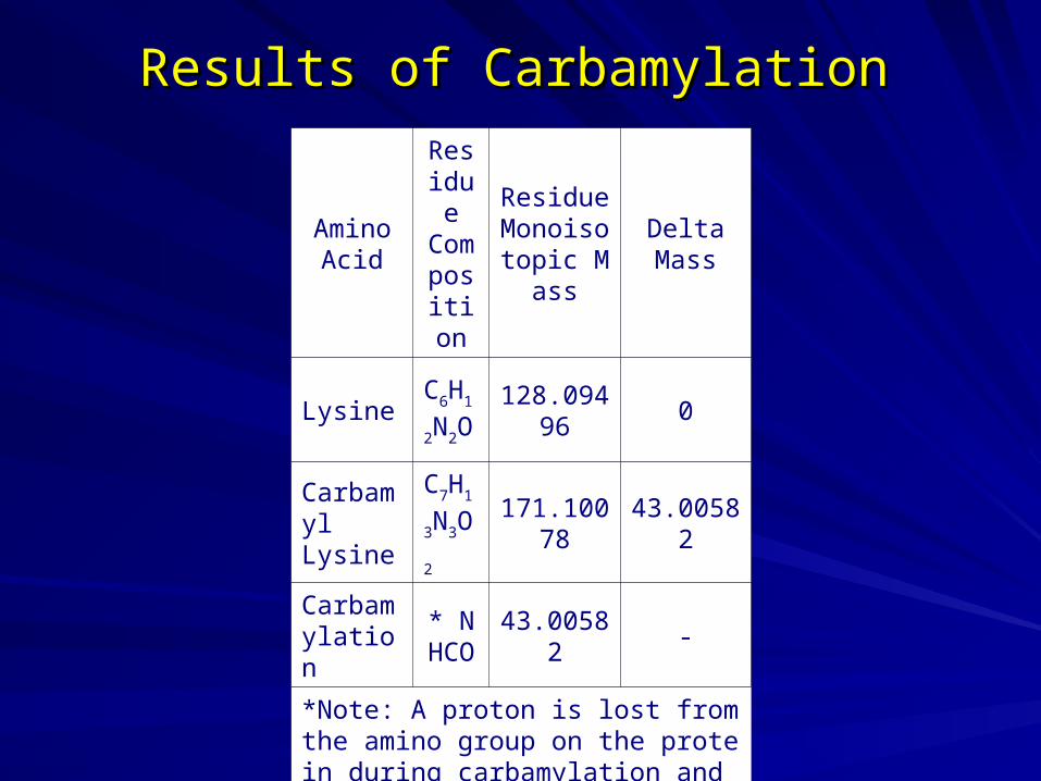
Results of CarbamylationResults of Carbamylation
Amino Acid
Residue Composition
ResidueMonoisotopic Mas
s
Delta Mass
Lysine
C6H1
2N2
O
128.09496
0
CarbamylLysine
C7H1
3N3
O2
171.10078
43.00582
Carbamylation
* NHCO
43.00582 -
*Note: A proton is lost from the amino group on the protein during carbamylation and thus the change in composition is NHCO.

Functions of detergent (CHAPS)Functions of detergent (CHAPS)
To Combine all the advantages of polar, sulfobetaine-coTo Combine all the advantages of polar, sulfobetaine-containing detergents and hydrophobic, bile salt, anionic dentaining detergents and hydrophobic, bile salt, anionic detergents into a single molecule with superior membrane tergents into a single molecule with superior membrane protein solubilization propertiesprotein solubilization properties
Non-denaturingNon-denaturing
Able to disrupt nonspecific protein interactionsAble to disrupt nonspecific protein interactions
Less protein aggregation than non-ionic detergentsLess protein aggregation than non-ionic detergents
Electrically neutralElectrically neutral
Easily removed by dialysisEasily removed by dialysis
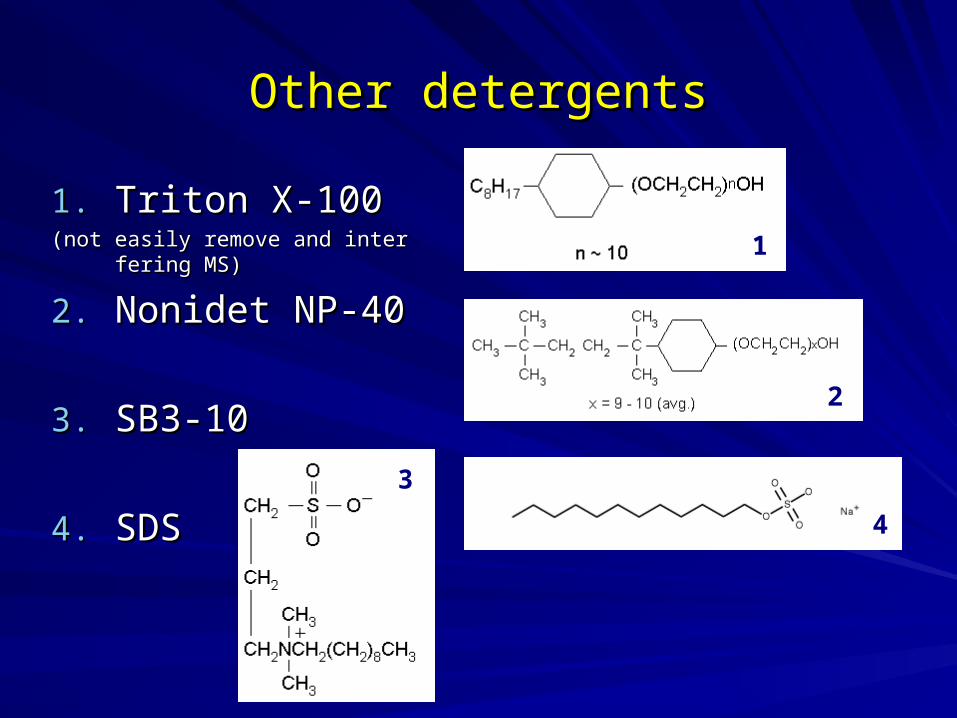
Other detergentsOther detergents
1.1. Triton X-100Triton X-100(not easily remove and interfering M(not easily remove and interfering M
S)S)
2.2. Nonidet NP-40Nonidet NP-40
3.3. SB3-10SB3-10
4.4. SDSSDS
1
2
4
3

Functions of reductant Functions of reductant
To prevent different oxidation steps of proteins.To prevent different oxidation steps of proteins.
2-mercaptoethanol 2-mercaptoethanol should not be used because its buffershould not be used because its buffering effect above pH 8.ing effect above pH 8.
Keratin contamination might from 2-mercaptoethanol.Keratin contamination might from 2-mercaptoethanol.
DTT (dithiothreitol) or DTE (dithioerythritol) DTT (dithiothreitol) or DTE (dithioerythritol) are used widare used widely.ely.
DTT and DTE ionized above pH8. They move toward anDTT and DTE ionized above pH8. They move toward anode during IEF in basic pH gradient.ode during IEF in basic pH gradient.
It leads to horizonal streaking at basic area.It leads to horizonal streaking at basic area.

Other reduction methods Other reduction methods
TBP (tributylphosphine): very unstablTBP (tributylphosphine): very unstable.e.
An alternative way to adequate and rAn alternative way to adequate and reproducible 2-DE patterns in basic areproducible 2-DE patterns in basic area:ea:
1.1. Addition of higher amount of DTT to the Addition of higher amount of DTT to the gelgel
2.2. Addition of more DTT to a cathodal papeAddition of more DTT to a cathodal paper strip.r strip.

Function of carrier ampholyte Function of carrier ampholyte
They do not disturb IEF like buffer addition, because they beThey do not disturb IEF like buffer addition, because they become uncharged when migrating to their pI.come uncharged when migrating to their pI.
1.1. To generate pH gradientsTo generate pH gradients
2.2. To substituting ionic bufferTo substituting ionic buffer
3.3. To improve the solubility of proteinTo improve the solubility of protein
4.4. Dedicated pH intervals, prepared for the addition to immDedicated pH intervals, prepared for the addition to immobilized pH gradients, are called obilized pH gradients, are called IPG buffer.IPG buffer.

Function of dyes Function of dyes
To visualize the sample solutionTo visualize the sample solution
To monitor the 2-DE running condition.To monitor the 2-DE running condition.
Bromophenol blue is interchangeable with Orange G.Bromophenol blue is interchangeable with Orange G.
++ --

Other considerations Other considerations
Protease inhibitorsProtease inhibitors
– 1. Some proteases are also active in presence of urea and deter1. Some proteases are also active in presence of urea and detergent.gent.
– 2. PMSF is frequently used (8mM), toxic and short half-life.2. PMSF is frequently used (8mM), toxic and short half-life.– 3. Pefabloc (AEBSF) can also be used but modified proteins.3. Pefabloc (AEBSF) can also be used but modified proteins.– 4. NO complete insurance against protease activity4. NO complete insurance against protease activity– 5. Boiling sample in SDS buffer for a few seconds can inactive p5. Boiling sample in SDS buffer for a few seconds can inactive p
rotease. rotease. – 6. Precipitate proteins with TCA/acetone at -20C might inactivati6. Precipitate proteins with TCA/acetone at -20C might inactivati
on protease activity.on protease activity.
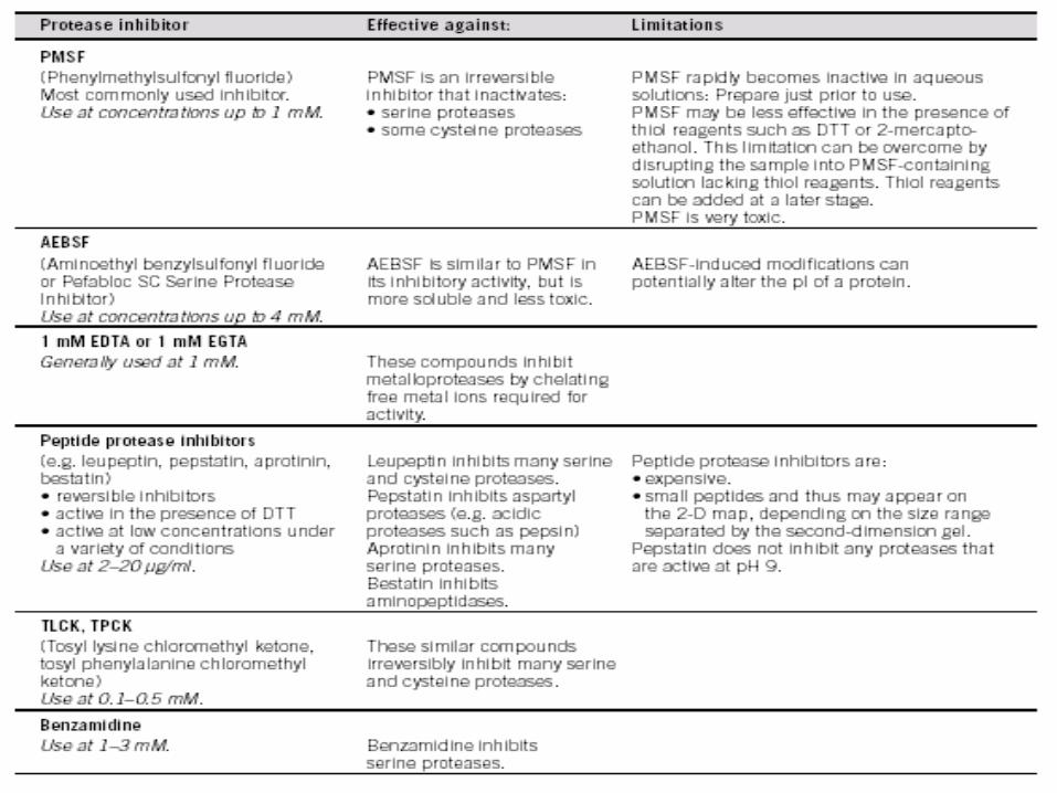
Types of protease inhibitor Types of protease inhibitor

Other considerations Other considerations
Alkaline conditionAlkaline conditionTris base (40mM) or spermidine (25mM) sometimes add to lysis buffer to Tris base (40mM) or spermidine (25mM) sometimes add to lysis buffer to
maximize protein extraction. maximize protein extraction.
– Pros:Pros:
1.1. They can also precipitate DNA/RNA. They can also precipitate DNA/RNA.
2.2. They keep proteasse activity low.They keep proteasse activity low.
– Cons:Cons:
1.1. Precipitation of basic protein.Precipitation of basic protein.
2.2. Ionic contamination is to high.Ionic contamination is to high.

Measure the protein conc. in your samples. Measure the protein conc. in your samples.
– Widely used protein assay methodsWidely used protein assay methods
1.1. BiuretBiuret
2.2. Lowry methods. Lowry methods.
3.3. Bradford methods.Bradford methods.
4.4. UV methods.UV methods.
5.5. Special methods Special methods
6.6. Other commercial methods.Other commercial methods.1.1. BCA assay (bicinchoninic acid assay, Pierce)BCA assay (bicinchoninic acid assay, Pierce)
2.2. DC protein assay (detergent compatible, Bio-rad)DC protein assay (detergent compatible, Bio-rad)
3.3. DC/RC protein assay (detergent/reducing agent compatible, Bio-raDC/RC protein assay (detergent/reducing agent compatible, Bio-rad)d)
Before runninng IEF, you should…Before runninng IEF, you should…

– Principle: Principle: The reactivity of the peptide bonds with the copper [II] The reactivity of the peptide bonds with the copper [II] ions under alkaline conditions to form purple biuret complex. ions under alkaline conditions to form purple biuret complex.
– Interfering substance:Interfering substance: Ammonium sulfate, Tris, etc. Ammonium sulfate, Tris, etc.
– Sensitivity:Sensitivity: >mg >mg
1. Biuret method1. Biuret method
A white, crystalline, nitrogenous substance, C2O2N3H5, formed by heating urea. It is intermediate between urea and cyanuric acid.

– Principle: Principle: The reactivity of the peptide nitrogen[s] with the coppeThe reactivity of the peptide nitrogen[s] with the copper [II] ions under alkaline conditions and the subsequent reductior [II] ions under alkaline conditions and the subsequent reduction of the Folin-Ciocalteay phosphomolybdicphosphotungstic acid n of the Folin-Ciocalteay phosphomolybdicphosphotungstic acid to heteropolymolybdenum blue by the copper-catalyzed oxidatioto heteropolymolybdenum blue by the copper-catalyzed oxidation of aromatic acids (Try, Try). n of aromatic acids (Try, Try).
– Interfering substance:Interfering substance: amino acid derivatives, certain buffers, dr amino acid derivatives, certain buffers, drugs, lipids, sugars, salts, nucleic acids, ammonium ions, zwitteriugs, lipids, sugars, salts, nucleic acids, ammonium ions, zwitterionic buffers, nonionic buffers and thiol compounds.onic buffers, nonionic buffers and thiol compounds.
– Sensitivity:Sensitivity: > 0.1 mg > 0.1 mg
2. Lowry method2. Lowry method

– Principle: Principle: The assay is based on the observation that the absorbThe assay is based on the observation that the absorbance maximum for an acidic solution of ance maximum for an acidic solution of Coomassie Brilliant Blue Coomassie Brilliant Blue G-250G-250 shifts from 465 nm to 595 nm when binding to protein occ shifts from 465 nm to 595 nm when binding to protein occurs. The Coomassie® dye binds primarily with basic and aromatiurs. The Coomassie® dye binds primarily with basic and aromatic side chains. The interaction with arginine is very strong and lec side chains. The interaction with arginine is very strong and less strong with histidine, lysine, tyrosine, tryptophan, and phenylalss strong with histidine, lysine, tyrosine, tryptophan, and phenylalanine. About 1.5 to 3 molecules of dye bind per positive charge oanine. About 1.5 to 3 molecules of dye bind per positive charge on the protein. n the protein.
– Interfering substance:Interfering substance: amino acid derivatives, certain buffers, dru amino acid derivatives, certain buffers, drugs, lipids, sugars, salts, nucleic acids, ammonium ions, zwitteriongs, lipids, sugars, salts, nucleic acids, ammonium ions, zwitterionic buffers, nonionic buffers and thiol compounds.ic buffers, nonionic buffers and thiol compounds.
– Sensitivity:Sensitivity: >10 -100 ug >10 -100 ug
3. Bradford method3. Bradford method

4. UV methods4. UV methods
– Principle: Principle: The aromatic groups (Phe, Tyr, Trp) and the peptide bThe aromatic groups (Phe, Tyr, Trp) and the peptide bonds have maximum UV absorbance around 280nm and 200nm.onds have maximum UV absorbance around 280nm and 200nm. 280nm was used most frequently. 280nm was used most frequently.
– Interfering substance:Interfering substance: anything containing anything containing
– Sensitivity:Sensitivity: >mg >mg

5. Special methods5. Special methods
– Principle: Principle: Some proteins contain functional groups, eg: Heme in Some proteins contain functional groups, eg: Heme in peroidase, hemoglobin and transferrin can be detected at 403nperoidase, hemoglobin and transferrin can be detected at 403nm, Cd2+ in some phytochelatins.m, Cd2+ in some phytochelatins.
– Interfering substance:Interfering substance: similar functional groups. similar functional groups.
– Sensitivity:Sensitivity: various various

6. Commercial methods6. Commercial methods
A.A. BCA assay (bicinchoninic acid assay, Pierce)BCA assay (bicinchoninic acid assay, Pierce)
B. DC protein assay (detergent compatible, Bio-raB. DC protein assay (detergent compatible, Bio-rad)d)
C. DC/RC protein assay (detergent/reducing agenC. DC/RC protein assay (detergent/reducing agent compatible, Bio-rad)t compatible, Bio-rad)
This process is a two-step reaction.
Protein + Cu2+ + OH- Cu1+
Cu1+ + 2 BCA Cu1+/BCA chromophore (562 nm).

Summary of protein quantitation methodsSummary of protein quantitation methods