Two-dimensional difference gel electrophoresis (DIGE ...
13
CLINICAL PROTEOMICS Two-dimensional difference gel electrophoresis (DIGE) analysis of sera from visceral leishmaniasis patients Rukmangadachar et al. Rukmangadachar et al. Clinical Proteomics 2011, 8:4 http://www.clinicalproteomicsjournal.com/content/8/1/4 (31 May 2011)
Transcript of Two-dimensional difference gel electrophoresis (DIGE ...
CLINICALPROTEOMICS
Two-dimensional difference gel electrophoresis(DIGE) analysis of sera from visceral leishmaniasispatientsRukmangadachar et al.
Rukmangadachar et al. Clinical Proteomics 2011, 8:4http://www.clinicalproteomicsjournal.com/content/8/1/4 (31 May 2011)
RESEARCH Open Access
Two-dimensional difference gel electrophoresis(DIGE) analysis of sera from visceral leishmaniasispatientsLokesh A Rukmangadachar1,2, Jitender Kataria1, Gururao Hariprasad1, Jyotish C Samantaray3 andAlagiri Srinivasan1*
* Correspondence: [email protected] of Biophysics, AllIndia Institute of Medical Sciences,New Delhi, 110029, IndiaFull list of author information isavailable at the end of the article
Abstract
Introduction: Visceral leishmaniasis is a parasitic infection caused by Lesihmaniadonovani complex and transmitted by the bite of the phlebotomine sand fly. It is anendemic disease in many developing countries with more than 90% of the casesoccurring in Bangladesh, India, Nepal, Sudan, Ethiopia and Brazil. The disease is fatal ifuntreated. The disease is conventionally diagnosed by demonstrating the intracellularparasite in bone marrow or splenic aspirates. This study was carried out to discoverdifferentially expressed proteins which could be potential biomarkers.
Methods: Sera from six visceral leishmaniasis patients and six healthy controls weredepleted of high abundant proteins by immunodepletion. The depleted sera werecompared by 2-D Difference in gel electrophoresis (DIGE). Differentially expressedproteins were identified the by tandem mass spectrometry. Three of the identifiedproteins were further validated by western blotting.
Results: This is the first report of serum proteomics study using quantitativeDifference in gel electrophoresis (DIGE) in visceral leishmaniasis. We identified alpha-1-acidglycoprotein and C1 inhibitor as up regulated and transthyretin, retinol bindingprotein and apolipoprotein A-I as down regulated proteins in visceral leishmaniasissera in comparison with healthy controls. Western blot validation of C1 inhibitor,transthyretin and apolipoprotein A-I in a larger cohort (n = 29) confirmed significantdifference in the expression levels (p < 0.05).
Conclusions: In conclusion, DIGE based proteomic analysis showed that severalproteins are differentially expressed in the sera of visceral leishmaniasis. The fiveproteins identified here have potential, either independently or in combination, asprognostic biomarkers.
IntroductionLeishmaniasis is a vector borne infection caused by the obligate intracellular protozoa
belonging to the genus Leishmania. It is endemic disease affecting people in the large
parts of tropical counties and the Mediterranean basin. Clinically, there are mainly
four subtypes, namely cutaneous, muco-cutaneous, visceral (kala azar) and post kala
azar dermal leishmaniasis. Visceral leishmaniasis is caused by the L. donovani group of
organisms and transmitted by the bite of the phlebotomine sand fly. There are an esti-
mated 500,000 new cases every year and more than 90% of these cases occur mainly in
Rukmangadachar et al. Clinical Proteomics 2011, 8:4http://www.clinicalproteomicsjournal.com/content/8/1/4 CLINICAL
PROTEOMICS
© 2011 Rukmangadachar et al; licensee BioMed Central Ltd. This is an Open Access article distributed under the terms of the CreativeCommons Attribution License (http://creativecommons.org/licenses/by/2.0), which permits unrestricted use, distribution, andreproduction in any medium, provided the original work is properly cited.
Bangladesh, India, Nepal, Sudan, Ethiopia and Brazil [1]. The disease has an incubation
period between 2 to 6 months and presents with fever, anaemia and enlargement of
liver, spleen and lymph nodes. It is almost always fatal if untreated within two years of
onset. Diagnosis of the disease is conventionally made by demonstrating the amastigote
form of the organism in spleen or bone marrow [2]. Presence of anti-rK39 antibody is
also used for the serodiagnosis of leishmania species. The test is highly sensitive but
there is a possibility of cross reactivity. Also, the test remains positive even after treat-
ment and may not be useful in immunocompromised patients [3]. The PCR based
approaches are expensive, require technical expertise and need to be cost-effective to
be useful, especially in areas where visceral leishmaniasis is endemic [4].
Studying the serum protein profile during the infection will yield important information
regarding the pathogenesis. This information can be used for identifying biomarkers and
therapeutic targets. Detailed proteomic studies of human body fluids in the recent past
have resulted in identifying potential biomarker candidates for many conditions [5]. In the
past, studies examining the electrophoretic pattern of serum proteins in visceral leishma-
niasis have reported decreased serum albumin and increased serum immunoglobulin levels
which are used as a supporting evidence for diagnosis [6]. Proteomic techniques have also
been used to investigate leishmaniasis in recent studies. Using 2-dimensional western blot
analyses with patient’s sera and parasites isolated from visceral leishmaniasis patients,
immune responses for 330 different leishmania antigens was detected and six antigens
were identified [7]. However, there has been no significant study examining the global pro-
teome profile of the visceral leishmaniasis patients’ sera. Here, we report, for the first time,
the difference-in-gel electrophoresis (DIGE) analysis of sera from visceral leishmaniasis
patients and the mass spectrometric identification of the differentially expressed proteins.
MethodsSample collection
The study population included groups of healthy controls and patients with confirmed
visceral leishmaniasis. Visceral leishmaniasis serum specimens were collected from the
Department of Microbiology, All India Institute of Medical Sciences. Patients were
admitted for evaluation of fever when they had fever over a mean duration of two months.
All subjects were negative for HIV and HBV. All the serum samples were collected before
initiation of the therapy for visceral leishmaniasis. Control serum specimens were from
healthy donors. Written informed consent was obtained from all participants before draw-
ing blood. The study was approved by the ethics committee of All India Institute of Medi-
cal Sciences and procedures followed were in accordance with the ethical standards
formulated in the Helsinki declaration. Serum was separated from 2 ml of blood, aliquoted
into separate eppendorf tubes and stored at -70°C.
Sample processing
Individual samples were treated with Multiple Affinity Spin Cartridge Hu PL 7 kit (Agilent
Technologies, USA) according to the manufacturer’s instruction for removal of seven high
abundant proteins (albumin, IgG, IgA, alpha-1-antitrypsin, haptoglobin, transferrin and
fibrinogen). Depleted serum fraction from affinity chromatography was concentrated to a
final volume of approximately 100 μl by ultra filtration (5 kDa cut-off). The protein con-
centration was estimated by Bradford method using bovine serum albumin as standard.
Rukmangadachar et al. Clinical Proteomics 2011, 8:4http://www.clinicalproteomicsjournal.com/content/8/1/4
Page 2 of 12
2D-DIGE
50 μg of protein from the each of the depleted serum fractions was precipitated with
80% acetone and the precipitate was solubilised in lysis solution (8M urea, 2M thiourea,
4% CHAPS). These samples were labelled with Cy dye flours according to Minimal
labelling protocol provided by the manufacturer (Amersham Biosciences, USA). Three
patient samples were labelled with Cy3 and three other patient samples with Cy5. Simi-
larly three controls were labelled with Cy5 and three controls with Cy3. This resulted in
equal distribution of Cy dyes in both patient and control groups. This dye swapping
strategy was adopted to avoid dye bias. Equal amount of protein from all the twelve
patient and control samples was mixed to generate an internal standard and 50 μg of
protein from this internal standard was labelled with 200 pmol of Cy2. Each gel there-
fore consisted of one patient (Cy3 or Cy5), one control (Cy5 or Cy3) and one internal
standard (Cy2) samples. Labelled serum samples of one patient, one control and one
internal standard were pooled together and rehydrating stock solution (8M urea, 2M
thiourea, 2% CHAPS, 0.002% bromophenol blue) was added to make up the final volume
to 250 μl. DTT and IPG buffer (pH 3-10) were added to a final concentrations of 0.003%
and 0.5% respectively. After 15 h of rehydration, IPG strips (13 cm, pH 3-10) were sub-
jected to iso-electric focusing in an Ettan IPGphor 3 system (Amersham Biosciences,
USA) for a total of 27,000 Volt-hours. Each electro focused strip was equilibrated, first
with 10 ml of SDS equilibration buffer containing 10 mg/ml DTT for 15 minutes. This
was followed by second equilibration with SDS equilibration buffer containing 25 mg/ml
iodoacetamide for 15 minutes. The strips were then transferred onto 10% homogenous
polyacrylamide gels cast on SE 600 Ruby gel apparatus (Amersham Biosciences, USA).
The strips were overlaid with 0.5% agarose sealing solution (0.5% agarose, 0.002% bro-
mophenol blue in Tris-glycine electrode buffer). Separation in SDS-PAGE was carried
out with constant running current set at 15 mA per gel at 20°C for 30 minutes, followed
by 30 mA per gel at 20°C until the bromophenol blue dye front ran off from the bottom
of the gels. Six such gels were run corresponding to six biological replicates.
Image acquisition and analysis
Labelled proteins were visualized using a Typhoon TRIO Variable Mode Imager (Amer-
sham Biosciences, USA). Cy2 images were scanned with 488 nm/520 nm, Cy3 images
were scanned with 532 nm/580 nm and Cy5 images were scanned with 633 nm/670 nm.
All gels were scanned with a PMT setting of 750 to 800 V with 200 μm/pixel resolution.
Images were cropped using Image-Quant™ v 5.5 (Amersham Biosciences, USA) to
remove areas extraneous to the gel image. Gel images were processed using DeCyder™
2D version 7.0 (Amersham Biosciences, USA). The images were imported to Differential
in-Gel Analysis (DIA) workspace to create six different workspaces for each of the six
gel pairs. The maximum number of spots for each co-detection procedure was set to
1500. The spots were co-detected and quantified automatically as 2-D DIGE image
pairs, intrinsically linking the samples to its in-gel standard. These six DIA workspaces
were then analyzed in the Biological Variation Analysis (BVA) workspace. In BVA work
space, each Cy3 or Cy5 gel image was assigned an experimental condition, either control
or visceral leishmaniasis and all Cy2 images were classified as standards. The gel with
the highest spot count was assigned as the master gel. Matching between gels was per-
formed utilizing the in-gel standard from each image pair. Matching was further
Rukmangadachar et al. Clinical Proteomics 2011, 8:4http://www.clinicalproteomicsjournal.com/content/8/1/4
Page 3 of 12

improved by land marking and manually confirming potential spots of interest. Student
t-test was performed for every matched spot-set, comparing the average and standard
deviation of protein abundance for a given spot.
Mass spectrometric analysis and protein identification
A preparative gel was run using 500 μg of pooled protein sample and stained with colloi-
dal coomassie blue. Matched spots of interest were picked manually from the preparative
gel. These spots were subjected to in-gel trypsinization according to the manufacturer’s
protocol (Promega, USA). After overnight digestion, digestion buffer containing the pep-
tides was recovered. Additional extraction of peptides was carried out with 100 μl of 50%
acetonitrile in 1% formic acid. The extracts were poled and vacuum-dried. For LC-MS/
MS, peptide mixtures were resuspended in 50% acetonitrile and 1% formic acid solution
and analyzed in a Tempo™ nano-LC system (Applied Biosystems) coupled to QSTAR XL
system (Applied Biosystems, USA). Some spots were analyzed by off-line nanospray
method. These peptides were dissolved in 20 μl of 50% acetonitrile in 0.1% formic acid.
Nanospray ionization was carried out using an ion spray voltage of 900. The spectra were
acquired in an information dependent manner utilizing the Analyst QS 2.0 software to
generate raw data. Database searching was done using Mascot search program (Version
1.6, Matrix Science, UK). Search parameters were as follows: 1 missed cleavage allowed,
carbamidomethylation set as fixed modification, methionine oxidation as variable modifi-
cation, peptide mass tolerance ± 1.2 Da, fragment mass tolerance: ± 0.6 Da, monoisotopic
mass values. Spectra were searched against NCBInr or MSDB database. Criteria for posi-
tive identification were a significant Mascot probability score (score >40; p < 0.05).
Western Blot analyses
Individual undepleted serum specimens were separated on 12% polyacrylamide gels and
transferred onto nitrocellulose membranes in a trans-blot electrophoresis transfer cell (Bio-
Rad, USA). Western blot analyses were performed by using polyclonal antibodies against
C1 inhibitor (diluted 1:200, Santa Cruz, USA), transthyretin (diluted 1:2500, Abcam, USA)
and apolipoprotein A-I (diluted 1:10000, Abcam, USA). Peroxidase-conjugated antibody
(diluted 1:5000, Abcam, USA) was used as secondary antibody. The reaction was detected
by chemiluminescence with ECL reagents (Pierce Biotechnology, USA). A semi quantitative
analysis based on optical density was performed by ImageJ software (available at http://
www.rsbweb.nih.gov/ij). Student t-test was used to determine mean differences between
two groups and a p < 0.05 was considered significant at a 95% confidence level.
ResultsClinical data
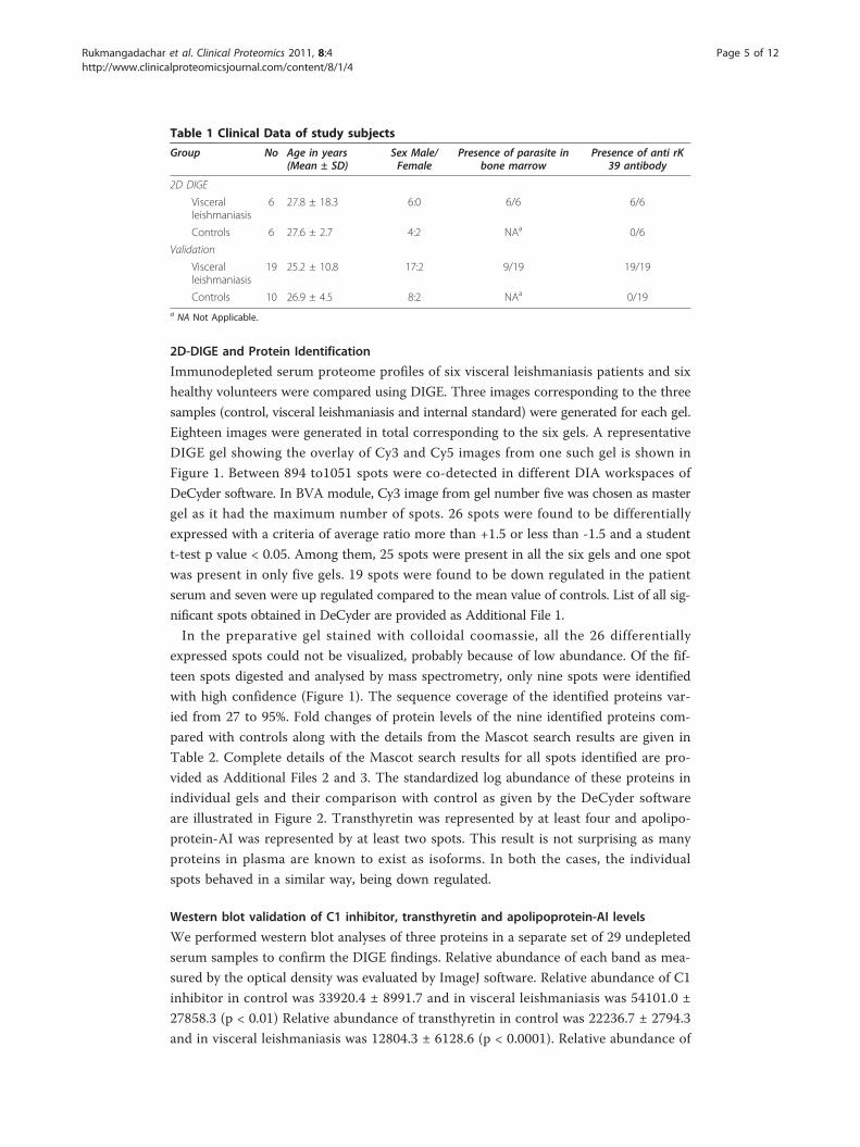
The clinical data from the study subjects are summarised in Table 1. Analysis of the
age distribution between the two groups showed that there was no significant differ-
ence among the patient and controls in both the DIGE study group and the validation
groups (p value > 0.05). Patients in DIGE study group had an established diagnosis of
visceral leishmaniasis as evidenced by the presence of the parasite in the bone marrow
aspirates and presence of anti rK39 antibody. Validation cohort consisted of patients
with clinical diagnosis of visceral leishmaniasis supported by the presence of anti rK-39
antibody.
Rukmangadachar et al. Clinical Proteomics 2011, 8:4http://www.clinicalproteomicsjournal.com/content/8/1/4
Page 4 of 12
2D-DIGE and Protein Identification
Immunodepleted serum proteome profiles of six visceral leishmaniasis patients and six
healthy volunteers were compared using DIGE. Three images corresponding to the three
samples (control, visceral leishmaniasis and internal standard) were generated for each gel.
Eighteen images were generated in total corresponding to the six gels. A representative
DIGE gel showing the overlay of Cy3 and Cy5 images from one such gel is shown in
Figure 1. Between 894 to1051 spots were co-detected in different DIA workspaces of
DeCyder software. In BVA module, Cy3 image from gel number five was chosen as master
gel as it had the maximum number of spots. 26 spots were found to be differentially
expressed with a criteria of average ratio more than +1.5 or less than -1.5 and a student
t-test p value < 0.05. Among them, 25 spots were present in all the six gels and one spot
was present in only five gels. 19 spots were found to be down regulated in the patient
serum and seven were up regulated compared to the mean value of controls. List of all sig-
nificant spots obtained in DeCyder are provided as Additional File 1.
In the preparative gel stained with colloidal coomassie, all the 26 differentially
expressed spots could not be visualized, probably because of low abundance. Of the fif-
teen spots digested and analysed by mass spectrometry, only nine spots were identified
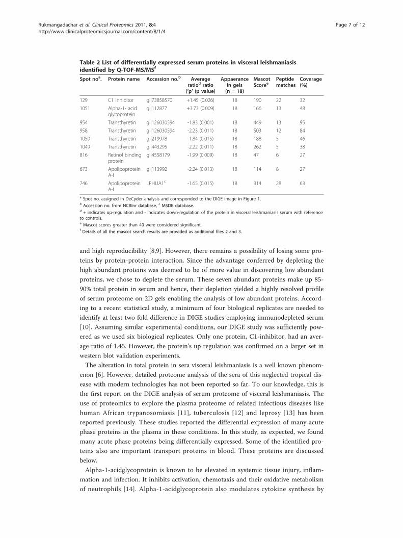
with high confidence (Figure 1). The sequence coverage of the identified proteins var-
ied from 27 to 95%. Fold changes of protein levels of the nine identified proteins com-
pared with controls along with the details from the Mascot search results are given in
Table 2. Complete details of the Mascot search results for all spots identified are pro-
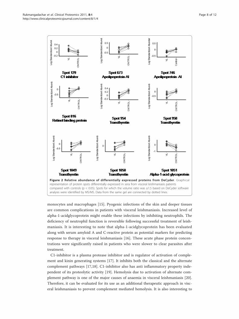
vided as Additional Files 2 and 3. The standardized log abundance of these proteins in
individual gels and their comparison with control as given by the DeCyder software
are illustrated in Figure 2. Transthyretin was represented by at least four and apolipo-
protein-AI was represented by at least two spots. This result is not surprising as many
proteins in plasma are known to exist as isoforms. In both the cases, the individual
spots behaved in a similar way, being down regulated.
Western blot validation of C1 inhibitor, transthyretin and apolipoprotein-AI levels
We performed western blot analyses of three proteins in a separate set of 29 undepleted
serum samples to confirm the DIGE findings. Relative abundance of each band as mea-
sured by the optical density was evaluated by ImageJ software. Relative abundance of C1
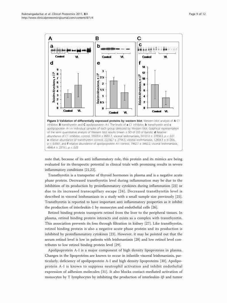
inhibitor in control was 33920.4 ± 8991.7 and in visceral leishmaniasis was 54101.0 ±
27858.3 (p < 0.01) Relative abundance of transthyretin in control was 22236.7 ± 2794.3
and in visceral leishmaniasis was 12804.3 ± 6128.6 (p < 0.0001). Relative abundance of
Table 1 Clinical Data of study subjects
Group No Age in years(Mean ± SD)
Sex Male/Female
Presence of parasite inbone marrow
Presence of anti rK39 antibody
2D DIGE
Visceralleishmaniasis
6 27.8 ± 18.3 6:0 6/6 6/6
Controls 6 27.6 ± 2.7 4:2 NAa 0/6
Validation
Visceralleishmaniasis
19 25.2 ± 10.8 17:2 9/19 19/19
Controls 10 26.9 ± 4.5 8:2 NAa 0/19a NA Not Applicable.
Rukmangadachar et al. Clinical Proteomics 2011, 8:4http://www.clinicalproteomicsjournal.com/content/8/1/4
Page 5 of 12

apolipoprotein A-I in control was 7962.7 ± 3462.2 and in visceral leishmaniasis was
4846.4 ± 2319.1 (p < 0.05). These results are illustrated graphically in Figure 3 and are
in agreement with the DIGE analysis. The up-regulation of C1 inhibitor and the down-
regulation of transthyretin and apolipoprotein A-I in visceral leishmaniasis were thus
confirmed in these samples.
DiscussionSerum is a rich source of disease-related information especially in a systemic infection
like visceral leishmaniasis. Since the dynamic range of human serum proteome is large,
we chose to deplete seven high abundant proteins from serum. Of all the methods
employed for depletion, immunoaffinity chromatography is more effective in removing
targeted proteins, with minimal carryover, high longevity, minimal nonspecific binding
Figure 1 Analysis of serum proteome by DIGE. A representative DIGE image (grey scale) showing theserum protein profile. Proteins identified as differentially expressed are shown by arrows with numbersassigned in the DeCyder analysis. Patient and control sera were labelled with Cy3 and Cy5 respectively inthis gel. The range of the horizontal dimension is isoelectric point (from pI = 3 to pI = 10); the range ofthe vertical dimension is molecular weight (from approx. 150 to 10 kD)
Rukmangadachar et al. Clinical Proteomics 2011, 8:4http://www.clinicalproteomicsjournal.com/content/8/1/4
Page 6 of 12
and high reproducibility [8,9]. However, there remains a possibility of losing some pro-
teins by protein-protein interaction. Since the advantage conferred by depleting the
high abundant proteins was deemed to be of more value in discovering low abundant
proteins, we chose to deplete the serum. These seven abundant proteins make up 85-
90% total protein in serum and hence, their depletion yielded a highly resolved profile
of serum proteome on 2D gels enabling the analysis of low abundant proteins. Accord-
ing to a recent statistical study, a minimum of four biological replicates are needed to
identify at least two fold difference in DIGE studies employing immunodepleted serum
[10]. Assuming similar experimental conditions, our DIGE study was sufficiently pow-
ered as we used six biological replicates. Only one protein, C1-inhibitor, had an aver-
age ratio of 1.45. However, the protein’s up regulation was confirmed on a larger set in
western blot validation experiments.
The alteration in total protein in sera visceral leishmaniasis is a well known phenom-
enon [6]. However, detailed proteome analysis of the sera of this neglected tropical dis-
ease with modern technologies has not been reported so far. To our knowledge, this is
the first report on the DIGE analysis of serum proteome of visceral leishmaniasis. The
use of proteomics to explore the plasma proteome of related infectious diseases like
human African trypanosomiasis [11], tuberculosis [12] and leprosy [13] has been
reported previously. These studies reported the differential expression of many acute
phase proteins in the plasma in these conditions. In this study, as expected, we found
many acute phase proteins being differentially expressed. Some of the identified pro-
teins also are important transport proteins in blood. These proteins are discussed
below.
Alpha-1-acidglycoprotein is known to be elevated in systemic tissue injury, inflam-
mation and infection. It inhibits activation, chemotaxis and their oxidative metabolism
of neutrophils [14]. Alpha-1-acidglycoprotein also modulates cytokine synthesis by
Table 2 List of differentially expressed serum proteins in visceral leishmaniasisidentified by Q-TOF-MS/MSf
Spot noa. Protein name Accession no.b Averageratiod ratio(’p’ (p value)
Appaerancein gels(n = 18)
MascotScoree
Peptidematches
Coverage(%)
129 C1 inhibitor gi|73858570 +1.45 (0.026) 18 190 22 32
1051 Alpha-1- acidglycoprotein
gi|112877 +3.73 (0.009) 18 166 13 48
954 Transthyretin gi|126030594 -1.83 (0.001) 18 449 13 95
958 Transthyretin gi|126030594 -2.23 (0.011) 18 503 12 84
1050 Transthyretin gi|219978 -1.84 (0.015) 18 188 5 46
1049 Transthyretin gi|443295 -2.22 (0.011) 18 262 5 38
816 Retinol bindingprotein
gi|4558179 -1.99 (0.009) 18 47 6 27
673 ApolipoproteinA-I
gi|113992 -2.24 (0.013) 18 114 8 27
746 ApolipoproteinA-I
LPHUA1c -1.65 (0.015) 18 314 28 63
a Spot no. assigned in DeCyder analysis and corresponded to the DIGE image in Figure 1.b Accession no. from NCBInr database, c MSDB database.d + indicates up-regulation and - indicates down-regulation of the protein in visceral leishmaniasis serum with referenceto controls.e Mascot scores greater than 40 were considered significant.f Details of all the mascot search results are provided as additional files 2 and 3.
Rukmangadachar et al. Clinical Proteomics 2011, 8:4http://www.clinicalproteomicsjournal.com/content/8/1/4
Page 7 of 12
monocytes and macrophages [15]. Pyogenic infections of the skin and deeper tissues
are common complications in patients with visceral leishmaniasis. Increased level of
alpha-1-acidglycoprotein might enable these infections by inhibiting neutrophils. The
deficiency of neutrophil function is reversible following successful treatment of leish-
maniasis. It is interesting to note that alpha-1-acidglycoprotein has been evaluated
along with serum amyloid A and C-reactive protein as potential markers for predicting
response to therapy in visceral leishmaniasis [16]. These acute phase protein concen-
trations were significantly raised in patients who were slower to clear parasites after
treatment.
C1-inhibitor is a plasma protease inhibitor and is regulator of activation of comple-
ment and kinin generating systems [17]. It inhibits both the classical and the alternate
complement pathways [17,18]. C1-inhibitor also has anti inflammatory property inde-
pendent of its proteolytic activity [19]. Hemolysis due to activation of alternate com-
plement pathway is one of the major causes of anaemia in visceral leishmaniasis [20].
Therefore, it can be evaluated for its use as an additional therapeutic approach in visc-
eral leishmaniasis to prevent complement mediated hemolysis. It is also interesting to
Figure 2 Relative abundance of differentially expressed proteins from DeCyder . Graphicalrepresentation of protein spots differentially expressed in sera from visceral leishmaniasis patientscompared with controls (p < 0.05). Spots for which the volume ratio was ±1.5 based on DeCyder softwareanalysis were identified by MS/MS. Data from the same gel are connected by dotted lines.
Rukmangadachar et al. Clinical Proteomics 2011, 8:4http://www.clinicalproteomicsjournal.com/content/8/1/4
Page 8 of 12
note that, because of its anti inflammatory role, this protein and its mimics are being
evaluated for its therapeutic potential in clinical trials with promising results in severe
inflammatory conditions [21,22].
Transthyretin is a transporter of thyroid hormones in plasma and is a negative acute
phase protein. Decreased transthyretin level during inflammation may be due to the
inhibition of its production by proinflammatory cytokines during inflammation [23] or
due to its increased transcapillary escape [24]. Decreased transthyretin level is
described in visceral leishmaniasis in a study with a small sample size previously [25].
Transthyretin is reported to have important anti inflammatory properties as it inhibit
the production of interleukin-1 by monocytes and endothelial cells [26].
Retinol binding protein transports retinol from the liver to the peripheral tissues. In
plasma, retinol binding protein interacts and exists as a complex with transthyretin.
This association prevents its loss through filtration in kidney [27]. Like transthyretin,
retinol binding protein is also a negative acute phase protein and its production is
inhibited by proinflammatory cytokines [23]. However, it may be pointed out that the
serum retinol level is low in patients with leishmaniasis [28] and low retinol level con-
tributes to low retinol binding protein level [29].
Apolipoprotein A-I is a major component of high density lipoproteins in plasma.
Changes in the lipoproteins are known to occur in infantile visceral leishmaniasis, par-
ticularly, deficiency of apolipoprotein A-I and high density lipoproteins [30]. Apolipo-
protein A-I is known to suppress neutrophil activation and inhibit endothelial
expression of adhesion molecules [31]. It also blocks contact-mediated activation of
monocytes by T lymphocytes by inhibiting the production of interleukin-1b and tumor
Figure 3 Validation of differentially expressed proteins by western blot. Western blot analysis of A C1inhibitor, B transthyretin and C apolipoprotein A-I. The levels of a C1 inhibitor, b transthyretin and capolipoprotein A-I in individual samples of each group detected by Western blot. Graphical representationof the semi quantitative analysis of Western blot results (mean ± SD of OD of bands). d Relativeabundance of C1 inhibitor: control, 33920.4 ± 8991.7, visceral leishmaniasis, 54101.0 ± 27858.3, p < 0.01.e relative abundance of transthyretin: control, 22236.7 ± 2794.3, visceral leishmaniasis, 12804.3 ± 6128.6,p < 0.0001. and f relative abundance of apolipoprotein A-I: control, 7962.7 ± 3462.2, visceral leishmaniasis,4846.4 ± 2319.1, p < 0.05
Rukmangadachar et al. Clinical Proteomics 2011, 8:4http://www.clinicalproteomicsjournal.com/content/8/1/4
Page 9 of 12
necrosis factor-a [32]. A decrease in apolipoprotein A-I and high density lipoprotein
therefore allows the uninhibited production of interleukin-1b and tumour necrosis
factor-a during inflammation.
Two up regulated proteins identified in this study, alpha-1-acidglycoprotein and C1-
inhibitor have anti inflammatory properties. Their elevated levels probably help
decrease the tissue injury during inflammation in visceral leishmaniasis. The low level
of apolipoprotein A-I leading to more proinflammatory cytokines may be seen as sys-
tem defence against infection. These cytokines inhibit the production of transthyretin
and retinol binding protein. Thus, there is a complex interplay among these proteins
and interpreting their biological significance needs identification of more differentially
expressed proteins. From the biomarker point of view, larger prospective studies incor-
porating appropriate controls like patients presenting with similar symptoms and
employing absolute quantitative methods are suggested to establish them as biomar-
kers. Moreover, since these proteins are related to the inflammatory process, they will
serve as good biomarkers for monitoring response to therapy. Longitudinal studies are
needed in this regard to evaluate their utility as prognostic biomarkers. Since visceral
leishmaniasis is endemic in resource constrained areas, simple and low cost methods
need to be developed to use these results in the clinical setting. Development of sim-
pler dipstick assays will enable such a possibility of testing these proteins in field
conditions.
ConclusionsIn conclusion, DIGE based proteomic analysis showed that several proteins are differ-
entially expressed in the sera of visceral leishmaniasis. The five proteins identified here
have potential, either independently or in combination, for prognostic biomarkers.
Further studies are suggested to establish their application potential.
Additional material
Additional file 1: List of differentially expressed spots in BVA. List of differentially expressed spots in BVA,showing details for each spot (master spot number, appearance in gels, average ratio and p value).
Additional file 2: Detailed Mascot search results for identified proteins. Detailed Mascot search results for theidentified proteins. Mowse score for the first five hits and peptides matched are shown.
Additional file 3: Detailed Mascot search results for identified proteins. Detailed Mascot search resultshowing the protein view. Score and sequence coverage for the identified protein and list of all the peptidesmatched is shown.
AcknowledgementsThis work was carried out at the Clinical Proteomics facility at All India Institute of Medical Sciences (supported byDepartment of Biotechnology, Ministry of Science and Technology, Government of India). GH thanks Council ofIndustrial and Scientific Research, Government of India for the Pool Officer Fellowship. The funding agency had norole in study design, collection, analysis and interpretation of data or in the decision to submit the paper forpublication.
Author details1Department of Biophysics, All India Institute of Medical Sciences, New Delhi, 110029, India. 2Department ofGastroenterology and Human Nutrition, All India Institute of Medical Sciences, New Delhi, 110029, India. 3Departmentof Microbiology, All India Institute of Medical Sciences, New Delhi, 110029, India.
Authors’ contributionsLAR wrote the main manuscript and designed and performed the most of the experiments. JK contributed to thedesign of the study, data collection and interpretation. GH contributed to the design of the study and revision of themanuscript draft. JCS participated in clinical sample and clinical data collection and contributed to the design of the
Rukmangadachar et al. Clinical Proteomics 2011, 8:4http://www.clinicalproteomicsjournal.com/content/8/1/4
Page 10 of 12
study. AS participated in the design of the experiments, supervised the data analysis and interpretation, andparticipated in manuscript writing. All authors read and approved the final manuscript.
Competing interestsThe authors declare that they have no competing interests.
Received: 4 April 2011 Accepted: 31 May 2011 Published: 31 May 2011
References1. Desjeux P: Leishmaniasis: current situation and new perspectives. Comp Immunol Microbiol Infect Dis 2004, 27:305-18.2. Chappuis F, Sundar S, Hailu A, et al: Visceral leishmaniasis: what are the needs for diagnosis, treatment and control?
Nat Rev Microbiol 2005, 5:873-82.3. Kumar R, Pai K, Pathak K, Sundar S: Enzyme-linked immunosorbent assay for recombinant K39 antigen in diagnosis
and prognosis of Indian visceral leishmaniasis. Clin Diagn Lab Immunol 2007, 8:1220-24.4. Reithinger R, Dujardin JC: Molecular Diagnosis of Leishmaniasis: Current Status and Future Applications. J Clin
Microbiol 2007, 45:21-25.5. Hu S, Loo JA, Wong DT: Human body fluid proteome analysis. Proteomics 2006, 6:6326-53.6. Shanker A: Electrophoretic differential serum protein pattern in kala-azar. British Medical journal 1959, 9:1221-23.7. Forgber M, Basu R, Roychoudhury K, et al: Mapping the antigenicity of the parasites in Leishmania donovani
infection byproteome serology. PLoS One 2006, 1:e40.8. Zolotarjova N, Martosella J, Nicol G, Bailey J, Boyes BE, Barrett WC: Differences among techniques for high-abundant
protein depletion. Proteomics 2005, 5:3304-13.9. Steel LF, Trotter MG, Nakajima PB, Mattu TS, Gonye G, Block T: Efficient and specific removal of albumin from human
serum samples. Mol Cell Proteomics 2003, 2:262-70.10. Corzett TH, Fodor IK, Choi MW, et al: Statistical analysis of the experimental variation in the proteomic
characterization of human plasma by two-dimensional difference gel electrophoresis. J Proteome Res 2006,5:2611-19.
11. Papadopoulos MC, Abel PM, Agranoff D, et al: A novel and accurate diagnostic test for human Africantrypanosomiasis. Lancet 2004, 363:1358-63.
12. Agranoff Fernandez-Reyes D, Papadopoulos MC, Rojas SA, Herbster M, Loosemore A: Identification of diagnosticmarkers for tuberculosis by proteomic fingerprinting of serum. Lancet 2006, 368:1012-21.
13. Gupta N, Shankernarayan NP, Dharmalingam K: Serum proteome of leprosy patients undergoing erythema nodosumleprosum reaction: regulation of expression of the isoforms of haptoglobin. J Proteome Res 2006, 6:3669-79.
14. Boutten A, Dehoux M, Deschenes M, Rouzeau JD, Bories PN, Durand G: Alpha 1-acid glycoprotein potentiateslipopolysaccharide-induced secretion of interleukin-1 beta, interleukin-6 and tumor necrosis factor-alpha byhuman monocytes and alveolar and peritoneal macrophages. Eur J Immunol 1992, 22:2687-95.
15. Libert C, Brouckaert P, Fiers W: Protection by alpha 1-acid glycoprotein against tumor necrosis factor-inducedlethality. J Exp Med 1994, 180:1571-75.
16. Wasunna KM, Raynes JG, Were JB, et al: Acute phase protein concentrations predict parasite clearance rate duringtherapy for visceral leishmaniasis. Trans R Soc Trop Med Hyg 1995, 89:678-81.
17. Davis AE III, Mejia P, Lu F: Biological activities of c1 inhibitor. Mol Immunol 2008, 45:4057-63.18. Jiang H, Wagner E, Zhang H, Frank MM: Complement 1 inhibitor is a regulator of the alternative complement
pathway. J Exp Med 2001, 194:1609-16.19. Thorgersen EB, Ludviksen JK, Lambris JD, Sfyroera G, Nielsen EW, Mollnes TE: Anti-inflammatory effects of C1-Inhibitor
in porcine and human whole blood are independent of its protease inhibition activity. Innate Immun 2010,16:254-64.
20. Chava AK, Chatterjee M, Sharma V, Sundar S, Mandal C: Variable degree of alternative complement pathway-mediated hemolysis in Indian visceral leishmaniasis induced by differential expression of 9-O-acetylatedsialoglycans. J Infect Dis 2004, 189:1257-64.
21. Kirschfink M, Mollnes TE: C1-inhibitor: an anti-inflammatory reagent with therapeutic potential. Expert OpinPharmacother 2001, 2:1073-83.
22. Beinrohr L, Dobó J, Závodszky P, Gál P: C1, MBL MASPs and C1-inhibitor: novel approaches for targetingcomplement-mediated inflammation. Trends Mol Med 2008, 14:511-21.
23. Johnson AM, Merlini G, Sheldon J, Ichihara K: Clinical indications for plasma protein assays: transthyretin(prealbumin) in inflammation and malnutrition. Clin Chem Lab Med 2007, 45:419-26.
24. Fleck A: Clinical and nutritional aspects of changes in acute-phase proteins during inflammation. Proc Nutr Soc1989, 48:347-54.
25. Bouree P, Botterel F, Lancon A: Study of protein profile in the visceral leishmaniasis. J Egypt Soc Parasitol 2000,30:885-93.
26. Borish L, King MS, Mascali JJ, Johnson S, Coll B, Rosenwasser LJ: Transthyretin is an inhibitor of monocyte andendothelial cell interleukin-1 production. Inflammation 1992, 16:471-84.
27. Naylor HM, Newcomer ME: The structure of human retinol-binding protein (RBP) with its carrier proteintransthyretin reveals an interaction with the carboxy terminus of RBP. Biochemistry 1999, 38:2647-53.
28. Maciel BL, Lacerda HG, Queiroz JW, et al: Association of nutritional status with the response to infection withLeishmania chagasi. Am J Trop Med Hyg 2008, 79:591-98.
29. Mourey MS, Siegenthaler G, Amédée-Manesme O: Regulation of metabolism of retinol-binding protein by vitamin Astatus in children with biliary atresia. Am J Clin Nutr 1990, 51:638-43.
30. Bekaert D, Kallel R, Bouma ME, et al: Plasma lipoproteins in infantile visceral leishmaniasis: deficiency ofapolipoproteins A-I and A-II. Clin Chim Acta 1989, 184:181-91.
31. Anantharamaiah GM, Engler JA, Borhani DW: Structural models of human apolipoprotein A-I: a critical analysis andreview. Biochim Biophys Acta 2001, 1531:4-46.
Rukmangadachar et al. Clinical Proteomics 2011, 8:4http://www.clinicalproteomicsjournal.com/content/8/1/4
Page 11 of 12

32. Hyka N, Dayer J, Modoux C, et al: Apolipoprotein A-I inhibits the production of interleukin-1β and tumor necrosisfactor-α by blocking contact-mediated activation of monocytes by T lymphocytes. Blood 2001, 97:2381-89.
doi:10.1186/1559-0275-8-4Cite this article as: Rukmangadachar et al.: Two-dimensional difference gel electrophoresis (DIGE) analysis of serafrom visceral leishmaniasis patients. Clinical Proteomics 2011 8:4.
Submit your next manuscript to BioMed Centraland take full advantage of:
• Convenient online submission
• Thorough peer review
• No space constraints or color figure charges
• Immediate publication on acceptance
• Inclusion in PubMed, CAS, Scopus and Google Scholar
• Research which is freely available for redistribution
Submit your manuscript at www.biomedcentral.com/submit
Rukmangadachar et al. Clinical Proteomics 2011, 8:4http://www.clinicalproteomicsjournal.com/content/8/1/4
Page 12 of 12