Turnip Mosaic Virus RNA Replication Complex Vesicles Are Mobile
12
JOURNAL OF VIROLOGY, Oct. 2009, p. 10460–10471 Vol. 83, No. 20 0022-538X/09/$08.000 doi:10.1128/JVI.00819-09 Copyright © 2009, American Society for Microbiology. All Rights Reserved. Turnip Mosaic Virus RNA Replication Complex Vesicles Are Mobile, Align with Microfilaments, and Are Each Derived from a Single Viral Genome † Sophie Cotton, 1 Romain Grangeon, 2 Karine Thivierge, 1 Isabelle Mathieu, 2 Christine Ide, 1 Taiyun Wei, 3 Aiming Wang, 3 and Jean-Franc ¸ois Laliberte ´ 2 * Department of Plant Science, McGill University, 21,111 Lakeshore, Ste-Anne-de-Bellevue, Quebec H9X 3V9, Canada 1 ; INRS-Institut Armand-Frappier, 531 Boulevard des Prairies, Laval, Quebec H7V 1B7, Canada 2 ; and Southern Crop Protection and Food Research Centre, Agriculture and Agri-Food Canada, 1391 Sandford Street, London, Ontario N5V 4T3, Canada 3 Received 22 April 2009/Accepted 1 July 2009 Nicotiana benthamiana plants were agroinoculated with an infectious cDNA clone of Turnip mosaic virus (TuMV) that was engineered to express a fluorescent protein (green fluorescent protein [GFP] or mCherry) fused to the viral 6K 2 protein known to induce vesicle formation. Cytoplasmic fluorescent discrete protein structures were observed in infected cells, corresponding to the vesicles containing the viral RNA replication complex. The vesicles were motile and aligned with microfilaments. Intracellular movement of the vesicles was inhibited when cells were infiltrated with latrunculin B, an inhibitor of microfilament polymerization. It was also observed that viral accu- mulation in the presence of this drug was reduced. These data indicate that microfilaments are used for vesicle movement and are necessary for virus production. Biogenesis of the vesicles was further investigated by infecting cells with two recombinant TuMV strains: one expressed 6K 2 GFP and the other expressed 6K 2 mCherry. Green- and red-only vesicles were observed within the same cell, suggesting that each vesicle originated from a single viral genome. There were also vesicles that exhibited sectors of green, red, or yellow fluorescence, an indication that fusion among individual vesicles is possible. Protoplasts derived from TuMV-infected N. benthamiana leaves were isolated. Using immunofluorescence staining and confocal microscopy, viral RNA synthesis sites were visualized as punctate structures distributed throughout the cytoplasm. The viral proteins VPg-Pro, RNA-dependent RNA polymerase, and cytoplasmic inclusion protein (helicase) and host translation factors were found to be associated with these structures. A single-genome origin and presence of protein synthetic machinery components suggest that translation of viral RNA is taking place within the vesicle. Positive-strand RNA viruses replicate their genomes on in- tracellular membranes. Extensive membrane rearrangements leading to cytoplasmic membranous structure production are observed during the infection cycle of many of these viruses (for a review, see reference 32). These virus-induced mem- brane structures vary greatly in origin, size, and shape. For instance, Flock House virus induces the formation of 50-nm vesicles (spherules), which are outer mitochondrial membrane invaginations with interiors connected to the cytoplasm by a necked channel of approximately 10-nm diameter (24). On the other hand, poxviruses replicate in 1- to 2-m cytoplasmic foci known as DNA factories (43), which are bounded by rough endoplasmic reticulum (ER). These factories are not only the site of DNA synthesis but also of DNA transcription and RNA translation (21). Similarly, mimiviruses are huge double- stranded DNA viruses that replicate in giant cytoplasmic virus factories (45). Three-dimensional electron microscopic imag- ing has shown that coronavirus-induced membrane alterations define a reticulovesicular network of modified ER that inte- grates convoluted membranes, numerous interconnected dou- ble-membrane vesicles, and vesicle packets (23), similar to what was observed for dengue viruses (52). These virus-in- duced structures are known to shelter the virus replication complex, which carries out viral RNA synthesis. The replica- tion complex contains the viral RNA-dependent RNA poly- merase (RdRp), positive- and negative-strand viral RNAs, ac- cessory nonstructural viral proteins, and host cell factors. The role of these virus-induced membrane vesicles in regard to viral RNA synthesis is not well understood. They have been proposed to increase the local concentration of components required for replication, to provide a scaffold for anchoring the replication complex, to confine the process of RNA replication to specific cytoplasmic locations, and to aid in preventing the activation of certain host defense functions. The mechanisms that are responsible for the formation of these structures have begun to be deciphered. Several studies have shown that the specific viral proteins are responsible for the formation of the membrane vesicles (3, 42). However, how individual proteins promote their formation is still unexplained. The full role of cellular factors also remains to be investigated in terms of both membrane vesicle formation and viral RNA synthesis. Finally, intracellular trafficking of these vesicles has been reported (15, 25, 29, 54). Turnip mosaic virus (TuMV) belongs to the genus Potyvirus in the family Potyviridae (44). The TuMV genome is composed * Corresponding author. Mailing address: Institut National de la Recherche Scientifique, Institut Armand-Frappier, 531 Boulevard des Prairies, Laval, Que ´bec H7V 1B7, Canada. Phone: (450) 687-5010. Fax: (450) 686-5501. E-mail: [email protected]. † Supplemental material for this article may be found at http://jvi .asm.org/. Published ahead of print on 5 August 2009. 10460 Downloaded from https://journals.asm.org/journal/jvi on 22 February 2022 by 90.149.191.46.
Transcript of Turnip Mosaic Virus RNA Replication Complex Vesicles Are Mobile

JOURNAL OF VIROLOGY, Oct. 2009, p. 10460–10471 Vol. 83, No. 200022-538X/09/$08.00�0 doi:10.1128/JVI.00819-09Copyright © 2009, American Society for Microbiology. All Rights Reserved.
Turnip Mosaic Virus RNA Replication Complex Vesicles Are Mobile,Align with Microfilaments, and Are Each Derived from a Single
Viral Genome�†Sophie Cotton,1 Romain Grangeon,2 Karine Thivierge,1 Isabelle Mathieu,2 Christine Ide,1 Taiyun Wei,3
Aiming Wang,3 and Jean-Francois Laliberte2*Department of Plant Science, McGill University, 21,111 Lakeshore, Ste-Anne-de-Bellevue, Quebec H9X 3V9, Canada1;
INRS-Institut Armand-Frappier, 531 Boulevard des Prairies, Laval, Quebec H7V 1B7, Canada2; andSouthern Crop Protection and Food Research Centre, Agriculture and Agri-Food Canada,
1391 Sandford Street, London, Ontario N5V 4T3, Canada3
Received 22 April 2009/Accepted 1 July 2009
Nicotiana benthamiana plants were agroinoculated with an infectious cDNA clone of Turnip mosaic virus (TuMV)that was engineered to express a fluorescent protein (green fluorescent protein [GFP] or mCherry) fused to the viral6K2 protein known to induce vesicle formation. Cytoplasmic fluorescent discrete protein structures were observedin infected cells, corresponding to the vesicles containing the viral RNA replication complex. The vesicles weremotile and aligned with microfilaments. Intracellular movement of the vesicles was inhibited when cells wereinfiltrated with latrunculin B, an inhibitor of microfilament polymerization. It was also observed that viral accu-mulation in the presence of this drug was reduced. These data indicate that microfilaments are used for vesiclemovement and are necessary for virus production. Biogenesis of the vesicles was further investigated by infectingcells with two recombinant TuMV strains: one expressed 6K2GFP and the other expressed 6K2mCherry. Green- andred-only vesicles were observed within the same cell, suggesting that each vesicle originated from a single viralgenome. There were also vesicles that exhibited sectors of green, red, or yellow fluorescence, an indication thatfusion among individual vesicles is possible. Protoplasts derived from TuMV-infected N. benthamiana leaves wereisolated. Using immunofluorescence staining and confocal microscopy, viral RNA synthesis sites were visualized aspunctate structures distributed throughout the cytoplasm. The viral proteins VPg-Pro, RNA-dependent RNApolymerase, and cytoplasmic inclusion protein (helicase) and host translation factors were found to be associatedwith these structures. A single-genome origin and presence of protein synthetic machinery components suggest thattranslation of viral RNA is taking place within the vesicle.
Positive-strand RNA viruses replicate their genomes on in-tracellular membranes. Extensive membrane rearrangementsleading to cytoplasmic membranous structure production areobserved during the infection cycle of many of these viruses(for a review, see reference 32). These virus-induced mem-brane structures vary greatly in origin, size, and shape. Forinstance, Flock House virus induces the formation of 50-nmvesicles (spherules), which are outer mitochondrial membraneinvaginations with interiors connected to the cytoplasm by anecked channel of approximately 10-nm diameter (24). On theother hand, poxviruses replicate in 1- to 2-�m cytoplasmic fociknown as DNA factories (43), which are bounded by roughendoplasmic reticulum (ER). These factories are not only thesite of DNA synthesis but also of DNA transcription and RNAtranslation (21). Similarly, mimiviruses are huge double-stranded DNA viruses that replicate in giant cytoplasmic virusfactories (45). Three-dimensional electron microscopic imag-ing has shown that coronavirus-induced membrane alterationsdefine a reticulovesicular network of modified ER that inte-
grates convoluted membranes, numerous interconnected dou-ble-membrane vesicles, and vesicle packets (23), similar towhat was observed for dengue viruses (52). These virus-in-duced structures are known to shelter the virus replicationcomplex, which carries out viral RNA synthesis. The replica-tion complex contains the viral RNA-dependent RNA poly-merase (RdRp), positive- and negative-strand viral RNAs, ac-cessory nonstructural viral proteins, and host cell factors. Therole of these virus-induced membrane vesicles in regard toviral RNA synthesis is not well understood. They have beenproposed to increase the local concentration of componentsrequired for replication, to provide a scaffold for anchoring thereplication complex, to confine the process of RNA replicationto specific cytoplasmic locations, and to aid in preventing theactivation of certain host defense functions. The mechanismsthat are responsible for the formation of these structures havebegun to be deciphered. Several studies have shown that thespecific viral proteins are responsible for the formation of themembrane vesicles (3, 42). However, how individual proteinspromote their formation is still unexplained. The full role ofcellular factors also remains to be investigated in terms of bothmembrane vesicle formation and viral RNA synthesis. Finally,intracellular trafficking of these vesicles has been reported (15,25, 29, 54).
Turnip mosaic virus (TuMV) belongs to the genus Potyvirusin the family Potyviridae (44). The TuMV genome is composed
* Corresponding author. Mailing address: Institut National de laRecherche Scientifique, Institut Armand-Frappier, 531 Boulevard desPrairies, Laval, Quebec H7V 1B7, Canada. Phone: (450) 687-5010.Fax: (450) 686-5501. E-mail: [email protected].
† Supplemental material for this article may be found at http://jvi.asm.org/.
� Published ahead of print on 5 August 2009.
10460
Dow
nloa
ded
from
http
s://j
ourn
als.
asm
.org
/jour
nal/j
vi o
n 22
Feb
ruar
y 20
22 b
y 90
.149
.191
.46.

of a positive-sense single-stranded RNA molecule of about 10kb in length (36). The 5� terminus of the viral RNA is linkedcovalently to a viral protein known as VPg and the 3� terminusis polyadenylated. The TuMV RNA is translated into a longpolyprotein of 358 kDa and is processed into at least 10 matureproteins by three different virus-encoded proteases. It wasdemonstrated for Tobacco etch virus (TEV) and Plum poxvirus, also members of the Potyvirus genus, that viral RNAsynthesis is associated with membranes of the ER (30, 42). Inthe case of TuMV, the 6K2-VPg-Pro polyprotein, through itshydrophobic 6K2 domain, was shown to be responsible for theformation of cytoplasmic vesicles derived from the ER (4),similar in structure to those observed during TEV infection(42, 51). Besides being involved in vesicle formation, 6K2-VPg-Pro binds a number of proteins of viral and host origin. Inter-action with the viral RdRp and the host translation eukaryoticinitiation factor (iso) 4E [eIF(iso)4E], poly(A)-binding protein(PABP), heat shock cognate 70–3 (Hsc70-3), and the eukary-otic elongation factor 1A (eEF1A) has been shown to takeplace within the 6K2-VPg-Pro-induced vesicles (4, 5, 9, 48).Although these vesicles have been referred to as sites forTuMV replication (48), the presence of viral RNA in thesevesicles has not been reported. The presence of translationfactors with virus replication proteins also brings the questionof the physical relationship between viral RNA translation andreplication.
In the present study, the biogenesis of the TuMV-inducedvesicles was investigated. It was observed that the TuMV-induced vesicles, tagged with a fluorescent protein fused to6K2, were mobile and aligned with microfilaments. In addition,results using depolymerizing compounds supported the con-tention that vesicles trafficked on microfilaments and that mi-crotubules were not involved. Evidence is also provided indi-cating that each vesicle is derived from a single viral genome.Finally, the association of host translation factors with viralRNA synthesis sites was confirmed by double immunofluores-cence staining in TuMV-infected protoplasts. The single-ge-nome origin and the presence of translation factors within thereplication complex vesicles suggest that viral translation istaking place within the vesicles.
MATERIALS AND METHODS
Plasmid constructions. pCambiaTunos was obtained by digesting p35Tunos(41) with KpnI and StuI, followed by ligation with pCambiaTunos/6KGFP (48),also digested with KpnI and StuI. Kanamycin-resistant Escherichia coli colonieswere screened for plasmids deleted in the region encoding 6K2GFP (i.e.,pCambiaTunos/6KGFP). pUC19/6KGFP was obtained by digestion ofp35Tunos/�Nsil-6KGFP (48) with BamHI and KpnI and the 1458-bp fragmentwas ligated in pUC19 digested with the same enzymes and in which the HindIIIrestriction site had previously been destroyed. The mCherry coding region wasamplified by PCR from pCambia/mCherry (9) by using the forward primer5�-ATTCGGATCCGTGAGCAAGGGCGAGGAG-3� and the reverse primer5�-ATTCAAGCTTCCTTGTACAGCTCGTCCATG-3� (the restriction sites areunderlined). The PCR product was digested with BamHI and HindIII andligated in pUC19/6KGFP (48), which was also digested with the same enzymes.Ampicillin-resistant E. coli colonies were screened for plasmids containing thefragment encoding 6K2mCherry (i.e., pUC19/6KmCherry). This plasmid wasthen cut with BamHI and KpnI and the 1451-bp fragment was ligated withp35Tunos/�Nsil-6KGFP (48) cut with the same enzymes. Ampicillin-resistant E.coli colonies were screened for plasmids containing the fragment encoding6K2mCherry (i.e., p35Tunos/�Nsil-6KmCherry). p35Tunos/�Nsil-6KmCherrywas digested with SmaI and KpnI and was ligated with pCambiaTunos/6KGFP,also digested with the same enzymes. Kanamycin-resistant E. coli colonies were
screened for plasmids containing the fragment encoding 6K2mCherry (i.e.,pCambiaTunos/ 6KmCherry). All plasmid constructs were verified by sequencing.
Antibodies. Rabbit antisera were used at the following dilutions: for immuno-blot analysis, anti-CP at 1:2,500; and for immunofluorescence labeling, anti-CP at1:300, anti-RdRp at 1:100 (9), anti-VPg-Pro at 1:200 (27), anti-PABP2 at 1:300(28), anti-eIF(iso)4E at 1:150 (7), anti-eEF1A at 1:500 (57), and anti-CI at 1:300.Recombinant clones pET-CP and pET-CI in E. coli BL21(DE3) cells were usedfor anti-CP and anti-CI serum production. Full-length coding sequences ofTuMV CP and CI were cloned in frame in the pET11d vector (Novagen). Therecombinant proteins were overproduced in E. coli and purified as insolubleinclusion bodies. Inclusion bodies were resuspended in Tris-buffered saline andused for rabbit injection and serum production at McGill University AnimalResources Center. The mouse monoclonal antibody dilutions were as follows:anti-actin8, 1:1000 (Sigma); anti-BrdU, 1:100 (Sigma); and anti-dsRNA, 1:300(English & Scientific Consulting Bt.). The secondary antibodies were goat anti-mouse conjugated to Alexa Fluor 568 at 1:500 (Molecular Probes) and goatanti-rabbit conjugated to Alexa Fluor 488 at 1:1,000 (Molecular Probes). Forimmunoblot analysis, the antigen-antibody complexes were visualized by using ahorseradish peroxidase-coupled goat anti-rabbit immunoglobulin G (IgG).
Protein expression in plants. Constructs containing genes for proteins fused tofluorescent tag were introduced by electroporation into Agrobacterium tumefa-ciens AGL1 and selected on LB ampicillin-kanamycin plates. The pellet of anovernight culture was gently resuspended in water supplemented with 10 mMMgCl2 and 150 �M acetosyringone and left at room temperature for 3 h. Thesolution was then diluted to an optical density at 600 nm of 0.6. Three-week-oldN. benthamiana plants were agroinfiltrated with the fusion construct. Plants werekept for 4 days in growth chamber until observation.
Drug treatments. Stock solutions of latrunculin B (LatB; 2.5 mM [Calbio-chem]), cytochalasin D (CytD; 20 mM [Calbiochem]), trifluralin (20 mM [Chem-Service]), and oryzalin (10 mM [ChemService]) were prepared in dimethyl sul-foxide (DMSO) and diluted to the desired concentration in water prior to theirinfiltration into 3-week-old N. benthamiana leaves.
Protoplast isolation and immunofluorescence labeling. N. benthamiana wasagroinfiltrated with pCambiaTunos/6KGFP. Leaves obtained at 4 days postin-fection were sliced in 1-mm wide stripes, followed by incubation in an enzymesolution (1.5% cellulose R10, 0.2% macerozyme R10, 0.5 M mannitol, 20 mMKCl, 20 mM MES [pH 5.7], 10 mM CaCl2, 0,1% bovine serum albumin [BSA])for 3 h in the dark under a vacuum. The protoplast suspension was filteredthrough a 45-�m-pore-size nylon filter and centrifuged for 4 min at 100 � g. Thesupernatant was removed. Protoplasts were incubated for 15 min at room tem-perature with 1 volume of fixing solution (4% paraformaldehyde, 0.25 M man-nitol, and 50 mM sodium phosphate in phosphate-buffered saline [PBS]). Theywere centrifuged and resuspended for 30 min with 2 volumes of fixing solution atroom temperature. Protoplasts were washed three times with PBS for 10 min.They were then put on cover slide pretreated with 0.1% poly-L-lysine (Sigma),treated with Triton X-100 0.5% in PBS for 10 min, and incubated for 20 min ina blocking solution of 5% BSA in PBS. The samples were then incubated for 1 hwith the primary antibody, washed three times with PBS for 10 min, incubated foranother hour with Alexa Fluor 568- or Alexa Fluor 488-conjugated secondaryantibody (Molecular Probes), and finally washed three times with PBS for 10min. Pro-long Gold Antifade (Invitrogen) was used to prepare the slides.
In vivo RNA labeling. Protoplasts were isolated from plants agroinfiltrated at4 days postinfection with A. tumefaciens containing pCambiaTunos. They wereincubated for 30 min with 10 �g/ml of actinomycin D in order to block RNAtranscription from cellular DNA-dependent RNA polymerases. Two mM 5-bro-mouridine 5�-triphosphate (BrUTP; Sigma) was then added for 3 h. The reactionwas stopped by the addition of fixing solution (described above) for 15 min,washed once with PBS, and fixed again for another 30 min. The immunofluo-rescence labeling was processed as described above using anti-BrdU (1:100), andthe detection was done using Alexa Fluor 568-conjugated secondary antibody(Molecular Probes) at a 1:500 dilution.
Immunoblot analysis. Total proteins were extracted from N. benthamianaagroinfiltrated with pCambiaTunos/6KGFP 4 days before and treated previouslywith drugs with an extraction buffer (50 mM Tris-HCl [pH 7.6], 50 mM KCl, 0.5mM EDTA, 20 mM NaCl, 5% glycerol, 0,1% Triton X-100, 0.01% sodiumdodecyl sulfate [SDS], and plant protease inhibitor cocktail [Sigma]). Plant tissue(0.5 g) was ground in 500 �l of extraction buffer with a Dounce homogenizer andcentrifuged for 1 min at 5,000 � g. The concentration of the supernatant wasdetermined by using a Bradford assay (Bio-Rad) using BSA as a standard. Then,2 �g of total protein preparation was used to perform an immunoblot analysisafter SDS-polyacrylamide gel electrophoresis. The antigen-antibody complexeswere visualized using a horseradish peroxidase-coupled goat anti-rabbit IgG orgoat anti-mouse IgG according to the manufacturer’s recommendations. Immu-
VOL. 83, 2009 TuMV RNA REPLICATION COMPLEX VESICLES 10461
Dow
nloa
ded
from
http
s://j
ourn
als.
asm
.org
/jour
nal/j
vi o
n 22
Feb
ruar
y 20
22 b
y 90
.149
.191
.46.
noreactions were detected with the SuperSignal West Pico chemiluminescentsubstrate (Pierce).
Confocal microscopy. Agroinfiltrated leaf sections were placed on a micro-scope coverslide using immersion oil. Coverslides were inverted on depressionslides, aligning the leaf tissue in the well. Cells were observed using a 40X oilimmersion objective on a Radiance 2000 confocal microscope (Bio-Rad). Argon-krypton laser was used to excite fluorescent proteins and data from both greenand red channels were collected at the same time. A charge-coupled-devicecamera was used to collect the images. Protoplasts were observed by using a 63Xoil immersion objective on a LSM 510 Meta confocal microscope (Zeiss). Argonand HeNe lasers were used to excite fluorescent proteins and data from both
green and red channels were collected at sequential time. After acquisition,images were processed by using Metamorph (6.2r6), ImageJ (1.41o), Carl ZeissLSM Image Browser, and/or Adobe Photoshop software. The Manual Trackingpluging (http://rsbweb.nih.gov/ij/plugins/track/track.html) was used for displayingvesicle movement paths.
RESULTS
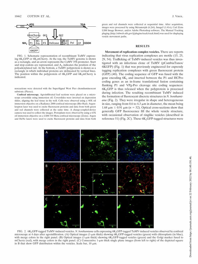
Movement of replication complex vesicles. There are reportsindicating that virus replication complexes are motile (15, 25,29, 54). Trafficking of TuMV-induced vesicles was thus inves-tigated with an infectious clone of TuMV (pCambiaTunos/6KGFP) (Fig. 1) that was previously engineered for expresslytagging replication complexes with green fluorescent protein(GFP) (48). The coding sequence of GFP was fused with thegene encoding 6K2 and inserted between the P1 and HCProcoding genes as an in-frame translational fusion containingflanking P1 and VPg-Pro cleavage site coding sequences:6K2GFP is thus released when the polyprotein is processedduring infection. The resulting recombinant TuMV inducedthe formation of fluorescent discrete structures in N. benthami-ana (Fig. 2). They were irregular in shape and heterogeneousin size, ranging from 0.6 to 4.3 �m in diameter, the mean being1.68 �m � 0.91 �m (n � 52). Optical cross-sections show thatgenerally GFP fluorescence fill the whole vesicle structure,with occasional observation of ringlike vesicles (described inreference 51) (Fig. 2C). These 6K2GFP-tagged structures were
FIG. 1. Schematic representation of recombinant TuMV express-ing 6K2GFP or 6K2mCherry. At the top, the TuMV genome is shownas a rectangle, and an arrow represents the CaMV 35S promoter. Startand stop codons are represented, and An indicates the position of thepolyadenylated tail. At the bottom, a TuMV polyprotein is shown as arectangle in which individual proteins are delineated by vertical lines.The position within the polyprotein of 6K2GFP and 6K2mCherry isindicated.
FIG. 2. 6K2GFP-tagged TuMV-induced vesicles. N. benthamiana cells expressing 6K2GFP-tagged TuMV-induced vesicles observed by confocalmicroscopy at 4 days after agroinfiltration. (A) Optical images (1-�m thick) showing 6K2GFP-tagged vesicles (green) with chloroplasts (in blue),with merge colors in the right panel. (B) Optical images (1-�m thick) showing 6K2GFP-tagged vesicles (green) and the Golgi marker fused tomCherry (red), with merge colors in the right panel. (C) Consecutive 1-�m thick single plane images (from left to right) of the depicted squarein B that show GFP distribution within the vesicles. Scale bar, 10 �m.
10462 COTTON ET AL. J. VIROL.
Dow
nloa
ded
from
http
s://j
ourn
als.
asm
.org
/jour
nal/j
vi o
n 22
Feb
ruar
y 20
22 b
y 90
.149
.191
.46.

previously shown to contain 6K2-VPg-Pro, RdRP, eIF(iso)4E,PABP, and eEF1a (9, 48) and were reminiscent of those in-duced by 6K2-VPg-Pro alone (4). To confirm that the 6K2GFPfluorescence matched with viral replication sites, these werevisualized by staining with antibodies directed against double-stranded RNA (dsRNA) (1, 10). N. benthamiana leaves wereagroinfiltrated with A. tumefaciens Agl1 strain containingpCambiaTunos/6KGFP, and protoplasts were isolated frominfiltrated leaves 4 days later. The fixed and permeabilizedprotoplasts were then reacted with a mouse antibody recog-nizing dsRNA and Alexa Fluor 586 (red)-conjugated anti-mouse antibodies. Protoplasts were subsequently observed byconfocal microscopy. dsRNA was associated with punctatestructures of 2.1 �m � 0.5 �m in diameter distributed through-out the cytoplasm in TuMV-infected protoplasts (see Fig. 8I).No immunofluorescence was observed in uninfected proto-plasts (data not shown). The presence of dsRNA coincidedwith fluorescence emitted by GFP, confirming that 6K2GFPtagging constitutes a good marker for the visualization ofTuMV replication complexes.
These discrete structures are likely to be membrane-boundvesicles and not protein aggregates. First, 6K2-VPg-Pro, whichhas been shown to colocalize with 6K2GFP (48), and RdRp“float” in membrane flotation experiments (5, 9), indicatingthat these replication-associated proteins are membrane asso-ciated and not aggregates or inclusions. Furthermore, Schaadet al. (42) have shown that the 6K2 protein of the relatedtobacco etch virus fused to GFP and expressed using the sameapproach as here was associated with large vesicular compart-ments derived from the ER. Although some are relatively largeand akin to mini-organelles, these TuMV-induced structureswill be designated as vesicles from now on.
The large size of the 6K2GFP-tagged TuMV-induced vesi-cles suggests that they are linked to chloroplasts or Golgibodies. The Bio-Rad confocal microscope used in the presentstudy possesses three detectors with filter sets calibrated toabsorb light at wavelengths of 515 nm � 15 nm (GFP fluores-cence [green channel]), of 600 nm � 20 nm (mCherry fluores-cence [red channel]), and of �660 nm (chlorophyll autofluo-rescence [blue channel]). Such setup allows chlorophyllfluorescence to be separated from the fluorescence emitted byGFP and mCherry. Observation of serial optical imagesshowed that the bulk of the chloroplasts were generally foundnear the surface of the plasma membrane, whereas the virus-induced vesicles were located more in the interior of the cell,often near the nucleus. Figure 2A shows a single 1-�m opticalslice where 6K2GFP-tagged vesicles can be seen concomitantlywith chloroplasts. Some vesicles were found in close associa-tion with chloroplasts, which possibly contribute along the ERto membrane structures at least in the case of plum pox virusreplication (30). However, more often, vesicles and chloro-plasts were seen as different entities. Golgi localization wasbased on the cytoplasmic tail and transmembrane domain (first49 amino acids) of the soybean -1,2-mannosidase I (40) fusedto mCherry (34). This Golgi marker consists of a large numberof small (1 �m) independent stacks (34) that appeared asround discs of uniform size (Fig. 2B). The Golgi marker alsoshowed ER labeling, resulting from the continuous recycling ofGolgi resident proteins through the ER round discs (6). The6K2GFP-tagged vesicles were found to align with the ER (as
expected) but generally did not colocalize with the Golgi bod-ies. Occasional association with Golgi is explained by the bio-genesis of potyvirus replication vesicles occurring at ER exitsites in a COPI- and COPII-dependent manner (51). The6K2GFP-tagged TuMV-induced vesicles are thus distinct fromGolgi bodies.
The intracellular trafficking of vesicles induced by TuMVand tagged with either fluorescent protein was then investi-gated. Three-week-old N. benthamiana leaves were agroinfil-trated with A. tumefaciens Agl1 containing pCambiaTunos/6KGFP, and movement of the fluorescing vesicles wasinvestigated by time-lapse imaging after 4 days. The vesicleswere motile, but each vesicle moved at different speed, withaverage velocities of 0.45 �m/s � 0.27 �m/s (n � 10) (Fig. 3and see Movie S2 in the supplemental material). Movementwas unidirectional, which was accompanied with stop-and-goactivity. Although the exact destination was not known, occa-sional fusion with perinuclear vesicles was observed, as shownin Fig. 3. Similar data were obtained when vesicles were taggedwith mCherry. In comparison, cauliflower mosaic virus(CaMV) P6 inclusion bodies move with an average velocity of2 �m/s (maximum of approximately 8 �m/s) (15), and theaverage velocities are �1 �m/s for tobacco mosaic virus 126Kbodies and viral replication complexes (maximum of 8 �m/s)(29).
Since 6K-VPg-Pro alone induces vesicle formation (4), theirtrafficking was also investigated. N. benthamiana plants wereagroinfiltrated with pGreen6KVPgProGFP, and the move-ment of the green fluorescing vesicles was observed after 3days. 6K2-VPg-Pro-induced vesicles were also motile (seeMovie S3 in the supplemental material), suggesting that thevirus molecular determinant for movement lies within 6K2-VPg-Pro.
Stop and go movement is indicative of Golgi and was con-sequently compared directly to that of TuMV-induced vesicles.The Golgi stacks can be seen moving and extensively along thepolygonal cortical ER network (see Movie S4 in the supple-mental material) with an estimated speed of 0.35 �m/s. Thesedata indicated that TuMV-induced 6K2GFP-tagged and Golgivesicles are distinct structures.
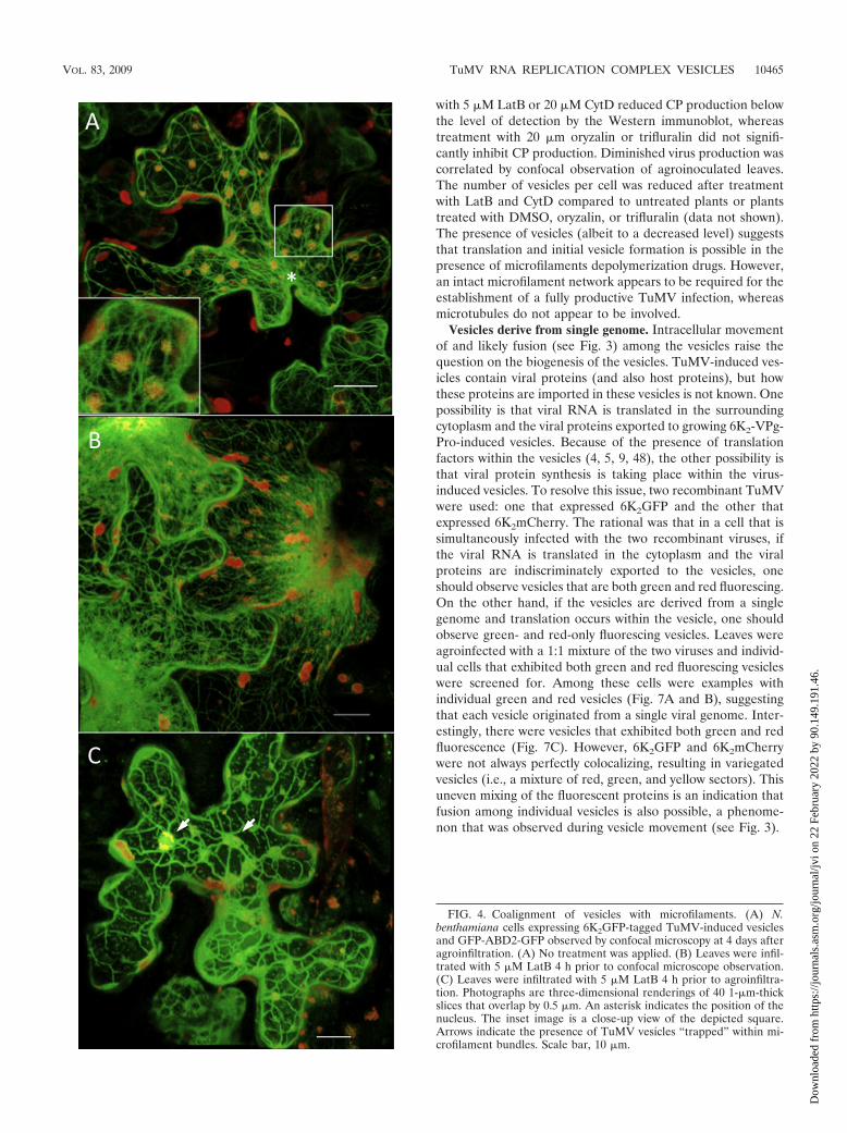
Vesicle alignment with microfilaments. Since the proteincontent and organized nature of the cytoplasm restrict diffu-sion of large molecular complexes, movement of virus-inducedvesicles is likely to require cytoskeleton elements (17). Todetermine whether TuMV vesicle movement is associated withmicrofilaments, an A. tumefaciens Agl1 mixture containingpCambiaTunos/6KmCherry (Fig. 1) and a pCambia vectorcontaining the 35S::GFP-ABD2-GFP construct (50) wasagroinfiltrated in N. benthamiana. The latter construct codesfor GFP fused to both termini of the actin-binding domain offimbrin (GFP-ABD2-GFP) and provides improved imaging ofactin filaments. Figure 4A shows the positioning of themCherry-tagged vesicles with respect to the GFP-ADB2-GFP-labeled actin filaments. A dense filament network is seen ra-diating from the nucleus (denoted by an asterisk in the figure)to the cell periphery, and the TuMV vesicles are randomlydistributed in the cell. Alignment of the vesicles with the ABD2filaments is readily observed, with each vesicle concurrentlyinteracting with several filaments. Vesicles seen not to alignwith ABD2 filaments are from adjoining cells that were not
VOL. 83, 2009 TuMV RNA REPLICATION COMPLEX VESICLES 10463
Dow
nloa
ded
from
http
s://j
ourn
als.
asm
.org
/jour
nal/j
vi o
n 22
Feb
ruar
y 20
22 b
y 90
.149
.191
.46.

expressing the latter protein. It must be noted that in cellsexpressing both proteins, no vesicles were observed in subcom-partments of the cell devoid of ABD2 filaments, suggestingthat alignment of the vesicles with the filaments is not theindirect consequence of the density of the network trappingany large structures. The red vesicles cannot be interpreted asbeing chloroplasts, as explained above. Moreover, no mCherryfluorescence is observed in the red channel in mock-inoculatedplants or in cells expressing GFP fusions only. Furthermore,experiments using mCherry fusions developed previously (9,48) showed that the red fluorescence does not result fromchlorophyll autofluorescence.
Movement of the vesicles was investigated in the presence ofGFP-ABD2-GFP. Trafficking of the vesicles was difficult todiscern, a phenomenon observed also for the movement of theCaMV P6 inclusion bodies (15). It has been reported that theuse of live markers to visualize actin in plants could affectmotility of cellular compounds because of the overexpressionof the markers (19). However, despite this inhibition, TuMVvesicles were seen to traffic along GFP-ABD2-GFP filaments(Fig. 5; see also Movie S5 in the supplemental material).
To confirm the implication of microfilaments in viral vesiclemovement, LatB was used to disassemble microfilaments.Since LatB’s effect on microfilaments is likely to affect severalprocesses, which indirectly might affect vesicle trafficking,movement was first evaluated on preestablished vesicles assoon as possible after drug application. N. benthamiana leaveswere agroinfiltrated with A. tumefaciens Agl1 suspensions con-taining pCambiaTunos/6KGFP and the 35S::GFP-ABD2-GFPconstruct. At 4 days postinfection and 4 h before confocalobservation, the agroinfiltrated leaves were treated with 5 �MLatB or 1% DMSO solvent control. The DMSO infiltration didnot affect the microfilament network (data not shown), butafter LatB treatment the ABD2 filaments had begun the de-
polymerization process, as noted by the blurred appearance ofthe filaments near the nucleus (Fig. 4B). When LatB wasinfiltrated 4 h prior to TuMV agroinfection, the network, whenobserved by confocal microscopy 4 days later, was sparser, thefilaments were thicker, and several microfilament bundles wereobserved (Fig. 4C). Within that time period, it is expected thatplant cells had metabolized some of the LatB molecules andthat the microfilament network is in the process of regaining itsnormal state. Interestingly, the 6K2mCherry-tagged TuMV-induced vesicles were tightly enclosed within the microfilamentbundles (4C).
Movement was assessed with N. benthamiana leaves agroin-filtrated with A. tumefaciens Agl1 suspensions containingpCambiaTunos/6KGFP or the GFP-labeled Golgi marker con-struct. The latter marker was used as a drug control since Golgitrafficking in dependent on myosin (2). DMSO had no inhib-itory effect on Golgi body and TuMV vesicle movement (datanot shown). On the other hand, LatB treatment 4 h beforeconfocal microscope observation abolished trafficking of Golgibodies (see Movie S6 in the supplemental material) and TuMVvesicles (see Movie S7 in the supplemental material).
TuMV infection is inhibited by LatB treatment. To assessthe effect of cytoskeleton-affecting drugs on the initiation ofTuMV infection, N. benthamiana leaves were infiltrated eitherwith LatB, CytD, oryzalin, trifluralin, or DMSO 24 h prior toagroinfiltration with A. tumefaciens Agl1 containing pCambia-Tunos/6KGFP. CytD depolymerizes microfilaments, whileoryzalin and trifluarin are agents that disassemble microtu-bules. Plant tissues were collected 4 days later, and the totalproteins were extracted, separated by SDS-polyacrylamide gelelectrophoresis, and subjected to immunoblot analysis. Virusaccumulation was assessed using a rabbit serum raised againstthe TuMV coat protein (CP). Figure 6 shows virus accumula-tion in plants following different drug treatments. Treatment
FIG. 3. Movement of TuMV-induced vesicles. N. benthamiana cells expressing 6K2GFP-tagged TuMV-induced vesicles observed 4 days afteragroinfiltration by confocal microscopy. Time-lapse images illustrate the movement of the vesicles over the indicated time periods. Lines depictthe path taken by individual vesicles. Scale bar, 10 �m.
10464 COTTON ET AL. J. VIROL.
Dow
nloa
ded
from
http
s://j
ourn
als.
asm
.org
/jour
nal/j
vi o
n 22
Feb
ruar
y 20
22 b
y 90
.149
.191
.46.
with 5 �M LatB or 20 �M CytD reduced CP production belowthe level of detection by the Western immunoblot, whereastreatment with 20 �m oryzalin or trifluralin did not signifi-cantly inhibit CP production. Diminished virus production wascorrelated by confocal observation of agroinoculated leaves.The number of vesicles per cell was reduced after treatmentwith LatB and CytD compared to untreated plants or plantstreated with DMSO, oryzalin, or trifluralin (data not shown).The presence of vesicles (albeit to a decreased level) suggeststhat translation and initial vesicle formation is possible in thepresence of microfilaments depolymerization drugs. However,an intact microfilament network appears to be required for theestablishment of a fully productive TuMV infection, whereasmicrotubules do not appear to be involved.
Vesicles derive from single genome. Intracellular movementof and likely fusion (see Fig. 3) among the vesicles raise thequestion on the biogenesis of the vesicles. TuMV-induced ves-icles contain viral proteins (and also host proteins), but howthese proteins are imported in these vesicles is not known. Onepossibility is that viral RNA is translated in the surroundingcytoplasm and the viral proteins exported to growing 6K2-VPg-Pro-induced vesicles. Because of the presence of translationfactors within the vesicles (4, 5, 9, 48), the other possibility isthat viral protein synthesis is taking place within the virus-induced vesicles. To resolve this issue, two recombinant TuMVwere used: one that expressed 6K2GFP and the other thatexpressed 6K2mCherry. The rational was that in a cell that issimultaneously infected with the two recombinant viruses, ifthe viral RNA is translated in the cytoplasm and the viralproteins are indiscriminately exported to the vesicles, oneshould observe vesicles that are both green and red fluorescing.On the other hand, if the vesicles are derived from a singlegenome and translation occurs within the vesicle, one shouldobserve green- and red-only fluorescing vesicles. Leaves wereagroinfected with a 1:1 mixture of the two viruses and individ-ual cells that exhibited both green and red fluorescing vesicleswere screened for. Among these cells were examples withindividual green and red vesicles (Fig. 7A and B), suggestingthat each vesicle originated from a single viral genome. Inter-estingly, there were vesicles that exhibited both green and redfluorescence (Fig. 7C). However, 6K2GFP and 6K2mCherrywere not always perfectly colocalizing, resulting in variegatedvesicles (i.e., a mixture of red, green, and yellow sectors). Thisuneven mixing of the fluorescent proteins is an indication thatfusion among individual vesicles is also possible, a phenome-non that was observed during vesicle movement (see Fig. 3).
FIG. 4. Coalignment of vesicles with microfilaments. (A) N.benthamiana cells expressing 6K2GFP-tagged TuMV-induced vesiclesand GFP-ABD2-GFP observed by confocal microscopy at 4 days afteragroinfiltration. (A) No treatment was applied. (B) Leaves were infil-trated with 5 �M LatB 4 h prior to confocal microscope observation.(C) Leaves were infiltrated with 5 �M LatB 4 h prior to agroinfiltra-tion. Photographs are three-dimensional renderings of 40 1-�m-thickslices that overlap by 0.5 �m. An asterisk indicates the position of thenucleus. The inset image is a close-up view of the depicted square.Arrows indicate the presence of TuMV vesicles “trapped” within mi-crofilament bundles. Scale bar, 10 �m.
VOL. 83, 2009 TuMV RNA REPLICATION COMPLEX VESICLES 10465
Dow
nloa
ded
from
http
s://j
ourn
als.
asm
.org
/jour
nal/j
vi o
n 22
Feb
ruar
y 20
22 b
y 90
.149
.191
.46.
Localization of host and viral proteins within virus replica-tion complexes. Formation of a large vesicle derived from asingle viral genome presupposes that a cis mechanism is in-volved: proteins synthesized from the same viral RNA areincorporated in the same vesicle, with no outside contributionfrom other genomes. This is possible if viral RNA translationis taking place within the vesicle. It was previously shown that6K2-VPg-Pro of TuMV induces the formation of vesicles thatcontain host translation factors such as eIF(iso)4E, PABP,eEF1A, and Hsc70 (4, 5, 9, 48). The question however remainswhether these same vesicles are also the sites of viral RNAsynthesis. RNA replication sites can be visualized by immuno-fluorescence staining using antibodies either directed againstdsRNA or neosynthesized 5-bromouridine-labeled RNA (1,10). To confirm that the above-listed host proteins colocalizewith viral replication sites, double immunofluorescence stain-ing of TuMV-infected protoplasts was performed as describedabove. The fixed and permeabilized protoplasts were then re-
acted with rabbit sera raised against viral or plant proteins andmouse antibodies recognizing dsRNA or bromouridine. Forbromouridine labeling, protoplasts were treated with actino-mycin D prior to BrUTP incorporation in order to block hostDNA transcription without affecting TuMV-directed RNA-dependent RNA synthesis. Rabbit and mouse antibodies werelabeled with Alexa Fluor 488 (green)- and Alexa Fluor 586(red)-conjugated secondary antibodies, respectively, and pro-toplasts were observed by confocal microscopy. To assess thebackground fluorescence, protoplasts from mock-inoculatedleaves were subjected to the same immunofluorescence label-ing conditions, and fluorescent signals were adjusted to set thebackground threshold level. For virus-specific elements, nosignificant background was detected in uninfected protoplasts(data not shown). Figure 8 shows the intracellular localizationof dsRNA or bromouridine-labeled RNA, along with plant andviral proteins in TuMV-infected protoplasts. As mentionedabove, dsRNA was associated with punctate structures distrib-uted throughout the cytoplasm. Similar structures were alsoobserved for bromouridine-labeled RNA, indicating that thepresence of dsRNA likely corresponds to area of active RNAsynthesis. No immunofluorescence was observed in uninfectedprotoplasts (data not shown). If the dsRNA punctate struc-tures are true indicators of the virus replication complex, viralproteins known to be involved in virus replication must beassociated with these structures. The anti-VPg-Pro antibodiesreact with VPg-Pro, its precursor (6K-VPg-Pro), as well asprocessed forms (VPg and Pro) (4). Staining with this serumwas observed throughout the cytoplasm, as expected from pre-vious cell fractionation data (4, 28). However, a subpopulationof the proteins recognized by the anti-VPg-Pro antibodies wasalso associated with the dsRNA or bromouridine-labeled RNApunctate structures (Fig. 8A and B). A similar fluorescentpattern was observed for RdRp (Fig. 8C). Interestingly, thecytoplasmic inclusion protein (CI), which has helicase activity
FIG. 5. Vesicle trafficking along microfilaments. N. benthamiana cell expressing 6K2GFP-tagged TuMV-induced vesicles and GFP-ABD2-GFPobserved 4 days after agroinfiltration by confocal microscopy. Time-lapse images illustrate the movement of a vesicle over the indicated timeperiods. A line depicts the path taken by the vesicle. An asterisk denotes the position of the nucleus, and arrows indicate the position of a singlemicrofilament. Scale bar, 10 �m.
FIG. 6. Effect of cytoskeleton-affecting drugs on the initiation ofTuMV infection. N. benthamiana leaves were infiltrated with 5 �MLatB or 20 �M CytD, oryzalin, or trifluralin 24 h prior to infiltrationwith A. tumefaciens Agl1 containing pCambiaTunos/6KGFP. Totalproteins were extracted 4 days after agroinfiltration, and 2 �g wasanalyzed by immunoblot analysis with antibodies raised against TuMVCP. Antibody against actin-8 was used as a loading control.
10466 COTTON ET AL. J. VIROL.
Dow
nloa
ded
from
http
s://j
ourn
als.
asm
.org
/jour
nal/j
vi o
n 22
Feb
ruar
y 20
22 b
y 90
.149
.191
.46.
(26), was associated with punctate structures that did not al-ways coincide with the presence of dsRNA. However, dsRNAstructures always correlated with the presence of CI (Fig. 8D).This suggests that CI may have additional roles besides beinginvolved with the replication complex. The CP, which is notknown to be directly involved in RNA replication, was veryrarely associated with dsRNA punctate structures (Fig. 8E),suggesting that RNA synthesis and capsid-related events (e.g.,virion assembly) are physically separated.
Host proteins that were shown to be enclosed within 6K-VPg-Pro-induced vesicles were then tested. eIF(iso)4E, PABP,and eEF1A were distributed throughout the cytoplasm (Fig.8F to H), as expected from the fractionation data (4, 28, 48),but subpopulations of the different proteins were found tocolocalize with the dsRNA punctate structures. Taken to-gether, these results indicate that TuMV RNA replication sitesnot only contained viral proteins expected to be involved inRNA synthesis but also host factors implicated in protein syn-thesis.
DISCUSSION
A major finding of this investigation is that a TuMV-inducedvesicle originates from a single viral genome. Infecting cellssimultaneously with two recombinant viruses, one tagging ves-icles with 6K2GFP and the other with 6K2mCherry, showedthat green- and red-only vesicles were observed within thesame cell. Vesicles tagged with both 6K2GFP and 6K2mCherrywere also found and were often characterized by uneven mix-ing of the fluorescent protein (i.e., the presence of green, red,
and yellow sectors). This indicates that fusion between vesiclesis taking place, a phenomenon that has been regularly ob-served while looking at vesicle trafficking. A single-genomeorigin means that there is a cis-acting mechanism that incor-porates the proteins resulting from the translation of a viralRNA into the same vesicle and also prevents the importationof viral proteins synthesized from surrounding viral RNAs. Thecis-acting mechanism that naturally comes to mind is that viralRNA translation is taking place within the vesicle. As soon asthe polyprotein is undergoing synthesis and processing, suffi-cient copies of 6K2-VPg-Pro are produced after a few roundsof translation to induce the formation of a membrane vesicle.Upon its formation, it encloses the viral RNA and the proteinsynthetic machinery through protein interaction with viral pro-teins, notably 6K2-VPg-Pro and RdRp (4, 5, 9, 48). Viral RNAtranslation continues within the vesicle and the viral proteinsproduced in situ contribute to the size increase of the vesicleand its maturation.
One requirement for this hypothesis is the presence of trans-lation initiation factors within the replication complex vesicles.PABP, eIF(iso)4E, Hsc70, and eEF1A have been shown tointeract with RdRp and/or VPg-Pro within 6K2-VPg-Pro-in-duced vesicles (4, 5, 9, 48, 53). PABP and Hsc70 undergointracellular redistribution and a population of the proteinsbecomes associated with membranes during TuMV infection(5, 9). Here, colocalization of viral RNA with these host pro-teins has been demonstrated. Viral RNA synthesis sites werevisualized as punctate structures distributed throughout thecytoplasm, likely enclosed within membrane vesicles inducedby 6K2-VPg-Pro. This dsRNA punctate distribution is com-
FIG. 7. Presence of TuMV-induced vesicles tagged with 6K2GFP and 6K2mCherry within the same cell. N. benthamiana leaves were agroin-filtrated with A. tumefaciens Agl1 containing pCambiaTunos/6KGFP and pCambiaTunos/6KmCherry in a 1:1 mixture and observed 4 days afteragroinfiltration. (A to C) Three different cells infected by both viruses. Photographs are consecutive 1-�m-thick single optical images (from leftto right) taken every 1 �m (A and C) or 2 �m (B). Arrowheads delimit cellular membrane between two cells in panel B. Arrows highlight chimericvesicles. Scale bar, 10 �m.
VOL. 83, 2009 TuMV RNA REPLICATION COMPLEX VESICLES 10467
Dow
nloa
ded
from
http
s://j
ourn
als.
asm
.org
/jour
nal/j
vi o
n 22
Feb
ruar
y 20
22 b
y 90
.149
.191
.46.

monly observed for plant positive-strand RNA viruses (10, 31).As expected, TuMV proteins suspected to be involved in RNAreplication, such as VPg-Pro, RdRp, and CI (helicase), werefound to colocalize with these same punctate structures. Thus,the presence of three translation proteins enclosed within ves-icles containing actively transcribing RNA was confirmed. As-
sociation of PABP or eEF1A with viral replication sites hasbeen reported (8, 18, 20, 55, 57), and this association hasgenerally been linked to replication of the viral genome. Here,at least three factors (and possibly Hsc70) were found withactively replicating RNA. Although a role in viral RNA syn-thesis cannot be excluded, the presence of so many translation
FIG. 8. Localization of dsRNA and BrUTP-labeled RNA with plant and viral proteins in TuMV-infected protoplasts. N. benthamiana leavesagroinfiltrated with A. tumefaciens Agl1 containing pCambiaTunos were collected for protoplast isolation. TuMV-infected protoplasts wereprocessed for double-label immunofluorescence with antisera raised against dsRNA and VPg-Pro (A), BrdU and VPg-Pro (B), dsRNA and RdRp(C), dsRNA and CI (helicase) (D), dsRNA and CP (E), dsRNA and eIF(iso)4E (F), dsRNA and PABP2 (G), and dsRNA and eEF1A (H). Goatanti-mouse conjugated to Alexa Fluor 568 (red, for visualization of dsRNA and BrdU) and goat anti-rabbit conjugated to Alexa Fluor 488 (green,for visualization of viral or host proteins) were used as secondary antibodies. (I) N. benthamiana leaves agroinfiltrated with A. tumefaciens Agl1containing pCambiaTunos/6KGFP and protoplasts were processed for dsRNA immunofluorescence detection as described above. The left panelsshow fluorescence emitted by the green channel only, the middle panels show fluorescence emitted by the red channel only, and the right panelsshow the merging of the red and green channels. The inset in panel E (left panel) is a close-up view of depicted square. Scale bar, 10 �m.
10468 COTTON ET AL. J. VIROL.
Dow
nloa
ded
from
http
s://j
ourn
als.
asm
.org
/jour
nal/j
vi o
n 22
Feb
ruar
y 20
22 b
y 90
.149
.191
.46.

factors is an indication that they are probably involved in viralprotein synthesis.
An electron microscopy tomography study showed thatcoronavirus replication is supported by a reticulovesicular net-work of modified ER (23). This network integrates convolutedmembranes, numerous interconnected double-membrane ves-icles and vesicle packets. Although not found within them, thepresence of ribosomes on the outer membrane of double-membrane vesicles and vesicle packets was noted. It is possiblethat the TuMV-induced vesicles at a higher resolution mayshow a similar assemblage of membrane structures and vesi-cles. Consequently, instead of translation taking place withinthe vesicles, viral proteins may be synthesized in the vicinity ofthe vesicle (i.e., on the cytoplasmic side) and being preferen-tially imported to the same vesicle.
In the case of positive-sense RNA viruses, virology textbooksgenerally depict viral RNA translation and synthesis as distinctprocesses that are physically separated (for example, see Fig.
12 and its legend in the appendix of reference 12). In thismodel, viral RNA is translated on ribosomes distributed ran-domly in the cytoplasm and resulting viral proteins necessaryfor viral RNA replication are exported to vesicle-enclosed rep-lication complexes. In the case of poliovirus, it was even sug-gested that the viral RNA intended to be translated is struc-turally different (i.e., it does not have a VPg) from the RNAfound associated with the replication complex (37). However,there are increasing reports indicating that viral RNA transla-tion and replication are tightly coupled events. This is the casefor picornaviruses (13) or ambisense viruses (35), where viralreplication and/or transcription necessitates continuous viralprotein synthesis. Egger et al. (11) demonstrated that pre-formed poliovirus vesicles could not accept viral RNA andproteins involved in replication and concluded that vesicleformation, viral RNA translation, and replication are cis-linked events. Inefficient complementation activity of poliovi-rus 2C and 3D proteins for the rescue of lethal mutations in the
FIG. 8—Continued.
VOL. 83, 2009 TuMV RNA REPLICATION COMPLEX VESICLES 10469
Dow
nloa
ded
from
http
s://j
ourn
als.
asm
.org
/jour
nal/j
vi o
n 22
Feb
ruar
y 20
22 b
y 90
.149
.191
.46.

viral genome indicates that poliovirus RNA replication showsmarked preference for proteins contributed in cis (47). Effi-cient Brome mosaic virus RNA1 replication requires 1a synthe-sis from RNA1 in cis (56), and eIF3 is associated with purifiedBrome mosaic virus replication complexes (38). Finally, it wasfound that coupling between translation and replication ofRNA2 may occur in cells infected with Red clover necroticmosaic virus (33). However, no physical mechanism responsi-ble for the coupling of viral RNA translation with replicationand/or transcription has been proposed for these studies.
Recently, Katsafanas and Moss showed that the host proteinsynthetic machinery can be sequestered within virus-inducedstructures (21). Poxviruses are large DNA viruses that replicatein cytoplasmic foci known as DNA factories. Poxvirus infectionredistributes associated components of eIF4F and concen-trates them within these factories (21, 49). To demonstrate thatthe factories were not only sites of DNA replication and tran-scription but also of viral RNA translation, these authors coin-fected cells with two engineered viruses, each of which ex-pressed a different fluorescent protein (yellow or cyan) fused toa late virion structural protein. The factories that staineduniquely with either yellow or cyan were readily observed, thusdemonstrating that protein synthesis was taking place withinthe factories. This observation parallels the one obtained forTuMV. It will be interesting to learn for other viruses whetherRNA translation is taking place in replication complex vesicles.
Another hallmark of TuMV-induced vesicles is their motil-ity, which is dependent on microfilaments. Numerous examplesof microtubule- or microfilament-mediated transport of viralcomponents have been reported (14, 17). In plant-virus inter-actions, viruses appears to utilize the plant cytoskeleton fordisease spread (46). For instance, viral movement proteinsinteract with actin microfilaments and microtubules and evi-dence has accumulated that both cytoskeletal systems may actas conduits not only for viral RNAs and virions but also forplant macromolecules to reach plasmodesmata (16). Recently,movement of RNA virus replication complexes has been doc-umented. Hepatitis C virus replication complexes showed mi-crotubule- and microfilament-dependent transport (25, 54).Tobacco mosaic virus replication complex trafficking is depen-dent on actin filaments (29) and is able to move in a cell-to-cellmanner (22). The CaMV protein P6 forms motile inclusionsthat traffic along actin microfilaments and stabilize microtu-bules (15). Furthermore, initiation of viral infection requiresan intact microtubule or microfilament network (15, 39). Thebiological function of TuMV vesicle movement is not clear butappears to be a requirement for the virus replication cyclesince disruption of microfilaments inhibited virus production.One possible mechanism for inhibition of virus productionappears to be the confinement of the vesicles in microfilamentbundles, which is perhaps preventing fusion of vesicles amongthemselves. Because microfilament disassembly results in acollapse of the ER network, replication defect can also beattributed to the inability of virus to recruit ER membranes forreplication or other processes.
ACKNOWLEDGMENTS
We thank Marcel Desrosiers and Christian Charbonneau for help-ing with confocal microscopy, Karen S. Browning for the anti-eEF1Aserum, Andreas Nebenfuhr for the Golgi organelle marker, and Elison
B. Blancaflor for the GFP-ABD2-GFP construct. We thank HeleneSanfacon for critical reading of the manuscript and the anonymousreviewers of a previous submission.
This study was supported the National Science and EngineeringResearch Council of Canada and from Le Fonds de la Recherche surla Nature et les Technologies from the Government of Quebec.
REFERENCES
1. Aizaki, H., K. S. Choi, M. Liu, Y. J. Li, and M. M. Lai. 2006. Polypyrimidine-tract-binding protein is a component of the HCV RNA replication complexand necessary for RNA synthesis. J. Biomed. Sci. 13:469–480.
2. Avisar, D., M. Abu-Abied, E. Belausov, E. Sadot, C. Hawes, and I. A.Sparkes. 2009. A comparative study of the involvement of 17 Arabidopsismyosin family members on the motility of Golgi and other organelles. PlantPhysiol. 150:700–709.
3. Barco, A., and L. Carrasco. 1995. A human virus protein, poliovirus protein2BC, induces membrane proliferation and blocks the exocytic pathway in theyeast Saccharomyces cerevisiae. EMBO J. 14:3349–3364.
4. Beauchemin, C., N. Boutet, and J.-F. Laliberte. 2007. Visualization of theinteraction between the precursors of VPg, the viral protein linked to thegenome of turnip mosaic virus, and the translation eukaryotic initiationfactor iso 4E in planta. J. Virol. 81:775–782.
5. Beauchemin, C., and J.-F. Laliberte. 2007. The poly(A) binding protein isinternalized in virus-induced vesicles or redistributed to the nucleolus duringturnip mosaic virus infection. J. Virol. 81:10905–10913.
6. Brandizzi, F., E. L. Snapp, A. G. Roberts, J. Lippincott-Schwartz, and C.Hawes. 2002. Membrane protein transport between the endoplasmic retic-ulum and the Golgi in tobacco leaves is energy dependent but cytoskeletonindependent: evidence from selective photobleaching. Plant Cell 14:1293–1309.
7. Browning, K. S., J. Humphreys, W. Hobbs, G. B. Smith, and J. M. Ravel.1990. Determination of the amounts of the protein synthesis initiation andelongation factors in wheat germ. J. Biol. Chem. 265:17967–17973.
8. Davis, W. G., J. L. Blackwell, P. Y. Shi, and M. A. Brinton. 2007. Interactionbetween the cellular protein eEF1A and the 3�-terminal stem-loop of WestNile virus genomic RNA facilitates viral minus-strand RNA synthesis. J. Vi-rol. 81:10172–10187.
9. Dufresne, P. J., K. Thivierge, S. Cotton, C. Beauchemin, C. Ide, E. Ubalijoro,J.-F. Laliberte, and M. G. Fortin. 2008. Heat shock 70 protein interactionwith turnip mosaic virus RNA-dependent RNA polymerase within virus-induced membrane vesicles. Virology 374:217–227.
10. Dunoyer, P., C. Ritzenthaler, O. Hemmer, P. Michler, and C. Fritsch. 2002.Intracellular localization of the Peanut clump virus replication complex intobacco BY-2 protoplasts containing green fluorescent protein-labeled en-doplasmic reticulum or Golgi apparatus. J. Virol. 76:865–874.
11. Egger, D., N. Teterina, E. Ehrenfeld, and K. Bienz. 2000. Formation of thepoliovirus replication complex requires coupled viral translation, vesicle pro-duction, and viral RNA synthesis. J. Virol. 74:6570–6580.
12. Flint, S. J., L. W. Enquist, V. R. Racaniello, and A. M. Skalka. 2009.Principles of virology: molecular biology, pathogenesis, and control of ani-mal viruses, 3rd ed. ASM Press, Washington, DC.
13. Gamarnik, A. V., and R. Andino. 1998. Switch from translation to RNAreplication in a positive-stranded RNA virus. Genes Dev. 12:2293–2304.
14. Greber, U. F., and M. Way. 2006. A superhighway to virus infection. Cell124:741–754.
15. Harries, P. A., K. Palanichelvam, W. Yu, J. E. Schoelz, and R. S. Nelson.2009. The cauliflower mosaic virus protein P6 forms motile inclusions thattraffic along actin microfilaments and stabilize microtubules. Plant Physiol.149:1005–1016.
16. Heinlein, M. 2002. The spread of tobacco mosaic virus infection: insights intothe cellular mechanism of RNA transport. Cell Mol. Life Sci. 59:58–82.
17. Henry, T., J.-P. Gorvel, and S. Meresse. 2006. Molecular motors hijacking byintracellular pathogens. Cell. Microbiol. 8:23–32.
18. Herold, J., and R. Andino. 2001. Poliovirus RNA replication requires ge-nome circularization through a protein-protein bridge. Mol. Cell 7:581–591.
19. Holweg, C. L. 2007. Living markers for actin block myosin-dependent mo-tility of plant organelles and auxin. Cell Motil. Cytoskelet. 64:69–81.
20. Johnson, C. M., D. R. Perez, R. French, W. C. Merrick, and R. O. Donis.2001. The NS5A protein of bovine viral diarrhoea virus interacts with the{alpha} subunit of translation elongation factor-1. J. Gen. Virol. 82:2935–2943.
21. Katsafanas, G. C., and B. Moss. 2007. Colocalization of transcription andtranslation within cytoplasmic poxvirus factories coordinates viral expressionand subjugates host functions. Cell Host Microbe 2:221–228.
22. Kawakami, S., Y. Watanabe, and R. N. Beachy. 2004. Tobacco mosaic virusinfection spreads cell to cell as intact replication complexes. Proc. Natl.Acad. Sci. USA 101:6291–6296.
23. Knoops, K., M. Kikkert, S. H. Worm, J. C. Zevenhoven-Dobbe, Y. van derMeer, A. J. Koster, A. M. Mommaas, and E. J. Snijder. 2008. SARS-coro-navirus replication is supported by a reticulovesicular network of modifiedendoplasmic reticulum. PLoS Biol. 6:e226.
10470 COTTON ET AL. J. VIROL.
Dow
nloa
ded
from
http
s://j
ourn
als.
asm
.org
/jour
nal/j
vi o
n 22
Feb
ruar
y 20
22 b
y 90
.149
.191
.46.

24. Kopek, B. G., G. Perkins, D. J. Miller, M. H. Ellisman, and P. Ahlquist.2007. Three-dimensional analysis of a viral RNA replication complex revealsa virus-induced mini-organelle. PLoS Biol. 5:e220.
25. Lai, C.-K., K.-S. Jeng, K. Machida, and M. M. C. Lai. 2008. Association ofhepatitis C virus replication complexes with microtubules and actin filamentsis dependent on the interaction of NS3 and NS5A. J. Virol. 82:8838–8848.
26. Lain, S., M. T. Martin, J. L. Riechmann, and J. A. Garcia. 1991. Novelcatalytic activity associated with positive-strand RNA virus infection: nucleicacid-stimulated ATPase activity of the plum pox potyvirus helicaselike pro-tein. J. Virol. 65:1–6.
27. Laliberte, J.-F., O. Nicolas, H. Chatel, C. Lazure, and R. Morosoli. 1992.Release of a 22-kDa protein derived from the amino-terminal domain of the49-kDa NIa of turnip mosaic potyvirus in Escherichia coli. Virology 190:510–514.
28. Leonard, S., C. Viel, C. Beauchemin, N. Daigneault, M. G. Fortin, and J.-F.Laliberte. 2004. Interaction of VPg-Pro of Turnip mosaic virus with thetranslation initiation factor 4E and the poly(A)-binding protein in planta.J. Gen. Virol. 85:1055–1063.
29. Liu, J.-Z., E. B. Blancaflor, and R. S. Nelson. 2005. The Tobacco mosaic virus126-kilodalton protein, a constituent of the virus replication complex, aloneof within the complex aligns with and traffics along microfilaments. PlantPhysiol. 138:1853–1865.
30. Martin, M. T., M. T. Cervera, and J. A. Garcia. 1995. Properties of the activeplum pox potyvirus RNA polymerase complex in defined glycerol gradientfractions. Virus Res. 37:127–137.
31. McCartney, A. W., J. S. Greenwood, M. R. Fabian, K. A. White, and R. T.Mullen. 2005. Localization of the tomato bushy stunt virus replication pro-tein p33 reveals a peroxisome-to-endoplasmic reticulum sorting pathway.Plant Cell 17:3513–3531.
32. Miller, S., and J. Krijnse-Locker. 2008. Modification of intracellular mem-brane structures for virus replication. Nat. Rev. Microbiol. 6:363–374.
33. Mizumoto, H., H.-O. Iwakawa, M. Kaido, K. Mise, and T. Okuno. 2006.Cap-independent translation mechanism of Red clover necrotic mosaic virusRNA2 differs from that of RNA1 and is linked to RNA replication. J. Virol.80:3781–3791.
34. Nelson, B. K., X. Cai, and A. Nebenfuhr. 2007. A multicolored set of in vivoorganelle markers for colocalization studies in Arabidopsis and other plants.Plant J. 51:1126–1136.
35. Nguyen, M., and A.-L. Haenni. 2003. Expression strategies of ambisenseviruses. Virus Res. 93:141–150.
36. Nicolas, O., and J.-F. Laliberte. 1992. The complete nucleotide sequence ofTurnip mosaic potyvirus RNA. J. Gen. Virol. 73:2785–2793.
37. Nomoto, A., N. Kitamura, F. Golini, and E. Wimmer. 1977. The 5�-terminalstructures of poliovirion RNA and poliovirus mRNA differ only in thegenome-linked protein VPg. Proc. Natl. Acad. Sci. USA 74:5345–5349.
38. Quadt, R., C. C. Kao, K. S. Browning, R. P. Hershberger, and P. Ahlquist.1993. Characterization of a host protein associated with brome mosaic virusRNA-dependent RNA polymerase. Proc. Natl. Acad. Sci. USA 90:1498–1502.
39. Roohvand, F., P. Maillard, J.-P. Lavergne, S. Boulant, M. Walic, U. Andreo,L. Goueslain, F. Helle, A. Mallet, J. McLauchlan, and A. Budkowska. 2009.Initiation of hepatitis C virus infection requires the dynamic microtubulenetwork: role of the viral nucleocapsid protein. J. Biol. Chem. 284:13778–13791.
40. Saint-Jore-Dupas, C., A. Nebenfuhr, A. Boulaflous, M.-L. Follet-Gueye, C.Plasson, C. Hawes, A. Driouich, L. Faye, and V. Gomord. 2006. Plant N-
glycan processing enzymes employ different targeting mechanisms for theirspatial arrangement along the secretory pathway. Plant Cell 18:3182–3200.
41. Sanchez, F., D. Martinez-Herrera, I. Aguilar, and F. Ponz. 1998. Infectivityof Turnip mosaic potyvirus cDNA clones and transcripts on the systemic hostArabidopsis thaliana and local lesion hosts. Virus Res. 55:207–219.
42. Schaad, M. C., P. E. Jensen, and J. C. Carrington. 1997. Formation of plantRNA virus replication complexes on membranes: role of an endoplasmicreticulum-targeted viral protein. EMBO J. 16:4049–4059.
43. Schramm, B., and J. K. Locker. 2005. Cytoplasmic organization of POXvirusDNA replication. Traffic 6:839–846.
44. Shukla, D. D., C. W. Ward, and A. A. Brunt. 1994. The Potyviridae. CABInternational, Wallingford, United Kingdom.
45. Suzan-Monti, M., B. L. Scola, L. Barrassi, L. Espinosa, and D. Raoult. 2007.Ultrastructural characterization of the giant volcano-like virus factory ofAcanthamoeba polyphaga mimivirus. PLoS ONE 2:e328.
46. Takemoto, D., and A. R. Hardham. 2004. The cytoskeleton as a regulator andtarget of biotic interactions in plants. Plant Physiol. 136:3864–3876.
47. Teterina, N., W. Zhou, M. Cho, and E. Ehrenfeld. 1995. Inefficient comple-mentation activity of poliovirus 2C and 3D proteins for rescue of lethalmutations. J. Virol. 69:4245–4254.
48. Thivierge, K., S. Cotton, P. J. Dufresne, I. Mathieu, C. Beauchemin, C. Ide,M. G. Fortin, and J.-F. Laliberte. 2008. Eukaryotic elongation factor 1Ainteracts with turnip mosaic virus RNA-dependent RNA polymerase andVPg-Pro in virus-induced vesicles. Virology 377:216–225.
49. Walsh, D., C. Arias, C. Perez, D. Halladin, M. Escandon, T. Ueda, R.Watanabe-Fukunaga, R. Fukunaga, and I. Mohr. 2008. Eukaryotic transla-tion initiation factor 4F architectural alterations accompany translation ini-tiation factor redistribution in poxvirus-infected cells. Mol. Cell. Biol. 28:2648–2658.
50. Wang, Y.-S., C.-M. Yoo, and E. B. Blancaflor. 2008. Improved imaging ofactin filaments in transgenic Arabidopsis plants expressing a green fluores-cent protein fusion to the C and N termini of the fimbrin actin-bindingdomain 2. New Phytologist 177:525–536.
51. Wei, T., and A. Wang. 2008. Biogenesis of cytoplasmic membranous vesiclesfor plant potyvirus replication occurs at endoplasmic reticulum exit Sites ina COPI- and COPII-dependent manner. J. Virol. 82:12252–12264.
52. Welsch, S., S. Miller, I. Romero-Brey, A. Merz, C. K. Bleck, P. Walther, S. D.Fuller, C. Antony, J. Krijnse-Locker, and R. Bartenschlager. 2009. Compo-sition and three-dimensional architecture of the dengue virus replication andassembly sites. Cell Host Microbe 5:365–375.
53. Wittmann, S., H. Chatel, M. G. Fortin, and J.-F. Laliberte. 1997. Interactionof the viral protein genome linked of Turnip mosaic potyvirus with thetranslational eukaryotic initiation factor (iso) 4E of Arabidopsis thalianausing the yeast two-hybrid system. Virology 234:84–92.
54. Wolk, B., B. Buchele, D. Moradpour, and C. M. Rice. 2008. A dynamic viewof hepatitis C virus replication complexes. J. Virol. 82:10519–10531.
55. Yamaji, Y., T. Kobayashi, K. Hamada, K. Sakurai, A. Yoshii, M. Suzuki, S.Namba, and T. Hibi. 2006. In vivo interaction between Tobacco mosaic virusRNA-dependent RNA polymerase and host translation elongation factor1A. Virology 347:100–108.
56. Yi, G., and C. Kao. 2008. cis- and trans-acting functions of brome mosaicvirus protein 1a in genomic RNA1 replication. J. Virol. 82:3045–3053.
57. Zeenko, V. V., L. A. Ryabova, A. S. Spirin, H. M. Rothnie, D. Hess, K. S.Browning, and T. Hohn. 2002. Eukaryotic elongation factor 1A interacts withthe upstream pseudoknot domain in the 3� untranslated region of tobaccomosaic virus RNA. J. Virol. 76:5678–5691.
VOL. 83, 2009 TuMV RNA REPLICATION COMPLEX VESICLES 10471
Dow
nloa
ded
from
http
s://j
ourn
als.
asm
.org
/jour
nal/j
vi o
n 22
Feb
ruar
y 20
22 b
y 90
.149
.191
.46.