Tumour inflammasome-derived IL-1b recruits neutrophils...
18
Tumour inflammasome-derived IL-1b recruits neutrophils and improves local recurrence-free survival in EBV-induced nasopharyngeal carcinoma Lih-Chyang Chen 1 , Li-Jie Wang 2 , Nang-Ming Tsang 3 , David M. Ojcius 4,5 , Chia-Chun Chen 1,2 , Chun-Nan OuYang 1 , Chuen Hsueh 1,6 , Ying Liang 1 , Kai-Ping Chang 7 , Chiu-Chin Chen 1 , Yu-Sun Chang 1,2 * Keywords: cancer; inflammasome; neutrophil; prognosis; therapy DOI 10.1002/emmm.201201569 Received May 14, 2012 Revised September 07, 2012 Accepted September 13, 2012 Inflammasomes sense infection and cellular damage and are critical for trigger- ing inflammation through IL-1b production. In carcinogenesis, inflammasomes may have contradictory roles through facilitating antitumour immunity and inducing oncogenic factors. Their function in cancer remains poorly character- ized. Here we show that the NLRP3, AIM2 and RIG-I inflammasomes are over- expressed in Epstein-Barr virus (EBV)-associated nasopharyngeal carcinoma (NPC), and expression levels correlate with patient survival. In tumour cells, AIM2 and RIG-I are required for IL-1b induction by EBV genomic DNA and EBV-encoded small RNAs, respectively, while NLRP3 responds to extracellular ATP and reactive oxygen species. Irradiation and chemotherapy can further activate AIM2 and NLRP3, respectively. In mice, tumour-derived IL-1b inhibits tumour growth and enhances survival through host responses. Mechanistically, IL-1b-mediated anti-tumour effects depend on infiltrated immunostimulatory neutrophils. We show further that presence of tumour-associated neutrophils is significantly associated with better survival in NPC patients. Thus, tumour inflammasomes play a key role in tumour control by recruiting neutrophils, and their expression levels are favourable prognostic markers and promising therapeutic targets in patients. INTRODUCTION Inflammasomes are multiprotein complexes consisting of the pattern recognition receptors NLRP3, NLRC4, AIM2 or RIG-I; the adaptor protein ASC; and caspase-1. Inflammasome assembly leads to caspase-1 activation, which cleaves intracellular pro-IL-1b into secretable IL-1b. Inflammasomes are activated by pathogen-associated molecular patterns (PAMPs) including viral DNA (Rathinam et al, 2010) and RNA (Poeck et al, 2010), and damage-associated molecular patterns (DAMPs) including extracellular ATP (Mariathasan et al, 2006) and reactive oxygen species (ROS; Dostert et al, 2008; Zhou et al, 2011). PAMPs and DAMPs are present in the tumour microenvironment of infection-associated cancers (Chen & Nunez, 2010; de Martel & Franceschi, 2009); however, the expression profile of OPEN ACCESS TRANSPARENT PROCESS Research Article Tumour inflammasomes as therapeutic targets (1) Chang Gung Molecular Medicine Research Center, Chang Gung Uni- versity, Taoyuan, Taiwan (2) Graduate Institute of Basic Medical Sciences, Chang Gung University, Taoyuan, Taiwan (3) Department of Radiation Oncology, Chang Gung Memorial Hospital at Lin-Kou, Taoyuan, Taiwan (4) Center for Molecular and Clinical Immunology, Chang Gung University, Taoyuan, Taiwan (5) Health Sciences Research Institute and Molecular Cell Biology, University of California, Merced, CA, USA (6) Department of Pathology, Chang Gung Memorial Hospital at Lin-Kou, Taoyuan, Taiwan (7) Department of Otolaryngology, Chang Gung Memorial Hospital at Lin- Kou, Taoyuan, Taiwan *Corresponding author: Tel: þ886 3 211 8800/5131; Fax: þ886 3 211 8683; E-mail: [email protected] 1276 ß 2012 The Authors. Published by John Wiley and Sons, Ltd on behalf of EMBO.This is an open access article under the terms of the Creative Commons Attribution License, which permits use, distribution and reproduction in any medium, provided the original work is properly cited. EMBO Mol Med (2012) 4, 1276–1293
Transcript of Tumour inflammasome-derived IL-1b recruits neutrophils...
OPENACCESS
TRANSPARENTPROCESS
Research ArticleTumour inflammasomes as therapeutic targets
1276
Tumour inflammasome-derived IL-1brecruits neutrophils and improves localrecurrence-free survival in EBV-inducednasopharyngeal carcinoma
Lih-Chyang Chen1, Li-Jie Wang2, Nang-Ming Tsang3, David M. Ojcius4,5, Chia-Chun Chen1,2,Chun-Nan OuYang1, Chuen Hsueh1,6, Ying Liang1, Kai-Ping Chang7, Chiu-Chin Chen1,Yu-Sun Chang1,2*
Keywords: cancer; inflammasome;
neutrophil; prognosis; therapy
DOI 10.1002/emmm.201201569
Received May 14, 2012
Revised September 07, 2012
Accepted September 13, 2012
(1) Chang Gung Molecular Medicine Research Cente
versity, Taoyuan, Taiwan
(2) Graduate Institute of Basic Medical Sciences, Cha
Taoyuan, Taiwan
(3) Department of Radiation Oncology, Chang Gung M
Lin-Kou, Taoyuan, Taiwan
(4) Center for Molecular and Clinical Immunology, Ch
Taoyuan, Taiwan
(5) Health Sciences Research Institute and Molecular Ce
of California, Merced, CA, USA
(6) Department of Pathology, Chang Gung Memorial
Taoyuan, Taiwan
(7) Department of Otolaryngology, Chang Gung Memo
Kou, Taoyuan, Taiwan
*Corresponding author: Tel:þ886 3 211 8800/5131; Fa
E-mail: [email protected]
� 2012 The Authors. Published by John Wiley and Sons,the terms of the Creative Commons Attribution License, wmedium, provided the original work is properly cited.
Inflammasomes sense infection and cellular damage and are critical for trigger-
ing inflammation through IL-1b production. In carcinogenesis, inflammasomes
may have contradictory roles through facilitating antitumour immunity and
inducing oncogenic factors. Their function in cancer remains poorly character-
ized. Here we show that the NLRP3, AIM2 and RIG-I inflammasomes are over-
expressed in Epstein-Barr virus (EBV)-associated nasopharyngeal carcinoma (NPC),
and expression levels correlatewith patient survival. In tumour cells, AIM2 andRIG-I
are required for IL-1b induction by EBV genomic DNA and EBV-encoded small RNAs,
respectively,whileNLRP3 responds to extracellular ATP and reactive oxygen species.
Irradiation and chemotherapy can further activate AIM2 andNLRP3, respectively. In
mice, tumour-derived IL-1b inhibits tumour growth and enhances survival through
host responses. Mechanistically, IL-1b-mediated anti-tumour effects depend on
infiltrated immunostimulatory neutrophils. We show further that presence of
tumour-associated neutrophils is significantly associated with better survival in
NPC patients. Thus, tumour inflammasomes play a key role in tumour control by
recruiting neutrophils, and their expression levels are favourable prognostic
markers and promising therapeutic targets in patients.
r, Chang Gung Uni-
ng Gung University,
emorial Hospital at
ang Gung University,
ll Biology, University
Hospital at Lin-Kou,
rial Hospital at Lin-
x:þ886 3 211 8683;
Ltd on behalf of EMBO. Thishich permits use, distributi
INTRODUCTION
Inflammasomes are multiprotein complexes consisting of the
pattern recognition receptors NLRP3, NLRC4, AIM2 or RIG-I; the
adaptor protein ASC; and caspase-1. Inflammasome assembly
leads to caspase-1 activation, which cleaves intracellular
pro-IL-1b into secretable IL-1b. Inflammasomes are activated
by pathogen-associated molecular patterns (PAMPs) including
viral DNA (Rathinam et al, 2010) and RNA (Poeck et al, 2010),
and damage-associated molecular patterns (DAMPs) including
extracellular ATP (Mariathasan et al, 2006) and reactive oxygen
species (ROS; Dostert et al, 2008; Zhou et al, 2011). PAMPs and
DAMPs are present in the tumour microenvironment of
infection-associated cancers (Chen & Nunez, 2010; de Martel
& Franceschi, 2009); however, the expression profile of
is an open access article underon and reproduction in any
EMBO Mol Med (2012) 4, 1276–1293

www.embomolmed.org Research ArticleLih-Chyang Chen et al.
Table 1. Gene expression profiles of various inflammasome-related
components in NPC
Genes Affymetrix HG U133
plus 2.0 set (n¼ 9)
Quantitative RT-PCR
(n¼7)
Fold change
(NPC/normal)
Fold change
(NPC/normal)
p-value
NOD-like receptors
CIITA 3.854 3.218 0.038�
NAIP 1.894 ND
NOD1 0.664 1.736 0.230
NOD2 1.093 1.351 0.552
NLRC3 1.031 1.579 0.313
NLRC4 2.887 2.040 0.202
NLRC5 1.071 1.125 0.765
NLRP1 0.672 1.328 0.573
NLRP2 2.127 1.169 0.644
NLRP3 ND 3.645 0.226
NLRP4 ND ND
NLRP5 ND ND
NLRP6 ND 1.964 0.560
NLRP7 7.223 3.367 0.170
NLRP8 ND ND
NLRP9 ND 0.806 0.664
NLRP10 ND ND
NLRP11 ND ND
NLRP12 ND ND
NLRP13 ND ND
NLRP14 0.598 1.704 0.504
NLRX1 0.653 1.259 0.428
DNA receptor
AIM2 7.238 8.974 0.038�
RNA receptors
RIG-I 1.966 2.212 0.185
MDA5 2.432 1.389 0.346
LGP2 1.814 0.783 0.381
ATP receptors
P2RX4 0.919 1.027 0.900
P2RX7 7.052 1.450 0.477
Pannexin 1 1.185 1.328 0.570
Core components
ASC 2.838 24.061 0.042�
Caspase1 1.393 0.612 0.115
Substrate cytokines
IL-1b 6.729 24.783 0.028�
IL-18 1.033 1.108 0.771
The relative fold change of mRNA expression between NPC and adjacent
normal tissues was determined by Affymetrix microchip analysis and quan-
titative RT-PCR. Affymetrix microarray analysis has been previously described
inflammasomes in malignant cells remains unclear. In addition,
the link between inflammasome function in malignant cells, on
the one hand, and infection- and stress-induced inflammation
and cancer, on the other, is unknown.
IL-1b is a proinflammatory cytokine and has been implicated
in carcinogenesis (Apte & Voronov, 2008). IL-1b is tightly
regulated by two steps: NF-kB-mediated transcriptional induc-
tion of non-secretable pro-IL-1b and inflammasome-mediated
cleavage of pro-IL-1b into the secretable form, IL-1b (Bryant &
Fitzgerald, 2009; Netea et al, 2009). Constitutively active NLRP3
inflammasome and IL-1b secretion were observed in late stage
melanoma cells (Okamoto et al, 2010). However, blocking IL-1b
signalling by a soluble truncated form of recombinant human
IL-1 receptor in a phase I study of patients with relapsed acute
myeloid leukaemia did not have any antileukaemic effect
(Bernstein et al, 1999). In addition, IL-1b has also been shown to
eliminate malignant cells by facilitating antitumour immunity
and enhancing the effects of chemotherapy (Apte & Voronov,
2008).
Persistent infection is associated with 18% of cancers
(Parkin, 2006). Nasopharyngeal carcinoma (NPC) is attributed
to infection of nasopharyngeal epithelial cells with Epstein-Barr
virus (EBV), a double-stranded DNA virus of the herpes virus
family; following infection, the NPC-infected site is infiltrated
with non-malignant lymphocytes (Huang et al, 1999; Shanmu-
garatnam et al, 1979). EBV establishes a latent infection in more
than 90% of the world’s population (Parkin, 2006). In tumour
cells, only a limited number of viral genes, including latent
membrane protein 1 (LMP1) and the EBV-encoded small RNAs,
EBER1 and EBER2 are expressed. NPC is relatively rare among
Caucasians, but its incidence is 20-fold higher among East Asian
populations (Wei & Sham, 2005). In NPC patients treated under
current guidelines, the tumour recurrence rate from residual
local disease is 20–25% (Chen et al, 2008, 2011); thus,
recurrence remains a major problem in NPC therapy.
Here, we report that inflammasome proteins overexpressed in
tumour cells play a key role in local tumour control, by
responding to stimulation from DAMPs, PAMPs and therapeutic
treatment, resulting in IL-1b secretion and neutrophil recruit-
ment. Our findings further highlight the potential for using
inflammasome overexpression as an independent favourable
prognostic marker for NPC, and suggest inflammasomes are
promising therapeutic targets in cancers.
(Chen et al, 2010b). p-values were calculated by Student’s t test. Abbreviations
and symbols: ND, not detected; �, statistically significant.
RESULTS
Overexpression of inflammasome genes in NPC
We analysed the expression of inflammasome components in
NPC tumours and adjacent normal tissues using Affymetrix
microchips (Chen et al, 2010b), followed by real-time PCR-(RT-
PCR)-based validation in a second group of samples (Table 1).
We found that 25 out of the 33 tested inflammasome-related
genes were detectable by quantitative RT-PCR, and 8 of them
(CIITA, NLRC4, NLRP3, NLRP7, AIM2, RIG-I, ASC and IL-1b)
were overexpressed (fold change, >2) in NPC tumour samples,
compared to adjacent normal samples. To search for the
EMBO Mol Med (2012) 4, 1276–1293 �
potentially functional genes involved in inflammasomes, we
selected the overexpressed genes whose expression levels are
>2 (tumour vs. adjacent normal tissues). The less significant
p-values of RIG-I, NLRP3, NLRC4 and NLRP7 are likely due to
the smaller sample size analysed in this Q-PCR study. We
nevertheless included RIG-I (2.212-fold), NLRP3 (3.645-fold),
NLRC4 (2.040-fold) and NLRP7 (3.367-fold) for further study.
We next used immunohistochemical staining to examine the
expression of the eight inflammasome proteins together with
caspase-1 in the third cohort of 104 NPC biopsy samples. Our
2012 The Authors. Published by John Wiley and Sons, Ltd on behalf of EMBO. 1277

Research Article www.embomolmed.orgTumour inflammasomes as therapeutic targets
Figure 1.
1278 � 2012 The Authors. Published by John Wiley and Sons, Ltd on behalf of EMBO. EMBO Mol Med (2012) 4, 1276–1293

www.embomolmed.org Research ArticleLih-Chyang Chen et al.
3
results confirmed that these proteins were highly expressed in
NPC tumour cells, but were weakly expressed in adjacent
normal cells of the nasopharyngeal epithelium (Fig 1A and
Supporting Information Fig S1A).
Association of NLRP3, AIM2 and RIG-I inflammasomes with
better survival in NPC patients
Kaplan–Meier survival analysis of the immunohistochemical
results showed that upregulation of ASC, caspase-1, IL-1b,
AIM2, RIG-I and NLRP3 correlated with better local recurrence-
free survival (LRFS; Fig 1B) and disease-free survival (DFS;
Supporting Information Fig S2). In contrast, the expression of
CIITA, NLRC4 and NLRP7 did not correlate with survival
(Supporting Information Fig S1B and S1C). No correlation was
found between inflammasome expression and clinicopatholog-
ical features (Supporting Information Tables S1–S6).
Multivariate analysis showed that upregulation of ASC,
caspase-1, IL-1b, AIM2, RIG-I and NLRP3 were strong
independent prognostic predictors for better LRFS (Fig 1C
and full information shown in Supporting Information
Tables S7–S12) and DFS (data not shown). Notably, patients
showing upregulation of all of these components in each
inflammasome showed even stronger correlation with better
LRFS (Fig 1D) and DFS (data not shown). None of the patients
whose tumours showed upregulation of AIM2 or NLRP3
inflammasomes had local recurrence within 5 years after
treatment (Fig 1D). Thus, overexpression of AIM2, RIG-I and
NLRP3 inflammasomes and IL-1b in tumour cells likely
contributes to local tumour control.
Induction of pro-IL-1b by EBV LMP1 in NPC cells
We have reported that EBV LMP1 is expressed in NPC tumour
cells (Tsai et al, 2006), which can activate NF-kB (Chen et al,
2010a; Hiscott et al, 1993) and AP-1 (Hurme & Matikainen,
1993). Since NF-kB activation is crucial for IL-1b expression
(Dinarello, 2009), we hypothesized that LMP1 is an endogenous
stimulator of pro-IL-1b in NPC. Thus, NPC-TW01, -TW02,
-TW04 and HK1 NPC cell lines were transiently transfected with
LMP1-expressing plasmid, and pro-IL-1b mRNA and protein
levels were determined. As demonstrated in Fig 2A, LMP1 can
significantly induce the expression of pro-IL-1b mRNA and
protein in all four NPC cell lines. In order to map the domain
of LMP1 responsible for pro-IL-1b induction, LMP1 and its
domain-specific mutants described previously were transfected
into NPC-TW02 cells (Chen et al, 2010a). According to the
Figure 1. Association of AIM2, RIG-I and NLRP3 inflammasomes with better s
A. Overexpression of ASC, caspase-1 and IL-1b proteins as well as the inflammas
containing tumour (T) and adjacent non-tumour (N) cells were immunohistoc
100�magnification (upper panel), and the ‘N’ areas are shown at 400�magnifi
the adjacent normal nasopharyngeal epithelial tissues. Bar, 100mm.
B. Kaplan–Meier survival analysis of LRFS as a function of elevated inflammasom
C. Multivariant analysis of the association of inflammasome components with L
D. Kaplan–Meier survival analysis of LRFS as a function of each AIM2, RIG-I and NLR
of all four AIM2 inflammasome components including AIM2, as well as three co
as high levels by immunohistochemistry analyses. Similarly, RIG-I inflammaso
combined with high levels of ASC, caspase-1 and IL-1b. �, statistically signific
EMBO Mol Med (2012) 4, 1276–1293 �
results shown in Fig 2B, deletion of the functional amino acids
within CTAR2 (DYYD) or mutation of the TRAF binding motif in
CTAR1 (mCTAR1) abolished LMP1-induced upregulation of
pro-IL-1b by approximately 65%, compared with the induction
levels achieved by the wild-type LMP1. The CTAR1 mutant
combined with the CTAR2 deletion mutant (mCTAR1þDYYD)
or the C-terminally deleted construct (DCTerm) almost
completely lost the ability to induce pro-IL-1b production.
However, combining the inactivating mutation of CTAR2 with
further deletion of CTAR3, which contains three putative JAK3
binding motifs (box1, -10 and -2) [DCTAR2þDCTAR30 (Dbox2)
and DCTAR2þDCTAR300 (Dbox10and Dbox2)], did not cause a
further decrease in LMP1-mediated activity. These experiments
revealed that the CTAR1 and CTAR2 domains of LMP1
cooperate to induce pro-IL-1b expression.
Since the CTAR1 and CTAR2 domains of LMP1 are involved
in NF-kB and MAPK activation (Chen et al, 2010a; Li & Chang,
2003; Zheng et al, 2007), we further determined the effect of
blocking the NF-kB and MAPK signalling pathways on LMP1-
mediated pro-IL-1b induction. As shown in Fig 2C and D,
inhibitors of NF-kB (BAY11-7082), JNK (SP600125), p38 MAPK
(SB203580) and ERK1/2 (PD98059), or knockdown of p65 NF-
kB, c-Jun, p38a/b MAPK and ERK1/2 significantly abolished
LMP1-induced pro-IL-1b expression. In addition, we analysed
the association of LMP1 and pro-IL-1b expression in NPC
biopsies by quantitative RT-PCR and found that the expression
of LMP1 was significantly correlated with pro-IL-1b (Fig 2E).
The results suggest that LMP1 can induce pro-IL-1b expression
in NPC tumours.
Activation of inflammasomes by PAMPs and DAMPs in
NPC cells
We thus investigated the mechanisms through which AIM2,
RIG-I and NLRP3 are activated by testing whether IL-1b
secretion from NPC cell lines could be induced by stimulation
with PAMPs (EBV genomic DNA, gDNA; and EBV-encoded
small RNAs, EBER1 and EBER2) or DAMPs present in the
tumour microenvironment (ATP and ROS), or could be affected
by the caspase-1 inhibitor, Z-VAD-FMK or depletion of
inflammasome components by RNA interference. After screen-
ing four NPC cell lines for expression of key inflammasome
components (Fig 3A), we chose HK1 cells for the in vitro analysis
because the basal expression level of pro-IL-1b in this cell line is
similar to that in NPC biopsies (Fig 1A), and inflammasome
activation was easily measured without additional induction of
urvival in NPC patients.
ome-receptor proteins in NPC tumour cells. Consecutive NPC tissue sections
hemically stained with protein-specific antibodies. The results are shown at
cation. The expression of these inflammasome receptors was relatively weak in
e component expression in NPC patients.
RFS.
P3 inflammasome in NPC patients. AIM2 inflammasome highmeans the levels
mmon components ASC, caspase-1 and IL-1b in NPC biopsy tissues are scored
me high or NLRP3 inflammasome high means high levels of RIG-I or NLRP3
ant as indicated.
2012 The Authors. Published by John Wiley and Sons, Ltd on behalf of EMBO. 1279

Research Article www.embomolmed.orgTumour inflammasomes as therapeutic targets
Figure 2. LMP1-mediated pro-IL1b induction through activation of the NF-kB and MAPK signalling pathways.
A. Induction of pro-IL-1b expression by LMP1 in NPC cell lines. NPC-TW01, -TW02, -TW04 and HK1 cells were transiently transfected with LMP1-expressing
plasmid (Flag-LMP1), and pro-IL-1b mRNA and protein levels were determined at 24 h post-transfection by quantitative RT-PCR and Western blotting. All
results are presented as the mean� SD of three independent experiments.
B. Mapping the domain of LMP1 responsible for pro-IL-1b induction. LMP1 and its domain-specific mutants were transfected into NPC-TW02 cells, and pro-IL-1b
mRNA and protein levels were determined at 24h post-transfection by quantitative RT-PCR andWestern blotting. All results are presented as themean� SD of
three independent experiments.
C. The effect of inhibitors on pro-IL-1b induction. NPC-TW02 cells transfected with Flag-LMP1 expressing plasmid were treated with the specific inhibitors of
NF-kB (BAY11-7082), JNK (SP600125), p38 MAPK (SB203580) and ERK1/2 (PD98059). The levels of pro-IL-1b protein were determined by Western blotting.
D. The effect of p65 NF-kB, c-Jun, p38a/b MAPK and ERK1/2 knockdown on pro-IL-1b induction. NPC-TW02 cells transfected with a siRNA targeting either p65
NF-kB, c-Jun, p38a/bMAPK or ERK1/2 were further transfected with an LMP1-expressing plasmid. The levels of pro-IL-1b protein were determined byWestern
blotting.
E. Correlation of LMP1 and pro-IL-1b mRNA expression in NPC biopsies. The relative fold change of LMP1 and pro-IL-1b mRNA expression between NPC and
adjacent normal tissues was determined by quantitative RT-PCR. The 20 tumour tissues were divided into two groups by their relative expression levels of
LMP1: high LMP1 (n¼ 10) and low LMP1 (n¼ 10). Normal tissues, n¼ 7. The results are presented as the mean� SD and analysed by Student’s t test.�p¼ 0.045.
1280 � 2012 The Authors. Published by John Wiley and Sons, Ltd on behalf of EMBO. EMBO Mol Med (2012) 4, 1276–1293
www.embomolmed.org Research ArticleLih-Chyang Chen et al.
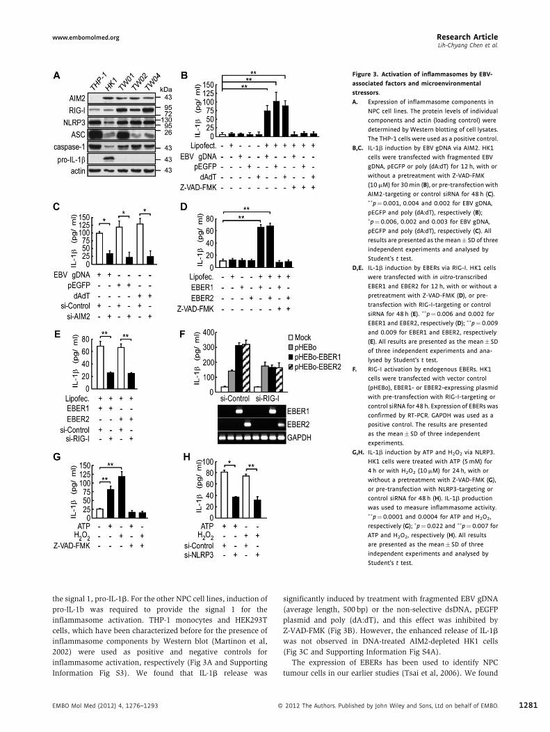
Figure 3. Activation of inflammasomes by EBV-
associated factors and microenvironmental
stressors.
A. Expression of inflammasome components in
NPC cell lines. The protein levels of individual
components and actin (loading control) were
determined by Western blotting of cell lysates.
The THP-1 cells were used as a positive control.
B,C. IL-1b induction by EBV gDNA via AIM2. HK1
cells were transfected with fragmented EBV
gDNA, pEGFP or poly (dA:dT) for 12 h, with or
without a pretreatment with Z-VAD-FMK
(10mM) for 30min (B), or pre-transfection with
AIM2-targeting or control siRNA for 48 h (C).��p¼0.001, 0.004 and 0.002 for EBV gDNA,
pEGFP and poly (dA:dT), respectively (B);�p¼0.006, 0.002 and 0.003 for EBV gDNA,
pEGFP and poly (dA:dT), respectively (C). All
results are presented as themean� SD of three
independent experiments and analysed by
Student’s t test.
D,E. IL-1b induction by EBERs via RIG-I. HK1 cells
were transfected with in vitro-transcribed
EBER1 and EBER2 for 12 h, with or without a
pretreatment with Z-VAD-FMK (D), or pre-
transfection with RIG-I-targeting or control
siRNA for 48 h (E). ��p¼ 0.006 and 0.002 for
EBER1 and EBER2, respectively (D); ��p¼ 0.009
and 0.009 for EBER1 and EBER2, respectively
(E). All results are presented as the mean� SD
of three independent experiments and ana-
lysed by Student’s t test.
F. RIG-I activation by endogenous EBERs. HK1
cells were transfected with vector control
(pHEBo), EBER1- or EBER2-expressing plasmid
with pre-transfection with RIG-I-targeting or
control siRNA for 48 h. Expression of EBERs was
confirmed by RT-PCR. GAPDH was used as a
positive control. The results are presented
as the mean� SD of three independent
experiments.
G,H. IL-1b induction by ATP and H2O2 via NLRP3.
HK1 cells were treated with ATP (5mM) for
4 h or with H2O2 (10mM) for 24 h, with or
without a pretreatment with Z-VAD-FMK (G),
or pre-transfection with NLRP3-targeting or
control siRNA for 48 h (H). IL-1b production
was used to measure inflammasome activity.��p¼0.0001 and 0.0004 for ATP and H2O2,
respectively (G); �p¼ 0.022 and ��p¼0.007 for
ATP and H2O2, respectively (H). All results
are presented as the mean� SD of three
independent experiments and analysed by
Student’s t test.
the signal 1, pro-IL-1b. For the other NPC cell lines, induction of
pro-IL-1b was required to provide the signal 1 for the
inflammasome activation. THP-1 monocytes and HEK293T
cells, which have been characterized before for the presence of
inflammasome components by Western blot (Martinon et al,
2002) were used as positive and negative controls for
inflammasome activation, respectively (Fig 3A and Supporting
Information Fig S3). We found that IL-1b release was
EMBO Mol Med (2012) 4, 1276–1293 �
significantly induced by treatment with fragmented EBV gDNA
(average length, 500 bp) or the non-selective dsDNA, pEGFP
plasmid and poly (dA:dT), and this effect was inhibited by
Z-VAD-FMK (Fig 3B). However, the enhanced release of IL-1b
was not observed in DNA-treated AIM2-depleted HK1 cells
(Fig 3C and Supporting Information Fig S4A).
The expression of EBERs has been used to identify NPC
tumour cells in our earlier studies (Tsai et al, 2006). We found
2012 The Authors. Published by John Wiley and Sons, Ltd on behalf of EMBO. 1281

Research Article www.embomolmed.orgTumour inflammasomes as therapeutic targets
1282
that IL-1b secretion was significantly induced by in vitro
synthesized-EBER1 and EBER2, and secretion was inhibited by
Z-VAD-FMK (Fig 3D) or RIG-I depletion in HK1 cells (Fig 3E and
Supporting Information Fig S4B). Further confirmation of the
specificity of the effect of endogenous EBER1 and EBER2 on RIG-
I-dependent IL-1b secretion was provided by transfection of the
expression plasmids for EBER1 and EBER2 into HK1 cells
(Gregorovic et al, 2011), indicating that endogenous EBER1 and
EBER2 are able to induce IL-1b secretion, which is blocked by
RIG-I depletion (Fig 3F). To test the effect of DAMPs, we
measured IL-1b in the culture medium of normal and NLRP3-
knockdown HK1 cells treated with ATP or an ROS inducer
(H2O2). IL-1b was significantly stimulated by ATP and H2O2,
and this effect could be completely blocked by Z-VAD-FMK
(Fig 3G). In contrast, no stimulation of IL-1b secretion was
observed in NLRP3-knockdown HK1 cells (Fig 3H and
Supporting Information Fig S4C). Furthermore, we used a
second NPC cell line, NPC-TW02 to examine whether
inflammasome activation was the general event in NPC cells.
As shown in Supporting Information Fig S5A and S5B, pro-IL-1b
was inducible by TNF-a, and the inflammasome and IL-1b
secretion were activated by various inflammasome stimulators,
poly (dA:dT), EBER, ATP and H2O2 in NPC-TW02 cells. The
interaction of endogenous AIM2, RIG-I and NLRP3 complexes
with ASC upon stimulation was confirmed by co-immunopre-
cipitation with an ASC-specific antibody (Supporting Informa-
tion Fig S6). Collectively, these findings indicate that AIM2, RIG-
I and NLRP3 inflammasomes in tumour cells are activated by
factors from the viral and tumour microenvironments, thus
contributing to IL-1b secretion.
Enhanced activation of inflammasomes by therapeutic
treatments
We next evaluated whether inflammasomes could be activated
in NPC cells by the current treatment remedies, irradiation and
cisplatin, which are known to induce ROS production
(Kruidering et al, 1997; Valerie et al, 2007) and DNA damage
(Siddik, 2003), resulting in cell death and the release of ATP
(Martins et al, 2009). In addition, intracellular cisplatin is
accumulated in lysosomes (Safaei et al, 2005), which are the
reservoir of the NLRP3 inflammasome activator, cathepsin B
(Schroder & Tschopp, 2010; Schroder et al, 2010).We found that
IL-1b secretion was dose-dependently induced by irradiation
(Fig 4A). Irradiation induced ROS production but not ATP
release (Supporting Information Fig S7A and S7B), and the
irradiation-induced IL-1b was specifically blocked by DPI (an
NADPH oxidase inhibitor) and Z-VAD-FMK, but not by oATP
(a P2X7-receptor antagonist) or apyrase (an ATP scavenger;
Fig 4B). Notably, irradiation-induced IL-1b was reduced by
depletion of AIM2, but not of NLRP3 or RIG-I (Fig 4C). To
identify the direct, endogenous ligand of AIM2, wemeasured the
levels of cytosolic DNA in NPC cells. As shown in Fig 4D,
irradiation treatment resulted in elevated cytosolic levels of both
nuclear (3.6-fold) and mitochondrial (4.1-fold) DNA. This level
of induction was similar to results reported previously for the
irradiation-mediated release of mitochondrial DNA into cytosol
(Nakahira et al, 2011; Shimada et al, 2012). Conversely, we
� 2012 The Authors. Published by John Wiley and Sons, Ltd on behalf of EMBO.
found that cisplatin treatment dose-dependently induced IL-1b
secretion (Fig 4E) and ATP release but not ROS production
(Supporting Information Fig S7C and S7D). Cisplatin-induced
IL-1b was specifically blocked by CA-074-Me (cathepsin B
inhibitor) and Z-VAD-FMK (Fig 4F), and was further demon-
strated in cathepsin B-depleted cells treated with cisplatin
(Fig 4G and Supporting Information Fig S4D). Interestingly,
cisplatin-induced IL-1b was significantly reduced by depletion
of NLRP3, but not of AIM2 or RIG-I (Fig 4H). Similar to HK1
cells, the inflammasome and IL-1b secretion were also activated
by irradiation and cisplatin in NPC-TW02 cells (Supporting
Information Fig S5C). The induction of AIM2 and NLRP3
inflammasome assembly in HK1 cells by irradiation and
cisplatin, respectively, was confirmed by immunoprecipitation
of endogenous ASC from irradiated and cisplatin-treated cell
lysates with an ASC-specific antibody (Supporting Information
Fig S8). Therefore, these results suggest that irradiation induces
ROS-dependent AIM2 activation, while cisplatin induces
cathepsin B-dependent NLRP3 activation (Schroder et al,
2010). Importantly, we demonstrate that therapeutic treatment
activates the inflammasome in NPC cells and can enhance IL-1b
production (from �174 to �492 pg/ml) in the presence of
tumour microenvironmental factors (Fig 4I).
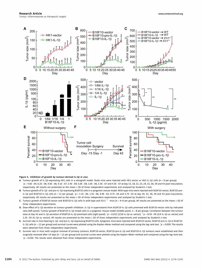
Tumour-derived IL-1b inhibits tumour growth in vivo
We examined the effect of tumour-derived IL-1b on tumour
growth in vivo by using HK1-IL-1b and HK1-vector cells
(Supporting Information Fig S9A and S9B). As shown in Fig 5A,
HK1-derived IL-1b secretion significantly abolished tumour
growth in nude mice in vivo starting at day 15, although tumour
growth between HK1-vector and HK1-IL-1b cells was similar
during the first week. The inhibitory effect of tumour-derived IL-
1b on tumour growth was further evaluated in immunocompe-
tent wild-type mice. B16F10 mouse melanoma cells, which do
not express pro-IL-1b, NLRP3 and AIM2 (data not shown), were
chosen to generate B16F10-IL-1b, B16F10-pro-IL-1b (expressing
non-secretable IL-1b) and B16F10-vector cells (Supporting
Information Fig S9C and S9D). B16F10 cell-derived IL-1b
significantly abolished tumour growth in wild-type syngeneic
C57BL/6 mice and nude mice at days 29 and 36, respectively,
although the tumour growth was similar during the first 3 weeks
for B16F10-IL-1b, B16F10-pro-IL-1b and B16F10-vector cells
(Fig 5B and Supporting Information Fig S10).
Since IL-1b does not directly inhibit tumour cell proliferation
(Supporting Information Fig S9B and S9D), the underlying
mechanisms involved in the IL-1b-mediated inhibitory effect
in vivo most likely occur through an indirect paracrine manner.
Therefore, the role of the host’s IL-1b receptor-mediated
signalling in tumour growth was further studied in Il1r1�/�
mice. As shown in Fig 5C, IL-1b negative B16F10-vector
tumours grew faster in wild-type mice than in Il1r1�/� mice,
suggesting that IL-1b produced by inflammatory cells and
stromal cells may promote tumour growth. In addition, the
tumour size of B16F10-IL-1b- and B16F10-vector tumours was
similar in Il1r1�/� mice, indicating that the inhibiting effect of
tumour-derived IL-1b on tumour growth was dependent on the
host response. Our data support the view that IL-1b has
EMBO Mol Med (2012) 4, 1276–1293

www.embomolmed.org Research ArticleLih-Chyang Chen et al.
Figure 4. Enhanced activation of inflammasomes by therapeutic treatments.
A. Dose-dependent induction of IL-1b by irradiation. HK1 cells were treated with various doses of irradiation for 24 h. ��p¼0.001; ���p¼2.7E�05, 9.8E�06 and
9.7E�08 for 20, 30 and 40Gy, respectively. All results are presented as the mean� SD of six independent experiments and analysed by Student’s t test.
B. ROS production is required for the induction of IL-1b by irradiation. HK-1 cells were irradiated (30Gy) with or without a 30min pretreatment with oATP
(100mM), apyrase (2.5 unit/ml), DPI (10mM) or Z-VAD-FMK. �p¼ 0.02 and 0.035 for DPI and Z-VAD-FMK, respectively. All results are presented as the
mean� SD of three independent experiments and analysed by Student’s t test.
C. Requirement of AIM2 for irradiation-induced IL-1b production. HK-1 cells transfected with NLRP3-targeting, AIM2-targeting, RIG-I-targeting, and control
siRNA for 48 h were irradiated. ���p¼0.0003, all results are presented as the mean� SD of three independent experiments and analysed by Student’s t test.
D. Quantitative PCR analysis of cytosolic DNA in irradiation-treated HK1 cells. HK1 cells were treated with irradiation (30Gy) for 24 h or ATP (5mM) for 4 h.
Nuclear DNA andmitochondrial DNA were indicated as nuDNA andmtDNA, respectively. �p¼ 0.005 (nuDNA analysis); �p¼ 0.016 and 0.017 for irradiation and
ATP, respectively (mtDNA analysis). All results are presented as the mean� SD of three independent experiments and analysed by Student’s t test.
E. Dose-dependent induction of IL-1b by cisplatin. HK1 cells were treated with various doses of cisplatin for 24 h. �p¼ 0.012. ��p¼ 0.007 and 0.003 for 40 and
50mM, respectively. All results are presented as the mean� SD of three independent experiments and analysed by Student’s t test.
F. Requirement of cathepsin B activity for the induction of IL-1b by cisplatin. HK-1 cells were incubated with 40mM cisplatin with or without a 30min
pretreatment with oATP, apyrase, DPI, CA-074Me (10mM) or Z-VAD-FMK. ��p¼0.0017 and 0.0005 for CA-074Me and Z-VAD-FMK, respectively. All results are
presented as the mean� SD of three independent experiments and analysed by Student’s t test.
G. Requirement of cathepsin B for cisplatin-induced IL-1b production. HK-1 cells transfected with cathepsin B-targeting and control siRNA for 48 h were treated
with cisplatin. �p¼0.037. The results are presented as the mean� SD of three independent experiments and analysed by Student’s t test.
H. Requirement of NLRP3 for cisplatin-induced IL-1b production. HK-1 cells transfected with NLRP3-targeting, AIM2-targeting, RIG-I-targeting and control siRNA
for 48 h were treated with cisplatin. �p¼ 0.029. The results are presented as themean� SD of three independent experiments and analysed by Student’s t test.
I. Enhanced induction of tumour microenvironmental factor-stimulated IL-1b by therapeutic treatments. HK1 cells were treated with tumour microenvi-
ronmental factors, EBV gDNA/EBERs/ATP/H2O2 as previously described with or without therapies, irradiation and cisplatin. IL-1b production was used to
measure inflammasome activity. �p¼0.00004, the results are presented as the mean� SD of four independent experiments and analysed by Student’s t test.
EMBO Mol Med (2012) 4, 1276–1293 � 2012 The Authors. Published by John Wiley and Sons, Ltd on behalf of EMBO. 1283
Research Article www.embomolmed.orgTumour inflammasomes as therapeutic targets
Figure 5. Inhibition of growth by tumour-derived IL-1b in vivo.
A. Tumour growth of IL-1b-expressing HK1 cells in a xenograft model. Nude mice were injected with HK1-vector or HK1-IL-1b cells (n¼ 9 per group).�p¼ 4.6E�04, 6.2E�06, 4.0E�06, 4.1E�07, 5.4E�09, 5.0E�08, 1.0E�06, 5.3E�07 and 4.3E�07 at day 15, 18, 22, 25, 29, 32, 36, 39 and 43 post-inoculation,
respectively. All results are presented as the mean� SD of three independent experiments and analysed by Student’s t test.
B. Tumour growth of IL-1b- and pro-IL-1b-expressing B16F10 cells in a syngeneic mouse model. Wild-type mice were injected with B16F10-vector, B16F10-pro-
IL-1b and B16F10-IL-1b cells (n¼12 per group). �p¼ 1.1E�06, 1.5E�06, 6.9E�03, 9.7E�04 and 5.7E�03 at day 29, 32, 36, 39 and 43 post-inoculation,
respectively. All results are presented as the mean� SD of three independent experiments and analysed by Student’s t test.
C. Tumour growth of B16F10-vector and B16F10-IL-1b cells in wild-type and Il1r1�/� mice (n¼ 4–6 per group). All results are presented as the mean� SD of
three independent experiments.
D. Dose effect of IL-1b secretion on tumour growth inhibition. IL-1b in supernatants from B16F10-IL-1b cells premixed with B16F10-vector cells by indicated
ratio (left panel). Tumour growth of B16F10-IL-1b mixed cells in a syngeneic mouse model (middle panel, n¼8 per group). Correlation between the tumour
sizes at day 43 and IL-1b secretion of B16F10-IL-1b premixed cells (right panel). �p¼0.022 (1/16 IL-1b vs. vector); ��p¼8.5E�04 (1/4 IL-1b vs. vector) and
1.2E�05 (IL-1b vs. vector). All results are presented as the mean� SD of three independent experiments and analysed by Student’s t test.
E. Survival rate in mice bearing IL-1b- and pro-IL-1b-expressing B16F10 cells. Syngeneic mice were injected with B16F10-vector, B16F10-pro-IL-1b or B16F10-
IL-1b cells (n¼12 per group) and survival curves were plotted using the Kaplan–Meier method and compared using the log-rank test. �p¼0.009. The results
were obtained from three independent experiments.
F. Survival rate in mice with surgical removal of primary tumours. B16F10-vector, B16F10-pro-IL-1b and B16F10-IL-1b tumours were established and then
surgically removed after 14 days (n¼12 per group) and survival curves were plotted using the Kaplan–Meier method and compared using the log-rank test.�p¼ 0.036. The results were obtained from three independent experiments.
1284 � 2012 The Authors. Published by John Wiley and Sons, Ltd on behalf of EMBO. EMBO Mol Med (2012) 4, 1276–1293

www.embomolmed.org Research ArticleLih-Chyang Chen et al.
contradictory roles in protumour and antitumour activity,
depending on the host immune response. Thus, in order to
determine the amount of tumour-derived IL-1b required for
inhibition of tumour growth, B16F10-IL-1b and B16F10-vector
cells were mixed at different ratios (0:1, 1:64, 1:16, 1:4 and 1:0)
to obtain low to high levels of secreted cytokine (0, 17.8, 64.2,
328.6 and 1580.3 pg/ml, respectively in Fig 5D, left panel). The
cell mixtures were inoculated into wild-type mice, and tumour
sizes were monitored for 43 days. The outgrowing tumours
indeed contain a mixture of IL-1b expressing and non-
expressing cells (Supporting Information Fig S11). As shown
in Fig 5D, middle panel, tumour growth was inhibited by
tumour-derived IL-1b in a dose-dependent manner. At day 43,
mice carrying cells secreting relatively low levels of IL-1b (1/16
B16F10-IL-1b) harboured significantly smaller tumours
(277mm2) than those of B16F10-vector cells (376mm2). Our
data showed that the threshold for the antitumour activity of
IL-1b was 64.2 pg/ml (Fig 5D, right panel).
The survival rates of mice carrying B16F10-IL-1b tumours
(83.3%) were significantly better than for mice carrying
tumours arising from B16F10-pro-IL-1b (25.0%, p¼ 0.003) or
B16F10-vector (33.3%, p¼ 0.006) cells (Fig 5E). We further
studied the effect of tumour-derived IL-1b on tumour recurrence
following surgical removal of the large primary tumours.
Mice with IL-1b-secreting B16F10-IL-1b tumours showed
better survival (83.3%) than those with tumours arising
from B16F10-pro-IL-1b (41.7%, p¼ 0.034) or B16F10-vector
(33.3%, p¼ 0.01) cells (Fig 5F). Collectively, the data show
that high levels of tumour-derived IL-1b can inhibit tumour
growth and local recurrence. This is consistent with the clinical
correlation between high levels of inflammasome protein and
IL-1b expression and better survival in NPC patients after
treatment (Fig 1D). Thus, our findings strongly suggest that
Figure 6. TANs as important effector cells for the IL-1b-mediated antitumour
A. Leukocyte subsets in tumours. Percentage of leukocytes (CD45þ), myeloid ce
dendritic cells (CD11cþ), T cells (CD3þ), B cells (B220þ) and NK cells (NK1.1þ)
(n¼3 per group) was determined by flow cytometry. �p¼ 0.0364, 0.0222 and 0.
presented as the mean� SD of three independent experiments and analysed
B. Induction of intratumoural neutrophils by IL-1b. Percentage of TANs (CD11bþ
group) in wild-type and Il1r1�/� mice was determined by flow cytometry. �p
experiments and analysed by Student’s t test.
C. Increased TANs in B16F10-IL-1b tumour. Ly6G immunohistochemistry perfor
D. The morphology of TANs. Neutrophils were sorted from CD11bþ/Ly6Gþ cells
E. Quantitative RT-PCR analysis. The fold change of gene expression in purified
using the expression level in TANs from B16F10-vector-tumours as the denom
independent experiments and analysed by Student’s t test.
F. Neutrophil depletion. Mice bearing B16F10-IL-1b- or B16F10-vector-tumour
intraperitoneally twice a week (n¼ 4 per group). After 2 weeks, the percentag
cytometry. �p¼ 0.005. The results are presented as the mean� SD of four ind
G. Effect of neutrophil depletion on tumour growth. Mice bearing B16F10-IL-1b
intraperitoneally (arrowheads) twice a week during the study period (n¼7–9 p
respectively. All results are presented as the mean� SD and analysed by Stud
H. TANs in NPC tumours. The H&E results are shown at 400� magnification (upp
TANs. Scale bars, 50mm.
I. Kaplan–Meier survival analysis of LRFS.
J. Correlation of TANs and pro-IL-1b and inflammasome components in NPC biop
RIG-I, NLRP3, ASC, caspase-1 and IL-1b versus the positive rates of TANs in pat
using Student’s t test (Supporting Information Table S13).
EMBO Mol Med (2012) 4, 1276–1293 �
tumour-derived IL-1b can contribute to local tumour control and
better survival.
Tumour-derived IL-1b can induce neutrophil infiltration
into tumours
Since the inhibitory effect of IL-1b on tumour growth was
dependent on IL-1b receptor-mediated signalling of the host
immune system (Fig 5B and C), we further investigated the
composition of tumour-infiltrated leukocyte subsets from
tumours by flow cytometry. We found that the infiltration of
leukocytes, myeloid cells and neutrophils was dramatically
higher in B16F10-IL-1b tumours, compared with B16F10-pro-IL-
1b and B16F10-vector tumours (Fig 6A). In contrast, the
proportion of macrophages, dendritic cells, T cells, B cells and
NK cells was not affected by the IL-1b-secreting tumours.
However, the effect of tumour-derived IL-1b on neutrophil
infiltration was completely abolished in Il1r1�/� mice (Fig 6B).
This suggested that the tumour-derived IL-1b induced neu-
trophil infiltration through the activation of IL-1b receptor
signalling in host cells. Further confirmation of neutrophil
infiltration into B16F10-IL-1b tumours was provided by
immunohistochemical staining of tumour tissues using a
specific antibody against neutrophil marker Ly6G. As shown
in Fig 6C, largely infiltrating neutrophils were observed in
B16F10-IL-1b tumours, but not in B16F10-pro-IL-1b and
B16F10-vector tumours. These intratumoural CD11bþ/Ly6Gþ
cells (tumour-associated neutrophils, TANs) indeed had a clear
neutrophil-like morphology (Fig 6D). Interestingly, most of the
TANs in B16F10-IL-1b tumours were more lobulated and
hypersegmented; in contrast, those in B16F10-vector tumours
maintained the characteristic banded appearance typical of
blood neutrophils. Our data indicate that neutrophils are the
mainly infiltrated cell type recruited by tumour-derived IL-1b.
activity.
lls (CD11bþ), neutrophils (CD11bþ/Ly6Gþ), macrophages (CD11bþ/F4/80þ),
in the B16F10-IL-1b-, B16F10-pro-IL-1b- or B16F10-vector-bearing tumours
0004 for leukocytes, myeloid cells and neutrophils, respectively. The results are
by Student’s t test.
/Ly6Gþ) in the B16F10-IL-1b and B16F10-vector-bearing tumours (n¼ 3 per
¼0.003. The results are presented as the mean� SD of three independent
med on tumour tissues. Scale bars, 100mm.
in B16F10-vector or B16F10-IL-1b tumours. Scale bars, 10mm.
neutrophils (CD11bþ/Ly6Gþ) of B16F10-IL-1b- and B16F10-vector-tumours,
inator, was calculated (n¼ 3 per group). The results were obtained from three
s were injected with either the anti-Ly6G 1A8 or a control IgG antibody
es of TANs (CD11bþ/Ly6Gþ) in whole tumour cells were determined by flow
ependent experiments and analysed by Student’s t test.
- or B16F10-vector-tumours were injected with antibodies described in (F)
er group). �p¼ 0.006, 0.003 and 0.002 at day 39, 43 and 46 post-inoculation,
ent’s t test. Each experiment was repeated at least twice.
er panel) and the enlarged box areas (lower panel). Arrowhead indicates the
sies. The positive rates of TANs in patients with high expression levels of AIM2,
ients with low expression levels of the above-described proteins are analysed
"
2012 The Authors. Published by John Wiley and Sons, Ltd on behalf of EMBO. 1285

Research Article www.embomolmed.orgTumour inflammasomes as therapeutic targets
Figure 6.
1286 � 2012 The Authors. Published by John Wiley and Sons, Ltd on behalf of EMBO. EMBO Mol Med (2012) 4, 1276–1293

www.embomolmed.org Research ArticleLih-Chyang Chen et al.
The TANs present in IL-1b-expressing tumours have an
immunostimulatory phenotype
Since TANs are composed of two subtype cells, antitumour N1
TANs (immunostimulator) and protumour N2 TANs (immu-
nosuppressor) (Fridlender et al, 2009; Hofman, 2010; Manto-
vani, 2009), we compared the mRNA levels of selected
cytokines, adhesion molecules and chemokines in TANs from
B16F10-IL-1b tumours versus B16F10-vector tumours by
quantitative RT-PCR (Fig 6E). The levels of arginase mRNA,
an important immunosuppressor of the adaptive immune
system (Rodriguez & Ochoa, 2008), were 5.0-fold lower in
TANs from B16F10-IL-1b tumours. In contrast, the mRNA levels
of the important immunostimulators, TNF-a, ICAM1 and iNOS
were significantly increased. The levels of neutrophil chemoat-
tractants, CCL2 and CCL5 were significantly reduced, whereas
CCL3 partially increased. Our data show that tumour-derived
IL-1b can polarize TANs towards an N1 TAN-like immuno-
stimulatory phenotype.
TANs are important effector cells for IL-1b-mediated
antitumour activity
We next studied the functional significance of TANs in IL-1b-
mediated tumour control by depleting the Ly6Gþ cells in
tumour-bearing animals. Tumour cells were inoculated in the
footpad, followed by intraperitoneal injection of an anti-Ly6G
monoclonal antibody 1A8 or isotype-matched control IgG
intraperitoneally, and tumour growth was then determined. A
significant reduction (66%) in TANs from B16F10-IL-1b
tumour-bearing mice was noted 2 weeks after treatment
(Fig 6F). Palpable tumours were observed at day 18, and
antibodies were introduced as described above twice per week
until day 43. As shown in Fig 6G, the tumour size of B16F10-IL-
1b tumour-bearing mice with neutrophil depletion increased
significantly at day 39 (p¼ 0.006) as compared to B16F10-IL-1b
tumour-bearing mice treated with isotope antibody. In contrast,
depletion of neutrophils in B16F10-vector tumour-bearing mice
did not affect tumour growth. Together, these data indicate that
depletion of TANs resulted in significant abrogation of the IL-1b-
mediated antitumour activity, suggesting that TANs contribute
to the antitumour activity of tumour-derived IL-1b.
Association of TANs with better survival in NPC patients
We further analyzed the significance of TANs on tumour
development in NPC patients. Thirteen out of the 140 tested NPC
specimens were TAN-positive as shown by hematoxylin and
eosin (H&E) staining (Fig 6H). Kaplan–Meier survival analysis
of the H&E results showed that the presence of TANs correlated
with better LRFS (Fig 6I). Notably, none of the patients whose
tumours were TAN-positive had local recurrence within 5 years
after treatment (Fig 6I). In addition, the correlation of the protein
levels of IL-1b and different inflammasome components with
the positive rate of TANs was analysed using the NPC samples
studied in Figs 1 and 6H. As shown in Fig 6J, the positive rates of
TANs were higher in the patients with high level of AIM2, RIG-I,
NLRP3, ASC, caspase-1 or IL-1b. Inflammasome genes were
significantly correlated with neutrophil infiltration in NPC
patients (Supporting Information Table S13). Taken together,
EMBO Mol Med (2012) 4, 1276–1293 �
our data inmice and humans are consistent with TANs playing a
major role in local tumour control.
Hierarchical cluster analysis of AIM2, RIG-I and NLRP3
inflammsome gene expression levels in 114 cancer cell lines
In order to delineate the function of inflammasomes in other
cancer types, we evaluated the expression patterns of AIM2,
RIG-I, NLRP3, ASC and caspase-1 in 114 cancer cell lines
involving 22 cancer types. For this analysis, we used 324
Affymetrix U133 Plus 2.0 array data sets from the eight NPC cell
lines generated in this study and 106 cell lines from the public
GSK Cancer Cell Line Genomic Profiling Data (Greshock et al,
2010), according to a distance tree obtained through hierarchical
cluster analysis. The cancer cell lines were separated into four
clusters (Supporting Information Fig S12A and Table S14), and
all eight NPC cell lines were clustered together and were closely
grouped with 9 of 17 lymphoma and 2 of 8 brain cancer cell lines
(designated as cluster 1) but not with cell lines of other cancer
types (Supporting Information Fig S12B and S12C). Since both
lymphoma and NPC are associated with EBV infection, we
found that high levels of AIM2, RIG-I and NLRP3 inflammasome
expression are significantly correlated with EBV infection in
lymphoma cell lines (p¼ 0.043; Supporting Information Fig
S12D), suggesting that EBV infection is a potential factor for
AIM2, RIG-I and NLRP3 inflammasome overexpression in
cancer cells of both epithelial and lymphocyte origin.
DISCUSSION
In this report, we demonstrated a link between neutrophils and
tumour suppression through inflammasome activation. In NPC,
NLRP3, AIM2 and RIG-I inflammasomes were overexpressed in
the tumour cells and were required for IL-1b production in
response to PAMPs and DAMPs in the tumour microenviron-
ment and therapeutic treatment. In addition, high levels of
tumour-derived IL-1b can recruit a large number of TANs,
which significantly inhibit tumour growth in mice. Importantly,
the expression of NLRP3, AIM2 and RIG-I inflammasomes and
IL-1b, as well as the presence of TANs, were associated with
better survival in NPC patients. This is the first study to
demonstrate that inflammasomes in the tumour cells can
respond to PAMPs, DAMPs and therapeutic treatment, and
contribute to TANs-dependent tumour suppression (Fig 7).
Inflammation has contradictory roles in carcinogenesis
through production of tumour-promoting proinflammatory
cytokines and facilitation of antitumour immune responses
(Hagemann et al, 2007; Zitvogel et al, 2012). In viral infection-
associated cancers, e.g. EBV-associated NPC, both PAMPs and
DAMPs exist in the tumour microenvironment, where they are
potential sources of stimuli for inflammasome activation in vivo.
In addition, the tumour cells express viral antigens that can
provide specificity for cytotoxic T cells (Berger et al, 2009;
Grivennikov et al, 2010). Thus, the activation of tumour
inflammasomes may facilitate antitumour immunity by initiat-
ing inflammation. Our findings show that the expression of
inflammasomes and the proinflammatory cytokine, IL-1b, in
2012 The Authors. Published by John Wiley and Sons, Ltd on behalf of EMBO. 1287

Research Article www.embomolmed.orgTumour inflammasomes as therapeutic targets
Figure 7. Role of inflammasomes in tumour cell
responses to microenvironmental factors and
therapy. Before treatment, tumour cells are latently
infected with EBV and express LMP1, which can
induce pro-IL-1b production through NF-kB and
MAPK signalling. In the tumour microenvironment,
tumour cells encounter PAMPs (EBV gDNA and EBV-
encoded RNAs, and EBER1 and EBER2) and DAMPs
(intracellular ROS or H2O2, and extracellular ATP)
produced by dying or stressed cells. The PAMPs and
DAMPs can stimulate AIM2, RIG-I and NLRP3, which
then form inflammasomes with ASC and caspase-1,
resulting in cleavage of pro-IL-1b and low-level
basal IL-1b secretion. Low levels of tumour-derived
IL-1b can facilitate tumour growth. Treatment of
NPC patients with irradiation leads to ROS
production that leads to the release of nuclear and
mitochondrial DNA into the cytosol of tumour cells,
while chemotherapy (cisplatin) causes cathepsin B to
be released from ruptured lysosomes due to cisplatin
accumulation. Thus, AIM2 and NLRP3 in tumour cells
are further activated to secret IL-1b at higher levels.
Finally, high levels of tumour-derived IL-1b achieved
by therapeutic treatment can inhibit tumour growth
and local relapse by recruiting neutrophils,
especially immunostimulatory N1 TANs.
1288
tumour cells is significantly correlated with better survival in
NPC patients (Fig 1), suggesting a role of tumour inflamma-
somes in tumour suppression. Although IL-1b has a protu-
mourigenic effect [Fig 5C in this study; and (Apte & Voronov,
� 2012 The Authors. Published by John Wiley and Sons, Ltd on behalf of EMBO.
2008)], inflammasome and IL-1b-mediated antitumour activity
have been reported recently by two other studies in gene-
deficient mice, which described the effect of the NLRP3
and IL-1b produced by hematopoietic cells against cancer
EMBO Mol Med (2012) 4, 1276–1293

www.embomolmed.org Research ArticleLih-Chyang Chen et al.
(Allen et al, 2010; Ghiringhelli et al, 2009). Consistent with their
findings, our data show that the inflammasomes in malignant
cells are responsive to PAMPs, DAMPs and therapeutic
treatment (Figs 3 and 4), and the interaction of tumour-derived
IL-1b (over 64.2 pg/ml) and its receptors on the host cells can
inhibit tumour growth and recurrence (Fig 5). In addition,
tumour growth and recurrence are inhibited by IL-1b, rather
than pro-IL-1b (Fig 5B), suggesting that cleavage of pro-IL-1b to
IL-1b by the inflammasome is critical for tumour suppression.
Taken together, our work reveals that tumour cells are as able as
immune cells to use the inflammasome for responding to
infection, stress and therapeutic treatment. Paradoxically, when
tumour-derived IL-1b reaches a certain threshold, it can also
limit tumour growth in vivo.
Our findings are in line with the emerging concept of tumour
inflammation, according to which cancer cells are able to
manipulate the host response by recruiting immune cells
(Demaria et al, 2010; Grivennikov et al, 2010; Hagemann et al,
2007). We demonstrate that tumour-derived IL-1b can alarm the
immune system to induce an influx of neutrophils to tumour
sites (Fig 6A and B). These findings are consistent with recent
reports that NLRP3 activation and IL-1b in experimental liver
injury can direct neutrophil trafficking by drawing neutrophils
out of the circulation to adhere to the vascular endothelium at
inflammation sites (McDonald et al, 2010). More significantly,
depletion of these neutrophils significantly blunts the anti-
tumour effect of IL-1b (Fig 6G), showing that IL-1b-induced
TANs act as the effectors of tumour elimination. These IL-1b-
induced TANs express more immunoactivating cytokines and
less immune-suppressing arginase (Fig 6E). Thus, besides the
recent reports showing the tumour cytotoxicity of N1 TANs
through TGF-b blockade (Fridlender et al, 2009) and tumour-
entrained neutrophils by CCL2 (Granot et al, 2011), we identify a
new mechanism mediated by IL-1b. Furthermore, the presence
of TANs is associated with better survival in NPC patients
(Fig 6I) and advanced gastric carcinoma (Caruso et al, 2002).
The partial effect of granulocyte depletion on recovering tumour
growth may be attributed to the activity of remaining
immunostimulatory TANs. In addition, tumour-derived IL-1b
may potentially induce dendritic cell-dependent adaptive
immunity against tumours (Ghiringhelli et al, 2009). Taken
together, our findings support the hypothesis that TANs are
recruited by tumour-produced cytokines, e.g. IL-1b, and exert a
host-mediated cytotoxic effect against the tumour.
A correlation between an elevated neutrophil to lymphocyte
ratio (NLR) in the blood stream and poor survival was recently
reported in NPC (An et al, 2011). While the underlying
mechanisms remain elusive, it may be attributed to the
following reasons. First, elevated NLR-associated lymphocyto-
penia may reduce lymphocyte-dependent cytotoxic cell death,
which is important for antitumour activity. Second, elevated
NLR-associated neutrophilic leukocytosis may increase the
production of angiogenic growth factors that function in
tumour-related angiogenesis and metastasis (An et al, 2011).
Improving the current treatment for patients with elevated NLR
is important. Our and Fridlender et al’s findings on the
recruitment and activation of TANs by IL-1b and TGF-b
EMBO Mol Med (2012) 4, 1276–1293 �
blockade shed some light on the possibility of conversion of
circulating protumour neutrophils to intratumoural antitumour
neutrophils (Fridlender et al, 2009). One might consider taking
advantage of the antitumour activity of N1 TANs to improve
the treatment of the patients with elevated NLR-associated
neutrophilic leukocytosis.
Irradiation and cisplatin are widely used for cancer treatment.
For the first time, we showed that irradiation can activate the
cytosolic DNA receptor, AIM2, via ROS production in malignant
cells, correlating with the release of nuclear and mitochondrial
DNA into the cytosol [Fig 4D and (Patrushev et al, 2006)].
Similar to a recent report indicating that the release of
mitochondrial DNA during ATP-triggered apoptosis can activate
NLRP3 inflammasome in macrophages (Nakahira et al, 2011;
Shimada et al, 2012), we found that inflammasomes activated
by ROS production and cytosolic DNA release during apoptosis
can facilitate antitumour immunity through IL-1b-mediated
inflammation. We also showed that cisplatin can induce IL-1b
secretion in NPC cells, which is in agreement with a previous
study indicating that cisplatin can activate caspase-1 (Kondo
et al, 1995) and accumulates in lysosomes (Safaei et al, 2005),
which are the reservoir of cathepsin B (Schroder & Tschopp,
2010; Schroder et al, 2010). Here, we link cisplatin and
inflammasomes and show that the cathepsin B-dependent
NLRP3 inflammasome can respond to cisplatin and further
induce caspase-1 activation and IL-1b production (Fig 4E–H).
Furthermore, cathepsin B is highly expressed in NPC (Chang
et al, 2010). Thus, our results suggest that therapeutic treatment
can cooperate with PAMPs and DAMPs in a tumour micro-
environment to induce more IL-1b secretion, which conse-
quently results in efficient tumour suppression by recruiting
neutrophils, especially immunostimulatory N1 TANs. The
association between therapeutic induction of IL-1b and
neutrophil recruitment cannot be demonstrated because the
collection of primary tumour biopsies from patients who had
recently undergone treatment is not ethical and would not be
allowed. Nonetheless, we provide strong evidence that the
chemoradiotherapeutic activation of the NLRP3 and AIM2
inflammasomes, via cathepsin B and ROS, is likely to emerge as
a crucial player in the regulation of cancer immunotherapy.
Compared with the results of clinical trials showing that IL-1b
treatment through intravenous infusion and subcutaneous
administration effectively increases the level of peripheral
neutrophils in cancer patients (Veltri & Smith, 1996), our
findings show that manipulating tumour-derived IL-1b by
chemoradiotherapy to induce N1 TANs should be considered as
a strategy for improving immunotherapy, and that inflamma-
somes are promising therapeutic targets in cancers.
Our results showed that pro-IL-1b is induced by LMP1 in 4
NPC cell lines, NPC-TW01, -TW02, -TW04 and HK1; similar
induction was previously reported in NPC-TW01 (Huang et al,
2010). Since inflammasome-mediated release of IL-1b can drive
chronic inflammation with protumour activity (Fig 5C; Apte &
Voronov, 2008), this might explain why in EBV-associated
cancers, the inflammasome genes are induced and activated by
viral factors such as LMP1 and EBERs (Fig 7). The practical
implications of this study are the discovery of new roles for
2012 The Authors. Published by John Wiley and Sons, Ltd on behalf of EMBO. 1289
Research Article www.embomolmed.orgTumour inflammasomes as therapeutic targets
1290
inflammasomes, which are favourable prognostic biomarkers
for cancer patients, and the identification of inflammasomes are
promising therapeutic targets in cancers.
MATERIALS AND METHODS
Patients and clinical characteristics
The retrospective cohort comprised 144 NPC patients who had been
admitted to Chang Gung Memorial Hospital (CGMH) at Lin-Kou from
1990 to 2000. Clinical stage was defined according to the 2002
cancer staging system revised by the American Joint Committee on
Cancer. Histological typing was done according to the World Health
Organization (WHO) classification criteria (Chen et al, 2008).
This study was reviewed and approved by the institutional review
board and ethics committee of CGMH. Informed consent was
obtained from all subjects and the experiments conformed to the
principles set out in the WMA Declaration of Helsinki (http://
www.wma.net/en/30publications/10policies/b3/) and the NIH
Belmont Report (http://ohsr.od.nih.gov/guidelines/belmont.html). Patients
diagnosed with distant metastatic disease at presentation (M1 stage)
and/or those who had undergone previous treatment at another
institute were excluded from the present study. Patient characteristics
and clinical features are summarized in Supporting Information Tables
S15–S17. The primary end-point was LRFS and DFS, which was
calculated from the date of diagnosis to the date of death or the last
follow-up.
Immunohistochemical staining
Immunohistochemical analyses were described previously (Chen et al,
2008, 2010b, 2011). The antibodies for individual proteins can be
found in Supporting Information Table S18. Protein expression was
assessed by quantitative scoring of the staining intensity and the
proportion of positively stained cells. The staining intensity was graded
as 0, 1, 2 or 3 to indicate undetectable, weak, moderate and strong
staining, respectively. These scores were multiplied by the percentage
of cells that showed positive staining; the resulting scores, which
were taken as reflecting protein expression, were used to classify
the specimens/patients into two groups: ‘high-level’ expression (ASC
scores>120, caspase-1 scores>100, IL-1b scores>40, NLRP3
scores>160, AIM2 scores>140, RIG-I scores>10, CIITA scores>50,
NLRC4 scores>100, NLRP7 scores>60) and ‘low-level’ expression
(ASC scores�120, caspase-1 scores�100, IL-1b scores�40, NLRP3
scores�160, AIM2 scores�140, RIG-I scores�10, CIITA scores�50,
NLRC4 scores�100, NLRP7 scores�60). ASC-, caspase-1-, IL-1b-,
NLRP3-, AIM2-, RIG-I-, CIITA-, NLRC4- and NLRP7-positive tumour
cells in representative microscopic fields were scored independently by
two experienced pathologists.
Definition of TANs in NPC patients
Hematoxylin and eosin-stained sections were examined to identify
TANs (Reid et al, 2011). Only foci with neutrophils within and/or
immediately adjacent to tumour cells were taken into consideration.
The areas immediately adjacent to necrotic tissue were disregarded.
Ten non-overlapping fields were examined. The number of TANs was
assessed and the areas with �10 neutrophils/100 epithelial cells were
considered negative and areas with >10 neutrophils/100 epithelial
� 2012 The Authors. Published by John Wiley and Sons, Ltd on behalf of EMBO.
cells were considered as positive for TANs. The cut-off value, >10
neutrophils/100 epithelial cells, was defined as TANs positive, which
was determined from Receiver Operating Characteristics curve
analysis. The criteria have also been used in a recent publication by
Reid MD et al (Reid et al, 2011).
Reagents
ATP, poly (dA:dT), oATP, apyrase, DPI, CA-074 Me, Z-VAD-FMK and
cisplatin were purchased from Sigma–Aldrich and H2O2, BAY11-7082,
SP600125, SB203580 and PD98059 from Calbiochem.
Cell culture and treatments
NPC-TW01, -TW02, -TW04 and HK1 cells were cultured under
conditions described before (Chen et al, 2010b, 2011). THP-1, CNE-
1, HONE-1 and HK1-EBV cells were grown in RPMI-1640 medium and
BM1 cells were grown in Dulbecco’s modified Eagle’s medium
(DMEM), supplemented with 10% fetal bovine serum, 100U/ml
penicillin and 100mg/ml streptomycin at 378C under 5% CO2. HK1-
EBV cells were maintained with 500mg/ml G418. Lentiviruses were
established by following the protocol of the RNAi Core, Taiwan (http://
rnail.genmed.sinica.edu.tw), and used to generate human IL-1b-
expressing or vector-control HK1 cells, as well as mouse pro-IL-1b-
expressing, mouse IL-1b-expressing or vector-control B16F10 cells.
For transfection assays, sonicated EBV gDNA (around 500 bp), poly
(dA:dT), pEGFP, EBER1/2, pHEBo, pHEBo-EBER1/2 were transfected by
Lipofectamine 2000 (Invitrogen). For irradiation treatment, HK1 cells
were irradiated at 10, 20, 30 or 40Gy using Gammacell 3000 Elan
(MDS Nordion) at 5.0 Gy/min.
Animal tumour models
Mouse experiments were performed under the approval of the
Institutional Animal Care and User Committee of Chang-Gung
University. C57BL/6 and nude mice (6-week-old male) were obtained
from National Laboratory Animal Center, Taiwan. Il1r1�/� mice in the
C57BL/6 background were purchased from The Jackson Laboratory,
and bred in our animal facility under SPF conditions. The tumours
were established at the intra-foot pad (i.f.p.) with 1�105 tumour
cells. Both survival and local tumour growth were determined twice
weekly. For the surgery treatment, the tumours were established with
1�106 cells by i.f.p. injection and the tumour-bearing leg was
amputated below the knee at day 14. For FACS, RNA and cell subset
isolation studies, tumours were harvested from the mice, and digested
with 60mg/ml DNase I (Sigma–Aldrich) and 1mg/ml collagenase D
(Roche) at 378C for 20min. All experiments were repeated at least
twice.
Flow cytometric analysis of tumour-infiltrating cells and
isolation of CD11bR/Ly6GR neutrophils
Tumour cells were analysed by FACSCalibur flow cytometer and
CellQuest pro software (BD Biosciences). The CD11bþ/Ly6Gþ neutro-
phils were sorted using a FACSAria cell sorter (BD Biosciences).
Fluorescently labelled antibodies were listed in Supporting Informa-
tion Table S19.
Evaluation of the morphology of tumour neutrophils
The sorted CD11bþ/Ly6Gþ cells from tumours were collected
using a Shandon Cytospin 4 (Shandon Southern Instrument, Inc.) at
EMBO Mol Med (2012) 4, 1276–1293

www.embomolmed.org Research ArticleLih-Chyang Chen et al.
The paper explained
PROBLEM:
Inflammasomes sense infection and cellular damage, and are
critical for triggering inflammation through IL-1b production. To
date, there has been no study on the roles of inflammasomes and
their clinical implications in virus-associated human cancers.
Inflammasomes are activated by two major groups of activators,
pathogen-associated molecular patterns (PAMPs) and damage-
associated molecular patterns (DAMPs), which are potentially
present in the tumour microenvironment of infection-associated
cancers. However, the expression profile of inflammasomes in
malignant cells remains unclear. NPC is closely associated with
EBV infection. The current treatment remedies are irradiation
and cisplatin; however, local recurrence after the treatment
remains a major clinical problem. In addition, the link between
inflammasome function in NPC cells, on the one hand, and EBV
infection- and stress-induced inflammation and cancer, on the
other, is unknown.
RESULTS:
We addressed the problem by carrying out comprehensive
analyses of inflammasomes using clinical samples and
mechanistic studies in vitro and in vivo. AIM2, RIG-I and NLRP3
inflammasomes in NPC tumour cells, as well as tumour-
associated neutrophils were identified as new markers for
favourable prognosis in NPC. Inflammasomes in tumour cells can
be activated by tumour microenvironmental factors including
microbial products (EBV genomic DNA and EBV-encoded RNAs,
EBERs) and tumour-derived danger signals (ATP and H2O2).
Importantly, the therapeutic treatments, irradiation and cis-
platin, can activate AIM2 and NLRP3 inflammasomes, respec-
tively, and further enhance IL-1b production in the presence of
tumour microenvironmental factors. Tumour-derived IL-1b can
mediate anti-tumour activity by recruiting immunostimulatory
neutrophils, which function as the effector cells for the inhibitory
effect of tumour-derived IL-1b on tumour growth. In addition,
high expression of AIM2, RIG-I and NLRP3 inflammasomes is
significantly correlated in EBV-positive lymphoma cell lines,
suggesting that EBV infection is a potential factor for
inflammasome overexpression in cancer cells.
IMPACT:
This is the first study to establish the roles of inflammasomes in
tumour cells responding to tumour microenvironmental factors
and therapeutic treatments, and contributing to tumour-
associated neutrophil-dependent tumour suppression. The
elevated expression of NLRP3, AIM2 and RIG-I inflammasomes
and IL-1b in malignant cells and tumour-associated neutrophils
can be useful as prognostic biomarkers in NPC patients and may
be important targets for developing treatments in EBV-
associated cancers.
800 rpm for 5min and stained with Liu’s stain solution (Muto Pure
Chemicals).
in vivo depletion of Ly6GR neutrophils
Neutrophils were depleted by using 100mg of purified monoclonal
anti-Ly6G antibody 1A8 or isotype-matched control IgG (BD
Biosciences) via intraperitoneal injections (i.p.) starting 18 days after
inoculation with tumour cells, and followed by i.p. injection twice a
week throughout the entire experimental period. Tumour neutrophil
depletion was confirmed by flow cytometry.
Immunoprecipitation and Western blotting
Immunoprecipitation and Western blotting have been previously
described (Chen et al, 2009). The detailed information of antibodies is
shown in Supporting Information Table S20.
Quantitative PCR
Quantitative RT-PCR analysis has been previously described (Chen
et al, 2009). Primers are presented in Supporting Information
Table S21. The gene expression results of human and mouse
specimens were normalized to the expression of GADPH and MRPL32,
respectively. For measurement of cytosolic DNA, we used the method
modified by Nakahira K. et al. (Nakahira et al, 2011). 5�106 cells
were homogenized with a Dounce homogenizer in 300ml of 100mM
EMBO Mol Med (2012) 4, 1276–1293 �
Tricine–NaOH solution, pH 7.4, containing 0.25M sucrose, 1mM EDTA
and protease inhibitor, then were centrifuged at 700g for 10min at
48C. 250ml of supernatant were taken and further centrifuged at
10,000g for 30min at 48C. Two hundred microlitres of supernatant
were taken as the cytosolic fraction. The amount of nuclear DNA
encoding 18S ribosomal RNA and mitochondrial DNA encoding
cytochrome c oxidase 1 were measured by quantitative RT-PCR with
same volume of the cytosolic fraction.
In vitro synthesis of EBER1 and EBER2
EBER1 and EBER2 cDNAs generated from C666-1 NPC cells by reverse
transcription were cloned into pGEMTeasy (Promega). Purified PCR
products of T7 promoter-fused EBER1 or EBER2 were used as a
template of in vitro transcription (Epicenter). The primers used for
cloning are listed in Supporting Information Table S21.
RNA interference
RNA interference has been previously described (Chen et al, 2010b).
The oligonucleotide sequences of dsRNA duplexes are presented in
Supporting Information Table S22.
IL-1b ELISA
Cell culture supernatants were assayed for human IL-1b (eBioscience)
and mouse IL-1b (R&D Systems).
2012 The Authors. Published by John Wiley and Sons, Ltd on behalf of EMBO. 1291

Research Article www.embomolmed.orgTumour inflammasomes as therapeutic targets
1292
Cell proliferation assay
5�104 cells were seeded, and total cell numbers were counted each
day for 4 days.
Transfection of LMP1-expressing plasmids
The LMP1 constructs and transfection methods have been described
previously (Chen et al, 2010a).
Statistical analysis
Statistical analyses were performed using the SPSS 13.0 statistical
software package. Clinicopathologic features were evaluated using
the Pearson Chi-Square test. Survival curves were plotted using the
Kaplan–Meier method and compared using the log-rank test. The Cox
proportional hazards model was applied for multivariate analysis.
The cut-off values to define high intensity of IHC staining in tumour
cells were determined from Receiver Operating Characteristics
curve analysis. In vitro data and tumour growth in mouse
were analysed with the two-tailed Student’s t test. Differences were
considered significant at p<0.05. The error bars were calculated and
represented in terms of mean� SD.
Author contributionsLCC and YSC designed this study; LCC, LJW, ChiaCC, CNO, CH,
YL and ChiuCC performed the experiments; NMT and KPC
oversaw collection of clinical samples; CH evaluated human
nasopharyngeal carcinoma tissue sections; YL supervised
histological staining and analysis; LCC, YSC and DMO evaluated
the data, and LCC and YSC wrote the manuscript; YSC
supervised the project.
AcknowledgementsWe wish to thank Prof. Paul J. Farrell for providing the EBER1
and EBER2 expression constructs. Grant support was provided
by the Ministry of Education, Taiwan (to Chang Gung
University), the National Science Council, Taiwan (grants
NSC 97-2320-B-182-001-MY3, 99-3112-B-182-006 and 100-
2320-B-182-020 to Y-S Chang; NSC 99-2321-B-182-005-MY2
to L-C Chen) and the Chang Gung Memorial Hospital,
Taiwan (Grant CMRPD190131 to Y-S Chang). We thank the
Pathology Core and Biostatistic Core of the Chang Gung
Molecular Medicine Research Center for technical support.
The public affymetrix microarray data are extracted from
GlaxoSmithKline.
Supporting Information is available at EMBO Molecular
Medicine online.
The authors declare that they have no conflict of interest.
ReferencesAllen IC, TeKippe EM, Woodford RM, Uronis JM, Holl EK, Rogers AB, Herfarth
HH, Jobin C, Ting JP (2010) The NLRP3 inflammasome functions as a
negative regulator of tumorigenesis during colitis-associated cancer. J Exp
Med 207: 1045-1056
� 2012 The Authors. Published by John Wiley and Sons, Ltd on behalf of EMBO.
An X, Ding PR, Wang FH, Jiang WQ, Li YH (2011) Elevated neutrophil to
lymphocyte ratio predicts poor prognosis in nasopharyngeal carcinoma.
Tumour Biol 32: 317-324
Apte RN, Voronov E (2008) Is interleukin-1 a good or bad ‘guy’ in tumor
immunobiology and immunotherapy? Immunol Rev 222: 222-241
Berger C, Turtle CJ, Jensen MC, Riddell SR (2009) Adoptive transfer of virus-
specific and tumor-specific T cell immunity. Curr Opin Immunol 21: 224-232
Bernstein SH, Fay J, Frankel S, Christiansen N, Baer MR, Jacobs C, Blosch C,
Hanna R, Herzig G (1999) A phase I study of recombinant human soluble
interleukin-1 receptor (rhu IL-1R) in patients with relapsed and refractory
acute myeloid leukemia. Cancer Chemother Pharmacol 43: 141-144
Bryant C, Fitzgerald KA (2009) Molecular mechanisms involved in
inflammasome activation. Trends Cell Biol 19: 455-464
Caruso RA, Bellocco R, Pagano M, Bertoli G, Rigoli L, Inferrera C (2002)
Prognostic value of intratumoral neutrophils in advanced gastric carcinoma
in a high-risk area in northern Italy. Mod Pathol 15: 831-837
Chang KP,Wu CC, ChenHC, Chen SJ, Peng PH, Tsang NM, Lee LY, Liu SC, Liang Y,
Lee YS, et al (2010) Identification of candidate nasopharyngeal carcinoma
serum biomarkers by cancer cell secretome and tissue transcriptome
analysis: potential usage of cystatin A for predicting nodal stage and poor
prognosis. Proteomics 10: 2644-2660
Chen CC, Chen LC, Liang Y, Tsang NM, Chang YS (2010a) Epstein-Barr virus
latent membrane protein 1 induces the chemotherapeutic target,
thymidine phosphorylase, via NF-kappaB and p38 MAPK pathways. Cell
Signal 22: 1132-1142
Chen GY, Nunez G (2010) Sterile inflammation: sensing and reacting to
damage. Nat Rev Immunol 10: 826-837
Chen LC, Chen CC, Liang Y, Tsang NM, Chang YS, Hsueh C (2011) A novel role
for TNFAIP2: its correlation with invasion andmetastasis in nasopharyngeal
carcinoma. Mod Pathol 24: 175-184
Chen LC, Chung IC, Hsueh C, Tsang NM, Chi LM, Liang Y, Chen CC, Wang LJ,
Chang YS (2010b) The antiapoptotic protein, FLIP, is regulated by
heterogeneous nuclear ribonucleoprotein K and correlates with poor overall
survival of nasopharyngeal carcinoma patients. Cell Death Differ 17: 1463-
1473
Chen LC, Hsueh C, Tsang NM, Liang Y, Chang KP, Hao SP, Yu JS, Chang YS (2008)
Heterogeneous ribonucleoprotein k and thymidine phosphorylase are
independent prognostic and therapeutic markers for nasopharyngeal
carcinoma. Clin Cancer Res 14: 3807-3813
Chen LC, Liu HP, Li HP, Hsueh C, Yu JS, Liang CL, Chang YS (2009) Thymidine
phosphorylase mRNA stability and protein levels are increased through
ERK-mediated cytoplasmic accumulation of hnRNP K in nasopharyngeal
carcinoma cells. Oncogene 28: 1904-1915
de Martel C, Franceschi S (2009) Infections and cancer: established
associations and new hypotheses. Crit Rev Oncol Hematol 70: 183-194
Demaria S, Pikarsky E, Karin M, Coussens LM, Chen YC, El-Omar EM, Trinchieri
G, Dubinett SM, Mao JT, Szabo E, et al (2010) Cancer and inflammation:
promise for biologic therapy. J Immunother 33: 335-351
Dinarello CA (2009) Immunological and inflammatory functions of the
interleukin-1 family. Annu Rev Immunol 27: 519-550
Dostert C, Petrilli V, Van Bruggen R, Steele C, Mossman BT, Tschopp J (2008)
Innate immune activation through Nalp3 inflammasome sensing of
asbestos and silica. Science 320: 674-677
Fridlender ZG, Sun J, Kim S, Kapoor V, Cheng G, Ling L, Worthen GS, Albelda SM
(2009) Polarization of tumor-associated neutrophil phenotype by TGF-beta:
‘‘N1’’ versus ‘‘N2’’ TAN. Cancer Cell 16: 183-194
Ghiringhelli F, Apetoh L, Tesniere A, Aymeric L, Ma Y, Ortiz C, Vermaelen K,
Panaretakis T, Mignot G, Ullrich E, et al (2009) Activation of the NLRP3
inflammasome in dendritic cells induces IL-1beta-dependent adaptive
immunity against tumors. Nat Med 15: 1170-1178
Granot Z, Henke E, Comen EA, King TA, Norton L, Benezra R (2011) Tumor
entrained neutrophils inhibit seeding in the premetastatic lung. Cancer Cell
20: 300-314
Gregorovic G, Bosshard R, Karstegl CE, White RE, Pattle S, Chiang AK, Dittrich-
Breiholz O, Kracht M, Russ R, Farrell PJ (2011) Cellular gene expression that
EMBO Mol Med (2012) 4, 1276–1293

www.embomolmed.org Research ArticleLih-Chyang Chen et al.
correlates with EBER expression in Epstein-Barr Virus-infected
lymphoblastoid cell lines. J Virol 85: 3535-3545
Greshock J, Bachman KE, Degenhardt YY, Jing J, Wen YH, Eastman S, McNeil E,
Moy C, Wegrzyn R, Auger K, et al (2010) Molecular target class is predictive
of in vitro response profile. Cancer Res 70: 3677-3686
Grivennikov SI, Greten FR, Karin M (2010) Immunity, inflammation, and
cancer. Cell 140: 883-899
Hagemann T, Balkwill F, Lawrence T (2007) Inflammation and cancer: a
double-edged sword. Cancer Cell 12: 300-301
Hiscott J, Marois J, Garoufalis J, D’Addario M, Roulston A, Kwan I, Pepin N,
Lacoste J, Nguyen H, Bensi G, et al (1993) Characterization of a functional
NF-kappa B site in the human interleukin 1 beta promoter: evidence for a
positive autoregulatory loop. Mol Cell Biol 13: 6231-6240
Hofman PM (2010) Pathobiology of the neutrophil-intestinal epithelial
cell interaction: role in carcinogenesis. World J Gastroenterol 16: 5790-
5800
Huang YT, Liu MY, Tsai CH, Yeh TH (2010) Upregulation of interleukin-1 by
Epstein-Barr virus latent membrane protein 1 and its possible role in
nasopharyngeal carcinoma cell growth. Head Neck 32: 869-876
Huang YT, Sheen TS, Chen CL, Lu J, Chang Y, Chen JY, Tsai CH (1999) Profile of
cytokine expression in nasopharyngeal carcinomas: a distinct expression of
interleukin 1 in tumor and CD4þ T cells. Cancer Res 59: 1599-1605
Hurme M, Matikainen S (1993) Okadaic acid, a phosphatase inhibitor,
enhances the phorbol ester-induced interleukin-1 beta expression via an
AP-1-mediated mechanism. Scand J Immunol 38: 570-574
Kondo S, Barna BP, Morimura T, Takeuchi J, Yuan J, Akbasak A, Barnett GH
(1995) Interleukin-1 beta-converting enzyme mediates cisplatin-induced
apoptosis in malignant glioma cells. Cancer Res 55: 6166-6171
Kruidering M, Van de Water B, de Heer E, Mulder GJ, Nagelkerke JF (1997)
Cisplatin-induced nephrotoxicity in porcine proximal tubular cells:
mitochondrial dysfunction by inhibition of complexes I to IV of the
respiratory chain. J Pharmacol Exp Ther 280: 638-649
Li HP, Chang YS (2003) Epstein-Barr virus latent membrane protein 1:
structure and functions. J Biomed Sci 10: 490-504
Mantovani A (2009) The yin-yang of tumor-associated neutrophils. Cancer
Cell 16: 173-174
Mariathasan S, Weiss DS, Newton K, McBride J, O’Rourke K, Roose-Girma M,
Lee WP, Weinrauch Y, Monack DM, Dixit VM (2006) Cryopyrin activates the
inflammasome in response to toxins and ATP. Nature 440: 228-232
Martinon F, Burns K, Tschopp J (2002) The inflammasome: a molecular
platform triggering activation of inflammatory caspases and processing of
proIL-beta. Mol Cell 10: 417-426
Martins I, Tesniere A, Kepp O, Michaud M, Schlemmer F, Senovilla L, Seror C,
Metivier D, Perfettini JL, Zitvogel L, et al (2009) Chemotherapy induces ATP
release from tumor cells. Cell Cycle 8: 3723-3728
McDonald B, Pittman K, Menezes GB, Hirota SA, Slaba I, Waterhouse CC, Beck
PL, Muruve DA, Kubes P (2010) Intravascular danger signals guide
neutrophils to sites of sterile inflammation. Science 330: 362-366
Nakahira K, Haspel JA, Rathinam VA, Lee SJ, Dolinay T, Lam HC, Englert JA,
Rabinovitch M, Cernadas M, Kim HP, et al (2011) Autophagy proteins
regulate innate immune responses by inhibiting the release of
mitochondrial DNA mediated by the NALP3 inflammasome. Nat Immunol
12: 222-230
Netea MG, Nold-Petry CA, Nold MF, Joosten LA, Opitz B, van der Meer JH,
van de Veerdonk FL, Ferwerda G, Heinhuis B, Devesa I, et al (2009)
Differential requirement for the activation of the inflammasome for
processing and release of IL-1beta in monocytes and macrophages. Blood
113: 2324-2335
EMBO Mol Med (2012) 4, 1276–1293 �
Okamoto M, Liu W, Luo Y, Tanaka A, Cai X, Norris DA, Dinarello CA, Fujita M
(2010) Constitutively active inflammasome in human melanoma cells
mediating autoinflammation via caspase-1 processing and secretion of
interleukin-1beta. J Biol Chem 285: 6477-6488
Parkin DM (2006) The global health burden of infection-associated cancers in
the year 2002. Int J Cancer 118: 3030-3044
Patrushev M, Kasymov V, Patrusheva V, Ushakova T, Gogvadze V, Gaziev AI
(2006) Release of mitochondrial DNA fragments from brain mitochondria of
irradiated mice. Mitochondrion 6: 43-47
Poeck H, Bscheider M, Gross O, Finger K, Roth S, Rebsamen M, Hannesschlager
N, Schlee M, Rothenfusser S, Barchet W, et al (2010) Recognition of RNA
virus by RIG-I results in activation of CARD9 and inflammasome signaling
for interleukin 1 beta production. Nat Immunol 11: 63-69
Rathinam VA, Jiang Z,Waggoner SN, Sharma S, Cole LE, Waggoner L, Vanaja SK,
Monks BG, Ganesan S, Latz E, et al (2010) The AIM2 inflammasome is
essential for host defense against cytosolic bacteria and DNA viruses. Nat
Immunol 11: 395-402
Reid MD, Basturk O, Thirabanjasak D, Hruban RH, Klimstra DS, Bagci P, Altinel
D, Adsay V (2011) Tumor-infiltrating neutrophils in pancreatic neoplasia.
Mod Pathol 24: 1612-1619
Rodriguez PC, Ochoa AC (2008) Arginine regulation by myeloid derived
suppressor cells and tolerance in cancer: mechanisms and therapeutic
perspectives. Immunol Rev 222: 180-191
Safaei R, Katano K, Larson BJ, Samimi G, Holzer AK, Naerdemann W, Tomioka
M, Goodman M, Howell SB (2005) Intracellular localization and trafficking
of fluorescein-labeled cisplatin in human ovarian carcinoma cells. Clin
Cancer Res 11: 756-767
Schroder K, Tschopp J (2010) The inflammasomes. Cell 140: 821-832
Schroder K, Zhou R, Tschopp J (2010) The NLRP3 inflammasome: a sensor for
metabolic danger? Science 327: 296-300
Shanmugaratnam K, Chan SH, de-The G, Goh JE, Khor TH, Simons MJ, Tye CY
(1979) Histopathology of nasopharyngeal carcinoma: correlations with
epidemiology, survival rates and other biological characteristics. Cancer 44:
1029-1044
Shimada K, Crother TR, Karlin J, Dagvadorj J, Chiba N, Chen S, Ramanujan VK,
Wolf AJ, Vergnes L, Ojcius DM, et al (2012) Oxidized mitochondrial
DNA activates the NLRP3 inflammasome during apoptosis. Immunity 36:
401-414
Siddik ZH (2003) Cisplatin: mode of cytotoxic action and molecular basis of
resistance. Oncogene 22: 7265-7279
Tsai CL, Li HP, Lu YJ, Hsueh C, Liang Y, Chen CL, Tsao SW, Tse KP, Yu JS, Chang YS
(2006) Activation of DNA methyltransferase 1 by EBV LMP1 involves c-Jun
NH(2)-terminal kinase signaling. Cancer Res 66: 11668-11676
Valerie K, Yacoub A, Hagan MP, Curiel DT, Fisher PB, Grant S, Dent P (2007)
Radiation-induced cell signaling: inside-out and outside-in. Mol Cancer
Ther 6: 789-801
Veltri S, Smith JW, II (1996) Interleukin 1 trials in cancer patients: a review of
the toxicity, antitumor and hematopoietic effects. Stem cells (Dayton, Ohio)
14: 164-176
Wei WI, Sham JS (2005) Nasopharyngeal carcinoma. Lancet 365: 2041-2054
Zheng H, Li LL, Hu DS, Deng XY, Cao Y (2007) Role of Epstein-Barr virus
encoded latent membrane protein 1 in the carcinogenesis of
nasopharyngeal carcinoma. Cell Mol Immunol 4: 185-196
Zhou R, Yazdi AS, Menu P, Tschopp J (2011) A role for mitochondria in NLRP3
inflammasome activation. Nature 469: 221-225
Zitvogel L, Kepp O, Galluzzi L, Kroemer G (2012) Inflammasomes in
carcinogenesis and anticancer immune responses. Nat Immunol 13:
343-351
2012 The Authors. Published by John Wiley and Sons, Ltd on behalf of EMBO. 1293