Tumor promotion by intratumoral plasmacytoid dendritic ...
32
Published OnlineFirst May 30, 2013. Cancer Res Isabelle Le Mercier, Dominique Poujol, Amelien Sanlaville, et al. is reversed by TLR7 ligand treatment Tumor promotion by intratumoral plasmacytoid dendritic cells Updated version 10.1158/0008-5472.CAN-12-3058 doi: Access the most recent version of this article at: Material Supplementary http://cancerres.aacrjournals.org/content/suppl/2013/05/30/0008-5472.CAN-12-3058.DC1.html Access the most recent supplemental material at: Manuscript Author edited. Author manuscripts have been peer reviewed and accepted for publication but have not yet been E-mail alerts related to this article or journal. Sign up to receive free email-alerts Subscriptions Reprints and . [email protected] Department at To order reprints of this article or to subscribe to the journal, contact the AACR Publications Permissions . [email protected] Department at To request permission to re-use all or part of this article, contact the AACR Publications on June 3, 2013. © 2013 American Association for Cancer Research. cancerres.aacrjournals.org Downloaded from Author manuscripts have been peer reviewed and accepted for publication but have not yet been edited. Author Manuscript Published OnlineFirst on May 30, 2013; DOI: 10.1158/0008-5472.CAN-12-3058
Transcript of Tumor promotion by intratumoral plasmacytoid dendritic ...

Published OnlineFirst May 30, 2013.Cancer Res Isabelle Le Mercier, Dominique Poujol, Amelien Sanlaville, et al. is reversed by TLR7 ligand treatmentTumor promotion by intratumoral plasmacytoid dendritic cells
Updated version
10.1158/0008-5472.CAN-12-3058doi:
Access the most recent version of this article at:
Material
Supplementary
http://cancerres.aacrjournals.org/content/suppl/2013/05/30/0008-5472.CAN-12-3058.DC1.html
Access the most recent supplemental material at:
Manuscript
Authoredited. Author manuscripts have been peer reviewed and accepted for publication but have not yet been
E-mail alerts related to this article or journal.Sign up to receive free email-alerts
Subscriptions
Reprints and
To order reprints of this article or to subscribe to the journal, contact the AACR Publications
Permissions
To request permission to re-use all or part of this article, contact the AACR Publications
on June 3, 2013. © 2013 American Association for Cancer Research. cancerres.aacrjournals.org Downloaded from
Author manuscripts have been peer reviewed and accepted for publication but have not yet been edited. Author Manuscript Published OnlineFirst on May 30, 2013; DOI: 10.1158/0008-5472.CAN-12-3058
1
Tumor promotion by intratumoral plasmacytoid dendritic cells is reversed by TLR7
ligand treatment
Isabelle Le Mercier 1,2,3, Dominique Poujol 1,2,3,4, Amélien Sanlaville 1,2,3, Vanja Sisirak 1,2,3,
Michael Gobert 1,2,3, Isabelle Durand 1,2,3,4, Bertrand Dubois 5,6, Isabelle Treilleux 4,
Jacqueline Marvel 5,6, Jaromir Vlach 7, Jean-Yves Blay 1,2,3,4,8, Nathalie Bendriss-Vermare 1,2,3,
Christophe Caux# 1,2,3,4,8 , Isabelle Puisieux# 1,2,3,4 and Nadège Goutagny# 1,2,3,4,8
1 Université de Lyon, Lyon, France; 2 Université Lyon 1, ISPB, Lyon, France; 3 Cancer
Research Cancer of Lyon, INSERM U1052-CNRS UMR5286, Lyon, France; 4 Centre Léon
Bérard, Lyon, France; 5 INSERM, U851, 21 avenue Tony Garnier, Lyon, France; 6 Université
Lyon1, UMS3444 / US8, France; 7 Merck Serono International SA, Geneva, Switzerland; 8
LabEx DEVweCAN. # Authors equally contributed to the work.
Running Title: TLR7L reverses pDC negative impact in murine breast tumor
Keywords: plasmacytoid dendritic cell, Toll-like receptor 7, breast cancer, tumor
progression, type I IFN.
Grant support: The BCRF, Ligue Contre le Cancer, Institut National du Cancer (INCa ACI-
63-04, METESCAPE), ANR-10-LABX-0061, ARC 3364, EML 2009, LYRIC, INCa_466) and
Lyon Biopole.
Corresponding Author: Dr Nadège Goutagny, Centre de Recherche en Cancérologie de
Lyon, 28 rue Laennec, 69373 Lyon, France; Tel : (33) 4 78 78 29 64; Fax : (33) 4 78 78 27
20; Email : [email protected]
Word count: 5193words, 7 figures
The authors have no financial conflict of interest.
on June 3, 2013. © 2013 American Association for Cancer Research. cancerres.aacrjournals.org Downloaded from
Author manuscripts have been peer reviewed and accepted for publication but have not yet been edited. Author Manuscript Published OnlineFirst on May 30, 2013; DOI: 10.1158/0008-5472.CAN-12-3058

2
ABSTRACT
Plasmacytoid dendritic cells (pDC) are key regulators of antiviral immunity. In previous
studies, we reported that pDC infiltrating human primary breast tumors represent an
independent prognostic factor associated with poor outcome. In order to understand this
negative impact of tumor-associated pDC (TApDC) we developed an orthotopic murine
mammary tumor model that closely mimics the human pathology, including pDC and
regulatory T cell (Treg) infiltration. We showed that TApDC are mostly immature and
maintain their ability to internalize antigens in vivo and to activate CD4+ T cells. Most
importantly, TApDC were specifically altered for cytokine production in response to toll-like
receptor (TLR)-9 ligands in vitro while preserving unaltered response to TLR7 ligands
(TLR7L). In vivo pDC depletion delayed tumor growth showing that TApDC provide an
immune-subversive environment most likely through Treg activation thus favoring tumor
progression. However, in vivo intratumoral administration of TLR7L led to TApDC
activation and displayed a potent curative effect. pDC depletion and type I IFN neutralization
prevented TLR7L antitumoral effect. Our results establish a direct contribution of TApDC to
primary breast tumor progression and rationalize the application of TLR7 ligands to restore
TApDC activation in breast cancer.
on June 3, 2013. © 2013 American Association for Cancer Research. cancerres.aacrjournals.org Downloaded from
Author manuscripts have been peer reviewed and accepted for publication but have not yet been edited. Author Manuscript Published OnlineFirst on May 30, 2013; DOI: 10.1158/0008-5472.CAN-12-3058

3
INTRODUCTION
Despite active immunosurveillance, some tumors still progress and escape through
immunosubversion processes (1, 2). The understanding of the paradoxical role of the immune
system during cancer development is a major challenge for new immunotherapy strategies.
Dendritic cells (DC), the most powerful antigen presenting cells (APC), play a key role in
orchestrating adaptive immune responses. Most cancers, including breast tumors, are highly
infiltrated by DC. Two main populations of DC, namely myeloid DC (mDC) and
plasmacytoid DC (pDC), are found in mouse and human tissues. Functional alteration of
mDC by the tumor microenvironment has been described as a mechanism to escape
immunosurveillance. However, there is increasing evidences implicating also pDC in tumor
immunity (3, 4).
pDC are key regulators of antiviral immunity at the interface of innate and adaptive immunity
(5). pDC secrete rapidly large amounts of type I interferons (IFN), inflammatory cytokines,
and chemokines in response to microbial and self-RNA or DNA recognized by endosomal
Toll-like receptor (TLR)-7 and TLR9 respectively (6-8). After their encounter with viruses,
pDC differentiate into mature DC and present viral antigens directing T cell responses with
considerable flexibility (9). Uncontrolled production of type I IFN by chronically activated
pDC contributes to autoimmune diseases (10). In contrast to immune activation, pDC were
also shown to suppress or limit inflammatory responses to allo-Ag, allergens or oral Ag (11-
14). In human breast cancer, we previously reported that pDC infiltrating the primary tumor
represent an independent prognostic factor associated with poor outcome (15). pDC infiltrate
other solid tumors with various consequences on immune response (16-18). We and others
reported that tumor-associated (TA)pDC are altered for IFNα production (17, 19, 20) and
favor Treg expansion via ICOS-ICOSL interaction (20, 20) that may contribute to tumor
progression and explain their negative impact on patient survival (15). In contrast, TApDC
on June 3, 2013. © 2013 American Association for Cancer Research. cancerres.aacrjournals.org Downloaded from
Author manuscripts have been peer reviewed and accepted for publication but have not yet been edited. Author Manuscript Published OnlineFirst on May 30, 2013; DOI: 10.1158/0008-5472.CAN-12-3058
4
were shown to become efficient therapeutic targets after recruitment and activation by TLR7L
in skin cancers (3, 21).
Depending on the context, TApDC could thus have negative or positive impact on anti-tumor
immune responses. The current study in orthotopic mouse mammary tumor model was
designed to understand whether TApDC directly contribute to breast tumor progression and
whether they could be mobilized to favor tumor regression. We showed that NEU15
mammary tumor cell line implanted in immunocompetent mice are highly infiltrated by both
TApDC and TATreg. Importantly, TApDC are functionally altered in their response to
TLR9L and their in vivo depletion delay tumor growth. However, TApDC can be activated in
vivo via TLR7L to induce tumor regression through a type I IFN-mediated mechanism. This
study demonstrates a direct contribution of TApDC to breast tumor progression and identifies
TLR7 ligands as new therapeutic strategies in breast cancer.
on June 3, 2013. © 2013 American Association for Cancer Research. cancerres.aacrjournals.org Downloaded from
Author manuscripts have been peer reviewed and accepted for publication but have not yet been edited. Author Manuscript Published OnlineFirst on May 30, 2013; DOI: 10.1158/0008-5472.CAN-12-3058

5
MATERIAL AND METHODS
Mice. Wild-type FVB/N and C57BL/6 (Charles River Laboratory, L’Arbresle, France),
homozygous or heterozygous (named MMTV-Neu F1) FVB/N-MMTVneu-202Mul
transgenic female mice ((22), Jackson Laboratory, USA) were used at 6-8 weeks of age. Mice
were maintained in pathogen-free animal facility “AniCan” at the CRCL. Experiments were
performed in accordance with the European and French laws and were validated by the local
animal ethical evaluation committee (CECCAPP, Lyon, France).
Tumor Cell line and reagents. The NEU15 cell line was established from a spontaneous
mammary tumor harvested from a MMTVneu transgenic female mouse. NEU15 cells were
grown in vitro with 5% CO2 in DMEM (Life Technologies, France) supplemented with 10%
heat inactivated fetal calf serum (PAA laboratories, France), 100U/ml penicillin, 100µg/ml
streptomycin and 1% L-glutamine (Sigma-Aldrich, France).
TLR7 ligands were formaldehyde-inactivated influenza virus (A/Wisconsin/67/05) (1000
HAU/mL) (Gift from Sanofi Pasteur), CL075 (3µg/mL) and R848 (5µg/mL) (Cayla SAS,
France), and SM360320 (5µg/mL, Janssen Infectious Diseases-Diagnostics BVBA, Belgium
(23)). TLR9 ligands were CpG-A/ODN-2336 and CpG-B/ODN-1826 (5µg/mL, Cayla SAS).
In vivo transplanted orthotopic mammary tumor models. WT FVB or MMTV-neu F1
mice were injected with 5 x 10⁶ NEU15 cells into the fourth mammary fat pad. Tumor volume
was calculated by using the ellipsoidal formula, π/6 × length × width2. Tumor-bearing mice
were euthanized at the experimental endpoint (volume > 2000mm3). TLR7L administration
was performed as intratumoral or subcutaneous contralateral injection (50μl) as indicated. In
pDC depletion experiment, mice were injected intraperitoneally with ascite-derived mab927
(200ul ½ diluted ascite, (24)) or purified 120G8 antibody (150μg/injection (25), BioXcell,
on June 3, 2013. © 2013 American Association for Cancer Research. cancerres.aacrjournals.org Downloaded from
Author manuscripts have been peer reviewed and accepted for publication but have not yet been edited. Author Manuscript Published OnlineFirst on May 30, 2013; DOI: 10.1158/0008-5472.CAN-12-3058

6
USA). Purified total IgG (Sigma-Aldrich) or rat IgG1 (BioXcell) were used as control. Anti-
mouse IFNAR1 and rat IgG1 (50 μg i.t.) were from eBioscience (France).
Enzymatic digestion and DC purification. After enzymatic digestion (30 min at 37°C, type
IA collagenase (1mg/mL) and DNase (0.1 mg/mL), red blood cell were lysed with Pharmlyse
Buffer (BD Biosciences). For cell sorting, DC were enriched by anti-CD11c microbeads
(Miltenyi Biotec), then stained with anti-CD11c-PE-Cy5, B220-PE and CD11b-FITC
antibodies in presence of Fc Block. pDC were sorted as CD11c+CD11b-B220+ cells among
live cells on a FACS Vantage sorter (BD Biosciences). Purity was routinely above 98%. For
infiltrate analysis, CD45+ cells were first enriched from tumor single cell suspension by
magnetic selection (Miltenyi Biotec) as detailed in supplementary Material and Methods.
Cytokine secretion assays. pDC were cultured at 0.25 x 10⁶ cells/ml with TLR-7 and -9
ligands in RPMI medium supplemented with 10% fetal calf serum, penicillin/streptomycin, L-
glutamine, non essential amino-acids and 20ng/mL of human recombinant Flt3-ligand
(PeproTech, USA). Supernatants were collected after 40h and assessed for cytokine
production. RANTES (R&D systems, USA) and IFNα (PBL, USA) levels were measured by
specific ELISA. IL-6 and MIP-1α were measured by Luminex multiplex bead cytokine assay
(MILLIPLEX Mouse Cytokine / Chemokine kit, Millipore).
Mixed lymphocyte reaction. Allogeneic CD4+ T cells were purified using anti-CD4 coated
microbeads (Miltenyi Biotec) from naïve spleen of C57BL/6 mice depleted of Gr1, MHC II,
CD11b, and CD8-expressing cells. CD4+ T cells (5 x 10⁴ per well) were cultured in triplicate
for 5 days with pDC using indicated TLR ligands. At day 4, half of the medium was collected
for IFN-γ determination by ELISA (R&D systems). T cell proliferation was assessed by [3H]-
on June 3, 2013. © 2013 American Association for Cancer Research. cancerres.aacrjournals.org Downloaded from
Author manuscripts have been peer reviewed and accepted for publication but have not yet been edited. Author Manuscript Published OnlineFirst on May 30, 2013; DOI: 10.1158/0008-5472.CAN-12-3058

7
thymidine incorporation during the last 18 hours (0.5µCi/well, GE healthcare, USA,). [3H]-
thymidine incorporation was measured as counts per minute (cpm) by liquid scintillation
counting (MicroBeta; PerkinElmer, USA).
Statistical analysis. Statistical analyses were performed using Prism 5 software. Differences
between groups were analyzed using the Mann-Whitney test for non-parametric and unpaired
samples. Gehan Breslow Wilcoxon test was used to compare survival curves. P values less
than 0.05 were considered significant (* p<0.05, ** p<0.01, *** p<0.001).
on June 3, 2013. © 2013 American Association for Cancer Research. cancerres.aacrjournals.org Downloaded from
Author manuscripts have been peer reviewed and accepted for publication but have not yet been edited. Author Manuscript Published OnlineFirst on May 30, 2013; DOI: 10.1158/0008-5472.CAN-12-3058
8
RESULTS
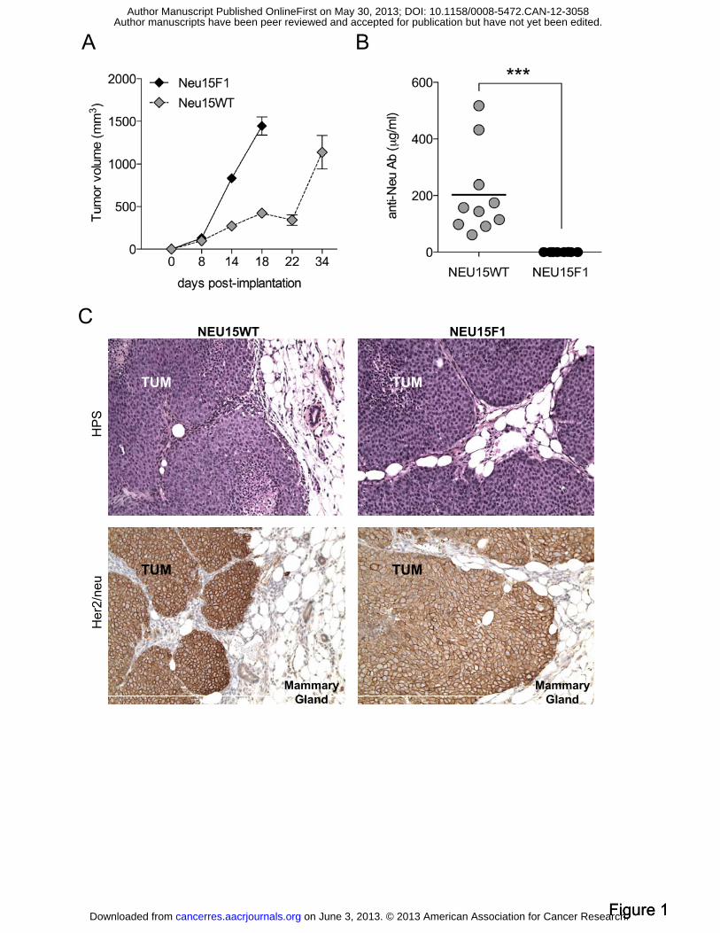
Transplantable HER2/neu-expressing tumors escape immune response in WT mice
while preserving HER2/neu expression. In order to characterize the potential role of pDC in
primary breast tumor progression, we selected a clinically relevant murine mammary tumor
model. HER-2/neu FVB/N transgenic mice express the rat proto-oncogene her2/neu under the
control of the mammary specific MMTV promoter. Those mice develop focal, poorly
differentiated ER-/neu+ spontaneous mammary carcinomas (Figure S1A/B). Due to long and
variable latency of tumor appearance, a stable HER-2/neu-expressing cell line (referred to as
NEU15) was derived in vitro from a spontaneous tumor. NEU15 was then transplanted by
orthotopic injection into syngeneic mice. Transgenic mice tolerated rat HER2/neu as a self-
protein as evidenced by the absence of specific immune responses due to central tolerance
mechanisms and the aggressive tumorigenicity of NEU15 tumor in MMTV-neu F1 mice
(named NEU15F1, Figure 1A). In contrast, rat HER2/neu was perceived as a foreign antigen
by the immune system of wild type (WT) mice, as evidenced by the induction of high levels
of anti-HER2/neu antibody of IgG1 isotype that resulted in delayed NEU15 tumor (named
NEU15WT) growth (Figure 1A/B). Accordingly, NEU15WT tumor-bearing mice displayed
(data not shown). Despite immune pressure, NEU15WT tumors maintained similar HER2/neu
expression to that of NEU15F1 tumors (Figure 1C). The resistance of NEU15WT tumor cells
to immunoediting implies that sustained HER2/neu expression is essential for NEU15 tumor
cell survival and suggests an immunosubversion by the tumor microenvironment.
NEU15WT tumors are highly infiltrated by pDC and Treg. While leukocytes are found in
all tumors, NEU15WT tumors appeared more infiltrated by CD45+ cells (22.9 ± 7,3%) than
NEU15F1 tumors (8.9 ± 5.3%) (Figure S2A).
on June 3, 2013. © 2013 American Association for Cancer Research. cancerres.aacrjournals.org Downloaded from
Author manuscripts have been peer reviewed and accepted for publication but have not yet been edited. Author Manuscript Published OnlineFirst on May 30, 2013; DOI: 10.1158/0008-5472.CAN-12-3058

9
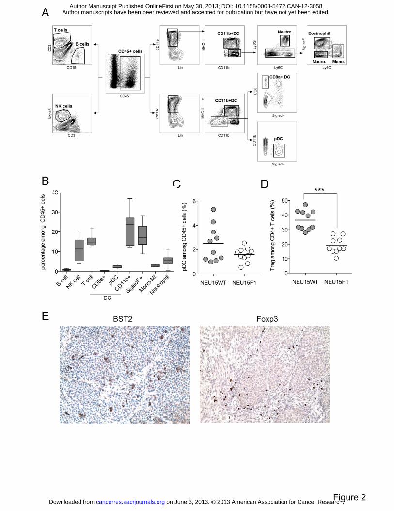
A thorough analysis of immune cell infiltrate was performed by multiparametric flow
cytometry on NEU15WT tumor single cell suspension (Figure 2A). T cells (CD3+) and NK
cells (NKp46+) represented about 10 to 15% of leukocytes, while B (CD19+) represented a
minor part of the infiltrate (Figure 2B). Macrophages (CD11b+MHCIIintLy6G-/C-),
monocytes (CD11b+MHCIIintLy6C+) and neutrophils (CD11b+CD11cintLy6G+) infiltration
(also described as MDSC) represented a moderate part of the immune infiltrate, in contrast to
more aggressive mammary tumor models (4T1 and TS/A). Interestingly, SiglecF+ myeloid
cells (most likely eosinophils or SiglecF+ macrophages) represented almost 20% of the
infiltrate. Remarkably, DC represented the most important infiltrating population in all tumors
thus identifying this model as particularly relevant for TADC functional characterization.
CD11b+ DC (CD11c+CD11b+MHCIIhi) represented the major part (about 20% of leukocytes)
when compared to CD8α+ DC (CD11c+CD8α+SiglecH-) and pDC (CD11c+ SiglecH+)
(Figure 2B).
Frequency of most immune cells was similar in both tumor types (data not shown) except for
pDC and Treg that were more abundant in NEU15WT than NEU15F1 tumors (Figure 2C/D,
and S2B). Finally, histological analyses confirmed that both TApDC and TATreg are found
within the tumor mass, with pDC mostly localized in the tumor bed and Treg in both tumor
bed and immune infiltrate areas (Figure 2E).
Taken together, these results show that NEU15WT tumor represents an interesting
immunosubversion model closely mimicking our observations in human breast cancer with
increased pDC and Treg recruitment possibly contributing to escape to immunosurveillance
(15, 20, 20, 26).
Tumor-associated pDC are functionally immature and can mediate CD4+ T cell
activation. TApDC were gated based on their high expression of CD11c and Siglec-H
on June 3, 2013. © 2013 American Association for Cancer Research. cancerres.aacrjournals.org Downloaded from
Author manuscripts have been peer reviewed and accepted for publication but have not yet been edited. Author Manuscript Published OnlineFirst on May 30, 2013; DOI: 10.1158/0008-5472.CAN-12-3058

10
(Figure 2A). They expressed high levels of BST2, B220 and Ly6C while lacking Ly6G and
CD11b thus confirming their identity (Figure 3A). Moreover, they showed heterogeneous
CD8α expression. TApDC were immature with no surface expression of CD40, CD80 and
CD86, and intermediate levels of MHC-II (Figure 3A). This phenotype resembled to the one
of pDC found in naive spleen (data not shown). Furthermore, in vivo phagocytic activity was
weak but similar to spleen-derived pDC (Figure S3) suggesting that immature TApDC may
uptake tumor Ags.
pDC were sorted from tumor or spleen and cultured with allogeneic naïve CD4+ T cells in the
presence or not of various TLR ligands. Regardless of their tissue of origin, freshly isolated
pDC did not activate CD4+ T cells (Figure 3B and C). However, pDC maturation through
TLR7 ligands, and to less extent TLR9 ligands, induced effector T cell proliferation (Figure
3B) as well as IFNγ production (mean IFNγ (pg/ml) for spleen vs tumor pDC respectively:
Ctrl, 30 vs 6; TLR7 ligands, 764-2312 vs 415-2275; TLR9 ligands, 84-278 vs 55-88 pg/ml)
(Figure 3C). In conclusion, TApDC are phenotypically and functionally immature and may
acquire abilities to activate anti-tumor effector T cells upon TLRL activation, as spleen-
derived pDC.
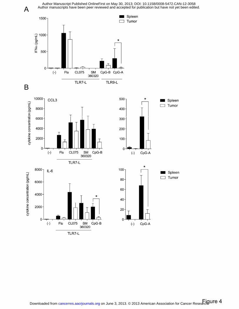
TApDC exhibit an abrogated cytokine response to TLR9 but not TLR7 ligands. As the
capacity of APC-derived cytokines is crucial to shape the immune response, we measured the
ability of TApDC to secrete cytokines after in vitro TLR stimulation. Interestingly, IFNα
production by TApDC in response to CpG-A was strongly inhibited, with 30-fold less IFNα
than in naive spleen pDC (294.4 ± 295.9 vs 10.65 ± 27.1) (Figure 4A). This alteration was
confirmed using CpG-B/ODN-1826 with a 5-fold decrease in IFNα production (Figure 4A).
In contrast, TApDC were as potent as naive spleen pDC to produce IFNα in response to Flu.
on June 3, 2013. © 2013 American Association for Cancer Research. cancerres.aacrjournals.org Downloaded from
Author manuscripts have been peer reviewed and accepted for publication but have not yet been edited. Author Manuscript Published OnlineFirst on May 30, 2013; DOI: 10.1158/0008-5472.CAN-12-3058

11
Other TLR7 ligands, such as CL075 or SM360320 (23), two synthetic TLR7 ligands, did not
trigger significant IFNα even in spleen pDC.
Similarly the production of inflammatory cytokines by TApDC was specifically altered in
response to CpG-A/B whereas responses to Flu, CL075 and SM360320 remained mostly
unchanged (Figure 4B). In particular, production of MIP-1α (323.7 ± 261.8 vs 83.4 ± 144.1
for CpG-A) and IL-6 (67.8 ± 60.9 vs 11.9 ± 17.5 for CpG-A; 1,999.8 ± 292.7 vs 292.7± 344.6
for CpG-B) by TApDC in response to CpG-A and/or CpG-B were significantly reduced when
compared to naive spleen pDC.
We then assessed a role for TLR9 downregulation in this alteration. Both TLR9 and TLR7
mRNA expression were slightly but similarly reduced in tumor versus spleen-derived pDC
(Figure S4). Furthermore, production of cytokine such as TNFα was not altered in tumor
versus spleen-derived pDC in response to TLR9L (data not shown). Altogether, this shows
that TLR9 receptor downregulation cannot merely explain the specific alteration of cytokine
production in response to TLR9L when compared to TLR7L.
pDC depletion delays tumor growth in vivo. To determine whether such TApDC
contributes to tumor growth, pDC were depleted in vivo using anti-BST2 depleting mAbs (24,
25). WT mice were treated every other day by i.p. injection from the day prior to tumor
implantation until the experimental endpoint. A significant decrease of the tumor volume was
observed upon pDC depletion from day 14 post-implantation (Figure 5A). Tumor growth was
followed over time and mice were euthanized when tumor size reached the endpoint. Survival
curve analysis showed an increase in median survival times from 35 to 43 days (Figure 5B).
Specific and effective pDC depletion in the tumor upward of 80% was validated by flow
cytometry (Figure 5D) and functional IFNα response to TLR7-L intratumoral injection at the
endpoint (Figure 5C). Those data show that effective pDC depletion in the tumor
on June 3, 2013. © 2013 American Association for Cancer Research. cancerres.aacrjournals.org Downloaded from
Author manuscripts have been peer reviewed and accepted for publication but have not yet been edited. Author Manuscript Published OnlineFirst on May 30, 2013; DOI: 10.1158/0008-5472.CAN-12-3058

12
microenvironment delays tumor growth and increases mice survival. These results are in
concordance with our observation in human breast cancer demonstrating that recruitment of
pDC within the tumor directly contributes to poor clinical outcome (15, 27).
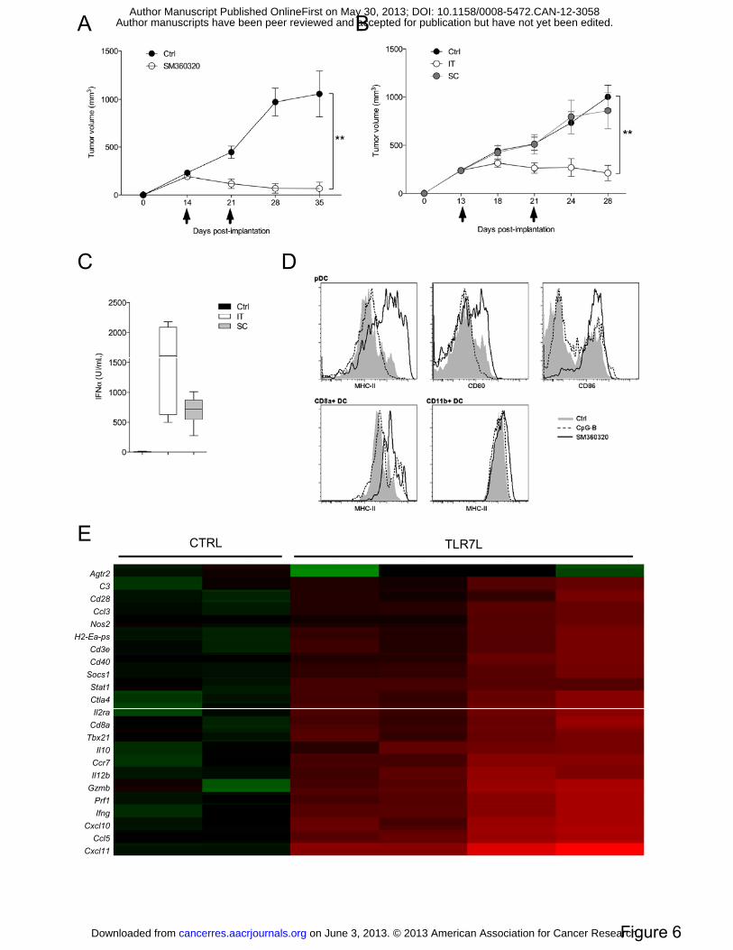
TLR7 triggering induces tumor regression in vivo. As breast TApDC respond to TLR7L in
vitro, we next assessed the possibility to activate TApDC via SM360320, a TLR7 agonist
shown to be in vivo a robust IFNα inducer and a potent adjuvant (23), in order to revert their
tumor-promoting ability. Intratumoral injections of TLR7-L (50μg) lead to potent tumor
regression when compared to vehicle-treated mice (Figure 6A). TLR7L treatment induced a
strong increase in complete response with 90% in TLR7L-treated group versus 30% of
spontaneous regression in the vehicle-treated group (p=0.0198, data not shown). Importantly,
100% of cured mice were protected against a subsequent orthotopic contralateral challenge of
NEU15 cells (3 months later) (data not shown). In contrast to i.t. injection, contralateral
subcutaneous injection of TLR7L did not induce significant tumor regression (Figure 6B).
Both i.t. and contralateral subcutaneous injections led however to similar range of plasmatic
IFNα levels suggesting that i.t. route is necessary to mobilize anti-tumor activity of TApDC
(Figure 6C).
Analyses of intratumoral immune infiltrates exhibited an increase in leukocyte frequency as
soon as 24h post TLR7L injection (Figure S6A). While no major changes in the frequency of
most immune cells could be noticed (data not shown), a significant and specific increase in
monocyte infiltration was observed (Figure S6B). As pDC are the most likely target of such
TLR7L in vivo, we explored whether i.t. TLR7L injection affected pDC frequency and
function in vivo. While pDC frequency remained unchanged (data not shown), TLR7L
induced strong increase in MHC-II expression and costimulatory molecules at their surface
(Figure 6D). In contrast, CpG did not activate TApDC thus confirming our in vitro data.
on June 3, 2013. © 2013 American Association for Cancer Research. cancerres.aacrjournals.org Downloaded from
Author manuscripts have been peer reviewed and accepted for publication but have not yet been edited. Author Manuscript Published OnlineFirst on May 30, 2013; DOI: 10.1158/0008-5472.CAN-12-3058

13
Specific activation of TApDC was confirmed, as neither TA-CD8α+ DC nor TA-CD11b+ DC
displayed increase in MHC-II (nor CD80, CD86, data not shown) expression upon TLR7
triggering (Figure 6D).
Finally, changes in gene expression were analyzed in NEU15WT tumors 8 hours post i.t.
injection of TLR7L by TaqMan® Low density array (TLDA) mouse immune assay
(Supplementary Material and Methods). Data are presented as fold changed over the non-
treated conditions and displayed as a Heat Map (Figure 6E) and detailed in Figure S5. Genes
that displayed fold changes higher than 10 were selected to highlight the most significant
changes in gene expression upon TLR7 i.t. injection. These data depicted an increased
infiltration in immune cells such as myeloid cells (H2-Ea-ps, Nos2) and in particular
cytotoxic T cells (Cd3e, Cd8, Tbx21), via chemokine-mediated recruitment (Cxcl11, 2413 ±
3409 fold changes; Ccr7, 58 ± 53 fold changes). The increase in perforin and granzyme genes
(125 ± 147 and 185 ± 209 fold changes respectively) also indicated a potent cytotoxic
response. A Th1-type T cell response was seen via the sharp increase in genes related to APC
maturation (Cd28, Cd40, IL12b), IFNγ (123 ± 151 fold changes) and IFNγ-induced genes
(Cxcl10, Ccl5, Stat1) production. Interestingly, the induction of this genes, with the exception
of IL10, was type I IFN-dependent as gene increase was no longer seen when anti-IFNAR1
antibody was co-injected with TLR7L (Figure S5A). In parallel, an increase in type I IFN
genes (IFNa and Mx1) was seen in samples treated by TLR7L by quantitative PCR (Figure
S5B).
Altogether these data demonstrated the induction of type I IFN response as well as Th1
cytotoxic T cells response leading to tumor regression.
In contrast to CpG, in vivo TLR7 antitumoral activity requires type I IFN production by
pDC. In order to explore whether TLR7L antitumoral activity was mediated by pDC, pDC
were depleted the two days prior to TLRL injection. As previously observed with a dose of
on June 3, 2013. © 2013 American Association for Cancer Research. cancerres.aacrjournals.org Downloaded from
Author manuscripts have been peer reviewed and accepted for publication but have not yet been edited. Author Manuscript Published OnlineFirst on May 30, 2013; DOI: 10.1158/0008-5472.CAN-12-3058

14
50μg, 10μg of TLR7L also lead to a significant decrease of tumor volume (day35) (Figure
7A). pDC depletion in such short-term schedule led to a delay in tumor growth as previously
observed (Figure 7A). Finally, pDC depletion completely abrogated TLR7L-mediated
antitumoral effect (1046 ± 221.58 mm3 for w/o pDC vs 1266.5 ± 335.67 mm3 for TLR7L w/o
pDC), demonstrating that pDC mediate the antitumor effect of TLR7L in vivo (Figure 7C).
The efficiency of pDC depletion was confirmed phenotypically (data not shown) and
functionally at the type I IFN systemic plasmatic level (Figure 7B).
We then assessed whether inhibition of type IFN signaling affected TLR7L activity. For that
matter, anti-IFNAR1 or control antibodies were co-administered i.t. with TLR7L (10μg).
IFNAR inhibition resulted in a significant decrease of TLR7L anti-tumoral activity showing
the requirement for type I IFN in TLR7L activity (752.1 ± 135.3 mm3 for TLR7L vs 1392.1 ±
181.8 mm3 for TLR7L w/o IFNAR). The efficacy of anti-IFNAR antibodies was confirmed
by qPCR on tumor extracts with a decrease in IFN stimulated gene expression upon TLR7L
treatment in the context of type I IFN blockade (data not shown).
As TApDC were shown to lack both in vivo and in vitro the ability to respond to CpG, the
effect of this TLR9L on tumor growth was assessed. Unexpectedly, intratumoral
administration of CpG-B induced tumor regression (771.7 ± 295.3 mm3 for CpG vs 1826.6 ±
196.8 mm3 for control). Interestingly, inhibition of tumor growth by CpG was delayed in
comparison with TLR7L, which started to decrease the tumor volume as soon as after the first
injection. However the mechanism seems different as intratumoral CpG injection did not lead
to any detectable IFNα production in contrast with TLR7L (data not shown). Surprisingly,
pDC depletion altered CpG antitumoral activity (Figure 7D), however inhibition of type I
IFN signaling did not (Figure 7E). These results suggest that pDC indirectly affect CpG
activity through a distinct type I IFN-independent mechanism.
on June 3, 2013. © 2013 American Association for Cancer Research. cancerres.aacrjournals.org Downloaded from
Author manuscripts have been peer reviewed and accepted for publication but have not yet been edited. Author Manuscript Published OnlineFirst on May 30, 2013; DOI: 10.1158/0008-5472.CAN-12-3058

15
Altogether, those data demonstrate that in contrast to CpG, TLR7L antitumoral activity is
mediated by pDC and their ability to secrete large amount of type I IFN.
on June 3, 2013. © 2013 American Association for Cancer Research. cancerres.aacrjournals.org Downloaded from
Author manuscripts have been peer reviewed and accepted for publication but have not yet been edited. Author Manuscript Published OnlineFirst on May 30, 2013; DOI: 10.1158/0008-5472.CAN-12-3058

16
DISCUSSION
In line with the negative prognostic value of pDC infiltration in human breast tumors (15), we
demonstrate for the first time in vivo, that mouse mammary TApDC favor primary tumor
growth but can be activated via TLR7 triggering to mediate antitumoral response and
subsequent tumor regression.
We developed a clinically relevant tumor model in which the HER2/neu+ NEU15 cell line
developed in WT hosts escapes from immunosurveillance through pDC and Treg-mediated
immunosubversion, thus closely mimicking our observations in human breast cancer (15, 20,
20, 26).
First, we showed in vitro and in vivo that TApDC from NEU15WT tumors were specifically
altered in their ability to respond to TLR9 but not TLR7 ligands. These data are consistent
with the specific alteration of TLR9 response previously reported by us (19, 20) and others on
both human tumor and immune cells (17, 28). While pDC hyporesponsiveness to TLR9
ligands might be explained by receptor downregulation (17, 28), we demonstrated that it is
rather due to a specific interference of the tumor microenvironment, for instance via IRF7
downregulation (29). Tumor-induced TLR9 loss of function might represent an
immunosubversion mechanism in order to avoid immune alert by putative endogenous ligands
for TLR9 within the tumor microenvironment, such as described in autoimmune disorders
(30-32).
Second, we demonstrated for the first time in primary breast tumors that TApDC favored
breast tumor progression. In vivo depletion of pDC indeed significantly slowed down tumor
growth. While antibody-mediated pDC depletion is quite efficient, variation in its efficacy
might account for discrepancies in the beneficial effect of pDC depletion in short-term
depletion settings (Figure 7B and D). The use of a pDC-deficient mouse model (33) will
formerly demonstrate the extent of pDC role in tumor progression. To our knowledge this is a
on June 3, 2013. © 2013 American Association for Cancer Research. cancerres.aacrjournals.org Downloaded from
Author manuscripts have been peer reviewed and accepted for publication but have not yet been edited. Author Manuscript Published OnlineFirst on May 30, 2013; DOI: 10.1158/0008-5472.CAN-12-3058

17
unique proof of a direct role of pDC in favoring primary tumor growth in solid tumors. pDC
were previously shown to regulate growth of multiple myeloma (MM) cells (34) and more
recently to favor bone metastasis of breast cancer cells (35).
Accumulating evidences have shown a specialized role of pDC in the induction of peripheral
tolerance (11-14, 36) through Ag-specific T cell deletion (12) or differentiation/expansion of
suppressive T cells (11, 14, 31, 37-44). NEU15WT tumors were indeed more infiltrated by
Treg. For instance, we observed in preliminary experiments a decrease in TATreg infiltration
after pDC depletion. This role of pDC would be consistent with our recent reports in human
breast cancer, showing that IFNα deficient TApDC from human breast tumors favor Treg
expansion via ICOS-ICOSL interaction (20, 27) that may contribute to tumor progression and
explain their negative impact on patient survival (15). Whether tumor pDC are able to
modulate Treg function/differentiation remains to be addressed.
Despite their negative impact on tumor progression, TApDC could be activated in vivo via
intra-tumoral injection of TLR7L, thus mediating a Th1 signature, as evidenced by TLDA
analysis, and subsequent tumor regression. TLR7L induced long term protective memory
response as 100% of cured mice were protected against subsequent tumor challenge.
In vivo depletion of pDC abrogated the therapeutic activity of TLR7L, demonstrating the
central role for TApDC in TLR7L-mediated antitumor response. Importantly, we
demonstrated that this therapeutic activity is mediated by locally-induced and not systemic
type I IFN. This points towards the importance of intratumoral TApDC activation leading to
type I IFN production and subsequent additional activities (cross-presentation, Treg
neutralization). We indeed observed that type I IFN neutralization led to the inhibition of the
intratumoral Th1 signature. The antitumor activity of Imiquimod, another TLR7L, is
dependent upon CD8α+ pDC that harbor direct tumor killing activity mediated by granzyme
B and/or TRAIL leading to subsequent capture and antigen cross-presentation (3, 21).
on June 3, 2013. © 2013 American Association for Cancer Research. cancerres.aacrjournals.org Downloaded from
Author manuscripts have been peer reviewed and accepted for publication but have not yet been edited. Author Manuscript Published OnlineFirst on May 30, 2013; DOI: 10.1158/0008-5472.CAN-12-3058

18
However we did not observe any increase in TRAIL expression on our TApDC upon TLR7L
treatment (data not shown) suggesting a different mechanism of action. However, considering
our results in human models that type I IFN led to the inhibition of TATreg proliferation (20),
the potential role of Treg inhibition in this TLR7L antitumoral activity will be carefully
assessed.
While we demonstrated both in vitro and in vivo that CpG could not activate TApDC, this
TLR9L unexpectedly induced tumor regression. While is antitumoral activity was reduced
upon pDC depletion, IFNAR inhibition did not affect their therapeutic action showing a
different mechanism of action than TLR7L. We hypothesize that pDC depletion somehow
impact on key effectors of CpG-mediated antitumoral response. We indeed observed an
increase in macrophage infiltration upon CpG treatment (Figure S6B) that was reduced upon
pDC depletion (data not shown). While tumor-associated macrophages (TAM) are commonly
associated with tumor development and progression, antitumor activity can be achieved by
targeting TAM recruitment and polarization toward M1 phenotype, a process to which CpG
was shown to participate (45, 46). CpG-mediated antitumoral mechanisms will need to be
further clarified.
Based on promising results of the literature, ongoing phase I/II clinical trials are currently
evaluating the therapeutic potential of TLR agonists for the treatment of various type of
cancer (47). In the light of our observation that TApDC remain responsive to TLR7L while
lacking response to CpG ODN, the therapeutic potential of TLR7 agonists in human breast
tumors shall be considered.
ACKNOWLEDGMENTS
on June 3, 2013. © 2013 American Association for Cancer Research. cancerres.aacrjournals.org Downloaded from
Author manuscripts have been peer reviewed and accepted for publication but have not yet been edited. Author Manuscript Published OnlineFirst on May 30, 2013; DOI: 10.1158/0008-5472.CAN-12-3058

19
We thank Sophie Goddard-Léon and Manon Pratviel for technical support, Marie Paturel and
Anthony Besse for precious advices for statistical analysis and Janssen Infectious Diseases-
Diagnostics BVBA for providing SM360320.
on June 3, 2013. © 2013 American Association for Cancer Research. cancerres.aacrjournals.org Downloaded from
Author manuscripts have been peer reviewed and accepted for publication but have not yet been edited. Author Manuscript Published OnlineFirst on May 30, 2013; DOI: 10.1158/0008-5472.CAN-12-3058
20
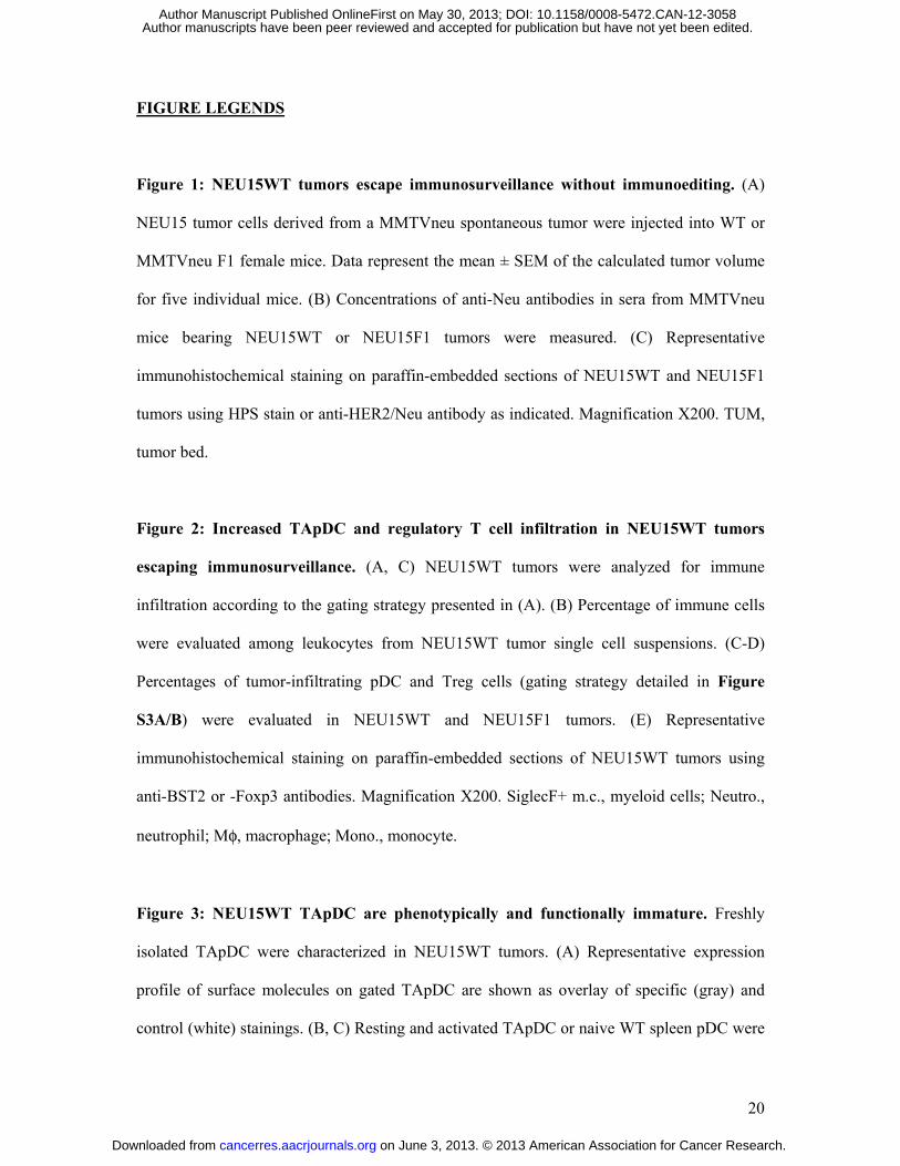
FIGURE LEGENDS
Figure 1: NEU15WT tumors escape immunosurveillance without immunoediting. (A)
NEU15 tumor cells derived from a MMTVneu spontaneous tumor were injected into WT or
MMTVneu F1 female mice. Data represent the mean ± SEM of the calculated tumor volume
for five individual mice. (B) Concentrations of anti-Neu antibodies in sera from MMTVneu
mice bearing NEU15WT or NEU15F1 tumors were measured. (C) Representative
immunohistochemical staining on paraffin-embedded sections of NEU15WT and NEU15F1
tumors using HPS stain or anti-HER2/Neu antibody as indicated. Magnification X200. TUM,
tumor bed.
Figure 2: Increased TApDC and regulatory T cell infiltration in NEU15WT tumors
escaping immunosurveillance. (A, C) NEU15WT tumors were analyzed for immune
infiltration according to the gating strategy presented in (A). (B) Percentage of immune cells
were evaluated among leukocytes from NEU15WT tumor single cell suspensions. (C-D)
Percentages of tumor-infiltrating pDC and Treg cells (gating strategy detailed in Figure
S3A/B) were evaluated in NEU15WT and NEU15F1 tumors. (E) Representative
immunohistochemical staining on paraffin-embedded sections of NEU15WT tumors using
anti-BST2 or -Foxp3 antibodies. Magnification X200. SiglecF+ m.c., myeloid cells; Neutro.,
neutrophil; Mφ, macrophage; Mono., monocyte.
Figure 3: NEU15WT TApDC are phenotypically and functionally immature. Freshly
isolated TApDC were characterized in NEU15WT tumors. (A) Representative expression
profile of surface molecules on gated TApDC are shown as overlay of specific (gray) and
control (white) stainings. (B, C) Resting and activated TApDC or naive WT spleen pDC were
on June 3, 2013. © 2013 American Association for Cancer Research. cancerres.aacrjournals.org Downloaded from
Author manuscripts have been peer reviewed and accepted for publication but have not yet been edited. Author Manuscript Published OnlineFirst on May 30, 2013; DOI: 10.1158/0008-5472.CAN-12-3058

21
incubated with allogeneic CD4+ T cells. T cell proliferation (B) and IFN-γ production (C)
were determined. Data are expressed as mean ± SEM of 7/ 8 experiments. Statistical analyses
of TLRL-treated versus medium ((-)) condition are shown for spleen and tumors.
Figure 4: NEU15WT TApDC exhibit an abrogated cytokine response to TLR9 but not
TLR7 ligands. TApDC and naive WT spleen pDC were incubated with indicated TLRL.
Supernatants were collected and analyzed for IFN-α (A), MIP-1α and IL-6 (B) production.
Data represent the mean ± SD from 3 to 12 independent samples.
Figure 5: pDC depletion delays tumor growth in vivo. (A-C) WT mice (n=20 per group)
were treated with purified pDC-depleting 120G8 or rat IgG1 control antibody from one day
prior to tumor implantation and every other day until tumors reached the experimental
endpoint. (A) Data represent the mean tumor volume mean ± SEM for twenty mice. (B)
Kaplan-Meier survival plot of mice stratified in those with pDC depletion versus none. Data
are from one representative out of 3 independent experiments. (C-D) In vivo depletion of pDC
was analyzed functionnaly (C) and phenotypically by flow cytometry in tumors (D). (C)
TLR7L (10µg) was administrated by i.t. injection at experimental endpoint and serum were
harvested 3 hours after for IFNα levels measurement (rat IgG1 control , pDC-depleted ).
(D) Dot plot representation of CD11b/SiglecF staining gated on CD45+CD11c+lin- cells after
dead cells and doublet exclusion for one representative mouse of each group.
Figure 6: Intratumoral TLR7L injection induces tumor regression. Mice were treated
with 50µg SM360320 TLR7L at day 13 and 20 post NEU15 tumor implantation by
intratumoral (A-E) or subcutaneous contralateral (B, C) injections. Controls were injected
with vehicle only (A, B) Data represent the mean tumor volume ± SEM for ten mice. (C)
on June 3, 2013. © 2013 American Association for Cancer Research. cancerres.aacrjournals.org Downloaded from
Author manuscripts have been peer reviewed and accepted for publication but have not yet been edited. Author Manuscript Published OnlineFirst on May 30, 2013; DOI: 10.1158/0008-5472.CAN-12-3058
22
Serum was harvested 3 hours after TLR7L injection for IFNα levels measurement. (D)
Intratumoral DC activation was assessed 24 hours post-intratumoral TLRL injection by flow
cytometry. Representative expression profiles of costimulatory molecules on gated TApDC,
CD8α+ or CD11b+ DC are shown. (E) Fold changes in gene expression upon intratumoral
TLR7L injection. TLDA mouse immune array were performed on tumor samples from
control and TLR7L treated mice. Data were calculated as fold changes over non-treated
control condition and presented as Heat map. Genes with fold changes over 10 are listed here.
Figure 7: Therapeutic activity of TLR7L depends on TApDC and type I IFN
production. NEU15WT-bearing mice were treated with 10µg TLR7L (A-C) or CpG-B (D-
E) at day 13 and 20 (n=10-15/groupe). (A, D) pDC were depleted from WT mice the two days
prior to TLRL injection. (B) Serum were harvested 3 hours after TLR7L it injection for
IFNα levels measurement. (C, E) Mouse IFNAR1 or rat IgG1 control antibodies were co-
injected with TLRL. (A, C-E) Data represent the mean tumor volume ± SEM.
on June 3, 2013. © 2013 American Association for Cancer Research. cancerres.aacrjournals.org Downloaded from
Author manuscripts have been peer reviewed and accepted for publication but have not yet been edited. Author Manuscript Published OnlineFirst on May 30, 2013; DOI: 10.1158/0008-5472.CAN-12-3058

23
REFERENCES
(1) de Visser KE, Eichten A, Coussens LM. Paradoxical roles of the immune system during cancer development. Nat Rev Cancer 2006;6:24-37.
(2) Fricke I, Gabrilovich DI. Dendritic cells and tumor microenvironment: a dangerous liaison. Immunol Invest 2006;35:459-83.
(3) Drobits B, Holcmann M, Amberg N, Swiecki M, Grundtner R, Hammer M, et al. Imiquimod clears tumors in mice independent of adaptive immunity by converting pDCs into tumor-killing effector cells. J Clin Invest 2012;122:575-85.
(4) Liu C, Lou Y, Lizee G, Qin H, Liu S, Rabinovich B, et al. Plasmacytoid dendritic cells induce NK cell-dependent, tumor antigen-specific T cell cross-priming and tumor regression in mice. J Clin Invest 2008;118:1165-75.
(5) Kadowaki N, Antonenko S, Lau JY, Liu YJ. Natural interferon alpha/beta-producing cells link innate and adaptive immunity. J Exp Med 2000;192:219-26.
(6) Colonna M, Trinchieri G, Liu YJ. Plasmacytoid dendritic cells in immunity. Nat Immunol 2004;5:1219-26. (7) Gilliet M, Cao W, Liu YJ. Plasmacytoid dendritic cells: sensing nucleic acids in viral infection and autoimmune diseases.
Nat Rev Immunol 2008;8:594-606. (8) Kawai T, Sato S, Ishii KJ, Coban C, Hemmi H, Yamamoto M, et al. Interferon-alpha induction through Toll-like
receptors involves a direct interaction of IRF7 with MyD88 and TRAF6. Nat Immunol 2004;5:1061-8. (9) Fonteneau JF, Gilliet M, Larsson M, Dasilva I, Munz C, Liu YJ, et al. Activation of influenza virus-specific CD4+ and
CD8+ T cells: a new role for plasmacytoid dendritic cells in adaptive immunity. Blood 2003;101:3520-6. (10) Blanco P, Palucka AK, Gill M, Pascual V, Banchereau J. Induction of dendritic cell differentiation by IFN-alpha in
systemic lupus erythematosus. Science 2001;294:1540-3. (11) de Heer HJ, Hammad H, Soullie T, Hijdra D, Vos N, Willart MA, et al. Essential role of lung plasmacytoid dendritic cells
in preventing asthmatic reactions to harmless inhaled antigen. J Exp Med 2004;200:89-98. (12) Goubier A, Dubois B, Gheit H, Joubert G, Villard-Truc F, Asselin-Paturel C, et al. Plasmacytoid dendritic cells mediate
oral tolerance. Immunity 2008;29:464-75. (13) Fugier-Vivier IJ, Rezzoug F, Huang Y, Graul-Layman AJ, Schanie CL, Xu H, et al. Plasmacytoid precursor dendritic
cells facilitate allogeneic hematopoietic stem cell engraftment. J Exp Med 2005;201:373-83. (14) Wei S, Kryczek I, Zou L, Daniel B, Cheng P, Mottram P, et al. Plasmacytoid dendritic cells induce CD8+ regulatory T
cells in human ovarian carcinoma. Cancer Res 2005;65:5020-6. (15) Treilleux I, Blay JY, Bendriss-Vermare N, Ray-Coquard I, Bachelot T, Guastalla JP, et al. Dendritic cell infiltration and
prognosis of early stage breast cancer. Clin Cancer Res 2004;10:7466-74. (16) Vermi W, Bonecchi R, Facchetti F, Bianchi D, Sozzani S, Festa S, et al. Recruitment of immature plasmacytoid dendritic
cells (plasmacytoid monocytes) and myeloid dendritic cells in primary cutaneous melanomas. J Pathol 2003;200:255-68. (17) Hartmann E, Wollenberg B, Rothenfusser S, Wagner M, Wellisch D, Mack B, et al. Identification and functional analysis
of tumor-infiltrating plasmacytoid dendritic cells in head and neck cancer. Cancer Res 2003;63:6478-87. (18) Zou W, Machelon V, Coulomb-L'Hermin A, Borvak J, Nome F, Isaeva T, et al. Stromal-derived factor-1 in human tumors
recruits and alters the function of plasmacytoid precursor dendritic cells. Nat Med 2001;7:1339-46. (19) Labidi-Galy SI, Sisirak V, Meeus P, Gobert M, Treilleux I, Bajard A, et al. Quantitative and Functional Alterations of
Plasmacytoid Dendritic Cells Contribute to Immune Tolerance in Ovarian Cancer. Cancer Res 2011. (20) Sisirak V, Faget J, Gobert M, Goutagny N, Vey N, Treilleux I, et al. Impaired IFN-alpha production by plasmacytoid
dendritic cells favors regulatory T-cell expansion that may contribute to breast cancer progression. Cancer Res 2012;72:5188-97.
(21) Stary G, Bangert C, Tauber M, Strohal R, Kopp T, Stingl G. Tumoricidal activity of TLR7/8-activated inflammatory dendritic cells. J Exp Med 2007;204:1441-51.
(22) Guy CT, Webster MA, Schaller M, Parsons TJ, Cardiff RD, Muller WJ. Expression of the neu protooncogene in the mammary epithelium of transgenic mice induces metastatic disease. Proc Natl Acad Sci U S A 1992;89:10578-82.
(23) Dharmapuri S, Aurisicchio L, Neuner P, Verdirame M, Ciliberto G, La MN. An oral TLR7 agonist is a potent adjuvant of DNA vaccination in transgenic mouse tumor models. Cancer Gene Ther 2009;16:462-72.
(24) Blasius AL, Giurisato E, Cella M, Schreiber RD, Shaw AS, Colonna M. Bone marrow stromal cell antigen 2 is a specific marker of type I IFN-producing cells in the naive mouse, but a promiscuous cell surface antigen following IFN stimulation. J Immunol 2006;177:3260-5.
(25) Asselin-Paturel C, Brizard G, Pin JJ, Briere F, Trinchieri G. Mouse strain differences in plasmacytoid dendritic cell frequency and function revealed by a novel monoclonal antibody. J Immunol 2003;171:6466-77.
(26) Gobert M, Treilleux I, Bendriss-Vermare N, Bachelot T, Goddard-Leon S, Arfi V, et al. Regulatory T cells recruited through CCL22/CCR4 are selectively activated in lymphoid infiltrates surrounding primary breast tumors and lead to an adverse clinical outcome. Cancer Res 2009;69:2000-9.
(27) Faget J, Bendriss-Vermare N, Gobert M, Durand I, Olive D, Biota C, et al. ICOS-ligand expression on plasmacytoid dendritic cells supports breast cancer progression by promoting the accumulation of immunosuppressive CD4+ T cells. Cancer Res 2012;72:6130-41.
(28) Hirsch I, Caux C, Hasan U, Bendriss-Vermare N, Olive D. Impaired Toll-like receptor 7 and 9 signaling: from chronic viral infections to cancer. Trends Immunol 2010;31:391-7.
(29) Sisirak V, Vey N, Goutagny N, Renaudineau S, Malfroy M, Thys S, et al. Breast cancer-derived TGF-beta and TNF-alpha compromise IFN-alpha production by tumor-associated plasmacytoid dendritic cells. Int J Cancer 2013.
(30) Barrat FJ, Meeker T, Gregorio J, Chan JH, Uematsu S, Akira S, et al. Nucleic acids of mammalian origin can act as endogenous ligands for Toll-like receptors and may promote systemic lupus erythematosus. J Exp Med 2005;202:1131-9.
(31) Vollmer J, Tluk S, Schmitz C, Hamm S, Jurk M, Forsbach A, et al. Immune stimulation mediated by autoantigen binding sites within small nuclear RNAs involves Toll-like receptors 7 and 8. J Exp Med 2005;202:1575-85.
(32) Ganguly D, Chamilos G, Lande R, Gregorio J, Meller S, Facchinetti V, et al. Self-RNA-antimicrobial peptide complexes activate human dendritic cells through TLR7 and TLR8. J Exp Med 2009;206:1983-94.
(33) Cisse B, Caton ML, Lehner M, Maeda T, Scheu S, Locksley R, et al. Transcription factor E2-2 is an essential and specific regulator of plasmacytoid dendritic cell development. Cell 2008;135:37-48.
(34) Chauhan D, Singh AV, Brahmandam M, Carrasco R, Bandi M, Hideshima T, et al. Functional interaction of plasmacytoid dendritic cells with multiple myeloma cells: a therapeutic target. Cancer Cell 2009;16:309-23.
(35) Sawant A, Hensel JA, Chanda D, Harris BA, Siegal GP, Maheshwari A, et al. Depletion of plasmacytoid dendritic cells inhibits tumor growth and prevents bone metastasis of breast cancer cells. J Immunol 2012;189:4258-65.
on June 3, 2013. © 2013 American Association for Cancer Research. cancerres.aacrjournals.org Downloaded from
Author manuscripts have been peer reviewed and accepted for publication but have not yet been edited. Author Manuscript Published OnlineFirst on May 30, 2013; DOI: 10.1158/0008-5472.CAN-12-3058
24
(36) Wang RF. Regulatory T cells and toll-like receptors in cancer therapy. Cancer Res 2006;66:4987-90. (37) Kuwana M. Induction of anergic and regulatory T cells by plasmacytoid dendritic cells and other dendritic cell subsets.
Hum Immunol 2002;63:1156-63. (38) Bilsborough J, George TC, Norment A, Viney JL. Mucosal CD8alpha+ DC, with a plasmacytoid phenotype, induce
differentiation and support function of T cells with regulatory properties. Immunology 2003;108:481-92. (39) Ito T, Yang M, Wang YH, Lande R, Gregorio J, Perng OA, et al. Plasmacytoid dendritic cells prime IL-10-producing T
regulatory cells by inducible costimulator ligand. J Exp Med 2007;204:105-15. (40) Gilliet M, Liu YJ. Human plasmacytoid-derived dendritic cells and the induction of T-regulatory cells. Hum Immunol
2002;63:1149-55. (41) Moseman EA, Liang X, Dawson AJ, Panoskaltsis-Mortari A, Krieg AM, Liu YJ, et al. Human plasmacytoid dendritic cells
activated by CpG oligodeoxynucleotides induce the generation of CD4+CD25+ regulatory T cells. J Immunol 2004;173:4433-42.
(42) Kang HK, Liu M, Datta SK. Low-dose peptide tolerance therapy of lupus generates plasmacytoid dendritic cells that cause expansion of autoantigen-specific regulatory T cells and contraction of inflammatory Th17 cells. J Immunol 2007;178:7849-58.
(43) Ouabed A, Hubert FX, Chabannes D, Gautreau L, Heslan M, Josien R. Differential control of T regulatory cell proliferation and suppressive activity by mature plasmacytoid versus conventional spleen dendritic cells. J Immunol 2008;180:5862-70.
(44) Sharma MD, Baban B, Chandler P, Hou DY, Singh N, Yagita H, et al. Plasmacytoid dendritic cells from mouse tumor-draining lymph nodes directly activate mature Tregs via indoleamine 2,3-dioxygenase. J Clin Invest 2007;117:2570-82.
(45) Guiducci C, Vicari AP, Sangaletti S, Trinchieri G, Colombo MP. Redirecting in vivo elicited tumor infiltrating macrophages and dendritic cells towards tumor rejection. Cancer Res 2005;65:3437-46.
(46) Colombo MP, Mantovani A. Targeting myelomonocytic cells to revert inflammation-dependent cancer promotion. Cancer Res 2005;65:9113-6.
(47) Goutagny N, Estornes Y, Hasan U, Lebecque S, Caux C. Targeting pattern recognition receptors in cancer immunotherapy. Target Oncol 2012;7:29-54.
on June 3, 2013. © 2013 American Association for Cancer Research. cancerres.aacrjournals.org Downloaded from
Author manuscripts have been peer reviewed and accepted for publication but have not yet been edited. Author Manuscript Published OnlineFirst on May 30, 2013; DOI: 10.1158/0008-5472.CAN-12-3058
A B
CNEU15WT NEU15F1
HP
S
TUM TUM
u
TUMTUM
Her
2/ne
u
MammaryGl d
MammaryGl dGland Gland
Figure 1Figure 1on June 3, 2013. © 2013 American Association for Cancer Research. cancerres.aacrjournals.org Downloaded from
Author manuscripts have been peer reviewed and accepted for publication but have not yet been edited. Author Manuscript Published OnlineFirst on May 30, 2013; DOI: 10.1158/0008-5472.CAN-12-3058
A
B C D
E BST2 Foxp3E BST2 Foxp3
Figure 2on June 3, 2013. © 2013 American Association for Cancer Research. cancerres.aacrjournals.org Downloaded from
Author manuscripts have been peer reviewed and accepted for publication but have not yet been edited. Author Manuscript Published OnlineFirst on May 30, 2013; DOI: 10.1158/0008-5472.CAN-12-3058

A
B C
Figure 3on June 3, 2013. © 2013 American Association for Cancer Research. cancerres.aacrjournals.org Downloaded from
Author manuscripts have been peer reviewed and accepted for publication but have not yet been edited. Author Manuscript Published OnlineFirst on May 30, 2013; DOI: 10.1158/0008-5472.CAN-12-3058
A
B
Figure 4on June 3, 2013. © 2013 American Association for Cancer Research. cancerres.aacrjournals.org Downloaded from
Author manuscripts have been peer reviewed and accepted for publication but have not yet been edited. Author Manuscript Published OnlineFirst on May 30, 2013; DOI: 10.1158/0008-5472.CAN-12-3058

A B
C D
Figure 5on June 3, 2013. © 2013 American Association for Cancer Research. cancerres.aacrjournals.org Downloaded from
Author manuscripts have been peer reviewed and accepted for publication but have not yet been edited. Author Manuscript Published OnlineFirst on May 30, 2013; DOI: 10.1158/0008-5472.CAN-12-3058
A B
C D
E CTRL TLR7L
Agtr2
C3
Cd28
Ccl3
Nos2
H2-Ea-ps
Cd3e
Cd40
Socs1
Stat1
Ctla4
Il2ra
Cd8a
Tbx21
Il10
Ccr7
Il12b
Gzmb
Prf1
Ifng
Cxcl10
Figure 6
Cxcl10
Ccl5
Cxcl11
on June 3, 2013. © 2013 American Association for Cancer Research. cancerres.aacrjournals.org Downloaded from
Author manuscripts have been peer reviewed and accepted for publication but have not yet been edited. Author Manuscript Published OnlineFirst on May 30, 2013; DOI: 10.1158/0008-5472.CAN-12-3058

A B
C
D E
Figure 7on June 3, 2013. © 2013 American Association for Cancer Research. cancerres.aacrjournals.org Downloaded from
Author manuscripts have been peer reviewed and accepted for publication but have not yet been edited. Author Manuscript Published OnlineFirst on May 30, 2013; DOI: 10.1158/0008-5472.CAN-12-3058