Tumor Presence Induces Global Immune Changes and Enhances Nanoparticle...
10
Tumor Presence Induces Global Immune Changes and Enhances Nanoparticle Clearance Marc P. Kai, † Hailey E. Brighton, ‡,§ Catherine A. Fromen, † Tammy W. Shen, ∥ J. Christopher Luft, ∥ Yancey E. Luft, # Amanda W. Keeler, ∥ Gregory R. Robbins, ‡ Jenny P. Y. Ting, ‡,⊥ William C. Zamboni, ‡,∥ James E. Bear,* ,‡,§,# and Joseph M. DeSimone* ,†,‡,∥,#,∇ † Department of Chemical and Biomolecular Engineering, North Carolina State University, Raleigh, North Carolina 27695, United States ‡ Lineberger Comprehensive Cancer Center, § Department of Cell Biology and Physiology, ∥ School of Pharmacy, # Department of Chemistry, ⊥ Department of Microbiology-Immunology, # Howard Hughes Medical Institute, and ∇ Department of Pharmacology, University of North Carolina at Chapel Hill, Chapel Hill, North Carolina 27599, United States * S Supporting Information ABSTRACT: Long-circulating nanoparticles are essential for increasing tumor accumulation to provide therapeutic efficacy. While it is known that tumor presence can alter the immune system, very few studies have explored this impact on nanoparticle circulation. In this report, we demonstrate how the presence of a tumor can change the local and global immune system, which dramatically increases particle clearance. We found that tumor presence significantly increased clearance of PRINT hydrogel nanoparticles from the circulation, resulting in increased accumulation in the liver and spleen, due to an increase in M2-like macrophages. Our findings highlight the need to better understand interactions between immune status and nanoparticle clearance, and suggest that further consideration of immune function is required for success in preclinical and clinical nanoparticle studies. KEYWORDS: nanoparticle, orthotopic, pharmacokinetic, immunology, intravital A dvances at the intersection of material science and biology have led to improved and expanded treatment options in the field of oncology. Research in nano- medicine has resulted in improved therapeutic index, targeting strategies, and biocompatibility of nanoparticle treatments against cancer in the clinic. 1 Major strides in parallel work have also resulted in benefits to the biology component of oncology research, including the development of a human cancer cell line library, which can be used to control variability and identify treatment platforms for preclinical studies. 2 This has led to a variety of selectively optimized particle platforms, which prove successful under narrow preclinical conditions using a particular therapy and model. In clinical settings, however, tumor heterogeneity in patients inhibits the effects of targeted nanotherapies that have proven successful in preclinical models. 3 For example, varying tumor cell growth, incongruous cancer cell genotypes, and different intratumoral immune responses can affect the properties, structure, and content of the tumor microenvironment. 4,5 Certain clinically relevant preclinical models can address and model tumor heterogeneity, such as some patient-derived xenografts and genetically engineered mice (GEM) models, but these systems still exhibit varied responses to different nanoparticle plat- forms. 6,7 Decades of research have shown that particles must demonstrate certain universal attributes for maximizing circulation persistence, a necessary tenet for passive tumor accumulation. 1 There is, however, an inherent variability among particle characteristics that may depend on the platform or fabrication technology, resulting in a variety of unique optimal combinations of properties for effective therapy. 8 This variability is further complicated by the status of the immune system, which adds a significant layer of complexity. 9 Regardless of how particle properties are manipulated, the eventual fate of a nanoparticle is typically the liver and spleen, which occurs largely through sequestration by the immune system, specifically by the mononuclear phagocyte system (MPS). 10,11 Interestingly, most studies investigate interactions between nanoparticles and the immune system in either healthy or in immune-compromised mice (e.g., tumor-bearing), but they ignore proper side-by-side comparisons in these models. A better understanding of nanoparticle-immune system inter- actions in healthy versus immune-compromised mice is Received: September 23, 2015 Accepted: November 23, 2015 Published: November 23, 2015 Article www.acsnano.org © 2015 American Chemical Society 861 DOI: 10.1021/acsnano.5b05999 ACS Nano 2016, 10, 861−870
Transcript of Tumor Presence Induces Global Immune Changes and Enhances Nanoparticle...

Tumor Presence Induces Global ImmuneChanges and Enhances Nanoparticle ClearanceMarc P. Kai,† Hailey E. Brighton,‡,§ Catherine A. Fromen,† Tammy W. Shen,∥ J. Christopher Luft,∥
Yancey E. Luft,# Amanda W. Keeler,∥ Gregory R. Robbins,‡ Jenny P. Y. Ting,‡,⊥ William C. Zamboni,‡,∥
James E. Bear,*,‡,§,# and Joseph M. DeSimone*,†,‡,∥,#,∇
†Department of Chemical and Biomolecular Engineering, North Carolina State University, Raleigh, North Carolina 27695, UnitedStates‡Lineberger Comprehensive Cancer Center, §Department of Cell Biology and Physiology, ∥School of Pharmacy, #Department ofChemistry, ⊥Department of Microbiology-Immunology, #Howard Hughes Medical Institute, and ∇Department of Pharmacology,University of North Carolina at Chapel Hill, Chapel Hill, North Carolina 27599, United States
*S Supporting Information
ABSTRACT: Long-circulating nanoparticles are essential for increasingtumor accumulation to provide therapeutic efficacy. While it is known thattumor presence can alter the immune system, very few studies haveexplored this impact on nanoparticle circulation. In this report, wedemonstrate how the presence of a tumor can change the local and globalimmune system, which dramatically increases particle clearance. Wefound that tumor presence significantly increased clearance of PRINThydrogel nanoparticles from the circulation, resulting in increasedaccumulation in the liver and spleen, due to an increase in M2-likemacrophages. Our findings highlight the need to better understandinteractions between immune status and nanoparticle clearance, andsuggest that further consideration of immune function is required forsuccess in preclinical and clinical nanoparticle studies.
KEYWORDS: nanoparticle, orthotopic, pharmacokinetic, immunology, intravital
Advances at the intersection of material science andbiology have led to improved and expanded treatmentoptions in the field of oncology. Research in nano-
medicine has resulted in improved therapeutic index, targetingstrategies, and biocompatibility of nanoparticle treatmentsagainst cancer in the clinic.1 Major strides in parallel workhave also resulted in benefits to the biology component ofoncology research, including the development of a humancancer cell line library, which can be used to control variabilityand identify treatment platforms for preclinical studies.2 Thishas led to a variety of selectively optimized particle platforms,which prove successful under narrow preclinical conditionsusing a particular therapy and model. In clinical settings,however, tumor heterogeneity in patients inhibits the effects oftargeted nanotherapies that have proven successful inpreclinical models.3 For example, varying tumor cell growth,incongruous cancer cell genotypes, and different intratumoralimmune responses can affect the properties, structure, andcontent of the tumor microenvironment.4,5 Certain clinicallyrelevant preclinical models can address and model tumorheterogeneity, such as some patient-derived xenografts andgenetically engineered mice (GEM) models, but these systemsstill exhibit varied responses to different nanoparticle plat-
forms.6,7 Decades of research have shown that particles mustdemonstrate certain universal attributes for maximizingcirculation persistence, a necessary tenet for passive tumoraccumulation.1 There is, however, an inherent variability amongparticle characteristics that may depend on the platform orfabrication technology, resulting in a variety of unique optimalcombinations of properties for effective therapy.8 Thisvariability is further complicated by the status of the immunesystem, which adds a significant layer of complexity.9
Regardless of how particle properties are manipulated, theeventual fate of a nanoparticle is typically the liver and spleen,which occurs largely through sequestration by the immunesystem, specifically by the mononuclear phagocyte system(MPS).10,11 Interestingly, most studies investigate interactionsbetween nanoparticles and the immune system in either healthyor in immune-compromised mice (e.g., tumor-bearing), butthey ignore proper side-by-side comparisons in these models. Abetter understanding of nanoparticle-immune system inter-actions in healthy versus immune-compromised mice is
Received: September 23, 2015Accepted: November 23, 2015Published: November 23, 2015
Artic
lewww.acsnano.org
© 2015 American Chemical Society 861 DOI: 10.1021/acsnano.5b05999ACS Nano 2016, 10, 861−870
necessary, as tumor burden has been shown to cause both localand systemic polarization of the normal balance in the immunesystem, depending on the model used.12 In humans, it is well-established that local immune suppression within the tumormicroenvironment prevents a natural intervention by the body,mainly through a shift from a Th1 (pro-inflammatory) to a Th2(anti-inflammatory) or Treg (regulatory) response (reviewedextensively by ref 13). Additionally, a multitude of studies inboth humans and animals have shown enhanced myelopoiesisin the marrow and spleen in response to tumor-burden,resulting in a system-wide shift and increased populations ofgranulocytes and monocytes.14−16 The bone marrow-derivedmyelomonocytic cells that reach the tumor often differentiateinto tumor-associated macrophages, which are polarized towarda M2-like phenotype (Th2-biased).17 Recently, Th2 bias inhealthy mice was shown to enhance nanoparticle clearance. Astudy by Jones et al. uncovered preferential particle uptake inTh2-prone murine strains.18 Monocytes, granulocytes, andmacrophages in the blood and spleen were responsible for thedifference in nanoparticle clearance. However, the final gapbetween the effect of tumor presence on the immune systemand nanoparticle behavior has yet to be bridged.Ultimately, anecdotal evidence is not sufficient to predict the
behavior of a given nanoparticle in a biological setting.Consequently, to increase the chance of successful translation,preclinical efforts should seek to understand particle behavior inthe most relevant preclinical animal models available inaddition to performing traditional efficacy and toxicity assays.The aim of this study was to investigate the effect of tumor-presence on the behavior of nanoparticles in several orthotopicallograft and xenograft models of cancer, and evaluate theinfluence of the tumor on immune status and resultant particleclearance. With the use of the Particle Replication inNonwetting Templates (PRINT) platform, particle parameterswere independently and systematically controlled to determineprecisely how the immune system affects nanoparticlebehavior.19,20 The modulus, shape, and surface chemistry ofPRINT hydrogels were previously optimized to achieve long-circulation.21,22 Herein, we report the effects of tumor presenceon the in vivo behavior of PEGylated PRINT hydrogelnanoparticles.
RESULTS
Tumor Burden Induces Pharmacokinetic Modulation.Intravital microscopy (IVM) was used to investigate thecirculation profile of nanoparticles in several tumor models.23,24
A far-red fluorescent dye was polymerized into PRINTparticles to facilitate imaging, and the mouse ear was chosensince the proximity of blood vessels to the surface minimizesthe effects of tissue autofluorescence and attenuation. Toensure a valid comparison between different animals, allparticles used in these studies were from the same batch, thesame dose was injected in each animal, and the instrumentsettings were kept constant. Three tumor models wereinvestigated and compared to naıve mice. Orthotopic locationswere used to mimic clinical conditions for tumor growth andmicroenvironment. A human lung xenograft (A549) and mouselung allograft (344SQ) were selected as examples of non-smallcell lung cancer (NSCLC), a typical nanomedicine target. Amouse melanoma (LKB498) allograft in the ear was included toaddress any effects caused by disrupting the physiology of thelung, an organ with prominent immune function.25
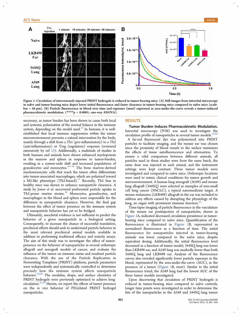
Time-lapse imaging of particle fluorescence in the vasculatureof the mouse ear postinjection of nanoparticles, shown inFigure 1A, indicated decreased circulation persistence in tumor-bearing mice compared to naıve mice. Quantification of thefluorescence is illustrated in Figure 1B, represented asnormalized fluorescence as a function of time. The initialfluorescence for nanoparticles injected in tumor-bearinganimals was lower compared to the naıve mice, despiteequivalent dosing. Additionally, the initial fluorescence leveldecreased as a function of tumor model; 344SQ lung was lowerthan LKB498 ear, and A549 lung was markedly lower than both344SQ lung and LKB498 ear. Analysis of the fluorescencecurves also revealed significantly lower particle exposure in theblood, represented by the area-under-the-curve (AUC), in thepresence of a tumor (Figure 1B, inset). Similar to the initialfluorescence trend, the A549 lung had the lowest AUC of thethree tumor models investigated.Upon discovering that circulation of PRINT hydrogels is
reduced in tumor-bearing mice compared to naıve controls,longer time points were investigated in order to determine thefate of the nanoparticles in the A549 and 344SQ lung tumor
Figure 1. Circulation of intravenously injected PRINT hydrogels is reduced in tumor-bearing mice. (A) Still images from intravital microscopyin naıve and tumor-bearing mice depict lower initial fluorescence and faster clearance in tumor-bearing mice compared to naıve mice (scale-bar = 50 μm). (B) Particle fluorescence in blood over time and exposure (inset) expressed as area-under-the-curve reveals a tumor-inducedpharmacokinetic modulation. (****p < 0.0001; one-way ANOVA).
ACS Nano Article
DOI: 10.1021/acsnano.5b05999ACS Nano 2016, 10, 861−870
862

models. To analyze pharmacokinetic (PK) behavior at longertime points, inductively coupled plasma mass spectroscopy(ICP-MS) was employed as an analytical technique.26 Platinumwas incorporated into the particles via cisplatin drug complex-ation and used to track particles in the blood, spleen, and liver.Limited release of the drug from the particle in plasma wasvalidated in previous studies, ensuring platinum detectioncorrelated directly to particle concentration.27 The circulationprofile was examined to extended time points (0.083, 0.5, 1, 6,and 24 h) using ICP-MS. Using this method, we observed thesame trend as in the IVM study, as both tumor-bearing modelsdemonstrated faster particle clearance compared to naıve mice(Figure 2A). The A549 lung model also exhibited an initialconcentration 2.8-times lower than that for the other particlearms, again despite initial equivalent dosing. This confirmed thephenomenon observed in the IVM fluorescence experiments.Additionally, the circulation behavior of free cisplatin was notaltered by the presence of a tumor. This indicated that themechanism responsible for the difference in clearance was size-dependent. Specific parameters of PK behavior were also
evaluated (Figure 2B). As previously discussed, the AUCrepresents particle exposure or presence in the blood, whichcorrelates to the amount of an entity reaching circulation. Therate of removal of an entity is measured as clearance (CL). Thevolume of distribution (Vd) is a theoretical value representingthe propensity for an entity to stay in the blood compartment(low Vd) compared to being tissue-bound and widelydistributed (high Vd). Examination of these PK parametersconfirmed the results from the fluorescence experiments. Alower AUC of particles in tumor-bearing mice indicateddecreased exposure compared to naıve mice. Increases in CLand Vd for particles in mice with tumors further pointed tomore efficient removal of particles from circulation. For allthree parameters, particles had favorable values compared tofree drug. Figure 2C illustrates the absence of a tumor-basedeffect on free drug PK parameters.The primary organs responsible for removing particles from
circulation are the liver and spleen. Consequently, these organswere also analyzed for platinum content via ICP-MS to evaluatethe fate of the PRINT hydrogel nanoparticles in mice with and
Figure 2. Tumor presence alters nanoparticle circulation and accumulation in organs with salient immune cell activity. The plasma profile (A)and pharmacokinetic parameters (B) of particles were significantly altered by the presence of a tumor, including exposure (AUC), clearancerate from circulation (CL), and volume of distribution (Vd). The behavior of free cisplatin, however, remained unaffected (C). Additionally,there was a significant increase in initial sequestration of particles in both liver (D) and spleen (E) in tumor-bearing mice compared to naıvemice. Measured as platinum (Pt) content via inductively coupled plasma mass spectroscopy (*p < 0.05, **p < 0.01, ***p < 0.001, ****p <0.0001; one-way ANOVA).
ACS Nano Article
DOI: 10.1021/acsnano.5b05999ACS Nano 2016, 10, 861−870
863
without lung tumors (Figure 2D,E). An increase in platinumwas observed at early time points in tumor-bearing mice,corresponding with higher particle concentrations. Theincreased activity by these organs with salient immune cellactivity inversely correlated to the plasma circulation profiles;the presence of a tumor decreased plasma circulation andincreased sequestration in the liver and spleen compared tonaıve mice. Additionally, a large portion of particles wereisolated within the first 5 min after injection in the A549 lungmice, indicated by the higher initial level in both the liver andspleen. As observed in plasma, the effect of a tumor on thedisposition of free cisplatin was also negligible.These surprising results suggest that tumor presence
dramatically impacts nanoparticle circulation, possibly due todifferences in immune status among the groups. The lung hasprominent immune function, and if its function is disrupted(e.g., surgery, tumor burden), this could provoke an unintendedimmune response.28 We validated that the shift in clearance wasnot caused by the surgical procedure for tumor inoculation, astumor-bearing and sham mice experienced the same exactsurgical procedure. Tumor-bearing mice received an injectionof PBS and Matrigel with A549 cells into the lung as describedin Methods, while sham mice received PBS and Matrigelwithout A549 cells. Naıve mice did not have any surgicalprocedure or lung injection. No significant difference incirculation was observed between the sham and naıve mice(Supporting Information Figure 1), confirming that the shift inparticle circulation was caused by the presence of cancer cellsand not by the surgery.Serum from Tumor-Bearing Mice Increases Macro-
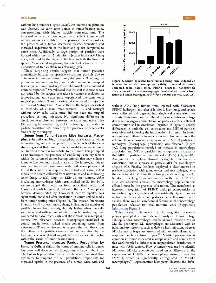
phage Activity ex Vivo. Increased particle clearance withintumor-bearing animals compared to naıve animals of the samestrain suggested that tumor presence might influence immunecell function even in regions distal to the tumor. This promptedus to investigate the presence of circulating secretory factorswithin the serum of tumor-bearing animals that may enhanceimmune function and particle clearance. To interrogate this exvivo, we harvested bone marrow-derived macrophages andtested their activity in response to control and serum-spikedmedia, with serum collected from naıve mice and mice bearingA549 lung, 344SQ lung, or LKB498 ear tumors. Afterincubating macrophages with serum-spiked media for 24 h,we exchanged this media for fresh, nonspiked media, andfluorescent particles were dosed onto the cells. Macrophageactivity, demonstrated by fluorescent particle uptake, wassignificantly enhanced after incubation in serum-spiked mediafrom tumor-bearing mice (Figure 3). The median fluorescentintensity (MFI) of each macrophage, indicating the number ofparticles internalized, was significantly higher when the cellswere incubated with serum collected from tumor-bearing micecompared to naıve mice. Only a slight increase in macrophageactivity was observed between macrophages incubated incontrol media versus spiked media containing serum fromnaıve mice. These ex vivo results support the hypothesis thatthe difference in particle clearance and sequestration by theliver and spleen is, at least in part, caused by a secreted factorwithin the serum of tumor-bearing mice.Tumor Presence Increases Particle Recognition by
Immune Cells. A shift in the status of immune cells in cancerhas been well documented, though less is known about theeffect of such polarization on particle behavior. We used flowcytometry to pinpoint the cell populations responsible forincreased uptake in the presence of a tumor. Mice with and
without A549 lung tumors were injected with fluorescentPRINT hydrogels, and after 2 h, blood, liver, lung, and spleenwere collected and digested into single cell suspensions foranalysis. This time point exhibited a balance between a largedifference in organ accumulation of particles and a sufficientconcentration still in circulation. Depicted in Figure 4, severaldifferences in both the cell association and MFI of particleswere observed following the introduction of a tumor. In blood,no significant difference in association was observed among thecell populations; however, an increase in the MFI of particles inmonocytes (macrophage precursors) was observed (Figure4A). Lung populations revealed an increase in macrophageassociation and MFI of particles, and a significant increase inthe MFI of particles in dendritic cells (DCs) (Figure 4B).Analysis of the spleen showed negligible differences inassociation, but an increase in particle MFI for granulocytes(Figure 4C). Finally, the liver displayed a clear increase inparticle association with granulocytes and macrophages, withthe same trend in MFI for those two populations (Figure 4D).Similar to the lung, a marked increase in the particle MFI inDCs was observed. Overall, the macrophage population wasaffected most by the presence of a tumor. This manifested asincreased recognition of PRINT hydrogel nanoparticles intumor-bearing mice, evidenced by consistently higher numbersin both cell association and particles per cell across organs.Finally, there was no significant difference in the macrophagepopulation relative to total immune cells (SupportingInformation Figure 2).This consistent difference in particle recognition by macro-
phages prompted a more detailed analysis of macrophagesubpopulations. Macrophages can be skewed toward a M1 orM2-like phenotype; M1 macrophages are associated with aninflammatory response, such as defense host infection, whereasM2-like macrophages are associated with an anti-inflammatoryresponse, such as tissue repair.29 M2-like polarization iscommon in tumor-associated macrophages,30 and results fromthis work revealed a difference in subpopulation distribution inmice with A549 tumors. Flow cytometry was used to identifyM1 versus M2-like phenotypes based on a difference in theexpression of CD206, the macrophage mannose receptor(MMR), which is significantly up-regulated in M2-likemacrophages.31 Representative histograms illustrate the differ-
Figure 3. Serum collected from tumor-bearing mice induced anincrease in ex vivo macrophage activity compared to serumcollected from naıve mice. PRINT hydrogel nanoparticleassociation with ex vivo macrophages incubated with serum fromnaıve and tumor-bearing mice (****p < 0.0001; one-way ANOVA).
ACS Nano Article
DOI: 10.1021/acsnano.5b05999ACS Nano 2016, 10, 861−870
864

ence in CD206 expression on macrophages isolated frommouse livers (Figure 5A). Quantification of this MMRexpression revealed a shift in the proportion of macrophagesubsets in the spleen and liver of mice with and without A549lung tumors (Figure 5B). In the spleen, the percentage ofmacrophages that expressed CD206 increased from 29.6% innaıve mice to 45.8% in tumor-bearing mice. The same trend inMMR expression was observed in liver macrophages, whichshifted from 13.7% to 37.1% in naıve and A549 lung mice,respectively. Overall, macrophages in the spleen and liver oftumor-bearing mice were skewed toward a M2-like phenotype.
There was no significant difference in populations among thelung macrophages.When the particle association was measured in these two
macrophage populations, differences were observed betweenthe organs of naıve and tumor-bearing mice (Figure 6). When atumor was present, particles associated more with M1macrophages in the lung and liver compared to the naıvemice (Figure 6A,C; top). In the liver, the M2-like populationalso had higher particle association when a tumor was present.This was consistent with the overall increase in macrophageassociation seen in Figure 4B,D. The spleen had no significant
Figure 4. Tumor presence alters immune cell interactions with particles. Immune cell distribution of PRINT hydrogel nanoparticles in blood(A), lung (B), spleen (C), and liver (D). Significant increases in association and MFI were seen for several populations, includingmacrophages and dendritic cells in the lung and liver. MFI = median fluorescence intensity (*p < 0.05, **p < 0.01, ***p < 0.001; two-wayANOVA).
Figure 5. Presence of A549 tumors skewed macrophage populations from M1 to M2-like. Representative flow cytometry histograms (A) ofCD206 expression in macrophages of the liver show an increase in M2-like phenotype in tumor-bearing mice. A significant increase in thepopulation of liver and spleen M2-like macrophages (B) was observed in tumor-bearing mice compared to naıve mice (**p < 0.01, ****p <0.0001; two-way ANOVA).
ACS Nano Article
DOI: 10.1021/acsnano.5b05999ACS Nano 2016, 10, 861−870
865
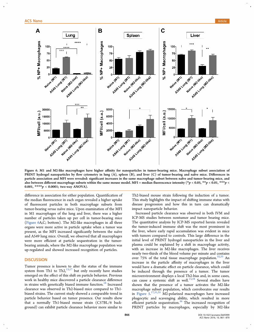
difference in association for either population. Quantification ofthe median fluorescence in each organ revealed a higher uptakeof fluorescent particles in both macrophage subsets fromtumor-bearing versus naıve mice. Upon examination of the MFIin M1 macrophages of the lung and liver, there was a highernumber of particles taken up per cell in tumor-bearing mice(Figure 6A,C; bottom). The M2-like macrophages in all threeorgans were more active in particle uptake when a tumor waspresent, as the MFI increased significantly between the naıveand A549 lung mice. Overall, we observed that all macrophageswere more efficient at particle sequestration in the tumor-bearing animals, where the M2-like macrophage population wasup-regulated and showed increased recognition of particles.
DISCUSSION
Tumor presence is known to alter the status of the immunesystem from Th1 to Th2,13,17 but only recently have studiesemerged on the effect of this shift on particle behavior. Previouswork in healthy mice discovered a particle clearance differencein strains with genetically biased immune function.18 Increasedclearance was observed in Th2-biased mice compared to Th1-biased strains. The current study showed a comparable trend inparticle behavior based on tumor presence. Our results showthat a normally Th1-biased mouse strain (C57BL/6 back-ground) can exhibit particle clearance behavior more similar to
Th2-biased mouse strain following the induction of a tumor.This study highlights the impact of shifting immune status withdisease progression and how this in turn can dramaticallyimpact nanoparticle behavior.Increased particle clearance was observed in both IVM and
ICP-MS studies between nontumor and tumor bearing mice.The quantitative analysis by ICP-MS reported herein revealedthe tumor-induced immune shift was the most prominent inthe liver, where early rapid accumulation was evident in micewith tumors compared to controls. This large difference in theinitial level of PRINT hydrogel nanoparticles in the liver andplasma could be explained by a shift in macrophage activity,with an increase in M2-like macrophages. The liver receivesnearly two-thirds of the blood volume per minute and containsover 75% of the total tissue macrophage population.32,33 Anincrease in the particle affinity of macrophages in the liverwould have a dramatic effect on particle clearance, which couldbe induced through the presence of a tumor. The tumormicroenvironment displays a local Th2-bias and, in some cases,can cause a systemic shift as well.13,34 Several studies haveshown that the presence of a tumor activates the M2-likemacrophage subset population, which corroborates our resultsin Figure 5.17,31,35 M2-polarized macrophages have increasedphagocytic and scavenging ability, which resulted in moreefficient particle sequestration.36 The increased recognition ofPRINT particles by macrophages, especially by M2-like
Figure 6. M1 and M2-like macrophages have higher affinity for nanoparticles in tumor-bearing mice. Macrophage subset association ofPRINT hydrogel nanoparticles by flow cytometry in lung (A), spleen (B), and liver (C) of tumor-bearing and naıve mice. Differences inparticle association and MFI were revealed: significant increases in the same macrophage subset between naıve and tumor-bearing mice, andalso between different macrophage subsets within the same mouse model. MFI = median fluorescence intensity (*p < 0.05, **p < 0.01, ***p <0.001, ****p < 0.0001; two-way ANOVA).
ACS Nano Article
DOI: 10.1021/acsnano.5b05999ACS Nano 2016, 10, 861−870
866

macrophages, can account for the decrease in particlecirculation in the presence of a tumor; however, the increasein both subsets suggests other factors may also be responsiblefor the difference in clearance. For instance, in a study by Caronet al., increased phagocytic activity of circulating monocytes wascorrelated with increased nanoparticle clearance in humans.37
This trend was also observed in our results of Figure 4A.Another study investigated the effect of certain chemokines onthe circulation and accumulation of PEGylated liposomaldoxorubicin (Doxil) in both naıve and tumor-bearing animals,with results similar to those reported here.38 These studiesprovide further evidence that multiple factors can beresponsible for a shift in particle behavior.The changes in immune status and the activation of M2-like
macrophages led us to hypothesize that the tumor cells ormicroenvironment was secreting a factor capable of skewingphagocytic capacity. The ability to mimic the difference inparticle recognition in ex vivo macrophages showed a serum-resident factor was at least partially responsible for the increasein liver and spleen sequestration upon tumor presence.Literature evidence suggests that tumor cells secrete factorsthat can directly affect and change macrophage activity.Previous studies have shown the ability of cancer cells topolarize macrophages, utilizing cytokine analysis, immunohis-tochemistry, and gene amplification to characterize the effectsof coculturing macrophages with cancer cells.39−41 In a study byMuller-Quernheim et al., both the coculture of tumor cells withmacrophages and the treatment of macrophages with mediataken from tumor cell cultures resulted in an increased activityfor both M1 and M2-like macrophages, with a slight skewingtoward a M2-like phenotype.42 Furthermore, this study usedA549 cells, the same line investigated in the work presentedhere. Similar results would be expected in the other two tumormodels, although potentially to a lesser degree due to thesyngeneic nature of the cancer cells. In the A549 modelinvestigated, the initial bias toward a M1 macrophagephenotype in the liver and spleen is supported by the geneticpredisposition of the C57BL/6 background nude mice used inthis study toward a Th1-bias immune system.43 More in-depthstudies to identify the specific factor(s) responsible for the shiftin immune cell activity are needed and currently underway. Thetumor microenvironment is heterogeneous, and identifying aspecific factor among the myriad of cytokines, chemokines,growth factors, cell signaling molecules, and countless othermoieties presents unique challenges. However, recent work hasprovided several areas to focus on in the search, such asgalectins, MHC protein homologues, CTLA-4, PD-1, and avariety of anti-inflammatory interleukins (i.e., IL-4, IL-13).44
The results presented here demonstrate that the presence ofa tumor can alter a local and global immune system, which inturn has dramatic effects on nanoparticle circulation andpotential efficacy. Interestingly, we observed a decrease inparticle circulation between tumor-bearing and nontumor-bearing mice regardless of tumor type and location. However,there were slight differences observed between the variousmodels chosen; the human lung xenograft (A549) showed thelargest increase in particle clearance, with the mouse lungallograft (344SQ) and mouse melanoma allograft (LKB498) inthe ear also showing an increase compared to naive controls. Allof these models were induced in female Foxn1nu (athymicnude, C57BL/6J background) mice, yet a considerabledifference was still apparent based on tumor presence. Wehypothesize that a similar shift from a Th1- to Th2-biased
immune status was responsible for the increased clearance ineach of these tumor models. Understanding the role of thesechanging phenotypes on nanoparticle interaction offers anopportunity for increasing nanoparticle efficacy; mitigation orreversal of the systemic shift could be achieved by incorporatinga scavenging moiety onto the particle to sequester thecirculating factor and reduce immune evasion by the tumor.Alternative approaches using immunotherapy could harness themacrophage’s affinity for particles and co-opt a change in statusby delivering a stimulant to the macrophage, reversing thepolarization to promote an antitumor M1 phenotype.
CONCLUSION
Our work highlights the important interplay between immunestatus, disease progression, and nanoparticle properties. Toevaluate nanoparticle efficacy, an animal model must be chosenwhich has an immune status similar to the actual disease. For agiven tumor cell line, mouse strain, and tumor location,differences in secreted factors and resultant changes in cellularfunction will affect particle clearance properties. Thus, priorcharacterization of the model itself will be essential toevaluating the preclinical success of a given nanoparticletherapeutic. Furthermore, a given immune status may bepredictive of nanoparticle success. This has important clinicalramifications. Further evaluation of a patient’s immune functionfollowing similar evaluations to those presented here may beimportant predictors of nanoparticle success in a clinical setting.
METHODSMaterials. Commercially available polyethylene glycol diacrylate
(PEG700-DA) (Mn = 700 Da), 2-aminoethyl methacrylate hydro-chloride (AEM), diphenyl(2,4,6-trimethylbenzoyl)-phosphine oxide(TPO), poly(vinyl alcohol) (PVOH, Mn = 2000 Da), succinicanhydride, cis-diaminedichloroplatinum(II) (CDDP), and sucrosewere purchased from Sigma-Aldrich. PTFE syringe filters (13 mmmembrane, 0.220 μm pore size), Dylight 488 maleimide, Dylight 650maleimide, dimethylformamide (DMF), triethylamine (TEA), pyr-idine, sterile water, borate buffer (pH 8.6), methanol, trace-metal gradeconcentrated nitric acid (HNO3), Corning Matrigel Membrane Matrix(LDEV-free), EDTA-treated collection tubes, cell strainers, 4%paraformaldehyde (PFA), and ACK buffer were obtained from FisherScientific. Methoxy PEG (5k)-succinimidyl carboxy methyl ester(mPEG5k-SCM) was purchased from Creative PEGWorks. Tetra-ethylene glycol monoacrylate (HP4A) was synthesized in-house aspreviously described.45 Conventional filters (2 μm) were purchasedfrom Agilent, and poly(vinyl alcohol) (Mw 2000) (PVOH) waspurchased from Acros Organics. PRINT molds (80 nm × 320 nm)were obtained from Liquidia Technologies (Morrisville, NC).Polyethylene terephthalate (PET) was purchased in 1000-foot rollsfrom 3M. Cisplatin was acquired from the University of NorthCarolina Pharmacy. Water, where used, was sterile-grade and 0.2-μmfiltered. A549-luc, LKB498, and L929 cells were purchased fromAmerican Type Culture Collection. 344SQ cells were a gift from TheUniversity of Texas M.D. Anderson Cancer Center (Jon Kurie Lab).All cells were maintained per vendor specifications. Fetal bovine serumwas purchased from Atlanta Biologicals. Hank’s Balanced Salt Solution(HBSS), RPMI 1640 Medium, and Dulbecco’s Modified EagleMedium (DMEM) were purchased from Gibco. All commerciallyavailable materials were used as received. Anti-mouse antibodies(CD45-Pac Blue, CD11c-PE, CD206-PE, CD11b-Brilliant Violet 605,CD19-PE-Cy7, F4/80-APC, Ly6G/C-PE-Cy5) were purchased fromBioLegend, Inc. Lymphoprep, DNase, and collagenase were purchasedfrom STEMCELL Technologies, Inc. AbC Anti-Mouse Bead Kit andanti-CD16/32 (Fc-block) were purchased from Invitrogen. FACSbuffer was prepared as HBSS plus 2% FBS.
ACS Nano Article
DOI: 10.1021/acsnano.5b05999ACS Nano 2016, 10, 861−870
867
Particle Fabrication and Characterization. PRINT 80 nm ×320 nm hydrogel particles were fabricated and functionalized with aPEG mushroom surface as described in ref 22, and a more detaileddescription can be found in Supporting Information. Particles werethen succinylated by reaction with an excess of pyridine and succinicanhydride (100× molar excess with respect to amine groups). Thereaction was carried out in a sonicator bath (Branson UltrasonicCleaner 1.4 A, 160 W) for 30 min. Following succinylation, theparticles were washed by centrifugation one time in DMF, followed bya borate buffer wash to neutralize any succinic acid side product, andthen three washes with sterile water.Cisplatin complexation was achieved by incubating the particles in a
solution of CDDP (2× molar excess with respect to carboxyl groups)in water at room temperature for >24 h under constant agitation(Eppendorf, 1400 rpm). After incubation in the complexation solution,particles were washed with sterile water by centrifugation andresuspended in 9.25 wt % sucrose (aq) at the appropriate doseconcentration. Aliquots were flash frozen in liquid nitrogen and storedat −20 °C until needed. Dylight 650 and 488 were used for theintravital and flow cytometry studies, respectively. PRINT-cisplatinwas used without a fluorophore for the inductively coupled-massspectroscopy (ICP-MS) studies.Particle concentrations were determined by thermogravimetric
analysis (TGA) using a TAInstruments Q5000 analyzer. Particle sizeand zeta potential were verified by dynamic light scattering (DLS) on aZetasizer Nano ZS (Malvern Instruments, Ltd.) at 37 °C. Cisplatinloading was assessed using an Agilent 1200 series high-performanceliquid chromatography system with an ultraviolet detector. The mobilephase consisted of 90% 0.9-wt % NaCl (aq) and 10% methanol, byvolume. A 5 min isocratic elution protocol was used with a ZORBAXEclipse Plus C18 column (Agilent Technologies). The product waseluted at a flow rate of 1 mL/min and monitored at a wavelength of210 nm. Drug loading was determined by analysis of the complexationsolution pre- and postincubation. The net difference in cisplatinconcentration was calculated as weight percent in the particle.Animals. All experiments involving mice were performed in
accordance with the National Research Council’s Guide to Care andUse of Laboratory Animals (1996), under an animal use protocolapproved by the University of North Carolina Animal Care and UseCommittee. All studies used female Foxn1nu (athymic nude, C57BL/6Jbackground) mice (5 weeks old, 17−27 g, Jackson Laboratory). Forthe tumor-bearing mice, two orthotopic non-small cell lung models(A549-luc and 344SQ) and an orthotopic melanoma (LKB498) modelwere used. Cell cultures were prepared and maintained per vendorspecifications. The orthotopic lung surgery was performed by injectionof a 40 μL suspension of either A549 cells (5 × 10⁶ cells per mouse) or344SQ cells (5000 cells per mouse) in a 50:50 Matrigel:PBS blendinto the lung via intrathoracic inoculations, as per publishedprotocol.46 Tumors were grown to 100−200 mm3 total cumulativevolume (caliper measurement for LKB498, orthotopic A549luc and344SQ verified by measurement following thoracotomy). Sham miceunderwent the surgical procedure and received the Matrigel:PBSsuspension injection without cells. For the orthotopic ear allograft, asingle spheroid of approximately 4000 LKB498 cells was injectedintradermally as previously described.47 All nanoparticles were dosedin 9.25 wt % sucrose to maintain isotonicity upon intravenousadministration.Ex Vivo Macrophage Association. Primary macrophages were
isolated from the bone marrow of nude mice per establishedprotocol.48 Briefly, mice were euthanized, and the femur and tibiabones were resected. Bone marrow cells were collected by flushing themarrow cavity with HBSS + 2% FBS. Cells were filtered and platedwith L929-conditioned medium (containing GM-CSF) to promotedifferentiation and growth of macrophages from precursor cells withinthe marrow. Cells were incubated with or without serum collectedfrom naıve and tumor-bearing mice. After 24 h, the serum-spikedmedia was replaced with fresh, nonspiked media, and fluorescentPRINT hydrogel nanoparticles were dosed at 1 mg/mL for 3 h. Cellswere then collected and fixed with 4% PFA for analysis by flowcytometry.
Intravital Microscopy Study. Particles containing Dylight 650were injected and analyzed by intravital microscopy as described in ref22. Briefly, experiments were performed using an IV 100 laser scanningmicroscope (Olympus). Mice (n = 5) were anesthetized withisofluorane, a tail vein catheter was applied, and animals were placedon a 37 °C heated stage in the prone position and kept underanesthesia. The ear was immobilized to an aluminum block withdouble-sided tape, and vasculature was visualized with a 488 nm laser;the nontumor-bearing ear was used in studies with the LKB498 mice.Mice were then dosed with a bolus injection of Dylight 650-labelednanoparticles. Fluorescence was measured using a 633 nm laser, andimaging scans were captured every 5 s for 2 h. For circulation analysis,the image files from each scan were exported to ImageJ. Followingliterature procedures, the images were stacked in groups of four, andfluorescent signal in each stack was analyzed in the region of interest(ROI).23,49 Background corrections were obtained using the initialfluorescence in the ROI before injection. All instrument settings werekept constant for the duration of the study. Particles were dosed at18.75 mg per kg of body weight.
ICP-MS study. Quantification of cisplatin and PRINT-cisplatin wasperformed using ICP-MS. Cisplatin and PRINT particles were dosedwith a bolus intravenous injection at an equivalent of 3 mg per kg drug,which corresponded to a particle dose of 18.75 mg/kg. Liver, spleen,lung, and kidney were harvested and flash-frozen in liquid nitrogen at0.083, 0.5, 1, 6, and 24 h postinjection. Tumor accumulation was notdetermined due to tumor cell heterogeneity with the lung cells withinthe organ in the orthotopic A549 model. Four mice per arm wereexamined at each time point. Blood was collected in EDTA by cardiacpuncture at the same time points and centrifuged (300g, 5 min, 4 °C)to isolate plasma from the cell fraction. Tissue sample preparation wasperformed as previously described.6 Briefly, tissue and plasma sampleswere digested in concentrated HNO3 spiked with 200 ng/mL Iridium(Ir; analytical internal standard, Inorganic Ventures) for 60−90 min at90 °C. Deionized water was added to bring sample to volume andHNO3 concentration of 3.5%, and the samples were stored at 4 °Cuntil platinum (Pt) analysis was completed. ICP-MS analysis (Agilent7500cx) was performed and validated as previously described.6,26
Pharmacokinetic analysis of the measured plasma concentrations wasperformed using PKSolver.50 Data was fit to either a one- or two-compartment model, and the Akaike information criterion (AIC) wasused to compare goodness of fit for each nanoparticle type.51
Tissue Preparation for Immune Cell Analysis. Once the A549lung tumors had grown to sufficient size, both tumor-bearing andnaıve mice were injected with a bolus intravenous suspension ofPRINT hydrogel particles containing Dylight 488 at 18.75 mg/kg (n =5). Additional tumor-bearing and naıve mice were given a bolusintravenous saline injection for use as controls (n = 3). Two hourspostinjections, mice were euthanized and blood was collected viacardiac puncture and stored on ice in EDTA-treated collection tubes.The thoracic cavity was opened to visualize the heart. The right atriumwas nicked, and organs were perfused with a HBSS flush via the leftventricle. The lung, spleen, and liver were resected and stored on ice.Spleens were mechanically forced through a cell strainer to dissociatetissue into FACS buffer. Lungs and livers were incubated at 37 °C, 5%CO2 in digestion media (HBSS + 2% FBS + 0.02 mg/mL DNase and1 mg/mL collagenase) until dissociated. ACK buffer was used to lysered blood cells in blood, lung, and spleen, and samples weresubsequently passed through a cell strainer to remove aggregates andexcess debris. Immune cells were isolated from livers usingLymphoprep, per manufacturer’s guidelines. This resulted in singlecell suspensions of blood, lung, spleen, and liver tissues. Samples wereblocked with anti-CD16/32 (Fc-block) and stained with a panel ofantibodies. Lung, spleen, and liver samples were split into two equalaliquots. One aliquot, along with blood samples, received Panel A:CD45-Pac Blue, CD11b-Brilliant Violet 605, CD11c-PE, Ly6G-PE-Cy5, F4/80-APC, and CD19-PE-Cy7. The remaining lung, spleen, andliver aliquots received Panel B: CD45-Pac Blue, CD11b-Brilliant Violet605, F4/80-APC, and CD206-PE. All samples were fixed with 4% PFAand stored for analysis by flow cytometry.
ACS Nano Article
DOI: 10.1021/acsnano.5b05999ACS Nano 2016, 10, 861−870
868

Flow Cytometry. Samples of single cell suspensions from blood,lung, spleen, and liver of naıve and tumor-bearing mice were analyzedwith an LSRII (BD Biosciences) flow cytometer. AbC Anti-MouseBeads were labeled with each of the fluorophore-antibody and used tocompensate for each fluorescence channel. Fluorescence minus one(FMO) samples were prepared from untreated cell suspensions fordelineation of positive and negative antibody expression on cells.Representative sample gating for Panel A and Panel B is depicted inSupporting Information Figures 3 and 4, respectively. Samples fromthe ex vivo macrophage association study were analyzed as described,and utilized only forward scatter, side scatter, and the Dylight 488fluorescence channel and did not require compensation or FMOsamples. FlowJo software (Tree Star) was used to analyze data perliterature precedence and guidance.18,52
Statistics. Analysis of Variance (ANOVA) was performed inGraphPad Prism using the Bonferroni post-test. All error barsrepresent of standard error mean.
ASSOCIATED CONTENT*S Supporting InformationThe Supporting Information is available free of charge on theACS Publications website at DOI: 10.1021/acsnano.5b05999.
The effect of surgery on particle circulation, flowcytometry gating schemes, and PRINT hydrogel nano-particle fabrication and characterization details (PDF)
AUTHOR INFORMATIONCorresponding Authors*E-mail: [email protected].*E-mail: [email protected] authors declare no competing financial interest.
ACKNOWLEDGMENTSThe authors would like to thank Charlene Santos and the UNCAnimal Studies Core for their help with the murineexperiments. We also thank Peter Cable for his expertise withICP-MS analysis, Sara O’Neal for help with pharmacokineticdata collection and analysis, and Dr. Ashish Pandya for thesynthesis of HP4A. This work was supported by LiquidiaTechnologies, the Carolina Center for Cancer NanotechnologyExcellence U54CA151652 and U54CA198999 (to J.P.-Y.T. andJ.M.D.), NIH Pioneer Award 1DP1OD006432 (to J.M.D.),NIH Grant U19AI109784 (to J.P.-Y.T. and J.M.D.), andDefense Threat Reduction Agency (DTRA) Award HDTRA1-13-1-0045 (to J.M.D.).
REFERENCES(1) Wang, A. Z.; Langer, R.; Farokhzad, O. C. Nanoparticle Deliveryof Cancer Drugs. Annu. Rev. Med. 2012, 63, 185−198.(2) Fidler, I. J. Rationale and Methods For the Use of Nude Mice toStudy the Biology and Therapy of Human Cancer Metastasis. CancerMetastasis Rev. 1986, 5, 29−49.(3) Marusyk, A.; Almendro, V.; Polyak, K. Intra-TumourHeterogeneity: A Looking Glass For Cancer? Nat. Rev. Cancer 2012,12, 323−334.(4) Balkwill, F. R.; Capasso, M.; Hagemann, T. The TumorMicroenvironment at a Glance. J. Cell Sci. 2012, 125, 5591−5596.(5) Whiteside, T. L. The Tumor Microenvironment and Its Role inPromoting Tumor Growth. Oncogene 2008, 27, 5904−5912.(6) Combest, A. J.; Roberts, P. J.; Dillon, P. M.; Sandison, K.; Hanna,S. K.; Ross, C.; Habibi, S.; Zamboni, B.; Muller, M.; Brunner, M.; et al.Genetically Engineered Cancer Models, But Not Xenografts, FaithfullyPredict Anticancer Drug Exposure in Melanoma Tumors. Oncologist2012, 17, 1303−1316.
(7) Richmond, A.; Su, Y. Mouse Xenograft Models vs GEM ModelsFor Human Cancer Therapeutics. Dis. Models & Mech. 2008, 1,78−82.(8) Nel, A. E.; Madler, L.; Velegol, D.; Xia, T.; Hoek, E. M. V.;Somasundaran, P.; Klaessig, F.; Castranova, V.; Thompson, M.Understanding Biophysicochemical Interactions at the Nano-BioInterface. Nat. Mater. 2009, 8, 543−557.(9) Albanese, A.; Tang, P. S.; Chan, W. C. W. The Effect ofNanoparticle Size, Shape, and Surface Chemistry on BiologicalSystems. Annu. Rev. Biomed. Eng. 2012, 14, 1−16.(10) Owens, D. E., III; Peppas, N. A. Opsonization, Biodistribution,and Pharmacokinetics of Polymeric Nanoparticles. Int. J. Pharm. 2006,307, 93−102.(11) Sun, X.; Rossin, R.; Turner, J. L.; Becker, M. L.; Joralemon, M.J.; Welch, M. J.; Wooley, K. L. An Assessment of the Effects of ShellCross-Linked Nanoparticle Size, Core Composition, and SurfacePEGylation on In Vivo Biodistribution. Biomacromolecules 2005, 6,2541−2554.(12) Kiessling, R.; Wasserman, K.; Horiguchi, S.; Kono, K.; Sjoberg,J.; Pisa, P.; Petersson, M. Tumor-Induced Immune Dysfunction.Cancer Immunol. Immunother. 1999, 48, 353−362.(13) Shurin, M. R.; Lu, L.; Kalinski, P.; Stewart-Akers, A. M.; Lotze,M. T. Th1/Th2 Balance in Cancer, Transplantation and Pregnancy.Springer Semin. Immunopathol. 1999, 21, 339−359.(14) Bronte, V.; Serafini, P.; Apolloni, E.; Zanovello, P. Tumor-Induced Immune Dysfunctions Caused by Myeloid Suppressor Cells.J. Immunother. 2001, 24, 431−446.(15) Kusmartsev, S.; Gabrilovich, D. I. Immature Myeloid Cells andCancer-Associated Immune Suppression. Cancer Immunol. Immun-other. 2002, 51, 293−298.(16) Serafini, P.; De Santo, C.; Marigo, I.; Cingarlini, S.; Dolcetti, L.;Gallina, G.; Zanovello, P.; Bronte, V. Derangement of ImmuneResponses by Myeloid Suppressor Cells. Cancer Immunol. Immunother.2004, 53, 64−72.(17) Sica, A.; Bronte, V. Altered Macrophage Differentiation andImmune Dysfunction in Tumor Development. J. Clin. Invest. 2007,117, 1155−1166.(18) Jones, S. W.; Roberts, R. A.; Robbins, G. R.; Perry, J. L.; Kai, M.P.; Chen, K.; Bo, T.; Napier, M. E.; Ting, J. P. Y.; DeSimone, J. M.;et al. Nanoparticle Clearance is Governed by Th1/Th2 Immunity andStrain Background. J. Clin. Invest. 2013, 123, 3061−3073.(19) Euliss, L. E.; DuPont, J. A.; Gratton, S.; DeSimone, J. ImpartingSize, Shape, and Composition Control of Materials for Nanomedicine.Chem. Soc. Rev. 2006, 35, 1095−1104.(20) Rolland, J. P.; Maynor, B. W.; Euliss, L. E.; Exner, A. E.;Denison, G. M.; DeSimone, J. M. Direct Fabrication and Harvesting ofMonodisperse, Shape-Specific Nanobiomaterials. J. Am. Chem. Soc.2005, 127, 10096−10100.(21) Kersey, F. R.; Merkel, T. J.; Perry, J. L.; Napier, M. E.;Desimone, J. M. Effect of Aspect Ratio and Deformability onNanoparticle Extravasation Through Nanopores. Langmuir 2012, 28,8773−8781.(22) Perry, J. L.; Reuter, K. G.; Kai, M. P.; Herlihy, K. P.; Jones, S.W.; Luft, J. C.; Napier, M.; Bear, J. E.; DeSimone, J. M. PEGylatedPRINT Nanoparticles: The Impact of PEG Density on ProteinBinding, Macrophage Association, Biodistribution, and Pharmacoki-netics. Nano Lett. 2012, 12, 5304−5310.(23) Merkel, T. J.; Jones, S. W.; Herlihy, K. P.; Kersey, F. R.; Shields,A. R.; Napier, M.; Luft, J. C.; Wu, H.; Zamboni, W. C.; Wang, A. Z.;et al. Using Mechanobiological Mimicry of Red Blood Cells to ExtendCirculation Times of Hydrogel Microparticles. Proc. Natl. Acad. Sci. U.S. A. 2011, 108, 586−591.(24) Hughes, E. L.; Gavins, F. N. E. Troubleshooting methods: UsingIntravital Microscopy in Drug Research. J. Pharmacol. Toxicol. Methods2010, 61, 102−112.(25) Hu, W.; Pasare, C. Location, Location, Location: Tissue-SpecificRegulation of Immune Responses. J. Leukocyte Biol. 2013, 94, 409−421.
ACS Nano Article
DOI: 10.1021/acsnano.5b05999ACS Nano 2016, 10, 861−870
869

(26) Morrison, J. G.; White, P.; McDougall, S.; Firth, J. W.; Woolfrey,S. G.; Graham, M. A.; Greenslade, D. Validation of a Highly SensitiveICP-MS Method for the Determination of Platinum in Biofluids:Application to Clinical Pharmacokinetic Studies With Oxaliplatin. J.Pharm. Biomed. Anal. 2000, 24, 1−10.(27) Kai, M. P.; Keeler, A. W.; Perry, J. L.; Reuter, K. G.; Luft, J. C.;O’Neal, S. K.; Zamboni, W. C.; DeSimone, J. M. Evaluation of DrugLoading, Pharmacokinetic Behavior, and Toxicity of a Cisplatin-Containing Hydrogel Nanoparticle. J. Controlled Release 2015, 204,70−7.(28) Kwon, M.; Berns, A. Mouse Models for Lung Cancer. Mol.Oncol. 2013, 7, 165−177.(29) Mantovani, A.; Locati, M. Tumor-Associated Macrophages as aParadigm of Macrophage Plasticity, Diversity, and Polarization:Lessons and Open Questions. Arterioscler., Thromb., Vasc. Biol. 2013,33, 1478−1483.(30) Mantovani, A.; Sozzani, S.; Locati, M.; Allavena, P.; Sica, A.Macrophage Polarization: Tumor-Associated Macrophages as aParadigm for Polarized M2Mononuclear Phagocytes. Trends Immunol.2002, 23, 549−555.(31) Gabrilovich, D. I.; Ostrand-Rosenberg, S.; Bronte, V.Coordinated Regulation of Myeloid Cells by Tumours. Nat. Rev.Immunol. 2012, 12, 253−268.(32) Jenne, C. N.; Kubes, P. Immune Surveillance by the Liver. Nat.Immunol. 2013, 14, 996−1006.(33) Sheth, K.; Bankey, P. The Liver as an Immune Organ. Curr.Opin Crit Care 2001, 7, 99−104.(34) Finn, O. J. Cancer Immunology. N. Engl. J. Med. 2008, 358,2704−2715.(35) Van Ginderachter, J.; Meerschaut, S.; Liu, Y.; Brys, L.; DeGroeve, K.; Hassanzadeh Ghassabeh, G.; Raes, G.; De Baetselier, P.Peroxisome Proliferator-Activated Receptor Gamma (PPARgamma)Ligands Reverse CTL Suppression by Alternatively Activated (M2)Macrophages in Cancer. Blood 2006, 108, 525−535.(36) Biswas, S. K.; Mantovani, A. Macrophage Plasticity andInteraction With Lymphocyte Subsets: Cancer as a Paradigm. Nat.Immunol. 2010, 11, 889−896.(37) Caron, W. P.; Lay, J. C.; Fong, A. M.; La-Beck, N. M.; Kumar,P.; Newman, S. E.; Zhou, H.; Monaco, J. H.; Clarke-Pearson, D. L.;Brewster, W. R.; et al. Translational Studies of Phenotypic Probes forthe Mononuclear Phagocyte System and Liposomal Pharmacology. J.Pharmacol. Exp. Ther. 2013, 347, 599−606.(38) Song, G.; Tarrant, T. K.; White, T. F.; Barrow, D. A.; Santos, C.M.; Timoshchenko, R. G.; Hanna, S. K.; Ramanathan, R. K.; Lee, C.R.; Bae-Jump, V. L.; et al. Roles of Chemokines CCL2 and CCL5 inthe Pharmacokinetics of PEGylated Liposomal Doxorubicin in vivo andin Patients With Recurrent Epithelial Ovarian Cancer. Nanomedicine2015, 11, 1797−1807.(39) Yuan, Z.; Mehta, H. J.; Mohammed, K.; Nasreen, N.; Roman,R.; Brantly, M.; Sadikot, R. T. TREM-1 is Induced in TumorAssociated Macrophages by Cyclo-oxygenase Pathway in Human Non-Small Cell Lung Cancer. PLoS One 2014, 9, e94241.(40) Cho, H. J.; Jung, J. I.; Lim, D. Y.; Kwon, G. T.; Her, S.; Park, J.H.; Park, J. H. Bone Marrow-Derived, Alternatively ActivatedMacrophages Enhance Solid Tumor Growth and Lung Metastasis ofMammary Carcinoma Cells in a Balb/C Mouse Orthotopic Model.Breast Cancer Res. 2012, 14, R81.(41) Han, W.; Joo, M.; Everhart, M. B.; Christman, J. W.; Yull, F. E.;Blackwell, T. S. Myeloid Cells Control Termination of LungInflammation Through the NF-kappaB Pathway. Am. J. Physiol LungCell Mol. Physiol 2009, 296, L320−327.(42) Muller-Quernheim, U. C.; Potthast, L.; Muller-Quernheim, J.;Zissel, G. Tumor-Cell Co-Culture Induced Alternative Activation ofMacrophages is Modulated by Interferons In Vitro. J. InterferonCytokine Res. 2012, 32, 169−177.(43) Petkova, S. B.; Yuan, R.; Tsaih, S.; Schott, W.; Roopenian, D. C.;Paigen, B. Genetic Influence on Immune Phenotype Revealed Strain-Specific Variations in Peripheral Blood Lineages. Physiol. Genomics2008, 34, 304−314.
(44) Burkholder, B.; Huang, R.-Y.; Burgess, R.; Luo, S.; Jones, V. S.;Zhange, W.; Lv, Z.-Q.; Gao, C.-Y.; Wang, B.-L.; Zhang, Y.-M.; et al.Tumor-Induced Perturbations of Cytokines and Immune CellNetworks. Biochim. Biophys. Acta, Rev. Cancer 2014, 1845, 182−201.(45) Guzman, J.; Iglesias, M. T.; Riande, E.; Compan, V.; Andrio, A.Synthesis and Polymerization of Acrylic Monomers With HydrophilicLong Side Groups. Oxygen Transport Through Water SwollenMembranes Prepared From These Polymers. Polymer 1997, 38,5227−5232.(46) Peng, L.; Feng, L.; Yuan, H.; Benhabbour, S. R.; Mumper, R. J.Development of a Novel Orthotopic Non-Small Cell Lung CancerModel and Therapeutic Benefit of 2′-(2-Bromohexadecanoyl)-Docetaxel Conjugate Nanoparticles. Nanomedicine 2014, 10, 1497−1506.(47) Rozenberg, G. I.; Monahan, K. B.; Torrice, C.; Bear, J. E.;Sharpless, N. E. Metastasis in an Orthotopic Murine Model ofMelanoma is Independent of RAS/RAF Mutation. Melanoma Res.2010, 20, 361−371.(48) Weischenfeldt, J.; Porse, B. Bone Marrow-Derived Macrophages(BMM): Isolation and Applications. Cold Spring Harb. Protoc. 2008, 3,1−6.(49) Tong, L.; He, W.; Zhang, Y.; Zheng, W.; Cheng, J.-X. VisualizingSystemic Clearance and Cellular Level Biodistribution of GoldNanorods by Intrinsic Two-Photon Luminescence. Langmuir 2009,25, 12454−12459.(50) Zhang, Y.; Huo, M.; Zhou, J.; Xie, S. PKSolver: An Add-inProgram for Pharmacokinetic and Pharmacodynamic Data Analysis inMicrosoft Excel. Comput. Methods Programs Biomed 2010, 99, 306−314.(51) Akaike, H. A New Look at the Statistical Model Identification.IEEE Trans. Autom. Control 1974, 19, 716−723.(52) Herzenberg, L.; Tung, J.; Moore, W.; Herzenberg, L.; Parks, D.R. Interpreting Flow Cytometry Data: A Guide for the Perplexed. Nat.Immunol. 2006, 7, 681−685.
ACS Nano Article
DOI: 10.1021/acsnano.5b05999ACS Nano 2016, 10, 861−870
870