Tumor interstitial fluid formation, characterization, and clinical ...
12
REVIEW published: 26 May 2015 doi: 10.3389/fonc.2015.00115 Edited by: Gianfranco Baronzio, Kines Medical Centre, Italy Reviewed by: Christian Stock, University of Münster, Germany Isabel Freitas, University of Pavia, Italy *Correspondence: Helge Wiig, Department of Biomedicine, University of Bergen, Jonas Lies vei 91, N-5009 Bergen, Norway [email protected] Specialty section: This article was submitted to Molecular and Cellular Oncology, a section of the journal Frontiers in Oncology Received: 20 March 2015 Accepted: 06 May 2015 Published: 26 May 2015 Citation: Wagner M and Wiig H (2015) Tumor interstitial fluid formation, characterization, and clinical implications. Front. Oncol. 5:115. doi: 10.3389/fonc.2015.00115 Tumor interstitial fluid formation, characterization, and clinical implications Marek Wagner and Helge Wiig* Department of Biomedicine, University of Bergen, Bergen, Norway The interstitium, situated between the blood and lymph vessels and the cells, consists of a solid or matrix phase and a fluid phase representing the tissue microenvironment. In the present review, we focus on the interstitial fluid phase of solid tumors, the tumor interstitial fluid (TIF), i.e., the fluid bathing the tumor and stroma cells, also including immune cells. This is a component of the internal milieu of a solid tumor that has attracted regained attention. Access to this space may provide important insight into tumor development and therapy response. TIF is formed by transcapillary filtration, and since this fluid is not readily available we discuss available techniques for TIF isolation, results from subsequent characterization and implications of recent findings with respect to fluid filtration and uptake of macromolecular therapeutic agents. There appear to be local gradients in signaling substances from neoplastic tissue to plasma that may provide new understanding of tumor biology. The development of sensitive proteomic technologies has made TIF a valuable source for tumor specific proteins and biomarker candidates. Potential biomarkers will appear locally in high concentrations in tumors and may eventually be found diluted in the plasma. Access to TIF that reliably reflects the local tumor microenvironment enables identification of substances that can be used in early detection and monitoring of disease. Keywords: extracellular matrix, extracellular space, biomarkers, proteomics, tumor microenvironment, tumor extracellular fluid, interstitial space Introduction The interstitium, or interstitial space, is a general term applied for connective and supporting tissues in the body. This space is located outside the blood and lymph vessels and parenchymal cells, and consists of two major phases: the interstitial fluid (IF) and the structural molecules comprising the extracellular matrix (ECM). The tumor interstitial fluid (TIF) is not only a transport medium for nutrients and waste products between cells and capillary blood, but also contains an abundance of substances that are either produced locally or transported to the organ by the blood circulation. Cells have traditionally not been included in this concept of the interstitium (1). Cells in the interstitium, however, are active in continuous bi-directional cell–matrix interactions that result in microenvironmental changes, secrete substances to the IF and have important roles in initiating immune responses (2), and are a central element of the tumor interstitium. All of these are good reasons for including cells in the term “interstitium” here, notably those that are not organ specific, e.g., fibroblasts or immune cells, but rather an integrated part of the ECM. Whereas in previous years, the focus has been on the tumor cell per se, during recent years, there has been an increasing Frontiers in Oncology | www.frontiersin.org May 2015 | Volume 5 | Article 115 1
Transcript of Tumor interstitial fluid formation, characterization, and clinical ...

REVIEWpublished: 26 May 2015
doi: 10.3389/fonc.2015.00115
Edited by:Gianfranco Baronzio,
Kines Medical Centre, Italy
Reviewed by:Christian Stock,
University of Münster, GermanyIsabel Freitas,
University of Pavia, Italy
*Correspondence:Helge Wiig,
Department of Biomedicine,University of Bergen, Jonas Lies vei
91, N-5009 Bergen, [email protected]
Specialty section:This article was submitted to
Molecular and Cellular Oncology,a section of the journal Frontiers in
Oncology
Received: 20 March 2015Accepted: 06 May 2015Published: 26 May 2015
Citation:Wagner M and Wiig H (2015) Tumor
interstitial fluid formation,characterization, and clinical
implications.Front. Oncol. 5:115.
doi: 10.3389/fonc.2015.00115
Tumor interstitial fluid formation,characterization, and clinicalimplicationsMarek Wagner and Helge Wiig*
Department of Biomedicine, University of Bergen, Bergen, Norway
The interstitium, situated between the blood and lymph vessels and the cells, consistsof a solid or matrix phase and a fluid phase representing the tissue microenvironment. Inthe present review, we focus on the interstitial fluid phase of solid tumors, the tumorinterstitial fluid (TIF), i.e., the fluid bathing the tumor and stroma cells, also includingimmune cells. This is a component of the internal milieu of a solid tumor that hasattracted regained attention. Access to this space may provide important insight intotumor development and therapy response. TIF is formed by transcapillary filtration, andsince this fluid is not readily available we discuss available techniques for TIF isolation,results from subsequent characterization and implications of recent findings with respectto fluid filtration and uptake of macromolecular therapeutic agents. There appear tobe local gradients in signaling substances from neoplastic tissue to plasma that mayprovide new understanding of tumor biology. The development of sensitive proteomictechnologies has made TIF a valuable source for tumor specific proteins and biomarkercandidates. Potential biomarkers will appear locally in high concentrations in tumors andmay eventually be found diluted in the plasma. Access to TIF that reliably reflects thelocal tumor microenvironment enables identification of substances that can be used inearly detection and monitoring of disease.
Keywords: extracellular matrix, extracellular space, biomarkers, proteomics, tumor microenvironment, tumorextracellular fluid, interstitial space
Introduction
The interstitium, or interstitial space, is a general term applied for connective and supporting tissuesin the body. This space is located outside the blood and lymph vessels and parenchymal cells, andconsists of two major phases: the interstitial fluid (IF) and the structural molecules comprising theextracellular matrix (ECM). The tumor interstitial fluid (TIF) is not only a transport medium fornutrients and waste products between cells and capillary blood, but also contains an abundance ofsubstances that are either produced locally or transported to the organ by the blood circulation.
Cells have traditionally not been included in this concept of the interstitium (1). Cells in theinterstitium, however, are active in continuous bi-directional cell–matrix interactions that resultin microenvironmental changes, secrete substances to the IF and have important roles in initiatingimmune responses (2), and are a central element of the tumor interstitium. All of these are goodreasons for including cells in the term “interstitium” here, notably those that are not organ specific,e.g., fibroblasts or immune cells, but rather an integrated part of the ECM. Whereas in previousyears, the focus has been on the tumor cell per se, during recent years, there has been an increasing
Frontiers in Oncology | www.frontiersin.org May 2015 | Volume 5 | Article 1151

Wagner and Wiig Tumor interstitial fluid
interest in the tumormicroenvironment shown to be of significantimportance for tumor growth and metastasis. The microenviron-ment consists of the insoluble elements of the ECM, the interstitialspace with its non-tumor cellular elements (frequently referred toas stroma), and the fluid phase containing dissolved substances.While tumor microenvironment studies have mostly been on thestroma and the cellular elements of the tumor, we will focus on thefluid phase that has received less attention (3–5).
Here, we will review in brief the structure of the tumor ECMas a part of a general description of the tumor interstitium beforewe turn to the formation of TIF and techniques for fluid isolationof most relevance for the secretome, i.e., substances secreted bythe tumor to the TIF. Our aim is to summarize recent studies onTIF where the focus has been locally secreted substances that willappear in the tumor at high concentrations, eventually appearingin the blood and thus reflecting processes at the tissue level. Inthe last part of the review, we will outline potential biologicaland clinical implications of new knowledge regarding secretedproteins and tissue microenvironment in tumors with respect tolocal signaling and the possible translation into new biomarkers.Although of interest in itself, fluids that are biologically moreproximal to the disease site and thereby called proximal fluids(e.g., TIF) are also important elements in a more integratedapproach toward biomarkers, also involving, e.g., tumor tissue,serum, and cancer cell lines (6). In a more extensive recent review,we have summarized literature on the formation of IF and TIF (7)and in another we have focused on the tumor secretome (8). Sincethe role of TIF as a source for biomarkers is an emerging and activefield we will here give an update particularly focusing on recentdevelopments in the area.
The Tumor Interstitium and InterstitialSpace – The Tumor Microenvironment
In general, the interstitium of normal tissue as well as tumorsconsist of a collagen fiber framework, a gel phase of glycosamino-glycans (GAGs), a salt solution, and plasma proteins. The struc-ture and composition of the tumor interstitium/stroma have beencovered in many recent comprehensive reviews, e.g., Ref. (9–15). A schematic picture of the tumor interstitium is shown inFigure 1. Because of the previous extensive literature on the topic,we therefore just discuss some salient features of importance forTIF pathophysiology here. As pointed out by Lu et al. (15), theECM directly or indirectly regulates almost all cellular behaviorandmoreover the availability and activation of growth factors (14)and is therefore highly relevant also when discussing TIF.
Even though the tumor interstitium consists of the same com-ponents as the interstitium of normal tissues as depicted inFigure 1A, it has its special features that will be addressed brieflyhere. Compared with normal interstitium, the tumor stroma is“reactive” (9), involving i.a. an increased number of inflammatorycells, endothelial cells, and fibroblasts, which evolve with andprovide support to tumor cells during the transition to malig-nancy (16). Macrophages are probably the most plastic among theinflammatory cells with tumor-associated macrophages (TAMs)serving as a paradigm for their functional polarization (17). Inestablished solid tumors, TAMs contribute to angiogenesis, tumor
FIGURE 1 | (A) Schematic overview of the interstitium with some of its majorextracellular matrix components. Fluid containing plasma proteins and othersolutes is filtered from the capillary percolates through the interstitium and isabsorbed and thus returned to the circulation by lymph. In addition to proteinsand solutes, immune cells migrate into lymphatic vessels and are transportedto lymph nodes where they may initiate an immune response. Reproducedfrom Wiig et al. (128) with permission. (B) Role of the extracellular matrix andmicroenvironment in lymphangiogenesis in tumors. Growth factors andcytokines produced by tumor cells and stroma are transported by fluid flowand down a diffusion gradient to lymphatics and blood capillaries. Tumor andimmune cells (expressing CCR7) are chemoattracted to and enter peritumoralinitial lymphatics expressing CCL19/21. + (plus) and − (minus) denotestimulating and inhibiting lymphangiogenesis, respectively. x-collagen,crosslinked collagen; Pif, interstitial fluid pressure; CAF, cancer-associatedfibroblast. Reproduced from Wiig et al. (128) with permission.
invasion, and metastasis by producing proangiogenic factorssuch as vascular endothelial growth factor (VEGF)-A, epidermalgrowth factor (EGF), and IL-8, and proteases such as cathepsins,serine proteases, and matrix metalloproteinases (MMPs) (18).Therefore, an abundance of TAMs in the tumor interstitium isoften associated with poor prognosis as revealed by analysis ofpre-clinical and clinical data (18, 19). Progress has been madein defining signaling molecules underlying macrophage polar-ization in vitro (17, 20). Classically activated (M1) macrophagesare induced by IFN-γ alone or in concert with microbialstimuli, such as lipopolysaccharide (LPS), or cytokines TNF-α and granulocyte-macrophage colony-stimulating factor (GM-CSF) and generally exert antitumoral functions (17). Conversely,IL-4 and IL-13 impose an alternative (M2) protumoral form of
Frontiers in Oncology | www.frontiersin.org May 2015 | Volume 5 | Article 1152

Wagner and Wiig Tumor interstitial fluid
macrophage activation (17). Additionally, other molecules, suchas macrophage colony-stimulating factor (M-CSF), can activatemacrophages toward M2 direction (17). In solid tumors, bi-directional interaction betweenmacrophages and the tumor inter-stitium shapes their phenotype. In response to various tumor- andstroma-derived cues, TAMs acquire M2-like state that shares avariable proportion of the signature features of M2 cells (17).
In contrast tomacrophages, tumor-infiltrating cytotoxic T lym-phocytes (TILs), including CD8+ T cells, are generally associatedwith good prognosis (21). CD4+ T cells, characterized by theproduction of IL-2 and IFN-γ, support CD8+ T cells and theirhigh numbers also correlate with good prognosis (21).
Another myeloid cell population characterized by the immunesuppressive activity has also been identified. These bonemarrow-derived cells defined as myeloid-derived suppressor cells(MDSCs) are able to suppress CD8+ T cells activation throughthe expression of arginase (ARG1) and nitric oxide synthase2 (NOS2), and induce the polarization of TAMs to M2-likestate (22, 23).
Additionally, an increased number of fibroblasts that are calledcancer-associated fibroblasts (CAFs) have a profound role withrespect to tumor ECMcomposition and dynamics (13–15), result-ing in a higher content of collagen, proteoglycans, and GAGs,notably hyaluronan and chrondroitin sulfate, e.g., Ref. (24–27).VEGF-A is a crucial inducer of reactive stroma formation (28) thatmay be secreted by inflammatory cells, by fibroblasts, or by thecancer cells themselves (29). The high levels of VEGF in tumorsresult in a high-microvascular permeability and extravasationof plasma proteins such as fibrin, again attracting fibroblasts,inflammatory cells, and endothelial cells (30, 31). These cellularresponses resemble those of wound healing; although the processis dysregulated in the case of tumor stroma (32). It is establishedthat stroma cells and fibroblasts are important for secretion ofangiogenetic factors, e.g., Ref. (29), less is known on lymphan-giogenic factors in this setting. Such secretion occurs, likely sinceinflammation has a pivotal role in tumor progression (33), andimmune as well as tumor cells are important sources for lym-phangiogenetic factors (34), again influencing the tumor stromastructure and function (Figure 1B). A very recent update on ECMbiology is given in two particularly relevant reviews (35, 36).
Tumor Interstitial Fluid Formation
As for normal tissues, the formation of IF in tumors is determinedby properties of the capillary wall, hydrostatic pressures, andprotein concentrations in the blood and interstitium accordingto basic principles for fluid exchange described by Starling morethan a century ago (37). He suggested that the capillaries aresemipermeablemembranes, and that transcapillary fluid filtrationis determined by the imbalance between oncotic (colloid osmotic)and hydrostatic forces. Later, important modifications have beenintroduced (38), resulting in the following expression for trans-membrane flux applicable also to tumors, known as the StarlingEquation:
JV = LpA [(Pc − Pif)− σ (COPc − COPif)] (1)
where Jv in the net capillary filtration, Lp is the hydraulic per-meability of the capillaries, A is the surface area available for
filtration, and σ is the capillary reflection coefficient. (Pc −Pif) isthe hydrostatic pressure difference between plasma in the capillar-ies (c) and IF, and (COPc −COPif) represents the correspondingdifference in colloid osmotic pressures. Solid tumors, however,have special features, notably a Pif that is elevated compared withnormal tissues, as reviewed in, e.g., Ref. (39–41). Skin and musclePif are in the range of −2 to 0 (42), while pressures in tumors arepositive both in experimental animals and humans, in the rangeof 10–40mm Hg in the latter (39, 40). Interestingly, a dramaticallyhigh mean Pif of 99mm Hg, and thus close to mean arterial pres-sure, has been observed in a model of pancreatic adenocarcinoma(43). The fact that tumor Pif is highmay dramatically influence thedelivery of therapeutic agents to tumors negatively, e.g., Ref. (41,44) and has resulted in various efforts to counteract this effect andenhance drug uptake, as recently reviewed in, e.g., Ref. (45, 46).
Several factorsmay contribute to the high tumorPif, notably thetumor vasculature (39, 40), which due to the effect of VEGF andother factors is irregular, convoluted, and highly permeable (47)and have no pericyte coverage (48). Accordingly, there will be lowrestriction of protein and transcapillary water transport, resultingin high Lp and low σ in Eq. 1, and high interstitial “counter-pressure” to filtration synonymous to Pif (49). A low restriction totranscapillary fluid and protein transport and lack of functioninglymphatics in central tumor areas will result in a high COPif (50,51), the latter factor also contributing to the high tumor Pif (52,53). Other factors contributing to the high tumor Pif would beintratumoral blood vessel compression due to solid stress due togrowth (54), and direct effects of growth factors such as PDGF,TGF-β, and VEGF (40). Collectively, these special features of thetumor microcirculation contribute to a TIF deviating from thenormal (7). Knowledge on these factors is of prime importancewhen attempting to overcome microenvironmental obstacles intherapy and to improve drug delivery to solid tumors (44, 55).
Isolation of Tumor Interstitial Fluid
Techniques for TIF IsolationWhen studying substances present in or secreted to the interstium,it is of prime importance to have methodologies that reflect thefluid microenvironment of the tissue cells, notably the local con-centration of substances of interest to be able to decide whethersubstances are produced locally or brought to the respective inter-stitium by the circulation. In most tissues and conditions, IF isnot readily available, and various methods have therefore beendeveloped for IF isolation. Isolation of TIF represents a particularchallenge due to the special properties of the tumor interstitium(see above), e.g., rich vascularization and high-cell content (4) andsome of these challenges will be given special attention.
We have recently discussed more extensively available methodsfor IF and TIF isolation and evaluated their inherent strengthsand weaknesses (7). Such an analysis is useful when decidingon a method for sampling of substrate for IF and, in particular,proteomic analysis. There have been no major developments inthis field since our previous analysis (7, 8), and the reader isreferred to these reviews for a more details. Available methodsmay be grouped according to whether the isolated fluid is nativeor derived, a fact that can be used to decide whether a substance
Frontiers in Oncology | www.frontiersin.org May 2015 | Volume 5 | Article 1153

Wagner and Wiig Tumor interstitial fluid
is produced locally and a part of the secretome or comes fromthe general circulation. It is generally accepted that IF and lymphhave the same composition and accordingly that IF and prenodallymph both represent the fluid microenvironment for cells in atissue (7). Tumor lymph collection might appear attractive, buteven though lymph vessels are present in tumor tissue [for reviewsee Ref. (52, 56–58)], these vessels appear to be non-functional,not draining any fluid (52, 53), and not cannulable, making lymphsampling inapplicable in tumors. Techniques that have been usedin tumors are tissue centrifugation, tissue elution, ultrafiltration,and microdialysis (59), as depicted schematically in Figure 2in Ref. (8).
Tissue CentrifugationTissue centrifugation (51) is one of the more recent methodsdeveloped to sample TIF for native fluid and secretome analysis. Itwas originally applied for cell-poor and collagen-rich tissues likecornea (60) and tail tendon (61), but it later turned out that TIFcould be extracted by exposing tumors to an increased G-force.Methodological studies using the extracellular tracer 51Cr-EDTAhave shown that provided application of a g-force of G≤ 424 thereis no dilution of extracellular fluid. Based on these and othervalidation experiments, we concluded that the isolated fluid wasrepresentative for TIF (51). The procedure has been used in othertumormodels (62, 63), andwas recently translated to human ovar-ian carcinomas (64) and validated using two “internal” markers,namely Na+ and creatinine, assumed to distribute predominantlyin the extracellular fluid phase.
Tissue ElutionA much-used method for TIF isolation is tissue elution, originallyintroduced by Celis and co-workers as a method when searchingfor a substrate for biomarker analysis (65). With this technique,fresh biopsies isolated from women with invasive breast cancerare cut into small pieces (1–3mm3), washed carefully, and incu-bated in phosphate buffered saline. The supernatant collected after1 h elution is named tumor IF. Although TIF collected this waycontained major serum proteins as might be expected, the generalprotein profile deviated strongly from that of serum.
A potential problem with the tissue elution method is that thepeptides and proteins found in isolated fluid may derive fromcell fluid released during sectioning for elution and thus be ofintracellular origin. This may not be a problem when searchingfor biomarkers, but may make it very challenging to calculate theexact tissue concentration in order to decide whether a substanceis produced locally or brought to the tissue by the circulation.
Capillary UltrafiltrationUltrafiltration, a technique mostly used for purification or sepa-ration of chemicals, has also been applied to sample tissue fluidafter implantation of capillary probes in vivo (66). With thismethod, negative pressure is applied to the probe. The recovery forsmall molecules is ~ 100%, and the in vitro recovery for albumin74–100% depending on sampling time (67). Membranes withMW cut-off of 400 kDa have been used to allow for collection ofproteins in TIF. For tumors, the technique has also been appliedfor collection of TIF from fibrosarcomas in mice (68), and it has
been claimed that the collected fluid directly reflects the tissueconcentration (69). Even if a high MW cut-off membrane isused, the protein concentration in the ultrafiltrate is very lowcompared with that found with alternative approaches, calculatedto be <1/2000 (7) of that in TIF of other tumors in mice (50).This is probably due to sieving of tissue proteins at the capil-lary membrane, in the tissue or at the tissue-membrane interfaceduring ultrafiltration (7), and will be accentuated with increasingprotein size. Ultrafiltration fluid will accordingly not representTIF composition.
MicrodialysisMicrodialysis, originally developed for fluid sampling from thebrain, is a method frequently applied for isolation of endoge-nous and exogenous substances from the extracellular space alsoin other organs. The technique has been used extensively tostudy TIF [for reviews see, e.g., Ref. (70–72)], although mostlyin pharmacokinetic and pharmacodynamics studies (73–75). Theunderlying principle of the method is that of passive diffusionof substances across a semipermeable membrane. Although ini-tially used for sampling of small molecules, microdialysis hasduring recent years also been applied to examine peptides andproteins in the extracellular fluid phase [for recent reviews see,e.g., Ref. (76–78)]. When applied for this purpose, the recovery ofmacromolecules in the dialyzate may, however, be very low (~1%)due to various physical restrictions (77). The dialyzate will thusnot reflect the concentration and molecular size distribution ofsubstances in TIF, a deviation that will increase with increasingmolecular size. This fact notwithstanding, the technique has beenapplied in studies of peptides and proteins dissolved in TIF (59,79), i.e., in a context where the movement of such substancesto the dialyzate may be severely restricted. Microdialysis is likelymore suitable for investigations of small molecules also in tumors,including the “metabolome” (80).
Composition of Tumor Interstitial Fluid
Characteristics of TIFThe composition of TIF has recently been addressed in a compre-hensive review by one of us (7) and moreover in a recent reviewby Baronzio et al. (5) and is therefore summarized just brieflyhere. When compared with plasma and subcutaneous IF, TIF hasa high PCO2 and lactate, and a low PO2 and pH (Table 1), withan ionic composition close to that of plasma (81). The interstitialacidity has been found to be related linearly to tumor size inrats, decreasing from pH of ~7.3–6.2 with increasing tumor massup to 50 g (82). High-capillary permeability and dysfunctionallymph vessels (53) have been suggested as explanation for therelatively high TIF protein concentration and thus a high TIFCOP, being ~80% of that in plasma and significantly higher thanthe corresponding ratio of 50–60% in subcutis (50, 51, 64). Itis likely that tumor specific proteins are found in TIF at highconcentrations.
Although to our knowledge not investigated directly, TIF con-ceivably contains a class of substances calledmatrikines (3). Theseare the result of limited enzymatic cleavage of numerous extracel-lular proteins and GAGs that exert biological activities (83, 84).
Frontiers in Oncology | www.frontiersin.org May 2015 | Volume 5 | Article 1154

Wagner and Wiig Tumor interstitial fluid
TABLE 1 | Composition of interstitial fluid in tumors.
Tumor type Host PO2
(mm Hg)PCO2
(mm Hg)PCO2
(mm Hg)PCO2
(mm Hg)pH pH pH Lactic acid
(mg/l)Reference
TIF TIF SIF Plasma TIF SIF Plasma(arterial)
TIF Plasma
Carcinoma (Walker 256) Rat 79±6 50±2 31±1 7.044±0.044 7.341±0.30 7.313±0.041 12±3 5.1±4 (81, 129)Chinese hamster lungfibroblasts
Mouse 76.9±7.9 6.85±0.05 20±1.2 (130)
Carcinoma (Walker 256) Rat 6.98±0.13 7.30±0.11 (82)Colon adenocarcinoma(LS174T)
Mouse 8.3±1.6 7.04±0.02 (131)
Cervical cancer Human <10 (132)Various Human <10 (133)
TIF, tumor interstitial fluid; SIF, subcutaneous interstitial fluid.Empty cells in table: value not determined. Reproduced from Haslene-Hox et al. (8).
Interestingly, their biological activity is usually different fromtheir parent full-length molecules (84), a property that may beexploited in anti-cancer therapy (85).
Tumor interstitial fluid likely harbors extracellular vesicles(EVs) [also called microparticles, e.g., Ref. (86)] that have beenisolated from most bodily fluids (87, 88). EVs have receivedconsiderable attention during the last years, shown by the almostexponential increase in published papers addressing this issue.Such vesicles are one likely component of the multifaceted TIFand are therefore just briefly considered here, but a recent broadand extensive review of the biogenesis, secretion, and intercellularinteractions can be found in Colombo et al. (88). EVs are a het-erogeneous population of cell-derived vesicles enclosed by a lipidbilayer with a diameter of 30–2000 nm released from cells thatappear to be involved not only in normal physiological processeslike tissue repair, immune surveillance, and blood coagulationbut also have a pathophysiological role, including that of tumorgrowth and progression, e.g., Ref. (87, 89). There are three mainclasses of EVs; exosomes,microvesicles, and apoptotic bodies (87),and their classification are based on cellular origin, size, biologicalfunction, or biogenesis. A considerable increase in EV generationis, however, found in various pathological conditions, includinginflammation and autoimmune diseases, vascular conditions, andmalignancies as discussed in several comprehensive reviews, e.g.,Ref. (86, 89–95). EVs may contain mRNA and microRNA, signal-ing proteins cytokines, and pro-thrombotic factors, and representa network for exchange of intercellular information and thusparacrine signaling. In tumors, EVs are shed from tumor as well asstroma cells to the surrounding microenvironment. Although notshown, it is highly likely that IF contains EVs that are enriched inTIF. Interestingly, EVs have been used to monitor tumor therapyin real time (96), and have emerged as possessing therapeuticopportunities (87). Although a normal phenomenon, EVs alsoreflect pathological processes and is a likely source for biomarkers.As stated earlier (8), there are apparently no studies on content orcomposition of EVs in TIF and this topic ought to be addressed infuture work.
Concentration Gradients in IFAnalysis of IF in normal tissue as well as tumors may enable theassessment of the quantitative importance of local production of,
e.g., signaling substances and thus a better knowledge on patho-physiological processes at the microenvironmental level. Localproduction in the respective interstitium will appear as a higherconcentration in IF than in plasma (P), i.e., IF/P> 1, since anysolute transported across the microvasculature from plasma willresult in an IF/P< 1.0.
An insight into such pathophysiological processes was given ina study on patients with acute myeloid leukemia. Iversen and Wiig(97) isolated bone marrow IF and could identify substances witha potential mechanistic role in leukemia development. Whereasfluid isolated from bone marrow repressed hematopoietic cellgrowth, there was no response to plasma. The IF repression effectwas, however, lost by successful induction treatment, suggestingthat the hematopoiesis inhibiting factor(s) was/were not presentin this situation, an assumption supported by the observation ofmaintained repression in cases where the treatment was unsuc-cessful. The IF/P ratio of adiponectin and TNF-α exceeded 1.0,thus showing local production. The cytokine concentrations fellin patients that went into remission, there was, however, nocorresponding reduction in plasma levels.
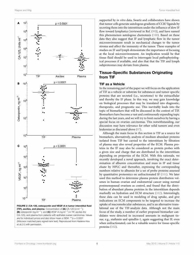
Gradients between the tumor interstitium and plasma havebeen presumed for tumor specific proteins and are an assumptionin most biomarker studies, as will be discussed in further detailbelow. In a recent study, we presented proof of this concept byisolation of native, undiluted, TIF by centrifugation from ovariancarcinomas (98). We assessed the TIF/P ratio for the knownovarian cancer biomarker cancer antigen (CA)-125 and the moregeneral tumor markers osteopontin and VEGF-A. All three weresignificantly up-regulated in TIF relative to plasma (Figure 2).Not surprisingly, this finding was most pronounced for CA-125,having a TIF/P ratio ranging from 1.4 to 24,300, with a medianconcentration 194 times that of plasma. This study documentspossible TIF/plasma gradients thatmay occur, and exemplifies theadvantage of using TIF as a source for biomarker and therapeutictarget discoveries.
As evident from the studies discussed above, there will beconcentration gradients between TIF and plasma for substancessecreted by tumor and stroma cells. Of interest, there may alsobe local gradients within the interstitium because of the flow ofIF toward the lymphatics that may be of considerable physiolog-ical and pathophysiological importance. In experiments in mice
Frontiers in Oncology | www.frontiersin.org May 2015 | Volume 5 | Article 1155
Wagner and Wiig Tumor interstitial fluid
FIGURE 2 | CA-125, osteopontin and VEGF-A in tumor interstitial fluid(TIF), ascites, and plasma. Concentration of (A) CA-125 (Uml−1);(B) osteopontin (ngml−1); and (C) VEGF-A (ngml−1) in TIF, ascites (forCA-125), and plasma from patients with epithelial ovarian carcinomas. Valuesare for individual tumors and also show mean±SEM. ***p= 0.0001(Wilcoxon matched pairs signed rank test). Reproduced from Haslene-Hoxet al.(98) with permission.
supported by in vitro data, Swartz and collaborators have shownthat tumor cells generate autologous gradients of CCR7 ligands bysecreting them into the interstitium under the influence of slow IFflow toward lymphatics [reviewed in Ref. (99)], and have namedthis phenomenon autologous chemotaxis (100). Based on thesedata they also suggest that IF and lymphatic flow in the tumormicroenvironment result in mechanical changes to the tumorstroma and affect the immunity of the tumor. These examples ofstudies on IF and lymph demonstrate the importance of focusingat the local microenvironment. An implication would be thattissue fluid should be used to interrogate local pathophysiolog-ical processes if available, and also that that the TIF and lymphsubproteomes may deviate from plasma.
Tissue-Specific Substances Originatingfrom TIF
TIF as a VehicleIn the remaining part of the paper we will focus on the applicationof TIF as a vehicle or substrate for substances and tumor-specificproteins that are secreted (i.e., secretome) to the extracellularand thereby the IF phase. In this way, we may gain knowledgeon biological processes that may be translated into diagnostic,therapeutic, and prognostic use. This inevitably leads into thetopic of biomarkers that will be discussed in the context of TIF.Biomarkers have become a vast and continuously expanding topicduring the last years, and we will try to limit ourselves by having aspecial focus on ovarian carcinomas. This notwithstanding, ourdiscussion may have relevance for other solid tumors and evenleukemias as discussed above (97).
Although the main focus in this section is TIF as a source forbiomarkers, alternatively, analysis of medium abundant proteinsisolated from TIF but carried to the interstitium by filtrationof plasma may also reveal properties of the ECM. Plasma pro-teins in the IF may also be considered as protein probes witha given size and charge that are distributed in the interstitiumdepending on properties of the ECM. With this rationale, werecently developed a novel approach, involving the exact deter-mination of albumin concentration and mass in IF and tissueeluate by HPLC and thereafter, expressing the correspondingnumbers relative to albumin for a set of probe proteins assessedby quantitative proteomics on unfractionated IF (101). We laterused this method to determine plasma protein distribution vol-umes in human ovarian and endometrial cancer using normalpostmenopausal ovarium as control, and found that the distri-bution of abundant plasma proteins in the interstitium dependsmarkedly on hydration and ECM structure (102). Interestingly,these data can be used in modeling of drug uptake, and giveindications on ECM components to be targeted to increase theuptake of macromolecular substances, and is an alternative trans-lational use of the TIF-analysis data. Although not the mainfocus of the study, a number of earlier proposed biomarker can-didates were detected in increased amounts in malignant tis-sue, e.g., stathmin and spindlin-1, again suggesting that IF, evenwhen unfractionated, can be a valuable source for tissue-specificproteins (102).
Frontiers in Oncology | www.frontiersin.org May 2015 | Volume 5 | Article 1156

Wagner and Wiig Tumor interstitial fluid
TIF and Secretome as Source for BiomarkersDuring the recent years, there have been rapid advances of massspectrometry techniques enabling the identification and quantifi-cation of thousands of proteins in biological samples. This fact,together with a corresponding improvement of bioinformatics,enabled the search for biomarkers with high throughput. In spiteof the considerable effort that has been invested in this search,identification of candidates fulfilling all the requirements of abiomarker has been sluggish, e.g., Ref. (103–106). Actually, asconcluded in a recent review (106), the “inconvenient truth” is thatno biomarker developed by proteomics has proven to be beneficialfor cancer patients.
Clearly, blood or plasma is the preferablematerial for a diagnos-tic test. In spite of significant technological advances, the presentproteomic technology, however, has limited power to detect a“needle” (low abundance disease biomarkers) in the “haystack”of high abundance plasma proteins. To reduce this problem, apossible strategy in a biomarker search might be to increasethe relative abundance of disease-associated proteins by moving“upstream,” to samples more proximal to the primary disease site(103, 105–107).
As recently shown in our study on the established biomarkerCA-125 (98) and most likely applying to all tumor-specificbiomarkers (104, 108, 109), there will be high concentrationslocally in the diseased tissue. The concentration will, however, bereduced in the perimeter of the lesion and the substance in ques-tion will be substantially diluted in blood. Accordingly, proximalfluids like TIF appear to be attractive substrates (107). Naturallysecreted proximal fluids, as cerebrospinal fluid, saliva, urine, andnipple aspirate fluid, have been substrates in proteomic discoverystudies [e.g., reviewed in Ref. (110)]. Examining TIF, however,will allow studies of shed and secreted proteins in tissues andconditions where natural secretion does not occur, e.g., in tumors.TIF is the best substrate to study proteins secreted by cancer cellsand other cells confined in the tumor microenvironment, i.e., thecancer secretome (111, 112).
Cell line supernatants andproximal (i.e., close to the anticipatedsource) biological fluids have been the two main substrates forstudies of the cancer secretome, where the conditioned mediacollected from in vitro cell cultures (112, 113) is themost commonsource. Evidently, it is debatable whether cell cultures can replicatethe complexity of the tumor microenvironment in vivo (114).This notwithstanding, such in vitro secretome studies have theadvantage of being able to simulate disease models and pertur-bations in the secretome due to altered physiological parametersor autocrine and/or paracrine secretion (115). Under these condi-tions, to distinguish between those proteins that are secreted andthose that are released into the conditioned media by cell deathand proteolysis due to serum-free media culturing conditions,may represent a challenge. Since the concentration of secretedproteins is low, lysis of a low fraction of cells will contaminatethe pool of truly secreted proteins due to a high intracellularprotein content and thus overshadow the small amount of secretedproteins in the sample (115).
Evidently, in vivo and/or ex vivo secretome studies are morecomplex since the microenvironment of the entire tissue isreflected, and due to challenges related to TIF isolation in these
situations, there are fewer studies (112, 115). Analysis of fluidharvested from tumor tissue is a powerful approach to bridgethe gap between cancer secretomes and tumor biology. Below weaddress studies performed on tissue fluid.
When studying the in vivo/ex vivo secretome, it may be ofimportance to be able to validate that the proteins in questiontruly originate from the extracellular fluid phase and thus todifferentiate between proteins that are of intracellular origin asrecently discussed in more detail (8). Extracellular markers canbe applied to validate the origin of the isolated fluid, and specificproteinsmay also represent intracellular fluid admixture andmorespecifically, defined intracellular compartments (116).
(More or Less) Specific Proteins and Peptidesin TIFProteomic profiling of TIF has been performed on samples col-lected by microdialysis (59, 117), capillary ultrafiltration (68, 69,118), incubation of tissue in a physiological buffer (65, 116, 119–122), tissue explants/elution (123), and tissue centrifugation (64,124). Table 2 summarizes TIF studies where human cancer sam-ples have been used as substrate for TIF isolation, and the resultingcandidate molecules and validation techniques.
Unfortunately, there are few common validated candidate pro-teins in the presented TIF studies. Of these, peroxiredoxin 1 andS100A8/9 have been suggested inmore than one study, and perox-iredoxin 1 is themost prevalent. It is, however, difficult to integrateresults from different studies. The methods for fluid isolationand data collection, analysis, and reporting and the selection of“secreted” proteins may influence the results in the various studiesand lead to discrepancies. Even in cell culture studies, wherethe complexity of an intact tissue is avoided that should reducebiological variation, the trend is similar. In addition, differentiallyexpressed proteins from different cancers and even within onecancer type although in a different model, appear to demonstratevery little overlap (113).
Common Proteins in TIFIn a recent publication (8), we investigated whether common“protein denominators” could be found in TIF, and examinedsix recently published TIF proteomes in more detail (64, 116–120), all deriving from different cancers and using different TIFextraction methods. Altogether, we found 1805 unique proteins,with 123 proteins (6.8%) discovered in five or six proteomes, andwith unique proteins in each proteome; 15% (116), 17% (120),23% (64), 30% (119), 31% (118), and 59% (117). The 123 commonproteins were intracellular enzymes, abundant plasma proteins,and several common cytoplasmic proteins highly conserved inexosomes (125, 126) and several proteins from the 14-3-3 familyand peroxiredoxins.
What might appear confusing considering that we were study-ing TIF, and thus extracellular fluid, was the finding of a substan-tial fraction of proteins that are classified as intracellular. That is aresult of the gene ontology (GO) system, assigning proteins to allthe compartments where they have been found. As a consequence,the extracellular compartment plasma will also contain a substan-tial fraction of proteins classified as intracellular (127). BecauseTIF contains many of the proteins referred as the exosome “core”
Frontiers in Oncology | www.frontiersin.org May 2015 | Volume 5 | Article 1157

Wagner
andWiig
Tumor
interstitialfluid
TABLE 2 | Summary of proteomic studies utilizing human tumor interstitial fluid, including candidate proteins that were chosen for validation.
Analyzedsample
Isolationtechnique
Samples Candidates Validation Published protein findings Reference
Mouse colorectalcarcinoma (human serum)
Elution TIF; NIF MCM4, S100A9 IHC 2172 proteins identified (1958 with humanhomologs), 52 suggested candidates
(134)
Serum (control; adenoma; CRC) CHI3L1, CEA ELISA
Hepatocellular carcinoma Elution TIF/NIF 381 (TIF) and 245 (NIF) identified proteins,111 unique for TIF
(116)
NIF 325 proteins identified in healthy liver
Hepatocellular carcinoma Elution TIF/NIF sERBB3 Western blot 72 proteins identified (135)Serum (HCC, cirrhosis, chronichepatitis)
sERBB3, AFP ELISA
Renal cell carcinoma Elution TIF ENO2, NNMT Western blot, SRM 539 proteins identified, 138 up-regulated (136)Serum (patient; normal pool) ENO2, TSP1 ELISA, SRM
Ovarian carcinoma Centrifugation TIF 769 proteins identified (64)Plasma (patient; control) 124 and 102 proteins identified in patient
and control plasma
Ovarian carcinoma Elution TIF/ascites PRDX1 Western blot 569 proteins identified (120)Serum (EOC; normal/benign) PRDX1 ELISA
Ovarian carcinoma Centrifugation TIF WDR1 MRM, SRM WB 6 proteins selected for validation (124)
Ovarian carcinoma Elution TIF/NIF S100-A8 IHC 58 proteins identified, 1 up-regulated, 5down-regulated proteins
(137)
Ovarian carcinoma Elution TIF; NIF STIP1, LAP3, TPI1, UCHL1 Western blot, IHC 8 proteins identified (138)BNDF, transferrin ELISA
Serum (patient; control) STIP1 ELISA
Breast carcinoma Elution with biotin TIF/NIF 93 up-regulated proteins (139)TIF; NIF CD276 IHC
Breast carcinoma Elution NIF/TIF Calreticulin, calumenin, TCPT,S100A9
IHC 832 proteins detected, 84 up-regulatedproteins
(119)
TIF; NIF Calreticulin, CRABP2, CLIC1,EF-1-beta, galectin-1, PRDX2,PD-ECGF, PDI, UCTH5
Tissue microarray 26 of protein candidates present in allpatients (gels compared)
Breast carcinoma Elution TIF/NIF YWHAZ, GDI-1, HNRNPD Western blot 1324 non-redundant proteins (121)
Non-small cell lung cancer Elution TIF/NIF PRDX1 Western blot/ELISA 24 proteins differentially expressed (122)
Colorectal tumor Explant eluate TIF/NIF Desmocollin, fibrinogen γ-chain 32 proteins differentially expressed (123)
Head and neck squamouscell carcinoma
Capillaryultrafiltration
TIF 525 proteins identified (118)
Oral squamous cellcarcinoma
Microdialysis TIF; NIF MMP-8, MMP-9, neurotrypsin,trypsin-1
IHC 217 proteins identified (117)
TIF, tumor interstitial fluid; NIF, normal tissue interstitial fluid; MCM4, minichromosome maintenance complex component 4; IHC, immunohistochemistry; CRC, colorectal carcinoma; CHI3L1, chitinase 3-like 1; CEA, carcinoembryonicantigen; ELISA, enzyme-linked immunosorbent assay; sERBB3, secreted receptor tyrosine-kinase erbB-3; HCC, hepatocellular carcinoma; AFP, alpha-fetoprotein; ENO2, enolase 2; NNMT, nicotinamide n-methyltransferase; SRM, selectedreaction monitoring; TSP1, thrombospondin-1; PRDX1, peroxiredoxin 1; EOC, epithelial ovarian carcinoma; STIP1, stress-induced phosphoprotein 1; LAP3, leucine aminopeptidase 3; TPI1, triosephosphate isomerase 1; UCHL1, ubiquitycarboxyl-terminal esterase L1; BNDF, brain-derived neurotrophic factor; TCPT, translationally controlled tumor protein; CRABP2, cellular retinoic acid-binding protein 2; CLIC1, chloride intracellular channel protein 1; EF-1-beta, elongationfactor 1-beta; PRDX2, peroxiredoxin 2; PD-ECGF, platelet-derived endothelial cell growth factor; PDI, protein disulfideisomerase; UCTH5, ubiquitin carboxyl-terminal hydrolase 5; MMP, matrix metalloprotease; WDR1, WD-repeat containingprotein 1; YWHAZ, 14-3-3 protein zeta/delta; GDI1, dissociation inhibitor alpha; HNRNPD, AU-rich element RNA-binding protein 1.
FrontiersinOncology
|www.frontiersin.org
May
2015|Volum
e5|A
rticle115
8

Wagner and Wiig Tumor interstitial fluid
proteins (125), intracellular proteins in TIF may actually derivefrom exosomes. Due to their size, these particles will be sieved offwhen using techniques involving membranes like microdialysisand ultrafiltration that may again lead to divergent proteomesdepending on the isolation techniques, a conclusion that wasactually supported by our analysis (8). Based on this evaluationwe concluded that there are many common proteins that appearin several proteomes, and moreover that there are many potentialunique candidates for each tumor type (8). Another implication ofthis analysis is that since the isolation method will influence theoverall composition of the identified proteome, proteomes fromdifferent studies should be evaluated with this inmind. These datamight actually suggest that more than one method should be usedto isolate TIF in the initial screening for biomarker candidates.
Summary and Conclusion
In spite of extensive efforts, economical as well as technical, “theinconvenient truth” is that up till now, no biomarker developedby proteomics has been proven to be of benefit for cancer patients(106). The many problems regarding proteomic analysis of serumare well known. This calls for alternative approaches and for newsubstrates in this endeavor. TIF represents a proximal fluid thatmay be enriched in tumor specific proteins. It may serve as anew substrate that could be used in a more targeted analysis ofthe proximal fluids in general. In the present review, we have
briefly summarized recent knowledge on the tumor interstitiumand the formation and composition of TIF. We have moreover, inparticular, addressed proteins secreted to the tumor fluid phase.While several proteomic secretome studies have been performedin cell cultures, only a few studies addressing the TIF proteomehave emerged in the recent years, and have been summarized inthis article. The isolation of TIF can be challenging per se, andthe choice of method may have a direct impact on the proteomicresults. Unfortunately, even when comparing a fluid that is moreproximal to the tumor, i.e., TIF, there are few common validatedcandidate proteins in the presented TIF studies. There appearto be an unexploited potential in using TIF proteomic data in afunctional context. It might appear as a more integrated systemsbiology biomarker discovery platform should be used. Such aplatform should also involve, e.g., cancer cell lines, animalmodels,tumor tissues, and transcriptomics in addition to proximal fluids(6). Such a strategy will provide new knowledge on tumor biologyand hopefully produce new biomarkers or treatment strategies forcancer.
Acknowledgments
Financial support from The Rakel and Otto Bruun foundationand The Western Norway Regional Health Authority is gratefullyacknowledged.
References1. Aukland K, Reed RK. Interstitial-lymphatic mechanisms in the control of
extracellular fluid volume. Physiol Rev (1993) 73:1–78.2. Randolph GJ, Angeli V, Swartz MA. Dendritic-cell trafficking to lymph nodes
through lymphatic vessels. Nat Rev Immunol (2005) 5:617–28. doi:10.1038/nri1670
3. Baronzio G, Schwartz L, Kiselevsky M, Guais A, Sanders E, Milanesi G, et al.Tumor interstitial fluid as modulator of cancer inflammation, thrombosis,immunity and angiogenesis. Anticancer Res (2012) 32:405–14.
4. Wiig H, Tenstad O, Iversen PO, Kalluri R, Bjerkvig R. Interstitial fluid: theoverlooked component of the tumor microenvironment? Fibrogenesis TissueRepair (2010) 3:12. doi:10.1186/1755-1536-3-12
5. Baronzio G, Parmar G, Baronzio M, Kiselevsky M. Tumor interstitial fluid:proteomic determination as a possible source of biomarkers. Cancer GenomicsProteomics (2014) 11:225–37.
6. Kulasingam V, Pavlou MP, Diamandis EP. Integrating high-throughput tech-nologies in the quest for effective biomarkers for ovarian cancer. Nat RevCancer (2010) 10:371–8. doi:10.1038/nrc2831
7. Wiig H, Swartz MA. Interstitial fluid and lymph formation and transport:physiological regulation and roles in inflammation and cancer. Physiol Rev(2012) 92:1005–60. doi:10.1152/physrev.00037.2011
8. Haslene-Hox H, Tenstad O, Wiig H. Interstitial fluid-a reflection of thetumor cell microenvironment and secretome. Biochim Biophys Acta (2013)1834:2336–46. doi:10.1016/j.bbapap.2013.01.028
9. Kalluri R, ZeisbergM. Fibroblasts in cancer.Nat Rev Cancer (2006) 6:392–401.doi:10.1038/nrc1877
10. Mueller MM, Fusenig NE. Friends or foes – bipolar effects of the tumourstroma in cancer. Nat Rev Cancer (2004) 4:839–49. doi:10.1038/nrc1477
11. Liotta LA, Kohn EC. The microenvironment of the tumour-host interface.Nature (2001) 411:375–9. doi:10.1038/35077241
12. Sund M, Kalluri R. Tumor stroma derived biomarkers in cancer. CancerMetastasis Rev (2009) 28:177–83. doi:10.1007/s10555-008-9175-2
13. Pickup MW, Mouw JK, Weaver VM. The extracellular matrix modulatesthe hallmarks of cancer. EMBO Rep (2014) 15:1243–53. doi:10.15252/embr.201439246
14. Miles FL, Sikes RA. Insidious changes in stromal matrix fuel cancerprogression. Mol Cancer Res (2014) 12:297–312. doi:10.1158/1541-7786.MCR-13-0535
15. Lu P,Weaver VM,Werb Z. The extracellular matrix: a dynamic niche in cancerprogression. J Cell Biol (2012) 196:395–406. doi:10.1083/jcb.201102147
16. Junttila MR, de Sauvage FJ. Influence of tumour micro-environment het-erogeneity on therapeutic response. Nature (2013) 501:346–54. doi:10.1038/nature12626
17. Sica A, Mantovani A. Macrophage plasticity and polarization: in vivo veritas.J Clin Invest (2012) 122:787–95. doi:10.1172/JCI59643
18. Woo SR, Corrales L, Gajewski TF. Innate immune recognition of can-cer. Annu Rev Immunol (2015) 33:445–74. doi:10.1146/annurev-immunol-032414-112043
19. Wynn TA, Chawla A, Pollard JW. Macrophage biology in development,homeostasis and disease.Nature (2013) 496:445–55. doi:10.1038/nature12034
20. Martinez FO, Gordon S. The M1 and M2 paradigm of macrophage activation:time for reassessment. F1000Prime Rep (2014) 6:13. doi:10.12703/P6-13
21. Fridman WH, Pages F, Sautes-Fridman C, Galon J. The immune contex-ture in human tumours: impact on clinical outcome. Nat Rev Cancer (2012)12:298–306. doi:10.1038/nrc3245
22. Gajewski TF, Schreiber H, Fu YX. Innate and adaptive immune cells inthe tumor microenvironment. Nat Immunol (2013) 14:1014–22. doi:10.1038/ni.2703
23. Sinha P, Clements VK, Bunt SK, Albelda SM, Ostrand-Rosenberg S. Cross-talkbetween myeloid-derived suppressor cells and macrophages subverts tumorimmunity toward a type 2 response. J Immunol (2007) 179:977–83. doi:10.4049/jimmunol.179.2.977
24. Mahfouz SM, Chevallier M, Grimaud JA. Distribution of the major con-nective matrix components of the stromal reaction in breast carcinoma. Animmunohistochemical study. Cell Mol Biol (1987) 33:453–67.
25. Takeuchi J, Sobue M, Sato E, Shamoto M, Miura K. Variation in glycosamino-glycan components of breast tumors. Cancer Res (1976) 36:2133–9.
26. Yeo TK, Brown L, Dvorak HF. Alterations in proteoglycan synthesis commonto healing wounds and tumors. Am J Pathol (1991) 138:1437–50.
27. Ronnov-Jessen L, Petersen OW, Bissell MJ. Cellular changes involved inconversion of normal to malignant breast: importance of the stromal reaction.Physiol Rev (1996) 76:69–125.
Frontiers in Oncology | www.frontiersin.org May 2015 | Volume 5 | Article 1159

Wagner and Wiig Tumor interstitial fluid
28. Brown LF, Guidi AJ, Schnitt SJ, Van De Water L, Iruela-Arispe ML, Yeo TK,et al. Vascular stroma formation in carcinoma in situ, invasive carcinoma, andmetastatic carcinoma of the breast. Clin Cancer Res (1999) 5:1041–56.
29. Fukumura D, Xavier R, Sugiura T, Chen Y, Park EC, Lu N, et al. Tumorinduction of VEGF promoter activity in stromal cells. Cell (1998) 94:715–25.doi:10.1016/S0092-8674(00)81731-6
30. Dvorak HF, Sioussat TM, Brown LF, Berse B, Nagy JA, Sotrel A, et al. Distri-bution of vascular permeability factor (vascular endothelial growth factor) intumors: concentration in tumor blood vessels. J Exp Med (1991) 174:1275–8.doi:10.1084/jem.174.5.1275
31. Senger DR, Galli SJ, Dvorak AM, Perruzzi CA, Harvey VS, Dvorak HF. Tumorcells secrete a vascular permeability factor that promotes accumulation ofascites fluid. Science (1983) 219:983–5. doi:10.1126/science.6823562
32. Dvorak HF. Tumors: wounds that do not heal. Similarities between tumorstroma generation and wound healing. N Engl J Med (1986) 315:1650–9.doi:10.1056/NEJM198612253152606
33. Mantovani A, Allavena P, Sica A, Balkwill F. Cancer-related inflammation.Nature (2008) 454:436–44. doi:10.1038/nature07205
34. Christiansen A, Detmar M. Lymphangiogenesis and cancer. Genes Cancer(2011) 2:1146–58. doi:10.1177/1947601911423028
35. Mouw JK, Ou G, Weaver VM. Extracellular matrix assembly: a multiscaledeconstruction.Nat RevMol Cell Biol (2014) 15:771–85. doi:10.1038/nrm3902
36. Bonnans C, Chou J, Werb Z. Remodelling the extracellular matrix in devel-opment and disease. Nat Rev Mol Cell Biol (2014) 15:786–801. doi:10.1038/nrm3904
37. Starling EH. On the absorption of fluids from the connective tissue spaces.J Physiol (1896) 19:312–26. doi:10.1113/jphysiol.1896.sp000596
38. Levick JR, Michel CC. Microvascular fluid exchange and the revised starlingprinciple. Cardiovasc Res (2010) 87:198–210. doi:10.1093/cvr/cvq062
39. Fukumura D, Jain RK. Tumor microenvironment abnormalities: causes, con-sequences, and strategies to normalize. J Cell Biochem (2007) 101:937–49.doi:10.1002/jcb.21187
40. Heldin CH, Rubin K, Pietras K, Ostman A. High interstitial fluid pressure –an obstacle in cancer therapy. Nat Rev Cancer (2004) 4:806–13. doi:10.1038/nrc1456
41. Goel S, Duda DG, Xu L, Munn LL, Boucher Y, Fukumura D, et al. Normaliza-tion of the vasculature for treatment of cancer and other diseases. Physiol Rev(2011) 91:1071–121. doi:10.1152/physrev.00038.2010
42. Wiig H. Evaluation of methodologies for measurement of interstitial fluidpressure (Pi): physiological implications of recent Pi data.Crit Rev Biomed Eng(1990) 18:27–54.
43. Provenzano PP, Cuevas C, Chang AE, Goel VK, Von Hoff DD, HingoraniSR. Enzymatic targeting of the stroma ablates physical barriers to treatmentof pancreatic ductal adenocarcinoma. Cancer Cell (2012) 21:418–29. doi:10.1016/j.ccr.2012.01.007
44. Khawar IA, Kim JH, KuhHJ. Improving drug delivery to solid tumors: primingthe tumor microenvironment. J Control Release (2015) 201:78–89. doi:10.1016/j.jconrel.2014.12.018
45. Ariffin AB, Forde PF, Jahangeer S, Soden DM, Hinchion J. Releasing pressurein tumors: what do we know so far and where do we go from here? A review.Cancer Res (2014) 74:2655–62. doi:10.1158/0008-5472.CAN-13-3696
46. Azzi S,Hebda JK,Gavard J. Vascular permeability and drug delivery in cancers.Front Oncol (2013) 3:211. doi:10.3389/fonc.2013.00211
47. Dvorak HF, Brown LF, Detmar M, Dvorak AM. Vascular permeability fac-tor/vascular endothelial growth factor, microvascular hyperpermeability, andangiogenesis. Am J Pathol (1995) 146:1029–39.
48. Baluk P, Morikawa S, Haskell A, Mancuso M, McDonald DM. Abnormalitiesof basement membrane on blood vessels and endothelial sprouts in tumors.Am J Pathol (2003) 163:1801–15. doi:10.1016/S0002-9440(10)63540-7
49. Wiig H, Tveit E, Hultborn R, Reed RK, Weiss L. Interstitial fluid pressurein DMBA-induced rat mammary tumours. Scand J Clin Lab Invest (1982)42:159–64. doi:10.3109/00365518209168068
50. Stohrer M, Boucher Y, Stangassinger M, Jain RK. Oncotic pressure in solidtumors is elevated. Cancer Res (2000) 60:4251–5.
51. WiigH, AuklandK, TenstadO. Isolation of interstitial fluid from ratmammarytumors by a centrifugation method. Am J Physiol Heart Circ Physiol (2003)284:H416–24. doi:10.1152/ajpheart.00327.2002
52. Leu AJ, Berk DA, Lymboussaki A, Alitalo K, Jain RK. Absence of functionallymphatics within a murine sarcoma: a molecular and functional evaluation.Cancer Res (2000) 60:4324–7.
53. Padera TP, Kadambi A, di Tomaso E, Carreira CM, Brown EB, Boucher Y,et al. Lymphaticmetastasis in the absence of functional intratumor lymphatics.Science (2002) 296:1883–6. doi:10.1126/science.1071420
54. Padera TP, Stoll BR, Tooredman JB, CapenD, di Tomaso E, Jain RK. Pathology:cancer cells compress intratumour vessels. Nature (2004) 427:695. doi:10.1038/427695a
55. Klemm F, Joyce JA. Microenvironmental regulation of therapeutic response incancer. Trends Cell Biol (2015) 25:198–213. doi:10.1016/j.tcb.2014.11.006
56. Alitalo K, Tammela T, Petrova TV. Lymphangiogenesis in development andhuman disease. Nature (2005) 438:946–53. doi:10.1038/nature04480
57. Thiele W, Sleeman JP. Tumor-induced lymphangiogenesis: a target for cancertherapy? J Biotechnol (2006) 124:224–41. doi:10.1016/j.jbiotec.2006.01.007
58. Stacker SA, Caesar C, Baldwin ME, Thornton GE, Williams RA, Prevo R, et al.VEGF-D promotes themetastatic spread of tumor cells via the lymphatics.NatMed (2001) 7:186–91. doi:10.1038/84635
59. Xu BJ, Yan W, Jovanovic B, Shaw AK, An QA, Eng J, et al. Microdial-ysis combined with proteomics for protein identification in breast tumormicroenvironment in vivo. Cancer Microenviron (2010) 4:61–71. doi:10.1007/s12307-010-0046-3
60. Wiig H. Cornea fluid dynamics. I: measurement of hydrostatic and colloidosmotic pressure in rabbits. Exp Eye Res (1989) 49:1015–30. doi:10.1016/S0014-4835(89)80023-5
61. Aukland K. Distribution volumes and macromolecular mobility in rat tailtendon interstitium. Am J Physiol (1991) 260:H409–19.
62. Choi J, Credit K, Henderson K, Deverkadra R, He Z, Wiig H, et al. Intraperi-toneal immunotherapy for metastatic ovarian carcinoma: resistance of intra-tumoral collagen to antibody penetration. Clin Cancer Res (2006) 12:1906–12.doi:10.1158/1078-0432.CCR-05-2141
63. Salnikov AV, Heldin NE, Stuhr LB, Wiig H, Gerber H, Reed RK, et al. Inhi-bition of carcinoma cell-derived VEGF reduces inflammatory characteristicsin xenograft carcinoma. Int J Cancer (2006) 119:2795–802. doi:10.1002/ijc.22217
64. Haslene-Hox H, Oveland E, Berg KC, Kolmannskog O, Woie K, Salvesen HB,et al. A new method for isolation of interstitial fluid from human solid tumorsapplied to proteomic analysis of ovarian carcinoma tissue. PLoS One (2011)6:e19217. doi:10.1371/journal.pone.0019217
65. Celis JE, Gromov P, Cabezon T, Moreira JM, Ambartsumian N, SandelinK, et al. Proteomic characterization of the interstitial fluid perfusing thebreast tumormicroenvironment: a novel resource for biomarker and therapeu-tic target discovery. Mol Cell Proteomics (2004) 3:327–44. doi:10.1074/mcp.M400009-MCP200
66. Leegsma-Vogt G, Janle E, Ash SR, Venema K, Korf J. Utilization of in vivoultrafiltration in biomedical research and clinical applications. Life Sci (2003)73:2005–18. doi:10.1016/S0024-3205(03)00569-1
67. Schneiderheinze JM, Hogan BL. Selective in vivo and in vitro sampling ofproteins using miniature ultrafiltration sampling probes. Anal Chem (1996)68:3758–62. doi:10.1021/ac960309u
68. Huang CM, Ananthaswamy HN, Barnes S, Ma Y, Kawai M, Elmets CA. Massspectrometric proteomics profiles of in vivo tumor secretomes: capillary ultra-filtration sampling of regressive tumor masses. Proteomics (2006) 6:6107–16.doi:10.1002/pmic.200600287
69. Yang S, Huang CM. Recent advances in protein profiling of tissues and tissuefluids. Expert Rev Proteomics (2007) 4:515–29. doi:10.1586/14789450.4.4.515
70. Dabrosin C. Microdialysis – an in vivo technique for studies of growth factorsin breast cancer. Front Biosci (2005) 10:1329–35. doi:10.2741/1622
71. Benjamin RK, Hochberg FH, Fox E, Bungay PM, Elmquist WF, Stewart CF,et al. Review of microdialysis in brain tumors, from concept to application:first annual Carolyn Frye-Halloran symposium. Neuro Oncol (2004) 6:65–74.doi:10.1215/S1152851703000103
72. Brunner M, Muller M. Microdialysis: an in vivo approach for measuring drugdelivery in oncology. Eur J Clin Pharmacol (2002) 58:227–34. doi:10.1007/s00228-002-0475-0
73. Liu L, Zhang X, Lou Y, Rao Y, Zhang X. Cerebral microdialysis in gliomastudies, from theory to application. J Pharm Biomed Anal (2014) 96:77–89.doi:10.1016/j.jpba.2014.03.026
74. Blakeley J, Portnow J. Microdialysis for assessing intratumoral drug disposi-tion in brain cancers: a tool for rational drug development. Expert Opin DrugMetab Toxicol (2010) 6:1477–91. doi:10.1517/17425255.2010.523420
75. Zhou Q, Gallo JM. In vivo microdialysis for PK and PD studies of anticancerdrugs. AAPS J (2005) 7:E659–67. doi:10.1208/aapsj070366
Frontiers in Oncology | www.frontiersin.org May 2015 | Volume 5 | Article 11510

Wagner and Wiig Tumor interstitial fluid
76. Ao X, Stenken JA. Microdialysis sampling of cytokines. Methods (2006)38:331–41. doi:10.1016/j.ymeth.2005.11.012
77. CloughGF.Microdialysis of largemolecules.AAPS J (2005) 7:E686–92. doi:10.1208/aapsj070369
78. Hersini KJ, Melgaard L, Gazerani P, Petersen LJ. Microdialysis of inflamma-tory mediators in the skin: a review. Acta Derm Venereol (2014) 94:501–11.doi:10.2340/00015555-1878
79. Bendrik C, Dabrosin C. Estradiol increases IL-8 secretion of normal humanbreast tissue and breast cancer in vivo. J Immunol (2009) 182:371–8. doi:10.4049/jimmunol.182.1.371
80. Wibom C, Surowiec I, Moren L, Bergstrom P, Johansson M, Antti H, et al.Metabolomic patterns in glioblastoma and changes during radiotherapy: aclinical microdialysis study. J Proteome Res (2010) 9:2909–19. doi:10.1021/pr901088r
81. Gullino PM, Clark SH, Grantham FH. The interstitial fluid of solid tumors.Cancer Res (1964) 24:780–98.
82. Jain RK, Shah SA, Finney PL. Continuous noninvasive monitoring of pHand temperature in rat walker 256 carcinoma during normoglycemia andhyperglycemia. J Natl Cancer Inst (1984) 73:429–36.
83. Grahovac J, Wells A. Matrikine and matricellular regulators of EGF receptorsignaling on cancer cell migration and invasion. Lab Invest (2014) 94:31–40.doi:10.1038/labinvest.2013.132
84. Ricard-Blum S, Salza R. Matricryptins and matrikines: biologically activefragments of the extracellular matrix. Exp Dermatol (2014) 23:457–63. doi:10.1111/exd.12435
85. Monboisse JC, Oudart JB, Ramont L, Brassart-Pasco S, Maquart FX.Matrikines from basement membrane collagens: a new anti-cancerstrategy. Biochim Biophys Acta (2014) 1840:2589–98. doi:10.1016/j.bbagen.2013.12.029
86. Voloshin T, Fremder E, Shaked Y. Small but mighty: microparticles as media-tors of tumor progression. Cancer Microenviron (2014) 7:11–21. doi:10.1007/s12307-014-0144-8
87. Andaloussi SE, Mager I, Breakefield XO, Wood MJ. Extracellular vesicles:biology and emerging therapeutic opportunities. Nat Rev Drug Discov (2013)12:347–57. doi:10.1038/nrd3978
88. Colombo M, Raposo G, Thery C. Biogenesis, secretion, and intercellularinteractions of exosomes and other extracellular vesicles. Annu Rev Cell DevBiol (2014) 30:255–89. doi:10.1146/annurev-cellbio-101512-122326
89. Antonyak MA, Cerione RA. Microvesicles as mediators of intercellular com-munication in cancer. Methods Mol Biol (2014) 1165:147–73. doi:10.1007/978-1-4939-0856-1_11
90. Mause SF, Weber C. Microparticles: protagonists of a novel communicationnetwork for intercellular information exchange. Circ Res (2010) 107:1047–57.doi:10.1161/CIRCRESAHA.110.226456
91. Morel O, Morel N, Jesel L, Freyssinet JM, Toti F. Microparticles: a criticalcomponent in the nexus between inflammation, immunity, and thrombosis.Semin Immunopathol (2011) 33:469–86. doi:10.1007/s00281-010-0239-3
92. D’Souza-Schorey C, Clancy JW. Tumor-derived microvesicles: shedding lighton novel microenvironment modulators and prospective cancer biomarkers.Genes Dev (2012) 26:1287–99. doi:10.1101/gad.192351.112
93. D’Asti E, Garnier D, Lee TH, Montermini L, Meehan B, Rak J. Oncogenicextracellular vesicles in brain tumor progression. Front Physiol (2012) 3:294.doi:10.3389/fphys.2012.00294
94. Loyer X, Vion AC, Tedgui A, Boulanger CM. Microvesicles as cell-cell mes-sengers in cardiovascular diseases. Circ Res (2014) 114:345–53. doi:10.1161/CIRCRESAHA.113.300858
95. Julich H, Willms A, Lukacs-Kornek V, Kornek M. Extracellular vesicle profil-ing and their use as potential disease specific biomarker. Front Immunol (2014)5:413. doi:10.3389/fimmu.2014.00413
96. Shao H, Chung J, Balaj L, Charest A, Bigner DD, Carter BS, et al. Protein typ-ing of circulating microvesicles allows real-time monitoring of glioblastomatherapy. Nat Med (2012) 18:1835–40. doi:10.1038/nm.2994
97. Iversen PO,WiigH. Tumor necrosis factor alpha and adiponectin in bonemar-row interstitial fluid from patients with acutemyeloid leukemia inhibit normalhematopoiesis. Clin Cancer Res (2005) 11:6793–9. doi:10.1158/1078-0432.CCR-05-1033
98. Haslene-Hox H, Madani A, Berg KCG, Woie K, Salvesen HB, Wiig H, et al.Quantification of the concentration gradient of biomarkers between ovarian
carcinoma interstitial fluid and blood. Biochim Biophys Acta Clin (2014)2:18–23. doi:10.1016/j.bbacli.2014.08.002
99. Swartz MA, Lund AW. Lymphatic and interstitial flow in the tumour microen-vironment: linking mechanobiology with immunity. Nat Rev Cancer (2012)12:210–9. doi:10.1038/nrc3186
100. Shields JD, Fleury ME, Yong C, Tomei AA, Randolph GJ, Swartz MA. Autol-ogous chemotaxis as a mechanism of tumor cell homing to lymphatics viainterstitial flow and autocrine CCR7 signaling. Cancer Cell (2007) 11:526–38.doi:10.1016/j.ccr.2007.04.020
101. Sagstad SJ, Oveland E, Karlsen TV, Haslene-Hox H, Tenstad O, Wiig H.Age-related changes in rat dermal extracellular matrix composition affect thedistribution of plasma proteins as a function of size and charge. Am J PhysiolHeart Circ Physiol (2015) 308:H29–38. doi:10.1152/ajpheart.00545.2014
102. Haslene-Hox H, Oveland E, Woie K, Salvesen HB, Tenstad O, Wiig H. Dis-tribution volumes of macromolecules in human ovarian and endometrialcancers-effects of extracellular matrix structure. Am J Physiol Heart CircPhysiol (2015) 308:H18–28. doi:10.1152/ajpheart.00672.2014
103. Polanski M, Anderson NL. A list of candidate cancer biomarkers for targetedproteomics. Biomark Insights (2007) 1:1–48.
104. Rifai N, GilletteMA, Carr SA. Protein biomarker discovery and validation: thelong and uncertain path to clinical utility. Nat Biotechnol (2006) 24:971–83.doi:10.1038/nbt1235
105. Veenstra TD. Global and targeted quantitative proteomics for biomarkerdiscovery. J Chromatogr B Analyt Technol Biomed Life Sci (2007) 847:3–11.doi:10.1016/j.jchromb.2006.09.004
106. Kondo T. Inconvenient truth: cancer biomarker development by using pro-teomics. Biochim Biophys Acta (2014) 1844:861–5. doi:10.1016/j.bbapap.2013.07.009
107. Gromov P, Gromova I, Olsen CJ, Timmermans-Wielenga V, Talman ML, Ser-izawa RR, et al. Tumor interstitial fluid – a treasure trove of cancer biomarkers.Biochim Biophys Acta (2013) 1834:2259–70. doi:10.1016/j.bbapap.2013.01.013
108. Sedlaczek P, Frydecka I, Gabrys M, Van Dalen A, Einarsson R, HarlozinskaA. Comparative analysis of CA125, tissue polypeptide specific antigen, andsoluble interleukin-2 receptor alpha levels in sera, cyst, and ascitic fluids frompatients with ovarian carcinoma.Cancer (2002) 95:1886–93. doi:10.1002/cncr.10917
109. Simpson RJ, Bernhard OK, Greening DW, Moritz RL. Proteomics-drivencancer biomarker discovery: looking to the future.CurrOpin ChemBiol (2008)12:72–7. doi:10.1016/j.cbpa.2008.02.010
110. Teng PN, Bateman NW, Hood BL, Conrads TP. Advances in proximalfluid proteomics for disease biomarker discovery. J Proteome Res (2010)9:6091–100. doi:10.1021/pr100904q
111. Gronborg M, Kristiansen TZ, Iwahori A, Chang R, Reddy R, Sato N, et al.Biomarker discovery from pancreatic cancer secretome using a differentialproteomic approach. Mol Cell Proteomics (2006) 5:157–71. doi:10.1074/mcp.M500178-MCP200
112. Xue H, Lu B, Lai M. The cancer secretome: a reservoir of biomarkers. J TranslMed (2008) 6:52. doi:10.1186/1479-5876-6-52
113. Makridakis M, Vlahou A. Secretome proteomics for discovery of cancerbiomarkers. J Proteomics (2010) 73:2291–305. doi:10.1016/j.jprot.2010.07.001
114. Karagiannis GS, Pavlou MP, Diamandis EP. Cancer secretomics reveal patho-physiological pathways in cancer molecular oncology. Mol Oncol (2010)4:496–510. doi:10.1016/j.molonc.2010.09.001
115. Stastna M, Van Eyk JE. Secreted proteins as a fundamental sourcefor biomarker discovery. Proteomics (2012) 12:722–35. doi:10.1002/pmic.201100346
116. Sun W, Ma J, Wu S, Yang D, Yan Y, Liu K, et al. Characterization of the livertissue interstitial fluid (TIF) proteome indicates potential for application inliver disease biomarker discovery. J Proteome Res (2010) 9:1020–31. doi:10.1021/pr9009172
117. Hardt M, Lam DK, Dolan JC, Schmidt BL. Surveying proteolytic processesin human cancer microenvironments by microdialysis and activity-basedmass spectrometry. Proteomics Clin Appl (2011) 5:636–43. doi:10.1002/prca.201100015
118. Stone MD, Odland RM, McGowan T, Onsongo G, Tang C, Rhodus NL, et al.Novel in situ collection of tumor interstitial fluid from a head and necksquamous carcinoma reveals a unique proteome with diagnostic potential.Clin Proteomics (2010) 6:75–82. doi:10.1007/s12014-010-9050-3
Frontiers in Oncology | www.frontiersin.org May 2015 | Volume 5 | Article 11511

Wagner and Wiig Tumor interstitial fluid
119. Gromov P, Gromova I, Bunkenborg J, Cabezon T, Moreira JM, Timmermans-Wielenga V, et al. Up-regulated proteins in the fluid bathing the tumourcell microenvironment as potential serological markers for early detectionof cancer of the breast.Mol Oncol (2010) 4:65–89. doi:10.1016/j.molonc.2009.11.003
120. Hoskins ER, Hood BL, Sun M, Krivak TC, Edwards RP, Conrads TP. Pro-teomic analysis of ovarian cancer proximal fluids: validation of elevated per-oxiredoxin 1 in patient peripheral circulation. PLoS One (2011) 6:e25056.doi:10.1371/journal.pone.0025056
121. Raso C, Cosentino C, Gaspari M, Malara N, Han X, McClatchy D, et al.Characterization of breast cancer interstitial fluids by TmT labeling, LTQ-orbitrap velos mass spectrometry, and pathway analysis. J Proteome Res (2012)11:3199–210. doi:10.1021/pr2012347
122. Li S, Wang R, Zhang M, Wang L, Cheng S. Proteomic analysis of non-smallcell lung cancer tissue interstitial fluids. World J Surg Oncol (2013) 11:173.doi:10.1186/1477-7819-11-173
123. Shi HJ, Stubbs R, Hood K. Characterization of de novo synthesized pro-teins released from human colorectal tumour explants. Electrophoresis (2009)30:2442–53. doi:10.1002/elps.200800767
124. Haslene-Hox H, Oveland E, Woie K, Salvesen HB, Wiig H, Tenstad O.Increased WD-repeat containing protein 1 in interstitial fluid from ovar-ian carcinomas shown by comparative proteomic analysis of malignant andhealthy gynecological tissue. Biochim Biophys Acta (2013) 1834:2347–59.doi:10.1016/j.bbapap.2013.05.011
125. Raimondo F, Morosi L, Chinello C, Magni F, Pitto M. Advances in mem-branous vesicle and exosome proteomics improving biological understandingand biomarker discovery. Proteomics (2011) 11:709–20. doi:10.1002/pmic.201000422
126. Simpson RJ, Lim JW, Moritz RL, Mathivanan S. Exosomes: proteomic insightsand diagnostic potential. Expert Rev Proteomics (2009) 6:267–83. doi:10.1586/epr.09.17
127. Pieper R, Gatlin CL, Makusky AJ, Russo PS, Schatz CR, Miller SS, et al.The human serum proteome: display of nearly 3700 chromatographicallyseparated protein spots on two-dimensional electrophoresis gels and identi-fication of 325 distinct proteins. Proteomics (2003) 3:1345–64. doi:10.1002/pmic.200300449
128. Wiig H, Keskin D, Kalluri R. Interaction between the extracellularmatrix and lymphatics: consequences for lymphangiogenesis andlymphatic function. Matrix Biol (2010) 29:645–56. doi:10.1016/j.matbio.2010.08.001
129. Gullino PM, Grantham FH, Smith SH, Haggerty AC. Modifications of theacid-base status of the internal milieu of tumors. J Natl Cancer Inst (1965)34:857–69.
130. Helmlinger G, Sckell A, Dellian M, Forbes NS, Jain RK. Acid production inglycolysis-impaired tumors provides new insights into tumormetabolism.ClinCancer Res (2002) 8:1284–91.
131. Helmlinger G, Yuan F, Dellian M, Jain RK. Interstitial pH and pO2 gradi-ents in solid tumors in vivo: high-resolution measurements reveal a lack ofcorrelation. Nat Med (1997) 3:177–82. doi:10.1038/nm0297-177
132. Hockel M, Schlenger K, Hockel S, Vaupel P. Hypoxic cervical cancers with lowapoptotic index are highly aggressive. Cancer Res (1999) 59:4525–8.
133. Vaupel P, Mayer A. Hypoxia in cancer: significance and impact onclinical outcome. Cancer Metastasis Rev (2007) 26:225–39. doi:10.1007/s10555-007-9055-1
134. Fijneman RJ, de Wit M, Pourghiasian M, Piersma SR, Pham TV, WarmoesMO, et al. Proximal fluid proteome profiling of mouse colon tumors revealsbiomarkers for early diagnosis of human colorectal cancer. Clin Cancer Res(2012) 18:2613–24. doi:10.1158/1078-0432.CCR-11-1937
135. Hsieh SY, He JR, Yu MC, Lee WC, Chen TC, Lo SJ, et al. Secreted ERBB3 iso-forms are serum markers for early hepatoma in patients with chronic hepatitisand cirrhosis. J Proteome Res (2011) 10:4715–24. doi:10.1021/pr200519q
136. Teng PN, Hood BL, Sun M, Dhir R, Conrads TP. Differential proteomicanalysis of renal cell carcinoma tissue interstitial fluid. J Proteome Res (2011)10:1333–42. doi:10.1021/pr101074p
137. Cortesi L, Rossi E, Della Casa L, Barchetti A, Nicoli A, Piana S, et al.Protein expression patterns associated with advanced stage ovarian cancer.Electrophoresis (2011) 32:1992–2003. doi:10.1002/elps.201000654
138. Wang TH, Chao A, Tsai CL, Chang CL, Chen SH, Lee YS, et al. Stress-inducedphosphoprotein 1 as a secreted biomarker for human ovarian cancer promotescancer cell proliferation. Mol Cell Proteomics (2010) 9:1873–84. doi:10.1074/mcp.M110.000802
139. Turtoi A, Dumont B, Greffe Y, Blomme A, Mazzucchelli G, Delvenne P, et al.Novel comprehensive approach for accessible biomarker identification andabsolute quantification from precious human tissues. J Proteome Res (2011)10:3160–82. doi:10.1021/pr200212r
Conflict of Interest Statement: The authors declare that the research was con-ducted in the absence of any commercial or financial relationships that could beconstrued as a potential conflict of interest.
Copyright © 2015 Wagner and Wiig. This is an open-access article distributed underthe terms of the Creative Commons Attribution License (CC BY). The use, distributionor reproduction in other forums is permitted, provided the original author(s) orlicensor are credited and that the original publication in this journal is cited, inaccordance with accepted academic practice. No use, distribution or reproduction ispermitted which does not comply with these terms.
Frontiers in Oncology | www.frontiersin.org May 2015 | Volume 5 | Article 11512


![ICT Presentation [Interstitial Fluid].pptx](https://static.fdocuments.net/doc/165x107/55cf970d550346d0338f7e16/ict-presentation-interstitial-fluidpptx.jpg)
![Research Paper Simulation of Interstitial Fluid Flow … › v09p1050.pdfDensity of interstitial fluid ρ /(kg·m-3) 1000[3] Length of capillary L/ µm 1000[3] Diameter of capillary](https://static.fdocuments.net/doc/165x107/5f22eb9f18b3c141e477817f/research-paper-simulation-of-interstitial-fluid-flow-a-density-of-interstitial.jpg)