Carboxylic Acids and Carboxylic Acid Derivatives Chapter 14.
1
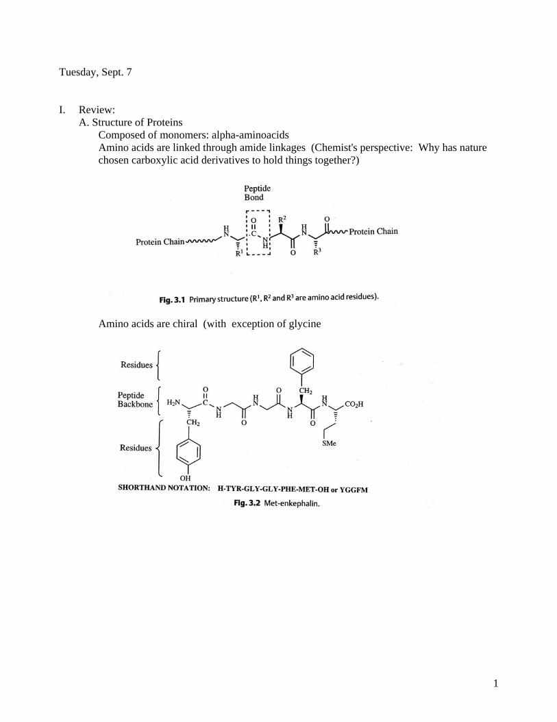
Tuesday, Sept. 7 I. Review: A. Structure of Proteins Composed of monomers: alpha-aminoacids Amino acids are linked through amide linkages (Chemist's perspective: Why has nature chosen carboxylic acid derivatives to hold things together?)
Amino acids are chiral (with exception of glycine

2
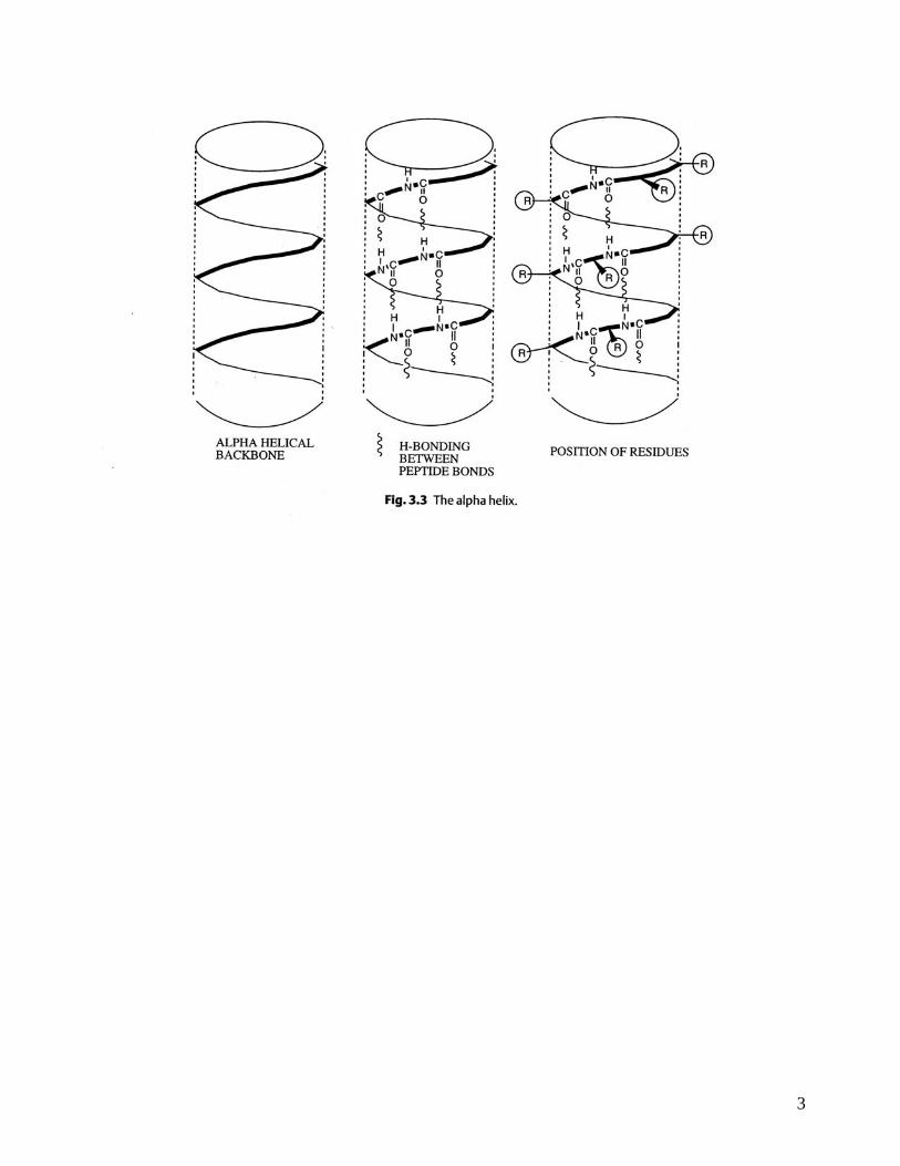
Proteins adopt three dimensional shape
3

4

5
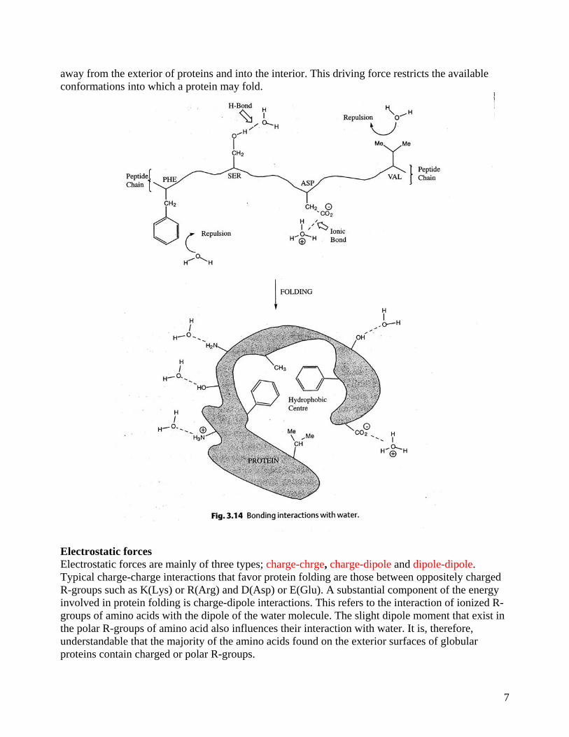
Forces holding three dimensional shape together include: non-polar interactions, charge interactions, hydrogen bonds, and covalent bonds (e.g. Cys-S-S-Cys) Forces controlling Protein Structure: Hydrogen Bonding Polypeptides contain numerous proton donors and acceptors both in their backbone and in the R-groups of the amino acids. The environment in which proteins are found also contains the ample H-bond donors and acceptors of the water molecule. H-bonding, therefore, occurs not only within and between polypeptide chains but with the surrounding aqueous medium

6
Hydrophobic Forces Proteins are composed of amino acids that contain either hydrophilic or hydrophobic R-groups. It is the nature of the interaction of the different R-groups with the aqueous environment that plays the major role in shaping protein structure. The spontaneous folded state of globular proteins is a reflection of a balance between the opposing energetics of H-bonding between hydrophilic R-groups and the aqueous environment and the repulsion from the aqueous environment by the hydrophobic R-groups. The hydrophobicity of certain amino acid R-groups tends to drive them
7
away from the exterior of proteins and into the interior. This driving force restricts the available conformations into which a protein may fold.
Electrostatic forces Electrostatic forces are mainly of three types; charge-chrge, charge-dipole and dipole-dipole. Typical charge-charge interactions that favor protein folding are those between oppositely charged R-groups such as K(Lys) or R(Arg) and D(Asp) or E(Glu). A substantial component of the energy involved in protein folding is charge-dipole interactions. This refers to the interaction of ionized R-groups of amino acids with the dipole of the water molecule. The slight dipole moment that exist in the polar R-groups of amino acid also influences their interaction with water. It is, therefore, understandable that the majority of the amino acids found on the exterior surfaces of globular proteins contain charged or polar R-groups.

8
van der Waals Forces There are both attractive and repulsive van der Waals forces that control protein folding. Attractive van der Waals forces involve the interactions among induced dipoles that arise from fluctuations in the charge densities that occur between adjacent uncharged non-bonded atoms. Repulsive van der Waals forces involve the interactions that occur when uncharged non-bonded atoms come very close together but do not induce dipoles. The repulsion is the result of the electron-electron repulsion that occurs as two clouds of electrons begin to overlap. Although van der Waals forces are extremely weak, relative to other forces governing conformation, it is the huge number of such interactions that occur in large protein molecules that make them significant to the folding of proteins.

9
Covalent Bonds
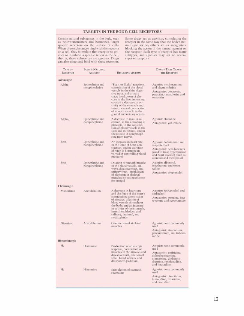
Protein Targets can take the form of either Receptors or Enzymes: Site Selectivity After being swallowed, injected, inhaled, or absorbed through the skin, most drugs enter the bloodstream and circulate throughout the body. Some drugs are administered directly to the area where they are wanted--for example, to the eyes in eyedrops. The drugs then interact with cells or tissues where they produce their intended effects (target sites). Some drugs are relatively nonselective; they affect many different tissues or organs. For example, atropine, a drug given to relax muscles in the digestive tract, may also relax muscles in the eyes and in the respiratory tract. Other drugs are relatively selective. For example, nonsteroidal anti-inflammatory drugs, such as aspirin and ibuprofen, target any area where inflammation is present. Still other drugs are highly selective; they affect mainly a single organ or system. For example, digoxin, a drug given to manage heart failure, affects mainly the heart, increasing its pumping efficiency. Sleep aids target certain nerve cells of the brain. How do drugs know where to exert their effects? The answer involves how they interact with cells or substances such as enzymes. Receptors on Cells On their surface, most cells have many different types of receptors. A receptor is a molecule with a specific three-dimensional structure, which allows only substances that fit precisely to attach to it--as a key fits in its lock. Receptors enable natural (originating in the body) substances outside the cell, such as neurotransmitters and hormones, to influence the activity of the cell. Drugs tend to mimic these natural substances and thus use receptors in the same way. For example, morphine and related pain-relieving drugs use the same receptors in the brain used by endorphins, which are substances produced by the body to help control pain. Some drugs attach to only one type of receptor; others, like a

10
master key, can attach to several types of receptors throughout the body. A drug's selectivity can often be explained by how selectively it attaches to receptors. Drugs that target receptors are classified as agonists or antagonists. Agonist drugs activate, or stimulate, their receptors, triggering a response that increases or decreases the cell's activity. Antagonist drugs block the access or attachment of the body's natural agonists, usually neurotransmitters, to their receptors and thereby prevent or reduce cell responses to natural agonists.
Agonist and antagonist drugs can be used together in patients with asthma. For example, albuterol can be used with ipratropium. Albuterol, an agonist, attaches to specific (adrenergic) receptors on cells in the respiratory tract, causing relaxation of smooth muscle cells and thus widening of the airways (bronchodilation). Ipratropium, an antagonist, attaches to other (cholinergic) receptors, blocking the attachment of acetylcholine, a neurotransmitter that causes contraction of smooth muscle cells and thus narrowing of the airways (bronchoconstriction). Both drugs widen the airways (and make breathing easier) but in different ways.

11
Beta-blockers, such as propranolol, are a widely used group of antagonists. These drugs are used to treat high blood pressure, angina (chest pain due to an inadequate blood supply to the heart muscle), and certain abnormal heart rhythms. They block or reduce stimulation of the heart by the agonist hormones epinephrine (adrenaline) and norepinephrine (noradrenaline), which are released during stress. Antagonists such as beta-blockers are most effective when the local concentration of the agonist is high. Similar to the way a roadblock stops more vehicles during the 5:00 pm rush hour than at 3:00 am, beta-blockers, given in doses that have little effect on normal heart function, may have a greater effect during sudden surges of hormones released during stress and thereby protect the heart from excess stimulation.
12
13
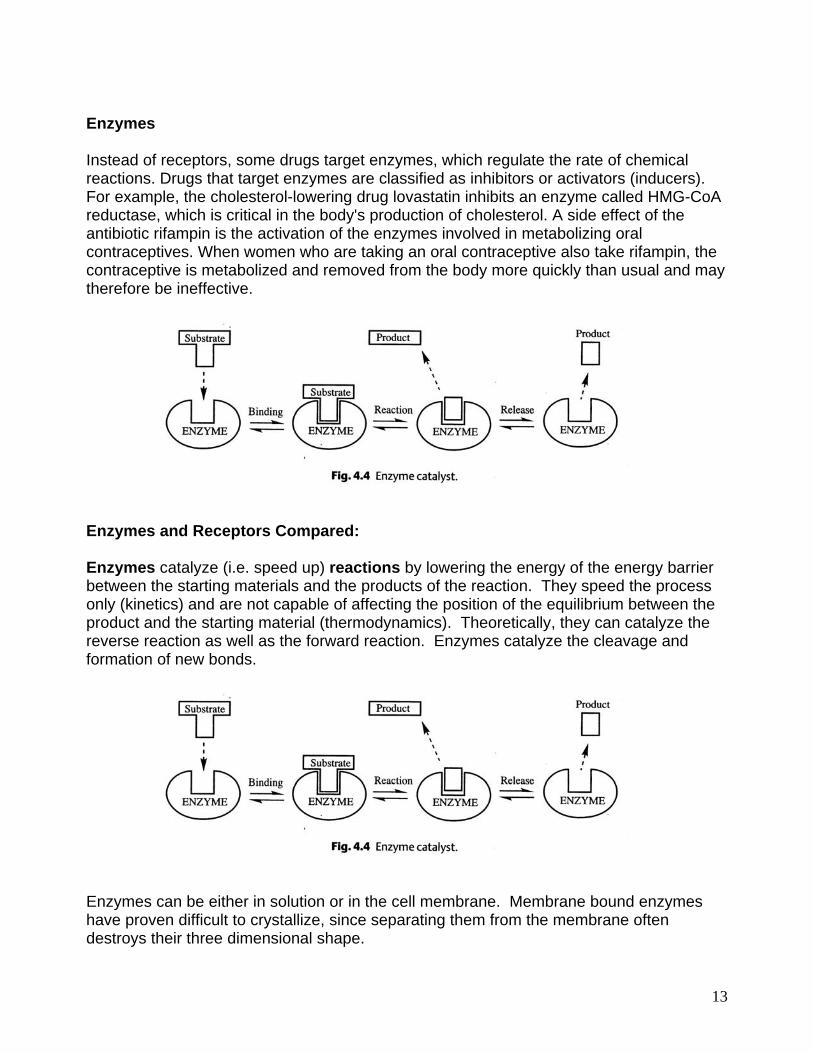
Enzymes Instead of receptors, some drugs target enzymes, which regulate the rate of chemical reactions. Drugs that target enzymes are classified as inhibitors or activators (inducers). For example, the cholesterol-lowering drug lovastatin inhibits an enzyme called HMG-CoA reductase, which is critical in the body's production of cholesterol. A side effect of the antibiotic rifampin is the activation of the enzymes involved in metabolizing oral contraceptives. When women who are taking an oral contraceptive also take rifampin, the contraceptive is metabolized and removed from the body more quickly than usual and may therefore be ineffective.
Enzymes and Receptors Compared: Enzymes catalyze (i.e. speed up) reactions by lowering the energy of the energy barrier between the starting materials and the products of the reaction. They speed the process only (kinetics) and are not capable of affecting the position of the equilibrium between the product and the starting material (thermodynamics). Theoretically, they can catalyze the reverse reaction as well as the forward reaction. Enzymes catalyze the cleavage and formation of new bonds.
Enzymes can be either in solution or in the cell membrane. Membrane bound enzymes have proven difficult to crystallize, since separating them from the membrane often destroys their three dimensional shape.

14
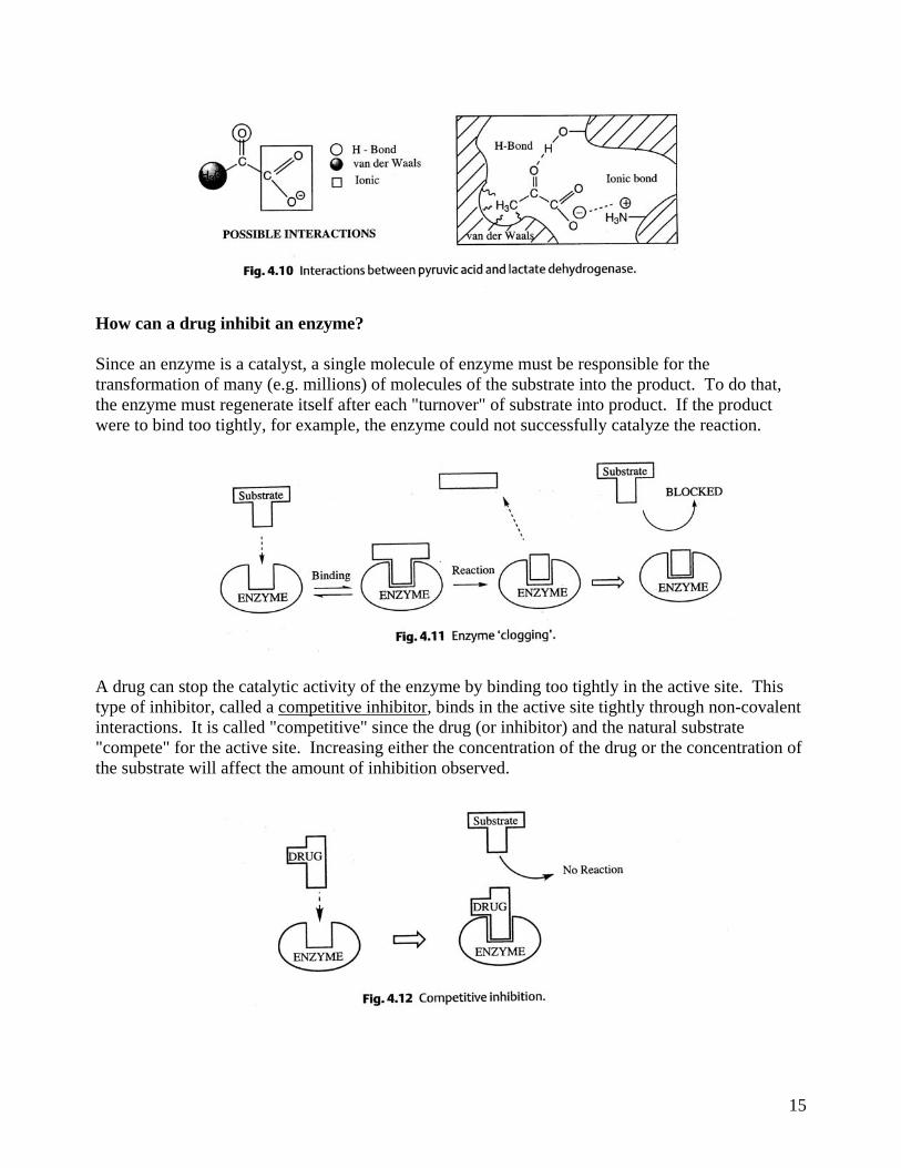
By contrast receptors recognize bind the messenger fleetingly and perform no chemistry on it. Instead, when the signal messenger binds to the receptor, a conformation change of the large receptor occurs, which somehow leads to the transmission of a signal to the cell (i.e. something is turned on or off). Receptors are located in a membrane. Few crystal structures of receptors have been obtained. How can an enzyme catalyze a reaction? The enzyme catalyzes the reaction at a pocket on its surface called the "active site".
Show example from the hydrolysis of the hydrolysis of the ester/amide bond. Chalkboard.
15
How can a drug inhibit an enzyme? Since an enzyme is a catalyst, a single molecule of enzyme must be responsible for the transformation of many (e.g. millions) of molecules of the substrate into the product. To do that, the enzyme must regenerate itself after each "turnover" of substrate into product. If the product were to bind too tightly, for example, the enzyme could not successfully catalyze the reaction.
A drug can stop the catalytic activity of the enzyme by binding too tightly in the active site. This type of inhibitor, called a competitive inhibitor, binds in the active site tightly through non-covalent interactions. It is called "competitive" since the drug (or inhibitor) and the natural substrate "compete" for the active site. Increasing either the concentration of the drug or the concentration of the substrate will affect the amount of inhibition observed.
16
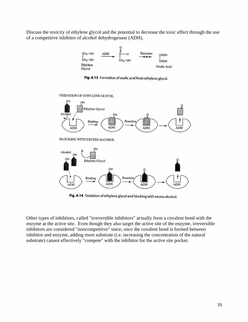
Discuss the toxicity of ethylene glycol and the potential to decrease the toxic effect through the use of a competitive inhibitor of alcohol dehydrogenase (ADH).
Other types of inhibitors, called "irreversible inhibitors" actually form a covalent bond with the enzyme at the active site. Even though they also target the active site of the enzyme, irreversible inhibitors are considered "noncompetitive" since, once the covalent bond is formed between inhibitor and enzyme, adding more substrate (i.e. increasing the concentration of the natural substrate) cannot effectively "compete" with the inhibitor for the active site pocket.

17
Yet a third type of inhibitor, called an allosteric inhibitor, will bind to a site removed from the active site. However, in so binding, the conformation of the entire protein is affected, causing enough of a change at the active site so that a molecule of substrate cannot bind. This type of inhibitor is also "noncompetitive" since adding more substrate will, once again, have no effect on the inhibition caused by the allosteric inhibitor.
Allosteric inhibition is utilized in the body to regulate complex, multistep, biochemical synthesis pathways. In such a multistep pathway, the final product of the pathway will not necessarily resemble the substrate or the product of the first step. Thus the final product will not be recognized at the active site of the first enzyme catalyst. Still the body needs a mechanism to shut down the pathway when enough of the final product has been produced. The below example shows an example of such "feedback inhibition".
18
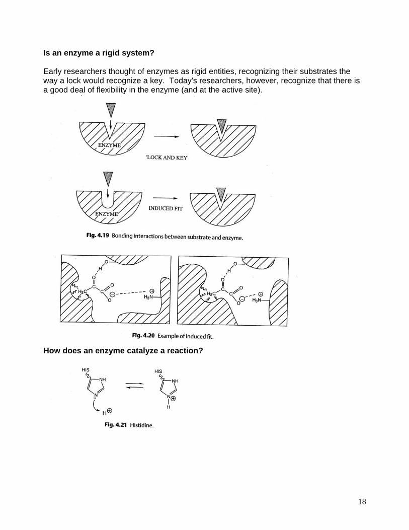
Is an enzyme a rigid system? Early researchers thought of enzymes as rigid entities, recognizing their substrates the way a lock would recognize a key. Today's researchers, however, recognize that there is a good deal of flexibility in the enzyme (and at the active site).
How does an enzyme catalyze a reaction?

19