Tuberculosis Clinical Presentation & Diagnosisnid... · Tuberculosis Clinical Presentation &...
82
Tuberculosis Clinical Presentation & Diagnosis Tuberculosis Clinical Intensive Thursday, June 16, 2016 Christopher Spitters, MD, MPH Public Health Seattle & King County Tuberculosis Clinic
Transcript of Tuberculosis Clinical Presentation & Diagnosisnid... · Tuberculosis Clinical Presentation &...

Tuberculosis Clinical Presentation & Diagnosis
Tuberculosis Clinical IntensiveThursday, June 16, 2016
Christopher Spitters, MD, MPHPublic Health Seattle & King County Tuberculosis Clinic

This slide has no title
“TB as a cause of the right upper lobe pneumonia in this 23 y/o male from Somaliapneumonia in this 23 y/o male from Somalia was ruled out with 3 negative sputum smears. The patient was released fromsmears. The patient was released from isolation and was discharged home to complete an outpatient course ofcomplete an outpatient course of levofloxacin and to follow-up with primary care.”care.
Anonymous

Missed TB DiagnosesCalifornia 2005-2011California, 2005-2011
Miller A, et al. Open Forum ID 2015. DOI: 10.1093/ofid/ofv171

Question
Which of the following statements is false about diagnosis of pulmonary tuberculosis?g p yA. The combination of cough, sputum and fever-or-
weight loss is up to 90% sensitive but <50% g pspecific.
B. The predictive value of a negative TST in a patient with a clinical presentation highly suggestive of severe TB could be as low as 50%
C. >95% of active pulmonary TB cases report cough

Learning Objectives
• Recognize clinical syndromes warranting evaluation for active TB.
• List the elements of a complete evaluation for active TBfor active TB.
• Upon completion of an initial evaluation, conduct medical decision making regardingconduct medical decision making regarding initiation of therapy.

Active TB Evaluation
• History and Examination• ImagingImaging• Mycobacteriology• Ancillary tests• Specialty evaluationSpecialty evaluation

Initial Clinical Evaluation• Symptoms• Prior TB diagnosis/treatment• Epidemiologic risk• Predisposing medical conditions/therapies• General temperature weightGeneral, temperature, weight• Lymphadenopathy
Ch l i b li i• Chest auscultation abnormalities• Abdominal distension• Neurologic abnormalities

Clinical Presentation: Signs and symptomsg y p
• Cough (dry/productive sputum) 75-80%g ( y p p )• Weight loss 45-75% • Fatigue 60-70%• Fatigue 60-70%• Fever 50-60%
i h• Night Sweats 50-55%• Hemoptysis 25-35%• No symptoms 10-20%
Source: Barnes 1988, Miller 2000

Accuracy of Symptom CheckAccuracy of Symptom Check
SENSITIVITY SPECIFICITY PPV NPVSENSITIVITY SPECIFICITY PPV NPV
Cough (any)Night sweats (24h)
93% 37% 21% 97%g ( )
Fever (2wks)Anorexia (4wks)
Good news: effectiveBad news: inefficient
Cain KP, et al. N Engl J Med 2010;362:707-16.

Clinical Presentation: Site of DiseaseCDC Reported TB Cases by Form of Disease United States, 2010
Extrapulmonary (22%)
Both (10%)
t apu o a y ( %)
Pleural (16%)
L h ti (40%)Pulmonary (68%)
Lymphatic (40%)
Bone/joint (10%) Genitourinary (5%
Other (18%)
y (Meningeal (6%)Peritoneal (5%)

Epidemiologic Risk Factors for TB
• Known contact to a pulmonary case• Nation of originNation of origin• Extended stays in high-risk areas
H l h k i i i• Health care work in certain settings• Incarceration in certain settings• Homelessness• Other occupational exposuresOther occupational exposures

Relative risk of TB reactivation by medical condition
Horsburgh, NEJM 2004 350; 20: 2060-7

Differential DiagnosisDifferential Diagnosis• Community acquired pneumonia• Malignanc• Malignancy• Lung abscess
N TB b t i• Non-TB mycobacteria• Fungal infection• Parasite (e.g., paragonimiasis)• Sarcoidosis• Rheumatologic disease (e.g., Wegener’s, RA)• Other systemic infections (e.g. brucellosis,
melioidosis, relapsing fever, etc.)

Radiographic Evaluation


Radiographic Patterns: Pulmonary TB
TB Pattern “Typical”/ R ti ti
“Atypical“/ PrimaryReactivation
Infiltrate 85% upper Upper:Lower 60:40U ll iUsually upper in
childrenCavitation Often present Rarep
Adenopathy RareChildren common
Adults ~30%il l bil lUnilateral > bilateral
Effusion May be present May be present
HIV CD4 >200 CD4 <200

TB Symptoms with Compatible CXRTB Symptoms with Compatible CXR
Sputum ExamAFB smear/culture x3
TB PCR x 1-2
PCR positive Smear/PCR Negative
Smear positive/PCR
negative
TB Treatment Public Health Pulmonary referral?
Observe?TB Rx?
Rx other?Rx other?

Clinical Specimens for ExaminationClinical Specimens for Examination• Sputum (x3)• Bronchoalveolar lavage, washings, brushings• Transbronchial biopsyp y• Fine needle or core needle aspirate• Tissue biopsy or excision (save some unfixed)Tissue biopsy or excision (save some unfixed)• Fluids
Cerebrospinal– Cerebrospinal– Pleural fluid– Peritoneal fluidPeritoneal fluid– Abscess

Collection of Respiratory Specimensp y p
• Sputum Expectoration: p p– 3 specimens (at least 8 hours apart)– 1 spot specimen1 spot specimen – 2 consecutive first-morning specimens
P b h• Post-bronchoscopy sputum

Bronchoscopy Indicationspy
• Unable to obtain specimen via induction or pgastric aspirate
• Sputum smear/PCR negative but clinicalSputum smear/PCR negative but clinical suspicion of TB still high
• Sputum smear negative and MDR is a high• Sputum smear negative and MDR is a high concernM li i d• Malignancy is suspected

Post-Bronchoscopy Sputumpy p
• 57 sputum smear-negative or non-productive1p g p– 33% AFB smear-positive PBS– 7% PBS sole culture-positive specimenp p
• 56 culture-confirmed cases with negative sputum AFB smears or non-productive2p– AFB smear sensitivity:
• BAL 57% • PBS 77%• BAL + PBS 84%
1George PM, et al. Respir Med 2011;105:1726.2Malekmohammad M, et. al. J Scand Infect Dis 2012;44:369.

Laboratory Evaluation

Acid Fast Staining
• Preparation– Centrifugation to concentrateCentrifugation to concentrate– NaOH, NaOCL wash to decontaminate
• Ziehl Neelsen• Ziehl-Neelsen– Carbolfuchsinacid alcoholmethylene blue
Fl t• Flourescent– Auramine-Rhodamine

Acid Fast Staining

AFB Smear Peformance
Steingart K, et al. Lancet ID 2006:6(10):664

AFB Culture• Broth (faster, more expensive, complex)
– Liberation of 14CO2 (defunct)Liberation of CO2 (defunct)– Emission of fluorescence (MGIT)
• Plates (slower, less expensive, “simpler”)( , p , p )– Lowenstein-Jensen slant– 7H11 plates
• Identification of AFB growth– Phenotypic characteristics– Nucleic acid hybridization– DNA sequencing
C– HPLC

MGIT

AFB Culture Performance
Caviedes L, et al. J Clin Microbiol 2000;38(3):1208-1209

AFB Culture Limitations
• False Positive (up to 3% of total)– Laboratory cross contaminationLaboratory cross contamination– Specimen mis-handling
• False Negative• False Negative– Small inoculum
D l i i l ti– Delay in inoculation– Difficult-to-grow strain

Mycobacteriologic Examinations for TB
Test Time
AFB stain <24 hoursAFB stain 24 hours
AFB culture 2-6 weeks
Phenotypic DST results 4-8 weeks
DNA fingerprinting Months

Culture-negative TBCulture negative TBDiagnostic Criteria
• Compatible clinical and radiographic syndromey
• AFB cultures negative– 10-15% pulmonaryp y– 25-30% extrapulmonary
• Clinical/radiographic improvement on therapy• Other causes reasonably excluded• Positive TST-or-IGRA

Presumptive Treatment forPresumptive Treatment for Smear-Negative Pulmonary TB
6 (8%) f 72 i ti ith i lt i d ff t6 (8%) of 72 inactive cases with regimen altering adverse effects
Gordin, et al. Am Rev Resp Dis 1989;139:1090-93

Mycobacteriologic Examinations for TB
Test Time
AFB stain <24 hoursAFB stain 24 hours
Nucleic acid amplification Hours-days
Molecular DST results Days-weeks
AFB culture 2-6 weeks
Phenotypic DST results 4-8 weeks
DNA fingerprinting Months

Nucleic Acid Amplification Tests (NAAT)
• Varieties– Amplified MTD (GenProbe)– GeneXpert Mtb/RIF (Cepheid)– Non FDA Approved
( i )• MTBDR (Hain)• Others
Laboratory developed– Laboratory developed
• Use Directly on processed specimen– Directly on processed specimen
– No current TB rx >7 days– No prior TB rx within past 12 monthsp p


Sensitivity of Xpert in Pulm TB Dx L & Hi h B d S iLow & High Burden Settings
TEST SENS SPEC PPV NPVTEST SENS SPEC PPV NPV
Xpert x 1 81.4% 98.7% 94.6% 99.7%
Smear pos 98.5%
Smear neg 54.8%
Xpert x 2 95 0%Xpert x 2 95.0%
Smear pos 100%
Smear neg 71.4%
• N = 992 (US, Brazil, S Africa)• Sensitivity of AFB smear: 60%
N diff b t hi h d l l tti• No difference between high and low prevalence settings
Leutkemeyer AF, et al. CID 2016

Caution with NAAT in P i l T d P iPreviously Treated Patients
Theron, et al. CID 2016•45/321 (14%) positive were culture negative( %) p g•Recency of prior treatment•Low DNA•Low DNA•CXR not suggestive of TBBoyles, et al. IJTLD 2014•4 false positive case reports p p•1, 2, 5 and 66 months after prior treatment

Mycobacteriology FlowMycobacteriology FlowSpecimen
/ iAFB smear/stain NAAT
MGIT broth and 7H10 plate cultivationp
Species identification
M. tuberculosis complex M. avium complexM d
Other MOTTM. gordonae MOTT
Broth sensitivitiesfor HRES + Z If resistance: plate confirmation/proportional p p p
method and key second line drugs

Other Laboratory ExaminationOther Laboratory Examination
Fl id h i ll d l• Fluids: chemistry, cell count and cytology• Tissues: routine histopathologynecrotizing
lgranulomata• Tuberculin skin test• Interferon gamma release assays• Adenosine deaminaseAdenosine deaminase

Granulomatous Inflammation

Typical Findings Extrapulmonary Specimens
• Protein elevatedPleural/peritoneal (>4 5gm/dL)– Pleural/peritoneal (>4-5gm/dL)
– CSF (>100-500mg/dL)• Moderately decreased glucose (~40-50mg/dL)Moderately decreased glucose ( 40 50mg/dL)• Pleocytosis
– Pleural (1,000-5,000 WBC/uL)– CSF (100-500/uL)
• Lymphocyte predominant differential• Necrotizing granulomata • NAAT 50-75% sensitive
AFB 10 50% iti• AFB smear: 10-50% sensitive • AFB culture: 60-90% sensitive

Pleural Effusion Evaluation SensitivityySpecimen Cultured AFB Culture SensitivitySputum only 48%Fluid only 63%Sputum + Fluid 79%
Ruan SY et al Thorax 2012;67:822 7Ruan SY, et al. Thorax 2012;67:822-7.
Pleural Biopsy• Closed
• Up to 40% of specimens contain no pleural tissue• Image guided gaining favor• S iti it ( th l + lt ) 80 90%• Sensitivity (pathology + culture): 80-90%
• Thoracoscopy/VATS: sensitivity approaches 100%
Koegelenberg CF et al Respirology 2011;16(5):738 46Koegelenberg CF, et al. Respirology 2011;16(5):738-46.Kirsch CM, et al. Chest 1997;112(3):702-6.Vorster, MJ, et al. J Thorac Dis 2015;7(6):981-991

TST & IGRA PerformanceTST & IGRA PerformanceTEST SENSITIVITY SPECIFICITY
RANGE MEDIAN POOLED RANGE MEDIAN POOLED
TST 53-95 82 95 -- -- 95TST 53 95 82 95 95
QFT 56-93 85 84 99-100 100 99
TSPOT 58-100 86 91 85-100 95 88
Mazurek GH et al MMWR 2010; 59(RR05);1 25Mazurek GH, et. al. MMWR 2010; 59(RR05);1-25Specificity of TST in BCG vaccinated children<1 / t i ti 91%
Sensitivity of TST in miliary vs LN TB22% vs. 73%
•<1 y/o at vaccination: 91%•>1 y/o at vaccination: 58%Farhat M, et al. IJTLD 2006;10(11):1192-204.
Lee, et al. CID 2013.
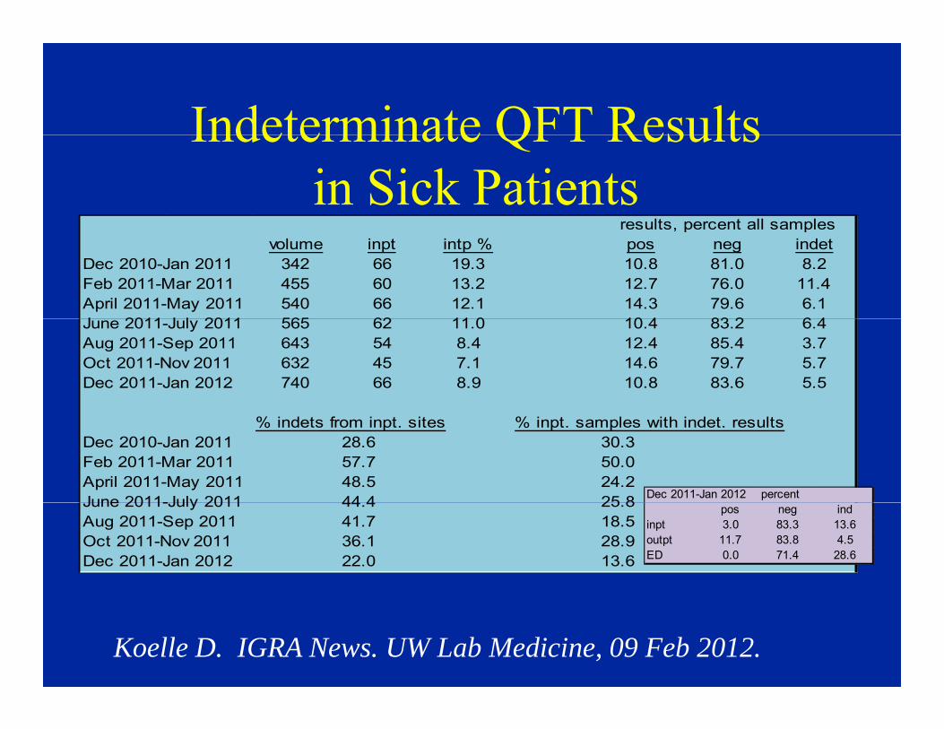
Indeterminate QFT ResultsIndeterminate QFT Results in Sick Patients
results percent all samplesresults, percent all samplesvolume inpt intp % pos neg indet
Dec 2010-Jan 2011 342 66 19.3 10.8 81.0 8.2Feb 2011-Mar 2011 455 60 13.2 12.7 76.0 11.4April 2011-May 2011 540 66 12.1 14.3 79.6 6.1J 2011 J l 2011 565 62 11 0 10 4 83 2 6 4June 2011-July 2011 565 62 11.0 10.4 83.2 6.4Aug 2011-Sep 2011 643 54 8.4 12.4 85.4 3.7Oct 2011-Nov 2011 632 45 7.1 14.6 79.7 5.7Dec 2011-Jan 2012 740 66 8.9 10.8 83.6 5.5
% indets from inpt. sites % inpt. samples with indet. resultsDec 2010-Jan 2011 28.6 30.3Feb 2011-Mar 2011 57.7 50.0April 2011-May 2011 48.5 24.2June 2011-July 2011 44 4 25 8 Dec 2011-Jan 2012 percentJune 2011-July 2011 44.4 25.8Aug 2011-Sep 2011 41.7 18.5Oct 2011-Nov 2011 36.1 28.9Dec 2011-Jan 2012 22.0 13.6
pos neg indinpt 3.0 83.3 13.6outpt 11.7 83.8 4.5ED 0.0 71.4 28.6
Koelle D. IGRA News. UW Lab Medicine, 09 Feb 2012.

Pleural Fluid ADAPleural Fluid ADALow Incidence Setting
•N=338 patients•Lymphocytic exudative y p ocyt c e udat ve•7 pleural TB cases•Typical cut-off: >40•Sensitivity: 85%Sensitivity: 85%•Specificity: 90%•PPV: 85%•NPV: 99%NPV: 99%
Arnold, et al. Thorax 2014;69:A62 doi:10.1136/thoraxjnl-2014-206260.121

ADA LimitationsADA Limitations• False negatives
– Early disease– Advanced age– Smokers
• False positivesFalse positives– Non-TB empyema, parapneumonic effusions– Mesothelioma lung and hematologicMesothelioma, lung and hematologic
malignancies– Rheumatologic conditionsRheumatologic conditions
Vorster MJ, et al. J Thorac Dis. 2015 Jun; 7(6): 981–991.

Pleural Fluid IGRA
“We conclude that commercial IGRAs, performed either on whole-blood or pleural fluid p psamples, have poor diagnostic accuracy in patients suspected to have TPE.”
Aggarawal AN, et al. J Clin Microbiol 2015 Aug;53(8):2451-9.

CSF IGRA for TB Meningitis• Meta analysis S iti it 77%• Meta-analysis• 6 CSF IGRA studies
included
• Sensitivity: 77%• Specificity: 88%
included
Yu J, et al. IJTLD 2016;20(4):494-499

Question
Which of the following statements is false about diagnosis of pulmonary tuberculosis?g p yA. The combination of cough, sputum and fever-or-
weight loss is up to 90% sensitive but <50% g pspecific.
B. The predictive value of a negative TST in a patient with a clinical presentation highly suggestive of severe TB could be as low as 50%
C. >95% of active pulmonary TB cases report cough

Medical Decision MakingMedical Decision MakingTreat for TB or Not?

i i f
Medical Decision Making Tool--1Smear positive for AFB
Culture and SpeciationCulture and Speciation
M. tuberculosis50-90%
Non-tuberculous mycobacteria10-50%
Predictive value of a positive smear is reduced in populations with increased prevalence of non-tuberculous mycobacterial p f y
infection

Medical Decision Making Tool--2Medical Decision Making Tool 2
Patient with smear-positive specimen
NAATNAAT
Positive NAAT Negative NAATPositive NAAT Negative NAAT
MTB NTM MTB NTM≥97% <3% 1-8% 92-99%
• 2009 CDC Guidelines: Test all AFB+/NAAT- specimens for inhibitors2009 CDC Guidelines: Test all AFB /NAAT specimens for inhibitors• Probably not necessary if using Xpert, which tests for PCR inhibitors

Medical Decision Making Tool--3
High Clinical Suspicion (e g 50% pre test probability)
Sputum Smear NegativeHigh Clinical Suspicion (e.g., 50% pre-test probability)
Perform NAAT
Positive NAAT Negative NAATPositive NAAT Negative NAAT
≥90% 10% 75%2525%MTB Not
MTBNot TBMTB
2009 CDC G id li C id t t t f fi ti if AFB NAAT+• 2009 CDC Guidelines: Consider repeat test for confirmation if AFB-, NAAT+ • May not be necessary if using Xpert, given high specificity and low risk of cross-
contamination

Medical Decision Making Tool--4
L Cli i l S i i 5% t t b bilit
Sputum Smear Negatives Low Clinical Suspicion; e.g., 5% pre-test probability
Perform NAAT??
Positive NAAT Negative NAAT
MTB20%
Not MTB80%
Not MTB99%
MTB<1%
• 2009 CDC Guidelines: Avoid NAAT in this clinical scenario• Same holds true for Xpert, which provides no added value over smear

Drug Susceptibility Testing

Culture-BasedCulture Based Drug Susceptibility Testing
• MGIT broth (2-3 weeks)– Control, INH, RIF, EMB, STR, PZAControl, INH, RIF, EMB, STR, PZA– Qualitative
• Agar plates (4 6 weeks)• Agar plates (4-6 weeks)– Quantitative: concentration, % resistant
Fi t d d li d– First and second line drugs

Molecular Drug Susceptibility TestingMolecular Drug Susceptibility TestingDetection of Common Resistance Conferring Mutations
• Requirements: sufficient DNA (smear positive sputum or culture growth)
• Time to results: ~1 week• Target sequences: rpoB, katG, inhA, othersTarget sequences: rpoB, katG, inhA, others
– Xpert MTB-RIF (rpoB only)– Hain GenoType MDRTB (rpoB, inhA, katG)Hain GenoType MDRTB (rpoB, inhA, katG)– WA DOH TB Lab (rpoB, katG, inhA, pncA)– CDC (all of the above, gyrA, emb, [injectables])( , gy , , [ j ])

Xpert MTB/RIF Test Performance for pDiagnosis of Pulmonary TB
S i i i S ifi iSensitivity Specificity
S TB 95 98%Smear pos. TB 95-98%99%
Smear neg TB 60 72%Smear neg. TB 60-72%
Rifampin “R” 98 99% 99 100%Rifampin R 98-99% 99-100%
Source: NEJM 2010; 361:1005,; Am J Crit Care Med 2011; 184:132

Molecular Drug Susceptibility TestingMolecular Drug Susceptibility TestingTypical Indications
• Treatment failure• Previous treatment for active TBPrevious treatment for active TB• Known contact to confirmed case of MDR
F hi hl MDR d i i (• From a highly MDR-endemic setting (e.g., S. Africa, Baltic states, Russian prison)

Drug resistance sequencing screenDrug resistance sequencing screenWA DOH PHL

Molecular detection of drug resistanceCDCCDC

BACTEC MGIT DSTBACTEC MGIT DST WA DOH PHL

DOH Plate DST Results

CDC Plates DST Results

TB Diagnosis Summary--1TB Diagnosis Summary 1• Cough, sputum, fever, night sweats, weight loss
sensiti e b t not specificsensitive but not specific• Initial diagnostic test is imaging• Collect appropriate specimens when clinical and
imaging findings suggest TB• Mycobacteriology: AFB smear, culture, and
nucleic acid amplificationp• Other tests: cytology/pathology, cell count,
differential protein LHD glucose pHdifferential, protein, LHD, glucose, pH

TB Diagnosis Summary--2TB Diagnosis Summary 2• Cautious use:
TST IGRA– TST, IGRA – ADA in serosal fluids
• Medical decision making ingredients: clinical, radiography, laboratory results AND epidemiology
• Predictive value of a test is driven by both test yaccuracy AND pre-test probability
• Specialty evaluation indicated for patients withSpecialty evaluation indicated for patients with inconclusive findings

Cases

Case 1 Clinical Presentation
• Cough, chest pain, fever, anorexia x 6 weeks
• What is your next step?

Case 1: What Would You Do?
A. Place a TSTB Collect sputum--AFB smear/culture/PCRB. Collect sputum AFB smear/culture/PCRC. CT chestD PA/LAT CXRD. PA/LAT CXRE. Levofloxacin 500mg po qd x 7d

Case 1: Chest RadiographsCase 1: Chest Radiographs 2007 vs 2013

Case 1—Evaluation
• Sputum AFB smears 1+ on 2 separate specimensp
• What else?

Case 1: NAAT
• Positive for MTB• Negative for MACNegative for MAC

Case 2—Clinical Presentation
• 33 y/o Ethiopian male • Visited home recently for 3 monthsy• Malaise, fatigue, fever x 1 month• Left chest pain worsening over past 2-3Left chest pain worsening over past 2 3
weeks• Weight 63kg60kgWeight 63kg60kg• T 38.5C; left base dull to percussion with
decreased breath soundsdecreased breath sounds

Case 2—Chest Radiograph

Case 2—Initial Evaluation
• HIV negative• Hgb 10 MCV 80 albumin 3 1Hgb 10, MCV 80, albumin 3.1• TST placed

Case 2—What Would You Do?What would you do next?What would you do next?A. Complete sputum collection and start 4-drug
therapytherapyB. Thoracentesis, sputum collection, and pleural
biopsy if other specimens AFB smear-negativebiopsy if other specimens AFB smear negativeC. Give azithromycin for presumed CAP and
discharge to outpatient pulmonary follow-upg p p y pD. Refer to thoracic surgery for tube drainage

Case 2—Pleural Fluid
• Protein 4.4 gm/dL, • WBC ~1500 (65% lymphocytes)WBC 1500 (65% lymphocytes) • No AFB seen
Pl l bi i i l• Pleural biopsy: necrotizing granulomata without visible AFB (culture pending, PCR
d )not done)• TST 18mm


![Melioidosis Mimicking Tuberculosis in an Endemic Zone · mimics tuberculosis [2]. Therefore, it is frequently treated with anti-tuberculosis drugs in an area where tuberculosis is](https://static.fdocuments.net/doc/165x107/605b21c8479bfc022b674719/melioidosis-mimicking-tuberculosis-in-an-endemic-zone-mimics-tuberculosis-2-therefore.jpg)

![Tuberculosis Clinical Presentation & Diagnosisnid]/3_spitters...Differential Diagnosis • Community acquired pneumonia • Malignancy • Lung abscess ... • 2009 CDC Guidelines:](https://static.fdocuments.net/doc/165x107/5ed5b4681e2a093f7737762f/tuberculosis-clinical-presentation-diagnosis-nid3spitters-differential.jpg)