Tsantan Sumtang Alleviates Chronic Hypoxia-Induced ...
14
Research Article Tsantan Sumtang Alleviates Chronic Hypoxia-Induced Pulmonary Hypertension by Inhibiting Proliferation of Pulmonary Vascular Cells Qilian He , 1,2,3 Xingmei Nan, 1,2 Silin Li , 4 Shanshan Su , 5 Ke Ma, 1,2 Zhanqiang Li , 1,2 Dianxiang Lu , 1,2 and Rili Ge 1,2 1 Research Center for High Altitude Medicine, Qinghai University, Xining 810001, China 2 Key Laboratory of Application and Foundation for High Altitude Medicine Research in Qinghai Province, Xining 810001, China 3 Medical Department, Medical College, Qinghai University, Xining 810001, China 4 Affiliated Hospital of Heilongjiang University of Traditional Chinese Medicine, Ha’erbin 150040, China 5 Qinghai Entry-Exit Inspection and Quarantine Bureau, Xining 810003, China Correspondence should be addressed to Zhanqiang Li; zhanqiang [email protected], Dianxiang Lu; [email protected], and Rili Ge; [email protected] Received 29 July 2018; Revised 23 October 2018; Accepted 1 November 2018; Published 28 November 2018 Academic Editor: Rui Liu Copyright © 2018 Qilian He et al. is is an open access article distributed under the Creative Commons Attribution License, which permits unrestricted use, distribution, and reproduction in any medium, provided the original work is properly cited. Hypoxia-induced pulmonary hypertension (HPH) is a severe condition associated with significant morbidity and mortality in people living at high altitude. Tsantan Sumtang, a traditional Tibetan medicine, has been routinely used for the treatment of cardiopyretic disease, as well as stenocardia. Interestingly, our previous research found that Tsantan Sumtang improved HPH in rats maintaining in a hypobaric chamber. We performed a series of experiments to test the indexes of vasoconstriction and vascular remodeling, the key pathophysiological characteristics of HPH. Our results showed that Tsantan Sumtang relaxed noradrenaline (NE)-precontracted rat pulmonary artery rings in a concentration-dependent manner in vitro. e PGI2-cAMP (prostaglandin I2- cyclic adenosine monophosphate) pathway, NO-cGMP (nitric oxide-cyclic guanosine monophosphate) pathway, and the opening of K + channels (inward rectifier K + channels, large conductance Ca 2+ -activated K + channels, and voltage-dependent K + channels) might play major roles in the vasorelaxation effect. In vivo, the administration of Tsantan Sumtang resulted in a substantial decrease in the rat mean pulmonary artery pressure (mPAP) and the right ventricular hypertrophy index (RVHI). e reduction of thickness of small pulmonary arterial wall and the WT% (the ratio of the vascular wall thickness to the vascular diameter) were observed. e smooth muscle muscularization of the arterials was alleviated by Tsantan Sumtang treatment at the same time. Tsantan Sumtang also reduced remodeling of pulmonary arterioles by suppressing the expression of proliferating cell nuclear antigen (PCNA), - smooth muscle actin (-SMA), cyclin D1, and cyclin-dependent kinase 4 (CDK4) through inhibition of p27Kip1 degradation. erefore, Tsantan Sumtang could be applied as a preventative medication for HPH, which would be a new use for this traditional medicine. 1. Introduction Pulmonary hypertension (PH) is a group of clinical disorders that feature an abnormal elevation in the pulmonary circula- tory pressure and are characterized by a sustained increase in pulmonary vascular resistance and vascular remodeling, which ultimately leads to right ventricular failure and death [1, 2]. HPH is a type of PH that occurs among residents and travelers at high altitude. It has been reported that people living on a plateau may suffer from minor to moderate pulmonary vasoconstriction and hypertension, and those with a maladaptation to hypoxia may induce hypoxia-related diseases, among which HPH appears in approximately 5–10% of cases [3–5]. Sustained pulmonary vasoconstriction and structural remodeling of small pulmonary arteries result Hindawi BioMed Research International Volume 2018, Article ID 9504158, 13 pages https://doi.org/10.1155/2018/9504158
Transcript of Tsantan Sumtang Alleviates Chronic Hypoxia-Induced ...

Research ArticleTsantan Sumtang Alleviates Chronic Hypoxia-InducedPulmonary Hypertension by Inhibiting Proliferation ofPulmonary Vascular Cells
Qilian He ,1,2,3 Xingmei Nan,1,2 Silin Li ,4 Shanshan Su ,5 KeMa,1,2 Zhanqiang Li ,1,2
Dianxiang Lu ,1,2 and Rili Ge 1,2
1Research Center for High Altitude Medicine, Qinghai University, Xining 810001, China2Key Laboratory of Application and Foundation for High Altitude Medicine Research in Qinghai Province, Xining 810001, China3Medical Department, Medical College, Qinghai University, Xining 810001, China4Affiliated Hospital of Heilongjiang University of Traditional Chinese Medicine, Ha’erbin 150040, China5Qinghai Entry-Exit Inspection and Quarantine Bureau, Xining 810003, China
Correspondence should be addressed to Zhanqiang Li; zhanqiang [email protected], Dianxiang Lu; [email protected],and Rili Ge; [email protected]
Received 29 July 2018; Revised 23 October 2018; Accepted 1 November 2018; Published 28 November 2018
Academic Editor: Rui Liu
Copyright © 2018 QilianHe et al.This is an open access article distributed under theCreative Commons Attribution License, whichpermits unrestricted use, distribution, and reproduction in any medium, provided the original work is properly cited.
Hypoxia-induced pulmonary hypertension (HPH) is a severe condition associated with significant morbidity and mortality inpeople living at high altitude. Tsantan Sumtang, a traditional Tibetan medicine, has been routinely used for the treatment ofcardiopyretic disease, as well as stenocardia. Interestingly, our previous research found that Tsantan Sumtang improved HPH inrats maintaining in a hypobaric chamber.We performed a series of experiments to test the indexes of vasoconstriction and vascularremodeling, the key pathophysiological characteristics of HPH. Our results showed that Tsantan Sumtang relaxed noradrenaline(NE)-precontracted rat pulmonary artery rings in a concentration-dependentmanner in vitro. The PGI2-cAMP (prostaglandin I2-cyclic adenosine monophosphate) pathway, NO-cGMP (nitric oxide-cyclic guanosine monophosphate) pathway, and the openingof K+ channels (inward rectifier K+ channels, large conductance Ca2+-activated K+ channels, and voltage-dependent K+ channels)might play major roles in the vasorelaxation effect. In vivo, the administration of Tsantan Sumtang resulted in a substantial decreasein the ratmean pulmonary artery pressure (mPAP) and the right ventricular hypertrophy index (RVHI).The reduction of thicknessof small pulmonary arterial wall and theWT% (the ratio of the vascular wall thickness to the vascular diameter) were observed.Thesmooth muscle muscularization of the arterials was alleviated by Tsantan Sumtang treatment at the same time. Tsantan Sumtangalso reduced remodeling of pulmonary arterioles by suppressing the expression of proliferating cell nuclear antigen (PCNA), 𝛼-smooth muscle actin (𝛼-SMA), cyclin D1, and cyclin-dependent kinase 4 (CDK4) through inhibition of p27Kip1 degradation.Therefore, Tsantan Sumtang could be applied as a preventative medication for HPH, which would be a new use for this traditionalmedicine.
1. Introduction
Pulmonary hypertension (PH) is a group of clinical disordersthat feature an abnormal elevation in the pulmonary circula-tory pressure and are characterized by a sustained increasein pulmonary vascular resistance and vascular remodeling,which ultimately leads to right ventricular failure and death[1, 2].
HPH is a type of PH that occurs among residents andtravelers at high altitude. It has been reported that peopleliving on a plateau may suffer from minor to moderatepulmonary vasoconstriction and hypertension, and thosewith a maladaptation to hypoxia may induce hypoxia-relateddiseases, among whichHPHappears in approximately 5–10%of cases [3–5]. Sustained pulmonary vasoconstriction andstructural remodeling of small pulmonary arteries result
HindawiBioMed Research InternationalVolume 2018, Article ID 9504158, 13 pageshttps://doi.org/10.1155/2018/9504158
2 BioMed Research International
in right ventricular hypertrophy and heart failure. HPH isassociated with significant morbidity and mortality, but aspecific and effective therapy remains unavailable [6–8]. Asthe key pathophysiological changes with HPH, vasocon-striction and vascular remodeling of the pulmonary arteryimpact the long-term prognosis of the patient, especiallyregarding myocardial damage, right ventricular hypertrophy,heart failure, and brain function decline [1, 2].
Tibetan medicament is a compoundmedicine with a longhistory of treatment effectiveness. This treatment approachattempts to harmonize the body, mind, and spirit [9]. Theinclusive and flexible remedy strategies of Tibetan medicinealways generate a prominent cure result based on herbalmedication, and an increasing number of people in the worldhave become interested in Tibetan medicine. Tsantan Sum-tang is recorded in the Tibetan medical classics Four Tantrasas a traditional cardiovascular therapeutic prescription andis composed of Choerospondias axillaris (Roxb.) Burtt etHill, Santalum album L., and Myristica fragrans Houtt [10].Tsantan Sumtang is traditionally decocted as a water solutionfor the treatment of cardiovascular disease, which is similarto stenocardia. Interestingly, our previous research found thatTsantan Sumtang could significantly attenuate the increase inthe mean pulmonary artery pressure (mPAP) and decreasethe right ventricular hypertrophy index (RVHI) in a HPH ratmodel established in a hypobaric cabin that simulates a highaltitude of 4500m. However, the mechanism is still unclear[11].
Therefore, based on the above background and ourprevious research results, this study aimed to explore theeffects and the underlying mechanisms of Tsantan Sumtangin pulmonary artery vasoconstriction and remodeling inHPH rats to provide evidence for a potential new clinical useof this traditional Tibetan medicine.
2. Materials and Methods
2.1. Medicine, Reagents, and Instruments. Tsantan Sumtangpowder composed of C. axillaris (Roxb.) Burtt et Hill (100 g),S. album L. (100 g), and M. fragrans Houtt (100 g) waspurchased fromQinghai Tibetan Medicine Hospital (Xining,Qinghai, China), the institute of authority in the area ofTibetan medicine. Noradrenaline (NE, catalog # H12020621)and acetylcholine (ACh, ≥98%, catalog # 60-31-1) werepurchased from Jin Yao Amino Acid Co., Ltd. (Tianjin,China). Indomethacin (IMT, catalog # 182810), N𝜔-nitro-L-arginine methyl ester hydrochloride (L-NAME, catalog #101150336), tetraethyl ammonium chloride (TEA, catalog #28460), barium chloride (BaCl
2, catalog # 1001900129), and
4-aminopyridine (4-AP, catalog # 275875) were purchasedfrom Sigma Chemical Co. (St. Louis, MO, USA); anti-PCNA (Antibody catalog # ab29), anti-𝛼-SMA (Antibodycatalog # ab7817), anti-CDK4 (Antibody catalog #AB199728),anti-cyclin D1 (Antibody catalog # ab134175), anti-p27Kip1(Antibody catalog # ab193379), and anti-𝛽-actin antibodies(Antibody catalog # ab8226) were purchased from AbcamBiotechnology (Cambridge, MA, USA). Primers for CDK4,cyclin D1, p27Kip1, and 𝛽-Actin were provided by the QIA-GENCompany (QIAGENChina Co., Ltd., Shanghai, China).
The following instruments were used: a rotary evaporator(RE-52, Ya Rong Biochemical Instrument Factory, Shanghai,China), force transducer (JH-2, Space Medico-EngineeringInstitute, Beijing, China), biological function experimentalsystem (BL-420, Tai Meng Technology Co., Ltd., Chengdu,China), and in vitro tissue and organ perfusion system (HV-4,Tai Meng Technology Co., Ltd., Chengdu, China). An auto-matically adjusted low-pressure hypobaric chamber (DYC-300, Guizhou Fenglei Oxygen Chamber Co., Ltd., Guizhou,China) was used in constructing the HPH animal model andData Acquisition System (MP100, Biopac Systems Inc., CA,USA) was used for pulmonary pressure measurement.
2.2. Animals. Theexperimental protocol was approved by theInstitutional Animal Care and Use Committee of QinghaiUniversity in compliance with the animal management rulesof the Chinese Ministry of Health. Specific pathogen-free(SPF) grade male Sprague Dawley (SD) rats weighing 250 ±20 g were supplied by the Animal Center of Xi’an JiaotongUniversity, China. The permit number is SCXK(Shan)2012-003.The animals were fed a standard pelleted diet ad libitum,and water was available without restriction at an ambienttemperature of 22 ± 2∘C and relative humidity of 45%–55%throughout the experiments. All efforts were made to reduceanimal distress by controlling factors and to limit the numberof rats used for experimentation.
2.3. In Vitro Pulmonary Artery Perfusion Experiments
2.3.1. Preparation of an Aqueous Extract of Tsantan Sumtang.Tsantan Sumtang, 30.0 g in a 500mL volumetric flask with150mL water, was reflux-extracted 3 times, for 60 minuteseach time. The extracted solution was collected for rotaryevaporation, concentrated to a paste, and then diluted withdeionized water to 400mL; the material was kept in a freezerat −20∘C in a separate package.
2.3.2. Preparation of Rat Pulmonary Arterial Rings. Ratswere anesthetized with urethane (1.0 g/kg) by intraperitonealinjection, and their hearts and lungs were then dissected andinstantly submerged in an ice-cold KH solution composedof the following ingredients (in mmol/L): 118.0 NaCl, 4.7KCl, 2.5 CaCl
2, 1.2 MgSO
4,⋅7 H2O, 1.2 KH
2PO4, 25 NaHCO
3,
and 11.1 glucose (pH 7.4). The intrapulmonary arterm wasseparated free from close connective tissue and then cutinto vessel rings of approximately 2-3mm in length. Therings were suspended in organ chambers filled with 10mL ofKH solution at 37∘C and gassed with 95% O
2and 5% CO
2;
isometric tension was measured using force transducer andbiological function experimental system.
2.3.3. Assessment of Vessel Ring Activity and EndothelialFunction. Arterial ringswere equilibrated for 2 hours under abasic pressure of 400mg while the KH solution was changedevery 15 minutes. Next, 1 𝜇mol/L of NE was added into thechamber to determine the percentage of vessel contraction.The vessels with a contraction percentage under 100% werenot used as samples. The integrity was measured as the rate of

BioMed Research International 3
relaxation produced byACh (10 𝜇mol/L) from the percentageof contraction induced by NE (1 𝜇mol/L). A relaxation ofmore than 80% by ACh indicated that the endotheliumremained in a good state, while rings with less than 80%relaxation were excluded as samples [12].
2.4. Effect and Mechanism of Tsantan Sumtang on NE-Precontracted Pulmonary Artery In Vitro. The effectivedosage of Tsantan Sumtangwas explored in our study andwasfinally determined to be 0.3-1.2mg/mL at every 0.3mg/mLinterval. After vasoconstriction of the intact endotheliumvessel rings caused by 1 𝜇mol/L NE reached a plateau(simulating the vasoconstriction in HPH), aqueous extractsof Tsantan Sumtang from 0.3 to 1.2mg/mL were cumulativelyadded. To determine the underlying mechanisms involvedin Tsantan Sumtang-induced relaxation, different inhibitors,including L-NAME (100 𝜇mol/L), IMT (10 𝜇mol/L), TEA(1mmol/L), BaCl
2(1mmol/L), and 4-AP (1mmol/L), were
individually added after the 1 𝜇mol/L NE-induced vesselring vasoconstriction reached a plateau; then, from 0.3 to1.2mg/mL of Tsantan Sumtang was added, cumulatively, afterthe new vasoconstriction reached a plateau.The same volumeof KH solution was added to the control chamber in bothexperiments.
2.5. Effect and Mechanism of Tsantan Sumtang on HPHRats In Vivo
2.5.1. Groups, Medicine Preparation, and HPH AnimalModel Establishment. Rats were randomly separated into fivegroups: a control group, a hypoxia group (rats were placedin a hypobaric chamber for 4 weeks with the inner pressureand oxygen content concomitantly adjusted to be equal tothose at an altitude of 4500m), and hypoxia + TsantanSumtang treatment groups. We spent approximately 1 hourevery day administering medicine, feeding, watering, andcleaning up. On the 28th day, the mPAP and RVHI weremeasured to detect the possible effect of Tsantan Sumtang andto determine whether the HPH model rats were successfullyestablished.
The recommended dosage of Tsantan Sumtang for adultsis 6-9 g per day. The equivalent dose in rats is approximately10 times the human dose. Hence, we chose 1.0 g/kg (equalto 6 g for an adult), 1.25 g/kg (equal to 7.5 g for an adult),and 1.5 g/kg (equal to 9 g for an adult) as low, middle,and high dosages, respectively. Because Tsantan Sumtangpowder is traditionally used in a solution, the medicinewas decocted with water 3 times, for 15 minutes each timeand the mixed decoctions were then adjusted to the neededconcentrations and administered by gavage to the hypoxia+ Tsantan Sumtang groups, while the same volume of 0.9%normal saline was given to the hypoxia group during the 4weeks.
For the mPAPmeasurement, a polystyrene microcatheterthoroughly soaked with heparin saline was used to connectthe pressure transducer to the Biopac MP100 Data Acquisi-tion System. After urethane (1.0 g/kg) was intraperitoneallyinjected based on the rat weight, the right external jugular
vein was carefully shunted, and the catheter was slowlypushed proximally; the location of the catheter was thendetermined based on the waveform until a typical pulmonaryartery pressure waveform appeared. After a stabilizationperiod of 4minutes, themean pulmonary artery pressurewasrecorded. The RVHI, the ratio of the right ventricular freewall (RV) to the left ventricle (LV) plus the interventricularseptum (S), was calculated as RV/(LV+S) after drying the twoparts with filter paper and weighing them separately.
2.5.2. Morphometric Evaluation of Lung Tissue. After themPAP and RVHI measurements, the rats were sacrificedand the left lung tissues were fixed in 4% paraformalde-hyde solution for 48 hours. The tissues were dehydratedand embedded in paraffin. Sections of 5𝜇m thick wereprepared and stained with hematoxylin and eosin (HE) forhistopathological evaluation. Morphological changes wereobserved with a BA400 Digital microscope (Motic ChinaGroup Co., Ltd., Chengdu, China) and measured by Image-Pro Plus 6.0 software (Media Cybernetics, Rockville, Mary-land, USA). Ultrastructural changes in small pulmonaryarteries were examined by means of transmission electronmicroscopy (TEM). The samples were immersion-fixed with3% buffered glutaraldehyde. After fixation, the tissues werestored in glutaraldehyde in a refrigerator overnight. Thetissue blocks were rinsed in 0.1M phosphate buffer and post-fixed for 2 h with 1% osmium tetroxide in 0.125M sodiumcacodylate buffer. They were then dehydrated in increasingconcentrations (30-100%) of ethanol, rinsed in acetone, andembedded in Araldite. Ultrathin-sections (50 nm) were cutand stained with uranyl acetate and lead citrate and examinedwith a Hitachi H-600 IV electron microscope (Hitachi HighTechnologies Co., Ltd., Shanghai, China).
2.6. Western Blot Analysis. The protein expression levelsof PCNA, 𝛼-SMA, CDK4, cyclin D1, and p27Kip1 in lungtissue were determined by Western blot analysis. The frozenlung tissue was homogenized in Radio ImmunoprecipitationAssay (RIPA) buffer and centrifuged at 10,000 g for 15minutesat a temperature of 4∘C. After centrifuging, the protein con-centration of the supernatant was measured with the Pierce�BCA Protein Assay Kit supplied by Beyotime Institute ofBiotechnology (Shanghai, China) with bovine serum albu-min as the standard sample. The proteins (50 𝜇g/lane) wereseparated using sodium dodecyl sulfate–polyacrylamide gelelectrophoresis (SDS-PAGE) and transferred to a polyvinyli-dene difluoride membrane. The membrane was blockedwith TBST containing 5% nonfat dry milk and incubatedwith anti-PCNA antibodies, anti-𝛼-SMA antibodies, anti-CDK4 antibodies, anti-cyclin D1 antibodies, anti-p27Kip1antibodies, and anti-𝛽-actin antibodies at a concentration of1:2500 (PCNA, 𝛼-SMA, CDK4, p27Kip1) or 1:10000 (cyclinD1), overnight at 4∘C. The membrane was then incubatedwith goat anti-mouse/anti-rabbit immunoglobulin G at aconcentration of 1:5000 and subsequently visualized with anenhanced chemiluminescence (ECL) kit (Biyuntian BiotechInstitute, Beijing, China). Equal lane loading was assessedusing 𝛽-actin.

4 BioMed Research International
0.2 0.4 0.6 0.8 1.0 1.2100
80
60
40
20
0Re
laxa
tion
(%)
Concentration (mg/mL)
CtrL-NAMEIMT
∗Δ
Δ
ΔΔ
(a)
Ctr L-NAME IMT0
20
40
60
80
100
Rela
xatio
n (%
)
SW (1.2 mg/mL)
Δ∗
Δ
(b)
Figure 1: The vasorelaxation effect of Tsantan Sumtang on intact endothelium pulmonary arterial rings and the effects of L-NAME andIMT on Tsantan Sumtang induced rat pulmonary artery vasorelaxation (n=6). Ctr: control group administered with Tsantan Sumtang butwithout any inhibitors. SW: Tsantan Sumtang. Tsantan Sumtang dilated the vessels gradually from 0.3 to 0.9mg/mL and roared to the maxrelaxation at 1.2mg/mL. To test the mechanisms involved in the vasorelaxation effect of Tsantan Sumtang, different inhibitors were used.Therings were constricted furthest with 1 𝜇mol/L of NE, and then L-NAME (eNOS inhibitor, 100 𝜇mol/L) and IMT (cyclooxygenase inhibitor, 10𝜇mol/L) were administered. After reaching a new stable phase, Tsantan Sumtang was added in an accumulative way (from 0.3 to 1.2mg/mL).The dosage reactions with or without the inhibitors were shown as (a) and column (b) giving a more visual image for the max relaxation rates(ΔP<0.05 compared to control group, ∗𝑃<0.05 compared to L-NAME).
2.7. Real-Time Quantitative Polymerase Chain Reaction (RT-qPCR). ATRIzol reagent was used to extract total RNA fromthe lung tissue. cDNAwas thenprepared from2 𝜇g of purifiedRNA using a Takara PrimeScript RT reagent kit with gDNAEraser according to the manufacturer’s protocol. The mRNAexpression of CDK4, cyclin D1, and p27Kip1 was determinedby Takara TB Green Premix Ex Taq with ABI 7500 Real-TimePCR system (Bio-Rad, CA, USA). The PCR conditions wereas follows: 95∘C for 30 seconds, and 40 cycles of 95∘C for 5seconds, 60∘C for 34 seconds, followed by a melting curveanalysis (95∘C for 15 seconds, 60∘C for 1 minute, and then95∘C for 30 seconds and 60∘C for 15 seconds). Experimentalgenes were normalized to 𝛽-actin. Primers were amplifiedwith equal efficiencies.The 2−ΔΔct methodwas used to analyzethe results.
2.8. Statistical Analysis. The results are expressed as the mean± SD. Statistical analysis was performed by means of one-way analysis of variance (ANOVA), followed by Student-Newman-Keuls test and Dunnett’s multiple comparison test.Significance was defined as P≤0.05.
3. Results
3.1. The Vasorelaxation Effect and Mechanism of TsantanSumtang on Pulmonary Arteries In Vitro. After the NE-induced vessel ring constriction reached a plateau, increasingdoses of Tsantan Sumtang were added (0.3-1.2mg/mL) to the
rings with an intact endothelium, which gradually dilated thevessels, reaching a maximum of 83.25±3.00% at 1.2mg/mL.Different doses of Tsantan Sumtang had dose-related vasore-laxation effects as shown in the control group, and therelaxation rates with 0.3mg/mL, 0.6mg/mL, and 0.9mg/mLof Tsantan Sumtang were 5.12±0.74%, 11.28±0.75%, and21.89±1.35%, respectively, but the relaxant effect plateaued ata maximum of 83.25±3.00% with 1.2mg/mL even when weadded more Tsantan Sumtang (Figures 1(a) and 2(a)). Next,after the different inhibitors were added, we found that thevasorelaxation caused by the Tsantan Sumtang treatment ofsmall pulmonary artery rings that had been contracted with 1𝜇mol/L NE was significantly blocked by L-NAME (an eNOSinhibitor, 100 𝜇mol/L), IMT (a cyclooxygenase inhibitor,10 𝜇mol/L), TEA (a large-conductance Ca2+-activated K+channel inhibitor, 1mmol/L), BaCl
2(an inward rectifier K+
channel inhibitor, 1mmol/L), and 4-AP (a voltage-dependentK+ channel inhibitor, 1mmol/L) withmaximum contractionsof 63.23±0.37%, 35.90±0.64%, 64.50±0.38%, 24.56±0.38%,and 40.87±0.40%, respectively, (P<0.05 versus the controlgroup of 83.25±3.00%, Figures 1 and 2). In detail, the vasore-laxation by Tsantan Sumtang was inhibited by L-NAME (100𝜇mol/L) and IMT (10 𝜇mol/L) at both the 0.9 and 1.2mg/mLlevels, but IMT had a greater inhibitory effect than L-NAMEat 1.2mg/mL of Tsantan Sumtang level (P<0.05, Figures 1(a)and 1(b)). Additionally, the pulmonary artery vasorelaxationat all 4 doses was also inhibited by the K+ channel inhibitors4-AP, BaCl
2, and TEA (all at 1mmol/L) (P<0.05, Figures
2(a) and 2(b)). Compared to 4-AP, BaCl2and TEA showed
BioMed Research International 5
0.2 0.4 0.6 0.8 1.0 1.2100
80
60
40
20
0 Δ
Δ
Δ
Δ
Rela
xatio
n (%
)
Concentration (mg/mL)
Ctr4-AP TEA
BaCL
Δ∗◆
Δ∗◆
Δ∗
Δ∗
Δ∗Δ∗Δ∗
Δ∗
(a)
Ctr 4AP BaCL2 TEA0
20
40
60
80
100
Rela
xatio
n (%
) Δ
SW (1.2 mg/mL)
Δ∗◆
Δ∗
(b)
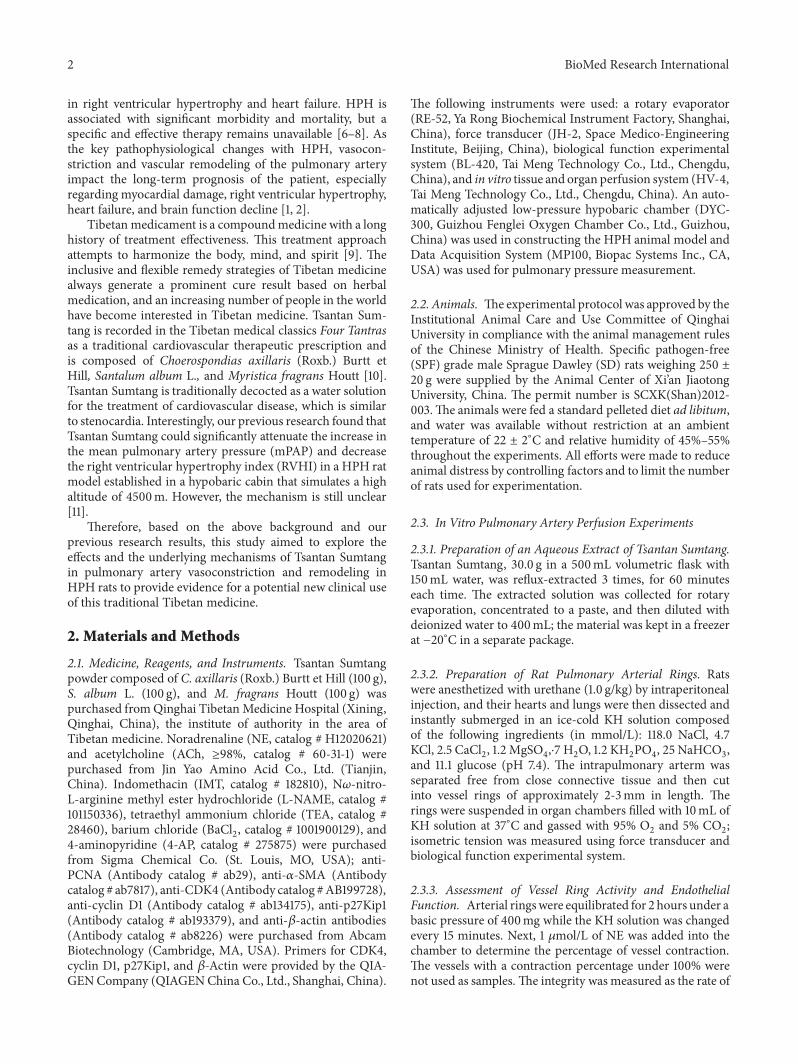
Figure 2:The influences of K+ channels inhibitors onTsantan Sumtang-induced ratpulmonary artery vasorelaxation (n=6).Ctr: controlgroup administered with Tsantan Sumtang but without the inhibitors. SW: Tsantan Sumtang. TEA (large-conductance Ca2+-activated K+channel inhibitor, 1mmol/L), BaCl
2(inward rectifier K+ channel inhibitor,1mmol/L), and 4-AP (voltage-dependent K+ channel inhibitor,
1mmol/L) were used to test the mechanisms involved in Tsantan Sumtang-induced rat pulmonary artery vasorelaxation too. The dosagereactions with or without the inhibitors were shown as (a) and column (b) giving a more visual image for the max relaxation rates. Thevasorelaxation effect of Tsantan Sumtang was markedly blocked by all three K+ channel inhibitors, with a max reduction to 64.5% ± 0.38%,24.56% ± 0.38%, and 40.87% ± 0.40%, respectively (ΔP<0.05 compared to control group). Among them, BaCl
2and TEA expressed stronger
blocking effect than 4-AP (∗𝑃<0.05 compared to 4-AP) and BaCl2is the strongest blocker (XP<0.05 compared to TEA).
stronger inhibitory effects at all 4 dose levels, while BaCl2
performed better than TEA at the 0.9mg/mL and 1.2mg/mLlevels (P<0.05, Figures 2(a) and 2(b)). The differences in themaximum effect of all the inhibitors at the high dose of1.2mg/mL can be visualized in greater detail in Figures 1(b)and 2(b).
3.2. Effects and Mechanisms of Tsantan Sumtang on HPHRats In Vivo
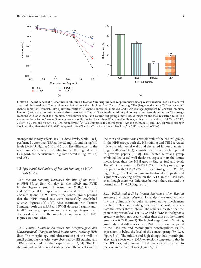
3.2.1. Tsantan Sumtang Decreased the Rise of the mPAPin HPH Model Rats. On day 28, the mPAP and RVHIin the hypoxia group increased to 32.81±3.58mmHgand 36.23±6.58%, respectively, compared with 15.89 ±2.54mmHg and 22.09±3.04% in the control group, provingthat the HPH model rats were successfully established(P<0.05, Figures 3(a)–3(c)). After treatment with TsantanSumtang, both the mPAP and RVHI decreased significantlyin all 3 dosage groups compared to the hypoxia group anddecreased greatly in the middle-dosage group (P< 0.05,Figures 3(a) and 3(b)).
3.2.2. Tsantan Sumtang Alleviated the Morphological andUltrastructural Changes in Small Pulmonary Arteries of HPHRats. The morphology and ultrastructure of the walls ofsmall pulmonary arteries were observed by HE staining andTEM, as reported in other experiments [13, 14]. The HEstaining indicated evenly distributed endothelial cells within
the thin and continuous arteriole wall of the control group.In the HPH group, both the HE staining and TEM revealedthicker arterial vessel walls and decreased lumen diameters(Figures 4(a) and 4(c)), consistent with the results reportedin previous papers [15–18]. The Tsantan Sumtang groupexhibited less vessel wall thickness, especially in the tunicamedia layer, than the HPH group (Figures 4(a) and 4(c)).The WT% increased to 43.92±2.37% in the hypoxia groupcompared with 15.15±3.97% in the control group (P<0.05,Figure 4(b)).The Tsantan Sumtang treatment groups showedsignificant alleviating effects on the WT% in the HPH rats,even though there was difference between these rats and thenormal rats (P< 0.05, Figure 4(b)).
3.2.3. PCNA and 𝛼-SMA Protein Expression after TsantanSumtang Treatment. Western blot analysis was used to iden-tify the pulmonary vascular antiproliferative mechanisminvolved in Tsantan Sumtang treatment that could substan-tiate the effects shown above. The results indicated that theprotein expression levels of PCNA and 𝛼-SMA in the hypoxiagroups were both noticeably higher than those in the controlgroups (P<0.05, Figure 5).The high-dosage Tsantan Sumtanggroup showed differences in PCNA expression comparedto the HPH rats and meaningfully downregulated PCNAexpression to below the level of the control group (P< 0.05,Figure 5(a)). The middle and high dosages had meaningfulalleviating effects on 𝛼-SMA expression compared to that inthe HPH rats, but there was still difference in comparison tothe level in the control rats (Figure 5(b)).
6 BioMed Research International
0
5
10
15
20
25
30
35
40
mPA
P (m
mH
g)
Ctr
Hyp
Hyp
+SW
1.0g
/kg
Hyp
+SW
1.25
g/kg
Hyp
+SW
1.5g
/kg
Δ∗Δ∗
Δ∗
Δ
(a)
Ctr
Hyp
Hyp
+SW
1.0g
/kg
Hyp
+SW
1.25
g/kg
Hyp
+SW
1.5g
/kg0
5
10
15
20
25
30
35
4045
RV/(
LV+S
)% R
VH
I Δ∗
Δ
Δ∗Δ∗
(b)
0.50.0
0.50.0
mPA
P (m
mH
g)
mPA
P (m
mH
g)
mPA
P (m
mH
g)
mPA
P (m
mH
g)
mPA
P (m
mH
g)
1.0 1.5 2.00
10
20
30
40
Time (second)
Ctr
0.0 0.5 1.0 1.5 2.00
10
20
30
40
Time (second)
Hyp
1.0 1.5 2.00
10
20
30
40
Time (second)
Hyp+ SW 1.0 g/kg
0.0 0.5 1.0 1.5 2.00
10
20
30
40
Time (second)
Hyp+ SW 1.25 g/kg
0.0 0.5 1.0 1.5 2.00
10
20
30
40
Time (second)
Hyp+ SW 1.5 g/kg
(c)
Figure 3:Chronic HPHmodel construction and alleviation effect caused by Tsantan Sumtang during 4 weeks (n=6). Ctr: control group,Hyp: untreated hypoxia group (rats exposed to hypoxia in hypobaric chamber, equal to the parameter in altitude 4500m), and Hyp + SW-treated groups (rats exposed to hypoxia in hypobaric chamber and received increasing doses of Tsantan Sumtang treatment, 1.0 g/kg, 1.25 g/kg,and 1.5 g/kg). (a, b) mPAP: mean pulmonary arterial pressure and RVHI: right ventricular hypertrophy index. Both mPAP and RVHI weremuch higher in Hyp group and all 3 levels of Tsantan Sumtang treatment decreased mPAP and RVHI significantly but still had differencewith the control rats (ΔP< 0.05 compared to Hyp; ∗𝑃 < 0.05 compared to Ctr). (c) Representative pictures of mPAP waves in Ctr, Hyp, andHyp+SW-treated groups.

BioMed Research International 7
Ctr Hyp Hyp+ SW 1.0 g/kg Hyp+ SW 1.25g/kg Hyp+ SW 1.5 g/kg
(a)
0
10
20
30
40
50
Δ
Δ
Δ
ΔWT
(%)
Ctr
Hyp
Hyp
+SW
1.0
g/k
g
Hyp
+SW
1.2
5 g/
kg
Hyp
+SW
1.5
g/k
g
(b)
Ctr Hyp Hyp+ SW 1.0 g/kg Hyp+ SW 1.25 g/kg Hyp+ SW 1.5 g/kg
Δ Δ
Δ
ΔΔ
(c)
Figure 4: Tsantan Sumtang alleviated chronic hypoxia-induced pulmonary vascular remodeling in 4 weeks (n=5). Ctr: control group;Hyp: untreated hypoxia group (rats exposed to hypoxia in hypobaric chamber, equal to the parameter in altitude 4500m), and Hyp+SW-treated groups (rats exposed to hypoxia in hypobaric chamber and received increasing doses of Tsantan Sumtang treatment, 1.0 g/kg, 1.25 g/kg,and 1.5 g/kg). (a) HE staining of the small pulmonary arterioles (diameters below100𝜇m, ×100). Triangle indicated that pulmonary smallvessels thickened in Hyp rats and were lessened notably by Tsantan Sumtang treatment. (b) The ratio of the vascular wall thickness to thevascular diameter (WT%) of pulmonary arterioles.WT%was increased obviously in Hyp group and alleviatedmarkedly by Tsantan Sumtangtreatment (ΔP< 0.05 compared to untreated hypoxic rats). (c) Ultrastructure of pulmonary arterioles by transmission electron microscope(TEM, ×20000) which indicated smooth muscle muscularization of the arterial vascular wall, especially in the tunica media induced bychronic hypoxia. Tsantan Sumtang treatment groups showed obvious alleviation of tunica media thickening (triangle indicated the changesof tunica media in pulmonary arterial vascular wall).
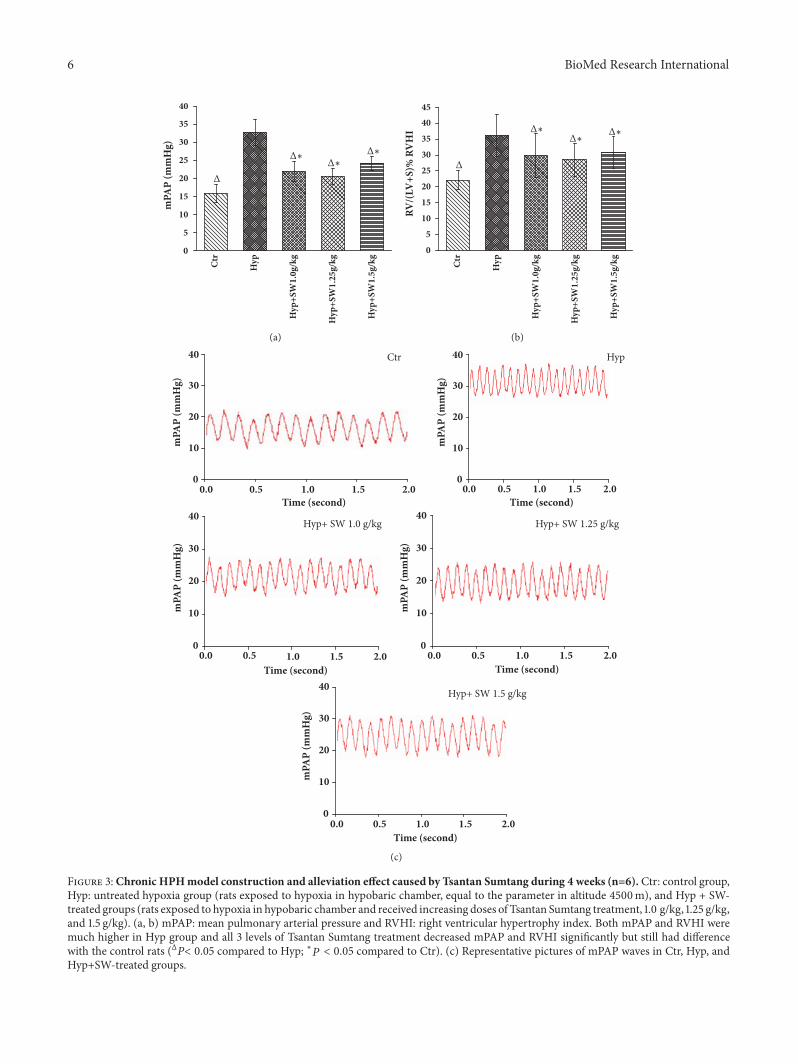
3.2.4. CyclinD1, CDK4, and p27Kip1 Protein and Gene Expres-sion Levels after Tsantan Sumtang Treatment. The Westernblot analysis showed that the cyclin D1 and CDK4 levelsin the hypoxia group were higher than those in the control
group (P< 0.05, Figures 6(a) and 6(b)). All 3 doses of TsantanSumtang significantly decreased the expression level of cyclinD1 compared to the hypoxia group, and the high dosageof medicine downregulated cyclin D1 expression to below
8 BioMed Research International
PCNA
0.0
0.1
0.2
0.3
0.4
0.5
0.6
0.7
0.8
0.9
Δ
PCN
A p
rote
in re
lativ
e exp
ress
ion
Ctr
Hyp
Hyp
+SW
1.0
g/kg
Hyp
+SW
1.2
5g/k
g
Hyp
+SW
1.5g
/kg
Ctr
Hyp
Hyp
+ SW
1.0
g/k
g
Hyp
+ SW
1.2
5g/k
g
Hyp
+ SW
1.5
g/kg
Δ∗
∗∗
-actin
(a)
Ctr
Hyp
Hyp
+SW
1.0
g/kg
Hyp
+SW
1.2
5g/k
g
Hyp
+SW
1.5g
/kg
Ctr
Hyp
Hyp
+ SW
1.0
g/k
g
Hyp
+ SW
1.2
5g/k
g
Hyp
+ SW
1.5
g/kg
0.0
0.1
0.2
0.3
0.4
0.5
0.6
0.7
0.8
0.9
Δ
Δ∗
Δ∗
∗
-actin
-S
MA
pro
tein
rela
tive e
xpress
ion
-SMA
(b)
Figure 5: Effects of Tsantan Sumtang on PCNA and 𝛼-SMA protein expression in lung tissue of HPHrats (n=5). Ctr: control group; Hyp:untreated hypoxia group (rats exposed to hypoxia in hypobaric chamber, equal to the parameter in altitude 4500m), and Hyp+SW-treatedgroups (rats exposed to hypoxia in hypobaric chamber and received increasing doses of Tsantan Sumtang treatment, 1.0 g/kg, 1.25 g/kg, and1.5 g/kg). Upper panel: relative expression levels of PCNA(a) and𝛼-SMA (b) protein (optical density of PCNAand𝛼-SMAnormalized against𝛽-actin expression). Lower panel: 𝛽-actin protein expression was used as control. (a) PCNA expression was significantly higher in Hyp groupand Tsantan Sumtang treatment of 1.5 g/kg significantly reduced protein expression of PCNA in HPH rats, even below the control rats (ΔP<0.05 compared to Hyp; ∗𝑃 < 0.05 compared to Ctr); (b) 𝛼-SMA expression was higher in Hyp group and Tsantan Sumtang treatment of1.25 g/kg and 1.5 g/kg significantly downregulated protein expression of 𝛼-SMA in HPH rats but still higher than the control rats (ΔP< 0.05compared to Hyp; ∗𝑃 < 0.05 compared to Ctr).
the control group level. CDK4 expression was inhibited inthe middle and high dosage groups (P< 0.05, Figure 6(b)),and expression in the high dosage group was not differentfrom that in the control group (Figure 6(b)). In addition,the p27Kip1 levels in the hypoxia and low dosage groupswere observably lower than that in the control group (P<0.05, Figure 7). With the administration of Tsantan Sumtang,the middle and high dosage groups showed meaningfuldifferences compared to the hypoxia rats, and there was nodifference compared with the control rats, implying that these2 dosages of Tsantan Sumtang could increase the p27Kip1expression to a nearly normal level (Figure 7).
In addition, the mRNA expression detected by RT-qPCRshowed that all 3 doses of Tsantan Sumtang meaningfullydownregulated CDK4 and cyclin D1 gene expression com-pared to the hypoxia group, and there was no differencecompared to the control group (Figure 8). p27Kip1 expressionwas significantly increased by the 1.25 g/kg and 1.5 g/kgTsantan Sumtang dosages, and expression with the 1.5 g/kg
dosage was not different from that of the control group(Figure 8).
4. Discussion
It is well known that chronic hypoxia causes pulmonaryvasoconstriction and pulmonary vascular cells proliferation,progressive right ventricular hypertrophy, and eventual rightheart failure, and the major pathological changes of HPHare the pulmonary vasoconstriction and remodeling resultingfrom pulmonary artery smooth muscle cell (PASMC) pro-liferation [19–23]. The pharmacological prevention of HPHhas focused on the modulation of PASMC proliferation,the activity of matrix metalloproteinases, and cell apoptosis.However, these medicines currently have limitations in clini-cal applications [6, 8].
Tsantan Sumtang, a Tibetan compound medicine, hasbeen used for cardiovascular diseases in Qinghai and theTibetan Plateau for years. The researches on effects and

BioMed Research International 9
Cyclin D1
Δ
∗Δ
Δ∗
Δ∗
-actin
Ctr
Hyp
Hyp
+ SW
1.0
g/k
g
Hyp
+ SW
1.2
5g/k
g
Hyp
+ SW
1.5
g/kg
Ctr
Hyp
Hyp
+SW
1.0
g/kg
Hyp
+SW
1.2
5g/k
g
Hyp
+SW
1.5g
/kg
0.0
0.1
0.2
0.3
0.4
0.5
0.6
0.7
0.8
0.9
Cycl
in D
1 pr
otei
n re
lativ
e exp
ress
ion
(a)
Δ Δ
CDK4
Δ∗∗
-actin
Ctr
Hyp
Hyp
+SW
1.0
g/kg
Hyp
+SW
1.2
5g/k
g
Hyp
+SW
1.5g
/kg
Ctr
Hyp
Hyp
+ SW
1.0
g/k
g
Hyp
+ SW
1.2
5g/k
g
Hyp
+ SW
1.5
g/kg
0.0
0.1
0.2
0.3
0.4
0.5
0.6
0.7
CDK
4 pr
otei
n re
lativ
e exp
ress
ion
(b)
Figure 6: Effects of Tsantan Sumtang on Cyclin D1 and CDK4 protein expression in lung tissue of HPH rats (n=5). Ctr: control group;Hyp: untreated hypoxia group (rats exposed to hypoxia in hypobaric chamber, equal to the parameter in altitude 4500m), and Hyp+SW-treated groups (rats exposed to hypoxia in hypobaric chamber and received increasing doses of Tsantan Sumtang treatment, 1.0 g/kg, 1.25 g/kg,and 1.5 g/kg). Upper panel: relative expression levels of CyclinD1 (a) andCDK4 (b) protein (optical density of CyclinD1 andCDK4normalizedagainst 𝛽-actin expression). Lower panel: 𝛽-actin protein expression was used as control. (a) Cyclin D1 expression was significantly higherin Hyp group and Tsantan Sumtang treatment of all 3 doses significantly downregulated protein expression of Cyclin D1 in HPH rats withthe best effect of high dose (ΔP< 0.05 compared to Hyp; ∗𝑃 < 0.05 compared to Ctr); (b) CDK4 expression was meaningfully higher in Hypgroup and Tsantan Sumtang treatment of 1.25 g/kg and 1.5 g/kg doses considerably downregulated protein expression of CDK4 in HPH rats,and the 1.5 g/kg Tsantan Sumtang treatment group showed best effect with no difference with the control rats on CDK4 expression (ΔP< 0.05compared to Hyp; ∗𝑃 < 0.05 compared to Ctr).
mechanismof components inTsantan Sumtang on cardiovas-culature showed that (+)-catechin, (+)-catechin-7-O-𝛽-D-glucopyranoside from C. axillaris inhibited the proliferationof K562 cells and (+)-catechin also showed antihypoxia effect[24]. And the pretreatmentwith total flavonoids ofC. axillaris(TFC) strongly improved cardiac function and obviouslyreduced heart pathologic lesion in ischemia/reperfusion(I/R) rat heart by increasing the levels of catalase, glu-tathione peroxidase, and superoxide dismutase in hearthomogenate and decreasing that of malondialdehyde level[24, 25]. Research on phenolic acids in C. axillaris showedthat gallic acid and protocatechuic acid showed obviouseffect of antioxidant, antihypoxia, scavenging free radicals,and inhibiting platelet aggregation [26]. It is reported thatmyristyl ether and elemene in M. fragrans may obviouslyslow down the ventricular activity, which is consistent withits traditional usage of heart disease [27]. Lahlou et al. foundthat intravenous injection of methyl eugenol into mice couldcause hypotension and the mechanism might be that methyleugenol acts directly on the vascular smooth muscle to
produce dilated blood vessels [28]. Our research findingsof Tsantan Sumtang on HPH showed the consistence withsome of the effects and mechanisms mentioned in the aboveresearches. Indeed, our previous research found that TsantanSumtang could significantly decrease the rise in the mPAP ofHPHmodel rats; however, the mechanism remained unclear[11]. Therefore, in this study, we evaluated the effect andmechanism of action of Tsantan Sumtang on pulmonaryvasoconstriction and vascular remodeling in HPH rats. Wefound that Tsantan Sumtang induced the vasorelaxation of ratpulmonary arteries precontracted with NE in a concentrationdependent manner in vitro while significantly reversingthe pulmonary arterial wall thickening induced by chronichypoxia in vivo. Histological observation also revealed that,with the Tsantan Sumtang treatment, there was an alterationin the dysplasia of the smooth muscle layer in the pulmonaryarterioles.
Our previous and current study both found that TsantanSumtang could significantly decrease the rise in the mPAPof HPH model rats. Additionally, in this study, pulmonary

10 BioMed Research International
0.0
0.1
0.2
0.3
0.4
0.5
0.6
0.7
0.8
0.9
ΔΔΔ
p27K
ip1 pr
otei
n re
lativ
e exp
ress
ion
∗ ∗
Ctr
Hyp
Hyp
+SW
1.0
g/kg
Hyp
+SW
1.2
5g/k
g
Hyp
+SW
1.5g
/kg
Ctr
Hyp
Hyp
+SW
1.0
g/kg
Hyp
+SW
1.2
5g/k
g
Hyp
+SW
1.5g
/kg
p27Kip1
-actin
Figure 7: Effects of Tsantan Sumtang on p27Kip1 protein expres-sion in lung tissue of HPH rats (n=5). Ctr: control group; Hyp:untreated hypoxia group (rats exposed to hypoxia in hypobaricchamber, equal to the parameter in altitude 4500m), and Hyp+SW-treated groups (rats exposed to hypoxia in hypobaric chamber andreceived increasing doses of Tsantan Sumtang treatment, 1.0 g/kg,1.25 g/kg, and 1.5 g/kg). Upper panel: relative expression levels ofp27Kip1 protein (optical density of p27Kip1 normalized against 𝛽-actin expression). Lower panel: 𝛽-actin protein expression was usedas control. p27Kip1 expression was notably decreased in Hyp groupand low dose of Tsantan Sumtang treatment rats, but the 1.25 g/kgand 1.5 g/kg of Tsantan Sumtang administration significantly ele-vated p27Kip1 protein expression and had no difference with thecontrol rats (ΔP< 0.05 compared to Hyp; ∗𝑃 < 0.05 compared toCtr).
artery ring perfusion in vitro, which simulated the vasocon-striction in HPH, proved the vasorelaxation effect of TsantanSumtang. Different inhibitors were used to clarify the mech-anisms involved. It has been suggested that the NO-cGMPand PGI2-cAMP pathways are the major ways to adjustendothelium-dependent vascular relaxation [12]. For furtherinvestigation, we used 100 𝜇mol/L L-NAME, an inhibitor ofNO integration into endothelial cells. The data showed thatL-NAME considerably diminished the vasodilation resultingfrom Tsantan Sumtang treatment at the 0.9 and 1.2mg/mLlevels, signifying that NO plays a role in the vasorelaxationinduced by Tsantan Sumtang, and themedicine improved theproduction of NO fromL-arginine by endothelial nitric oxidesynthase (eNOS) in the vascular endothelium and stimulated
guanylate cyclase, thereby catalyzing the transformation ofGTP to cGMP and reducing the internal Ca2+ flow throughcGMP-dependent protein kinase [12]. Thus, the pulmonaryarteries were dilated by increasing the intake of Ca2+ inthe sarcoplasmic reticulum by Ca2+-ATPase or by directlyleading to contractile protein dephosphorylation [29]. Fur-thermore, IMT, an inhibitor of cyclooxygenase (COX, a keyPGI2 synthetase), had an even stronger influence at the sameconcentration levels, indicating that PGI2 was more likely tobe involved in the vasorelaxation by Tsantan Sumtang andto play a more important role. This result also indicated thatTsantan Sumtang enhanced cAMP production through theeffect on smooth muscle cells by means of PGI2 in vascularendothelial cells, inducing pulmonary vessel dilation throughthe PGI2-cAMP pathway. An interesting phenomenon wasthat both the vasorelaxation effect of Tsantan Sumtang andthe inhibitory influences of 4-AP and IMT reached theirmaximum at a dose of 1.2mg/mL compared to the other3 doses from 0.3 to 0.9mg/mL, which demonstrated themedicine performed its strongest vasodilation effect at itshighest dose in vitro. Our previous drug safety evaluationresearch showed no evidence of toxicity, even in rats treatedwith 150 times the regular adult dosage for 90 days [11]. Thisresult may give some guidance for the future use of TsantanSumtang in HPH patients.
It is generally accepted that an increase in intracellularCa2+ initiates the contraction and proliferation of PASMCsand increases pulmonary vessel resistance; K+ channelsregulate pulmonary vessel tone by cytoplasmic K+ and Ca2+concentration and membrane potential [12]. The openingof K+ channels prompts the cell membrane to becomehyperpolarized, resulting in the closing of L-type calciumchannels and the decline of intracellular Ca2+ [30, 31]. Ourresearch showed that BaCl
2(an inhibitor of KIR channels),
4-AP (an inhibitor of Kv channels), and TEA (an inhibitorof BKCa channels) extraordinarily diminished the vasore-laxation effect of Tsantan Sumtang at all levels from 0.3 to1.2mg/mL, illustrating that the KIR, Kv, and BKCa channelswere all engaged in such influence. In addition, the resultshowed that Tsantan Sumtang-induced vasorelaxation wasconsiderably weaker at all 4 doses after exposure to BaCl2 andTEA compared to 4-AP (all at 1mmol/L), and BaCl
2demon-
strated strongest inhibitory effect at the 0.9 and 1.2mg/mLlevels. BKCa channels, with seven transmembrane domainsand a calcium-binding zone (bowl), are fairly structurallydistinctive, even among the KCa channel family [32–36].BKCa channels have also been suggested as probable remedialtargets for circulatory diseases based on their ability to adjustvascular tension [21, 33, 36].
Based on the above results, the PGI2-cAMP path-way, NO-cGMP pathway, and the opening of K+ chan-nels (inward rectifier K+ channels, large conductance Ca2+-activated K+ channels, and voltage-dependent K+ channels)seemed to be the main factors in Tsantan Sumtang-inducedconcentration-dependent pulmonary artery vasorelaxation.For monomer vascular dilators, acetylcholine shows weakermaximum vasorelaxation influence via the NO-cGMP path-way, but echinacoside is a very similar dilator at its highest

BioMed Research International 11
0.0
0.5
1.0
1.5
2.0Fo
ld ch
ange
of m
RNA
CDK4
Ctr
Hyp
Hyp
+SW
1.0
g/kg
Hyp
+SW
1.2
5g/k
g
Hyp
+SW
1.5g
/kg
Δ Δ Δ
∗
(a)
0.0
0.5
1.0
1.5
2.0
Fold
chan
ge of m
RNA
Cyclin D1
Ctr
Hyp
Hyp
+SW
1.0
g/kg
Hyp
+SW
1.2
5g/k
g
Hyp
+SW
1.5g
/kg
Δ ΔΔ
∗
(b)
0.0
0.5
1.0
1.5
Fold
chan
ge of m
RNA
p27Kip1
Ctr
Hyp
Hyp
+SW
1.0
g/kg
Hyp
+SW
1.2
5g/k
g
Hyp
+SW
1.5g
/kg
Δ
Δ∗
Δ∗
∗
(c)
Figure 8:Effects ofTsantan Sumtang onCDK4,CyclinD1, and p27Kip1mRNAexpressionby qRT-PCR in lung tissue ofHPHrats (n=5).Ctr: control group; Hyp: untreated hypoxia group (rats exposed to hypoxia in hypobaric chamber, equal to the parameter in altitude 4500m),and Hyp+SW-treated groups (rats exposed to hypoxia in hypobaric chamber and received increasing doses of Tsantan Sumtang treatment,1.0 g/kg, 1.25 g/kg, and 1.5 g/kg). 𝛽-actin gene expression was used as a control. Relative mRNA expression levels of CDK4, Cyclin D1, andp27Kip1 were shown. All 3 doses of Tsantan Sumtang meaningfully downregulated the CDK4 and Cyclin D1 gene expression compared tohypoxia groups and had no difference compared to control groups. Meanwhile, the p27Kip1 was increased significantly by Tsantan Sumtang,and the 1.5 g/kg dose showed no difference with the control group (ΔP< 0.05 versus untreated hypoxic rats; ∗𝑃 < 0.05 compared to Ctr).
concentration through the opening of NO-cGMP-PKG-BKCa channels [37, 38]. According to these reports, bothTsantan Sumtang and monomers exhibited concentration-dependent vasorelaxation effects and reached their max-imum rates at one specific dose, while as a compoundmedicine, the role of Tsantan Sumtang in vasorelaxationoccurs through a combination of more pathways than thatof monomers; however, further research is still needed[37, 38].
We successfully developed the animal model of HPHby using an automatically adjusted low-pressure hypobaricchamber. The mPAP and RVHI of the HPH model rats hadan increased average of 16.92mmHg and 14.14%, respectively,compared with the control group (P< 0.05, Figures 3(a) and3(b)). With the treatment of Tsantan Sumtang, both themPAP and RVHI decreased significantly in comparison withthe hypoxia group (P< 0.05, Figures 3(a) and 3(b)). However,as HPH is defined by mPAP ≥ 25mmHg at rest and rightventricular hypertrophy is a compensated outcome from thesustained increase of the mPAP in HPH, we propose that themedicine significantly decreased the mPAP while having aprotective influence regarding right ventricular hypertrophy[39, 40].
Regarding the morphometric experiments in vivo, thethickening and ultrastructural change of the pulmonaryartery wall proved the successful establishment of the HPHmodel rats (Figure 4). Histological observations revealed thatthe treatment with Tsantan Sumtang greatly corrected hyper-plasia of the smoothmuscle layer in the pulmonary arterioles,and the WT%, as the percentage of vascular wall thickness,was obviously decreased compared to the HPH model rats(P< 0.05, Figure 4). As the above results showed that TsantanSumtang significantly improved HPH, we further explored
the proliferation related protein expression levels of PCNAand 𝛼-SMA by Western Blot analysis.
PCNA, one of the molecular markers of cell proliferationthat is mainly detected in the S phase of the cell cycle, is anuclear protein and DNA polymerase cofactor, and restingcells hardly express PCNA [41, 42]. As a subtype of musclefibrocytes and a molecular marker of activated myofibrob-lasts, the presence of𝛼-SMA indicates the transition of cells tothe myofibroblast phenotype [43]. Compared to the controlgroup, the high expression levels of PCNA and 𝛼-SMAshown by the Western blot analysis indicated that PASMCproliferation occurred in the HPH model rats. The resultsalso revealed that Tsantan Sumtang significantly reduced thehypoxia-induced high protein expression levels of PCNAand 𝛼-SMA in the pulmonary artery (P< 0.05, Figure 5),which was in accord with the morphological results shownby HE staining and TEM, especially the alleviation of thetunica media thickening in the pulmonary arterial vascularwall as well as the decreased WT% data previously discussed(Figure 4).
To elucidate the antiproliferative mechanism of TsantanSumtang observed in Figures 4 and 5, the protein and geneexpression of CDK4, cyclin D1, and p27Kip1 were detectedby Western Blot analysis and qRT-PCR, respectively. CDK4and cyclin D1 are key regulators in the switch from the G
0/G1
phase to the S phase during cell proliferation. As cyclin-Cdkcomplexes, these regulators activate important transcriptionfactors in cell cycle progression. p27Kip1, as the key CDKinhibitor, plays major roles in cell-cycle regulation. p27Kip1binds cyclin-Cdkmultiplexes and inhibits the hyperphospho-rylation of the retinoblastoma protein, causes G
1blockage,
and limits cell proliferation [44, 45]. This result is in accor-dance with other studies and shows that p27Kip1 acted as a
12 BioMed Research International
negative regulator of cell proliferation by downregulating theexpression of CDK4 and cyclin D1 [16–18, 45, 46]. Comparedwith the hypoxia group, the CDK4 and cyclin D1 levels in theTsantan Sumtang groupswere distinctly decreased.The aboveresults combined to illustrate the underlying mechanismsmight be involved in the antiproliferation and antiremodelingeffect of Tsantan Sumtang on pulmonary arterioles (Figures5, 6, and 7).
Our study found that the opening of K+ channels wasgreatly involved in the dilation induced by the medicine andthat the decreased p27Kip1 expression in the hypoxia groupwas elevated with Tsantan Sumtang administration, whichmight demonstrate the underlying mechanisms involved inthe effect of Tsantan Sumtang against HPH. Accordingly, wemay presume that Tsantan Sumtang significantly improvedHPH in the rats by reducing hypoxic pulmonary vaso-constriction and diminishing PASMC proliferation throughdecreasing the expression levels of CDK4 and cyclin D1 withthe elevation of their negative regulator p27Kip1.
5. Conclusions
This study explored new effects and the underlying mech-anisms of Tsantan Sumtang on HPH. Our research showedthat Tsantan Sumtang could be a potential preventative med-ication for HPH, in addition to its traditional cardiovascularusage. We found that the administration of Tsantan Sumtangprevented HPH based on pulmonary artery vasorelaxationand the reversal of proliferation. The mechanism involvedin pulmonary artery vasorelaxation might be related to thePGI2-cAMP pathway, NO-cGMP pathway, and the openingof K+ channels (inward rectifier K+ channels, large conduc-tance Ca2+-activated K+ channels, and voltage-dependentK+ channels). Tsantan Sumtang also alleviated pulmonaryvascular cells proliferation in HPH rats by suppressingcyclin D1 and CDK4 expression through the inhibition ofp27Kip1 degradation. Further research of effects and thecorresponding mechanisms of Tsantan Sumtang related toHPH is needed and will be ongoing.
Data Availability
The research is funded by the National Natural Science Foun-dation of China (No. 81660308), www.nsfc.gov.cn, and Min-istry of Human Resources and Social Security of China (No.386,522), www.mohrss.gov.cn, and still has closely relatedexperiments ongoing, so the data are not available right nowbut could be reached by contacting the corresponding authorDianxiang Lu at [email protected] after 2021.
Conflicts of Interest
The authors declare that they have no conflicts of interest.
Acknowledgments
This study was supported by National Natural Science Foun-dation of China (No. 81660308), http://www.nsfc.gov.cn, and
Ministry of Human Resources and Social Security of China(No. 386,522), http://www.mohrss.gov.cn.The authors wouldlike to thank JinGuoen, YangQuanyu, andGaQin for helpingto operate the hypobaric chamber in this study.
References
[1] K. M. Chin and L. J. Rubin, “Pulmonary arterial hypertension,”Journal of the American College of Cardiology, vol. 51, no. 16, pp.1527–1538, 2008.
[2] P. Crosswhite and Z. Sun, “Molecular mechanisms of pul-monary arterial remodeling,” Molecular Medicine, vol. 20, no.1, pp. 191–201, 2014.
[3] R. Naeije and C. Dedobbeleer, “Pulmonary hypertension andthe right ventricle in hypoxia,” Experimental Physiology, vol. 98,no. 8, pp. 1247–1256, 2013.
[4] R. Naeije and R. Vanderpool, “Pulmonary hypertension andchronic mountain sickness,” High Altitude Medicine & Biology,vol. 14, no. 2, pp. 117–125, 2013.
[5] D. Penaloza and J. Arias-Stella, “The heart and pulmonarycirculation at high altitudes: healthy highlanders and chronicmountain sickness,” Circulation, vol. 115, no. 9, pp. 1132–1146,2007.
[6] E. Cahill, S. C. Rowan, M. Sands et al., “The pathophysiologicalbasis of chronic hypoxic pulmonary hypertension in themouse:Vasoconstrictor and structuralmechanisms contribute equally,”Experimental Physiology, vol. 97, no. 6, pp. 796–806, 2012.
[7] G. Simonneau, M. Gatzoulis, and I. Adatia, “Updated clinicalclassification of pulmonary hypertension,” Journal of the Amer-ican College of Cardiology, vol. 62, no. 25, supplement, 2013.
[8] T. F. Whayne, “Cardiovascular medicine at high altitude,”Angiology, vol. 65, no. 6, pp. 459–472, 2014.
[9] P. Roberti di Sarsina, L. Ottaviani, and J. Mella, “Tibetanmedicine: a unique heritage of person-centered medicine,”EPMA Journal, vol. 2, no. 4, pp. 385–389, 2011.
[10] W.-Z. Luo, Q.-E. Li, J. Chen et al., “Textual research for Tibetanmedicine Qumazi,” China journal of Chinese materia medica,vol. 40, no. 10, pp. 2047–2049, 2015.
[11] M. Yang, S. Zhang, S. Ren et al., “Effect of aqueous extractof Sanweitanxiang powder on calcium homeostasis proteinexpression in ischemic-reperfusion injury rat heart,” Journal ofTraditional Chinese Medicine, vol. 33, no. 3, pp. 355–360, 2013.
[12] M. Shen, L. Zhao, R.-X. Wu, S.-Q. Yue, and J.-M. Pei, “Thevasorelaxing effect of resveratrol on abdominal aorta from ratsand its underlying mechanisms,” Vascular Pharmacology, vol.58, no. 1-2, pp. 64–70, 2013.
[13] F. Dierick, T. Hery, B. Hoareau-Coudert et al., “Resident PW1+progenitor cells participate in vascular remodeling duringpulmonary arterial hypertension,”Circulation Research, vol. 118,no. 5, pp. 822–833, 2016.
[14] R. L. van Loon, B. Bartelds, F. A.Wagener et al., “Erythropoietinattenuates pulmonary vascular remodeling in experimentalpulmonary arterial hypertension through interplay betweenendothelial progenitor cells and heme oxygenase,” Frontiers inPediatrics, vol. 3, no. 71, 2015.
[15] B. Colleoni, S. Paternot, J. M. Pita et al., “JNKs function asCDK4-activating kinases by phosphorylating CDK4 and p21,”Oncogene, vol. 36, no. 30, pp. 4349–4361, 2017.
[16] H. Hino, P. Dai, T. Yoshida et al., “Interaction of Cx43 withHsc70 regulates G1/S transition through CDK inhibitor p27,”Scientific Reports, vol. 5, 2015.

BioMed Research International 13
[17] S. Orlando, E. Gallastegui, A. Besson et al., “P27Kip1 andp21Cip1 collaborate in the regulation of transcription by recruit-ing cyclin-Cdk complexes on the promoters of target genes,”Nucleic Acids Research, vol. 43, no. 14, pp. 6860–6873, 2015.
[18] P. Patel, B. Asbach, E. Shteyn et al., “ Brk/Protein TyrosineKinase 6 Phosphorylates p27 ,” Molecular and Cellular Biology,vol. 35, no. 9, pp. 1506–1522, 2015.
[19] N. D. Detweiler, L. Song, S. J. McClenahan et al., “BK channelsin rat and human pulmonary smooth muscle cells are BK,”Pulmonary Circulation, vol. 6, no. 4, pp. 563–575, 2017.
[20] A. Modgil, L. Guo, S. T. O’Rourke, and C. Sun, “Apelin-13 inhibits large-conductance Ca2+-Activated Kchannels incerebral artery smooth muscle cells via a PI3-Kinase dependentmechanism,” PLoS ONE, vol. 8, no. 12, 2013.
[21] M. Revermann, S. Neofitidou, T. Kirschning, M. Schloss, R. P.Brandes, and C. Hofstetter, “Inhalation of the BKCa -openerNS1619 attenuates right ventricular pressure and improvesoxygenation in the rat monocrotaline model of pulmonaryhypertension,” PLoS ONE, vol. 9, no. 1, 2014.
[22] X.Wang, T. Zhao, S. Zhou, L. Sun, L. Zhang, andG. Yu, “Mg2+-dependent modulation of BKCa channels by genistein in ratarteriolar smooth muscle cells,” Journal of Cellular Physiology,vol. 229, no. 12, pp. 1981–1989, 2014.
[23] J. Yan, R. Chen, P. Liu, and Y. Gu, “Docosahexaenoic acid atten-uates hypoxic pulmonary vasoconstriction by activating thelarge conductance Ca2+-activated K+ currents in pulmonaryartery smooth muscle cells,” Pulmonary Pharmacology andTherapeutics, vol. 28, no. 1, pp. 9–16, 2014.
[24] C.-W. Li and C.-B. Cui, “One new and nine known flavonoidsfrom Choerospondias axillaries and their In Vitro antitumor,anti-hypoxia and antibacterial activities,”Molecules, vol. 19, no.12, pp. 21363–21377, 2014.
[25] C. Li, J. He, Y. Gao, Y. Xing, J. Hou, and J. Tian, “Pre-ventive effect of total flavones of Choerospondias axillar-ies on ischemia/reperfusion-induced myocardial infarction-related MAPK signaling pathway,” Cardiovascular Toxicology,vol. 14, no. 2, pp. 145–152, 2014.
[26] J. M. Gee and I. T. Johnson, “Polyphenolic compounds: Interac-tions with the gut and implications for human health,” CurrentMedicinal Chemistry, vol. 8, no. 11, pp. 1245–1255, 2001.
[27] D. A. Kalbhen, “Nutmeg as a narcotic. a contribution to thechemistry and pharmacology of nutmeg myristica fragrans,”Angewandte Chemie International Edition, vol. 10, no. 6, pp.370–374, 2010.
[28] S. Lahlou, A. F. Figueiredo, P. J. C. Magalhaes, J. H.Leal-Cardoso, and P. D. Gloria, “Cardiovascular effects ofmethyleugenol, a natural constituent of many plant essentialoils, in normotensive rats,” Life Sciences, vol. 74, no. 19, pp. 2401–2412, 2004.
[29] L. d. Mendes-Junior, D. D. Guimaraes, D. D. Gadelha etal., “The new nitric oxide donor cyclohexane nitrate inducesvasorelaxation, hypotension, and antihypertensive effects viaNO/cGMP/PKG pathway,” Frontiers in Physiology, vol. 6, 2015.
[30] Y. Deng, E. S. K. Ng, Y. W. Kwan et al., “Cerebral vasodilatorproperties of Danshen and Gegen: A study of their combinedefficacy and mechanisms of actions,” Phytomedicine, vol. 21, no.4, pp. 391–399, 2014.
[31] Z. Qu, J. Zhang, W. Gao et al., “Vasorelaxant effects of Cerebral-care Granule� are mediated by NO/cGMP pathway, potassiumchannel opening and calcium channel blockade in isolated ratthoracic aorta,” Journal of Ethnopharmacology, vol. 155, no. 1, pp.572–579, 2014.
[32] H. C. Szappanos, H. Viola, and L. C. Hool, “L-type calciumchannel: Clarifying the “oxygen sensing hypothesis”,”The Inter-national Journal of Biochemistry & Cell Biology, vol. 86, pp. 32–36, 2017.
[33] K. J. Dunham-Snary, D. Wu, E. A. Sykes et al., “Hypoxicpulmonary vasoconstriction: from molecular mechanisms tomedicine,” CHEST, vol. 151, no. 1, pp. 181–192, 2017.
[34] Y. Hayabuchi, “The action of smooth muscle cell potassiumchannels in the pathology of pulmonary arterial hypertension,”Pediatric Cardiology, vol. 38, no. 1, 2017.
[35] D. Johar, “Cytoskeletal remodeling and regulation of cell fatein the hypertensive neonatal pulmonary artery in response tostress,” Journal of Cellular Physiology, vol. 233, no. 3, pp. 2146–2161, 2018.
[36] M. A. Lyle, J. P. Davis, and F. V. Brozovich, “Regulation ofpulmonary vascular smooth muscle contractility in pulmonaryarterial hypertension: Implications for therapy,” Frontiers inPhysiology, vol. 8, 2017.
[37] H. Christou,H.Hudalla, Z.Michael et al., “Impaired pulmonaryarterial vasoconstriction and nitric oxide–Mediated relaxationunderlie severe pulmonary hypertension in the sugen-Hypoxiarat model,” The Journal of Pharmacology and ExperimentalTherapeutics, vol. 364, no. 2, pp. 258–274, 2018.
[38] X.-Y. Gai, Y.-H. Wei,W. Zhang et al., “Echinacoside induces ratpulmonary artery vasorelaxation by opening the NO-cGMP-PKG-BKCa channels and reducing intracellular Ca2+ levels,”Acta Pharmacologica Sinica, vol. 36, no. 5, pp. 587–596, 2015.
[39] C. F. Barnett, P. Alvarez, and M. H. Park, “Pulmonary ArterialHypertension: Diagnosis and Treatment,” Cardiology Clinics,vol. 34, no. 3, pp. 375–389, 2016.
[40] P. R. Forfia and T. K. Trow, “Diagnosis of pulmonary arterialhypertension,”Clinics in Chest Medicine, vol. 34, no. 4, pp. 665–681, 2013.
[41] B. Lan, E.Hayama,N.Kawaguchi, Y. Furutani, andT.Nakanishi,“Therapeutic efficacy of valproic acid in a combined monocro-taline and chronic hypoxia rat model of severe pulmonaryhypertension,” PLoS ONE, vol. 10, no. 1, 2015.
[42] S. Zhang,M.Wang, Q. Li, and P. Zhu, “MiR-101 reduces cell pro-liferation and invasion and enhances apoptosis in endometrialcancer via regulating PI3K/Akt/mTOR,” Cancer Biomarkers,vol. 21, no. 1, pp. 189–196, 2017.
[43] S. R. Boser, T. Mauad, B. B. Araujo-Paulino et al., “Myofibrob-lasts are increased in the lung parenchyma in asthma,” PLoSONE, vol. 12, no. 8, p. e0182378, 2017.
[44] B. W. Fouty, B. Grimison, K. A. Fagan et al., “p27Kip1 isimportant in modulating pulmonary artery smooth musclecell proliferation,” American Journal of Respiratory Cell andMolecular Biology, vol. 25, no. 5, pp. 652–658, 2001.
[45] R. Ke, L. Liu, Y. Zhu et al., “Knockdown of AMPK𝛼2 pro-motes pulmonary arterial smooth muscle cells proliferation viamTOR/Skp2/p27Kip1 signaling pathway,” International Journalof Molecular Sciences, vol. 17, no. 6, p. 844, 2016.
[46] Y. Luo, D.-Q. Xu, H.-Y. Dong et al., “Tanshinone IIA inhibitshypoxia-induced pulmonary artery smooth muscle cell prolif-eration via Akt/Skp2/p27-associated pathway,” PLoS ONE, vol.8, no. 2, 2013.

Medicinal ChemistryInternational Journal of
Hindawiwww.hindawi.com Volume 2018
ToxicologyJournal of
Hindawiwww.hindawi.com Volume 2018
PainResearch and TreatmentHindawiwww.hindawi.com Volume 2018
Hindawiwww.hindawi.com Volume 2018
Arthritis
Neurology Research International
Hindawiwww.hindawi.com Volume 2018
StrokeResearch and TreatmentHindawiwww.hindawi.com Volume 2018
Drug DeliveryJournal of
Hindawiwww.hindawi.com Volume 2018
Hindawiwww.hindawi.com Volume 2018
Advances in Pharmacological Sciences
Tropical MedicineJournal of
Hindawiwww.hindawi.com Volume 2018
AddictionJournal of
Hindawiwww.hindawi.com Volume 2018
Hindawiwww.hindawi.com Volume 2018
BioMed Research International
Emergency Medicine InternationalHindawiwww.hindawi.com Volume 2018
Hindawiwww.hindawi.com Volume 2018
Anesthesiology Research and Practice
Journal of
Hindawiwww.hindawi.com Volume 2018
Pharmaceutics
Hindawi Publishing Corporation http://www.hindawi.com Volume 2013Hindawiwww.hindawi.com
The Scientific World Journal
Volume 2018
Infectious Diseases and Medical Microbiology
Hindawiwww.hindawi.com Volume 2018
Canadian Journal of
Hindawiwww.hindawi.com Volume 2018
Autoimmune DiseasesScienti�ca
Hindawiwww.hindawi.com Volume 2018
Hindawiwww.hindawi.com Volume 2018
MEDIATORSINFLAMMATION
of
Submit your manuscripts atwww.hindawi.com