Prevalência de Trichomonas vaginalis em uma população de ...
Upload
truongdienCategory
view
234download
0
Research ArticleTrichomonas vaginalis Transports VirulentMycoplasmahominis and Transmits the Infection to Human Cells afterMetronidazole Treatment: A Potential Role in Bacterial Invasionof Fetal Membranes and Amniotic Fluid
Tran Thi Trung Thu,1,2 ValentinaMargarita ,2 Anna Rita Cocco,2
AlessandraMarongiu ,2 Daniele Dessì ,2 Paola Rappelli ,2 and Pier Luigi Fiori 2
1Department of Biochemistry, Hue University of Medicine and Pharmacy, Vietnam2Department of Biomedical Sciences, University of Sassari, Italy
Correspondence should be addressed to Pier Luigi Fiori; [email protected]
Received 19 March 2018; Revised 9 July 2018; Accepted 26 July 2018; Published 2 August 2018
Academic Editor: Tamas Zakar
Copyright © 2018 Tran Thi Trung Thu et al. This is an open access article distributed under the Creative Commons AttributionLicense, which permits unrestricted use, distribution, and reproduction in any medium, provided the original work is properlycited.
Mycoplasma hominis is considered an opportunistic pathogen able to colonize the lower urogenital tract; in females the infection isassociated with severe pregnancy and postpartum complications, including abortion, endometritis, preterm delivery, and low birthweight.Molecularmechanisms of pathogenicity and virulence effectors remain poorly characterized.A number of studies in the lastdecade have demonstrated that M. hominis can establish an endosymbiotic relationship with Trichomonas vaginalis, a urogenitalparasitic protozoon, also associated with adverse pregnancy outcomes. Recently, two bacterial genes (alr and goiB) associated withamniotic cavity invasion and a single gene (goiC) associated with intra-amniotic infections and high risk of preterm delivery havebeen identified in M. hominis isolated from a group of pregnant patients. In this work we demonstrate that a high number of M.hominis intracellularly associated with T. vaginalis have goiC gene, in association with alr and goiB. In addition, we demonstratethat metronidazole treatment of M. hominis-infected T. vaginalis allows delivering viable intracellular goiC positive M. hominisfrom antibiotic-killed protozoa and that freeM. hominis can infect human cell cultures. Results suggest that molecular diagnosticstrategies to identify both pathogens and their virulence genes should be adopted to prevent severe complications during pregnancy.
1. Introduction
Preterm birth is a major cause of neonatal diseases andaccounts for 75% of perinatal mortality [1, 2]. Preterm laborand premature rupture of membranes can be initiated bymultiple mechanisms, but in most cases a precise causecannot be established. In the last years, several studies haveshown a significant and strong association between pretermbirth and intrauterine infections, accounting for at least 25-40% of cases [3, 4]. Infections induce a robust inflammatoryresponse that can stimulate uterine contractility and triggerspontaneous preterm labor [5, 6]. In most cases microorgan-isms reach the uterus and placental membranes via ascendingroute from vagina or through haematogenous spread fromdifferent sources [7].
Mycoplasma hominis is one of the microorganisms mostcommonly associated with preterm labor and it has beenisolated in 40% of amniotic fluids showing infection [8].
Recent studies have suggested the occurrence of geneticvariations among differentM. hominis isolates with regard totheir potential to invade the amniotic fluid and membranes.Three genes (alr, goiB, and goiC) have been identified inM. hominis isolated from amniotic fluids and the placentaof women with preterm labor, but not in the referencestrain PG21, isolated from human intestine [9]. The genealr encodes for alanine racemase and is involved in thepeptidoglycan synthesis, yet the function of this enzyme inMycoplasma species is not clear. goiB (gene of interest B) hasunknown functions and encodes a protein that aligns with an
HindawiJournal of PregnancyVolume 2018, Article ID 5037181, 6 pageshttps://doi.org/10.1155/2018/5037181
2 Journal of Pregnancy
Ureaplasma urealyticum hypothetical protein (41% identityand 63% similarity), while goiC (gene of interest C) encodesa 55 kDa polypeptide that appears to be strictly specific forM. hominis. Among the three genes, goiC is significantlyassociated with amniotic infection and preterm labor and canthus be considered a virulence trait of the M. hominis strainsable to infect the amniotic cavity and the placenta [9].
Interestingly, M. hominis can establish a symbiotic rela-tionship with T. vaginalis, a sexually transmitted protozoonassociated with adverse pregnancy outcomes [10].M. hominishas the ability to enter and survive in protozoan cytoplasm,where it multiplies in coordination with the eukaryotic host[11]. It has been demonstrated that the percentage of T.vaginalis infected by Mycoplasma hominis ranges from 5to 89% regardless of the geographic origin [12–15]. For arecent review see Fichorova et al. [16]. The M. hominis-T.vaginalis consortium strongly influences the pathobiologyof the protozoon and contributes to upregulating the hostinflammatory response to the infection [17–19].
The protozoon Trichomonas vaginalis has been alsolinked to pretermbirth, but in this case the infection is limitedto the vagina, without reaching the uterus and placentalmembranes [20]. The effective role of T. vaginalis in pretermlabor is debated, since the mechanisms involved are stillunclear. Data on protective effect of metronidazole treatmenton adverse pregnancy outcomes are contradictory. In somestudies antibiotic therapy seems to be effective in preventingadverse pregnancy complications, but several papers reporta failure to prevent preterm delivery by metronidazole treat-ment in pregnant women with T. vaginalis infection [21, 22].
The first objective of this work was to understand if goiC(in association with alr and goiB) is present in M. hominisable to establish intracellular symbiosis with T. vaginalisclinical isolates, thus representing an additional potential riskfactor for adverse maternal outcomes during trichomoniasis.In addition, we set up an in vitro model to assess thetransmissibility of virulent (i.e., goiC positive) M. hominisreleased from metronidazole-treated T. vaginalis to human-derived cells. Results obtained suggest an active role forprotozoa not only in transport but also in transmission ofbacterial infection to human tissues during pregnancy.
2. Material and Methods
2.1. Cells and Culture Conditions. A total of 34 T. vaginalisstrains were isolated in Italy and Mozambique from 1994 to2017. Protozoa were isolated from vaginal swabs of womenwith trichomoniasis by inoculation in Diamond’s TYMmedium (trypticase, yeast extract, maltose) supplementedwith 20% fetal bovine serum (FBS), penicillin (300 U.I/ml),and streptomycin (300mg/ml), in order to eliminate con-comitant vaginal flora [23]. Protozoa were then culturedby 1:16 daily passages in Diamond’s TYM medium withoutantibiotics at 37∘C in a 5% carbon dioxide atmosphere for atleast three weeks and stored at −80∘ until use.
The immortalized human cell line WISH (ECACC cat-alog code: 88102403) was maintained in RPMI 1640 sup-plemented with 10% FBS at 37∘C in a 5% carbon dioxideatmosphere, in T-25 flasks. Once cell cultures reached the
confluence (twice a week) adherent cellular monolayer wasenzymatically dissociated with trypsin, and detached cellswere passed, 1/10-1/20, in complete RPMI medium supple-mented with 10% fetal bovine serum.
M. hominis isolated from T. vaginalis and the bacterialreference strain PG21 were cultivated in SP4 agar plates andSP4 broth.
Minimal Inhibitory Concentration (MIC) for metron-idazole and gentamicin were calculated by incubating T.vaginalis SS-49 and M. hominis isolated from the sameprotozoan strain, with serial dilutions of drugs in liquidmedia (Diamond’s TYM and SP4 broth, respectively).
2.2. Selection of M. hominis-Parasitized T. vaginalis Isolates.The presence of M. hominis in T. vaginalis in the 34 strainsincluded in this study was assessed by specific PCR. Foreach strain, DNA was extracted from 106 mid–log-phasetrichomonad cells as previously described. DNA was thenresuspended in TE buffer (10mM Tris-HCl pH 8, 1mMEDTA) at a final concentration of 0.1 𝜇g/𝜇l and subjected to aMultiplex PCR assay for the detection of T. vaginalis and M.hominis [24].
M. hominis were isolated from each positive T. vaginalisstrain in selectivemedia. Briefly, protozoan cultureswere cen-trifuged at 350 x g, and T. vaginalis-free culture supernatantswere filtered through a 0.45-𝜇m filter membrane and finallyinoculated in SP4 agar plates. Plates were incubated at 37∘Cuntil the appearance of detectable fried egg shape colonies onthe agar surface. Bacterial DNA was extracted as described.
2.3. Identification of Virulence Genes inM. hominis Associatedwith T. vaginalis. The presence of genes alr, goiB, and goiCin Mycoplasma hominis isolated from protozoan strains wasassessed by specific PCR as described by Allen-Daniels et al.[9]. The intestinal M. hominis reference strain PG21, lackingthe alr, goiB, and goiC genes, and the T. vaginalis referencestrain G3 that is not parasitized by M. hominis, were usedas negative controls. In order to confirm PCR results, allamplicons were sequenced (BMR Genomics, Padova, Italy).
2.4. Transmissibility of M. hominis Infection from Metronid-azole-TreatedT. vaginalis toHuman-DerivedCells. The trans-missibility of intracellular M. hominis from Mycoplasma-infected T. vaginalis to human-derived cells was studied.Exponentially growing T. vaginalis SS-49, naturally infectedby M. hominis positive for alr, goiB, and goiC genes, wereextensively washed in phosphate buffered saline (PBS), inorder to eliminate nonadherent extracellular mycoplasmacells. Protozoa were then resuspended in 500𝜇l of RPMImedium supplemented with 10% FBS, added to a T-25 flask ofsemiconfluent WISH cells at 2:1 protozoa/human cells ratio,and incubated at 37∘C in a 5% CO
2atmosphere. After 30
minutes of incubation, cells were extensively washed withPBS to remove protozoa and WISH cells were trypsinized
Theeffect ofmetronidazole onMycoplasma transmissibil-ity was assessed in a second experiment: WISH cells, infectedwith T. vaginalis as described above for 30 minutes, weretreated by adding to the flask 25𝜇g/ml of metronidazole (tentimes the minimal lethal concentration for the strain SS-49,
Journal of Pregnancy 3
data not shown). After 24 hours of incubation, cells wereextensively washed in PBS to eliminate killed protozoa andcellular debris and trypsinized as described.
In order to demonstrate that M. hominis associated withT. vaginalis can invade human cells and survive intracellu-larly, the same experiment has been carried out adding afurther step. Briefly, after 24 hours of incubation of WISHcells in the presence of T. vaginalis SS-49 with metronida-zole, gentamicin was added to the cells at concentration of50𝜇g/ml (four times the minimal lethal concentration forthe M. hominis strain, data not shown) for 3 hours to killextracellular bacteria. Cultures were then extensively washedin PBS and cultured for three additional weeks in completeRPMI 1640 medium, without any drugs. In all experimentthe number of M. hominis cells associated with WISH cellswas quantified by qPCR according to the protocol describedby Ferandon et al. [25].
In all experiments, one aliquot of trypsinized cells wasinoculated into BE broth to reisolate Mycoplasma hominis,and a second aliquot was subjected to DNA extraction aspreviously mentioned. All the experiments were carried outin triplicate.
3. Results
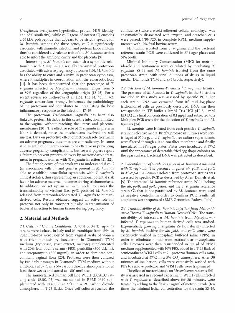
3.1. T. vaginalis Isolates Can Be Parasitized by goiC PositiveM.hominis Strains. We demonstrated by PCR that 29 out 34 T.vaginalis isolates included in this studywere stably parasitizedbyM. hominis.M. hominiswere isolated in SP4medium fromall T. vaginalis positive strains, and the presence of goiC, arl,and goiB virulence genes associated with adverse pregnancyoutcomes and preterm delivery was assessed by PCR. Among29 M. hominis tested the goiC gene is present in 17 strains(58.2%), while goiB and alr are detected, respectively, in 11(37.93%) and 28 (96.55%) isolates. The goiC gene is stronglyassociated with alr (16/17) and only partially with goiB (6/17).Only 6 out 29 (20.7%) M. hominis have all three genes. M.hominis reference strain PG21 and the T. vaginalis referencestrain G3, that is M. hominis free, do not possess alr, goiB,and goiC genes. Results are shown in Figure 1.
3.2. T. vaginalis Parasitized by Virulent M. hominis CanDeliver Bacteria Able to Infect Human Cells. We hypothesizethat the massive release of virulent intracellular M. hominisfrom metronidazole-killed T. vaginalis following antiproto-zoan therapy could mediate the infection of amniotic fluidand membranes. In order to demonstrate this hypothesis, weset up in vitro experiments coculturing a human-derived cellline WISH and T. vaginalis infected withM. hominis positivefor all virulence genes (alr, goiB, and goiC). First experimentwas carried out coincubating protozoa and WISH cells for30 minutes in absence of metronidazole. Results obtainedby qPCR show that M. hominis delivered fromMycoplasma-parasitized T. vaginalis live cells can infect less than 0.2% ofWISH cells after 30 minutes of incubation.
On the contrary, M. hominis were massively deliveredfrom metronidazole-killed protozoa after 24 hours of incu-bation and viable bacteria were able to infect human cellswith amultiplicity of infection (MOI) corresponding to 1.2M.
0
2
4
6
8
10
12
alr
goiB
goiC
alr/goiB
alr/goiC
goiB/goiC
arl/g
oiB/goiC
Figure 1: Association among alr, goiB, and goiC genes in a group of29 samples of DNA extracted by T. vaginalis.
hominis/3 WISH cells. These data suggest that free bacteriaquickly adhere to human target cells once delivered fromkilled protozoa. In all experiments, infecting M. hominiswere reisolated frommammalian cells by using mycoplasma-specific media.
A further experiment was assessed adding gentamicin inmedium cultures (gentamicin protection assay), in order toestablish if M. hominis released by T. vaginalis treated withmetronidazole can also invade and chronically survive intohuman cells. qPCR demonstrate that M. hominis can surviveintracellularly for three weeks in WISH cells exposed to gen-tamicin, with a MOI corresponding to 1 bacterium/4 humancells. Results were also confirmed by isolation of bacteriafrom gentamicin treated human cells in mycoplasma-specificmedia.
Results obtained suggest that M. hominis released byT. vaginalis after treatment with metronidazole can lead tochronic infection in fetal membranes and amniotic fluid,causing adverse pregnancy outcomes, and can also explainthe pharmacological failure in preventing adverse pregnancycomplications by use of metronidazole to treat subacutetrichomoniasis.
4. Discussion
Preterm birth is a major cause of neonatal illness anddeath, especially in developing countries. Local and systemicmicrobial infections are important causes of preterm laborand premature rupture of membranes [1, 26]. M. hominisand T. vaginalis infections are both associated with adversepregnancy outcomes. T. vaginalis limits its colonization tothe vagina [27, 28], and the infection seems to play arole in adverse pregnancy outcomes by inducing a massivelocal inflammation and the production of proinflamma-tory cytokines [29, 30]. On the contrary, M. hominis can
4 Journal of Pregnancy
invade the amniotic cavity, thus directly exploiting virulencemechanisms in this microenvironment. Even if mechanismsof pathogenicity and virulence genes involved in adversepregnancy complications associated M. hominis are not fullycharacterized, Allen-Daniels and colleagues recently identi-fied two genes (arl and goiB) in bacterial strains isolated inamniotic fluid and placental tissue and a third gene (goiC)that is significantly associated with amniotic fluid invasionand preterm labor risk [9]. Even if the effective function ofthe three genes is not clear, the authors hypothesize that goiCcould contribute to the colonization of the placenta and theamniotic fluid rather than the vagina [9].
M. hominis infection can be mediated in vivo by T.vaginalis, since the twomicroorganisms are able to establish asymbiotic relationship, andmost protozoan isolates are stablyinfected by the bacterium [10]. In a study conducted in Italy,78.6% of womenwith trichomoniasis were affected also byM.hominis [24]. So far, the presence of alr, goiB, and goiC geneshas been verified only in M. hominis isolated from patientsthat had no history of trichomoniasis. We investigated thepresence of alr, goiB, and goiC genes in M. hominis strainsthat live in symbiosis with T. vaginalis. Our data reveal thatnot only freeM. hominis but also those that live in symbiosiswith T. vaginalis can possess the three genes associated withamniotic membranes colonization and adverse pregnancyoutcomes.
The ability ofM. hominis isolates to locate intracellularlyhas been previously demonstrated by several authors, indifferent human-derived cell lines and spermatozoa [31–34]. Hopfe et al. demonstrated that mycoplasmal infectionof host cells is mediated by bacterial cytoadhesins [31]. Inaddition, Henrich et. al characterized several M. hominisgenes involved in HeLa cells intracellular infection [35].
We demonstrate that M. hominis released by T. vagi-nalis are able to infect WISH cells in vitro. However, sinceT. vaginalis is highly cytopathic and induces a massivedestruction of the cell monolayer in less than 2 hours, wehad to limit the coincubation to a very short time. Toprevent target cell lysis, we added metronidazole to the cellsafter 30 minutes of coincubation, demonstrating that M.hominis released by T. vaginalis and killed by the drug canefficiently and stably invade human cells. The quantificationby qPCR ofM. hominis associated (i.e., membrane associatedand intracellular bacteria) with human cells after 24 hoursof coincubation of metronidazole-treated T. vaginalis andWISH cells reveals that about 40% of mammalian cells areinfected by the bacteria.
Results obtained by gentamicin protection assays demon-strate the ability of M. hominis released by metronidazole-killed T. vaginalis to locate intracellularly in mammaliancells. In fact, since gentamicin kills only the extracellular M.hominis, the detection of bacteria in infected WISH afterthree weeks of cultivation in the presence of gentamicinindicates that human cells can be chronically infected byintracellular bacteria, as previously demonstrated by Hopfeby using HeLa cells [31]. These data confirm the ability ofmycoplasmas released by T. vaginalis to infect human hostcells and to locate intracellularly, suggesting a role of T.vaginalis infection in transmission of M. hominis. A similar
result has been reported by Fichorova et al.: in an elegantpaper the authors demonstrated that metronidazole-killedT. vaginalis can deliver intracellular endobiont dsRNA virus(TVV) and that free viruses, even if they are unable to directlyinfect human cells, can stimulate a massive proinflammatoryresponse: the production of cytotoxic cytokines can lead tosevere complications during pregnancy [36].
These results suggest that the role of protozoan infectionin adverse pregnancy outcome could not be limited to theinduction of vaginal inflammation. In fact, the peculiarT. vaginalis/M. hominis symbiosis represents an additionalpotential risk factor for adverse maternal outcomes andpretermdelivery during trichomoniasis.T. vaginalis can carryM. hominis possessing the alr, goiB, and goiC genes, protect-ing them intracellularly from the host immune response andantibacterial therapy, thus allowing their multiplication andtransmission. The intracellular localization of bacteria in T.vaginalis cells can explain the paradoxical results describedby some authors, reporting the failure of metronidazoletreatment of subclinical trichomoniasis to prevent pretermdelivery in pregnantwomen [21, 22]. In this scenario, the anti-T. vaginalis treatment with drugs selectively effective againsttrichomoniasis, probably together with cytolysis of protozoamediated by host immune response, could induce a massiverelease of M. hominis from killed T. vaginalis, leading tobacterial invasion of placentalmembranes and amniotic fluid.
5. Conclusions
The symbiotic consortium between T. vaginalis and M.hominis implies a role in infections during pregnancy: wecan hypothesize a primary role for T. vaginalis as “Trojanhorse”, able to transport virulent bacteria, protecting themnot only from local massive host innate and adaptive immuneresponse, but also from antimycoplasma antibiotics unable tocross the protozoan membrane. In consequence, moleculardiagnostic strategies to identify both pathogens and theirvirulence genes might be adopted to prevent severe compli-cations during pregnancy.
Data Availability
The data used to support the findings of this study areavailable from the corresponding author upon request.
Conflicts of Interest
The authors declare that they have no conflicts of interest.
Authors’ Contributions
Trung Thu Tran Thi and Valentina Margarita contributedequally to this work.
Acknowledgments
Financial support for this work was provided by MIUR(Ministero dell’Universita e della Ricerca, Italy), PRIN 2012,no. 2012WJSX8K 004, and PIA 2013, RAS.
Journal of Pregnancy 5
References
[1] R. L. Goldenberg, J. F. Culhane, J. D. Iams, and R. Romero,“Epidemiology and causes of preterm birth,” The Lancet, vol.371, no. 9606, pp. 75–84, 2008.
[2] WHO Fact sheet, “Preterm Birth,” http://www.who.int/news-room/fact-sheets/detail/preterm-birth, 19 February 2018.
[3] S. Guaschino, F. De Seta, M. Piccoli, G. Maso, and S. Alberico,“Aetiology of preterm labour: Bacterial vaginosis,” BJOG: AnInternational Journal of Obstetrics & Gynaecology, vol. 113, no.3, pp. 46–51, 2006.
[4] V. Agrawal and E. Hirsch, “Intrauterine infection and pretermlabor,” Seminars in Fetal andNeonatalMedicine, vol. 17, no. 1, pp.12–19, 2012.
[5] N. Hofer, R. Kothari, N. Morris, W. Muller, and B. Resch,“The fetal inflammatory response syndrome is a risk factor formorbidity in preterm neonates,” American Journal of Obstetrics& Gynecology, vol. 209, no. 6, pp. 542–E11, 2013.
[6] T. Cobo, M. Kacerovsky, and B. Jacobsson, “Amniotic fluidinfection, inflammation, and colonization in preterm laborwith intact membranes,” American Journal of Obstetrics &Gynecology, vol. 211, no. 6, p. 708, 2014.
[7] L. V. H. Hill, E. R. Luther, D. Young, L. Pereira, and J. A. Embil,“Prevalence of lower genital tract infections in pregnancy,”Sexually Transmitted Diseases, vol. 15, no. 1, pp. 5–10, 1988.
[8] A. P. Murtha and J. M. Edwards, “The role of Mycoplasma andUreaplasma in adverse pregnancy outcomes,” Obstetrics andGynecology Clinics of North America, vol. 41, no. 4, pp. 615–627,2014.
[9] M. J. Allen-Daniels, M. G. Serrano, L. P. Pflugner et al.,“Identification of a gene in Mycoplasma hominis associatedwith preterm birth and microbial burden in intraamnioticinfection,” American Journal of Obstetrics & Gynecology, vol.212, no. 6, pp. 779–779.e13, 2015.
[10] P. Rappelli, M. F. Addis, F. Carta, and P. L. Fiori, “Mycoplasmahominis parasitism of Trichomonas vaginalis,”The Lancet, vol.352, no. 9136, p. 1286, 1998.
[11] D. Dessı, G. Delogu, E. Emonte, M. R. Catania, P. L. Fiori, andP. Rappelli, “Long-term survival and intracellular replication ofMycoplasma hominis in Trichomonas vaginalis cells: Potentialrole of the protozoon in transmitting bacterial infection,”Infection and Immunity, vol. 73, no. 2, pp. 1180–1186, 2005.
[12] J. C. Xiao, L. F. Xie, S. L. Fang et al., “Symbiosis of Mycoplasmahominis in Trichomonas vaginalis may link metronidazoleresistance in vitro,”Parasitology Research, vol. 100, no. 1, pp. 123–130, 2006.
[13] S. E. Butler, P. Augostini, and W. E. Secor, “Mycoplasmahominis infection of Trichomonas vaginalis is not associatedwith metronidazole-resistant trichomoniasis in clinical isolatesfrom theUnited States,”Parasitology Research, vol. 107, no. 4, pp.1023–1027, 2010.
[14] J. Fraga, N. Rodrıguez, C. Fernandez et al., “Mycoplasmahominis in Cuban Trichomonas vaginalis isolates: Associationwith parasite genetic polymorphism,” Experimental Parasitol-ogy emphasizes, vol. 131, no. 3, pp. 393–398, 2012.
[15] D. da Luz Becker, O. dos Santos, A. P. Frasson, G. de VargasRigo, A. J.Macedo, and T. Tasca, “High rates of double-strandedRNA viruses and Mycoplasma hominis in Trichomonas vagi-nalis clinical isolates in South Brazil,” Infection, Genetics andEvolution, vol. 34, pp. 181–187, 2015.
[16] R. Fichorova, J. Fraga, P. Rappelli, and P. L. Fiori, “Trichomonasvaginalis infection in symbiosis with Trichomonasvirus and
Mycoplasma,” Research in Microbiology, vol. 168, no. 9-10, pp.882–891, 2017.
[17] V. Margarita, P. Rappelli, D. Dessı, G. Pintus, R. P. Hirt, andP. L. Fiori, “Symbiotic association with Mycoplasma hominiscan influence growth rate, ATP production, cytolysis andinflammatory response of Trichomonas vaginalis,” Frontiers inMicrobiology, vol. 7, 2016.
[18] P. L. Fiori, N. Diaz, A. R. Cocco, P. Rappelli, andD. Dessi, “Asso-ciation of Trichomonas vaginalis with its symbiontMycoplasmahominis synergistically upregulates the in vitro proinflam-matory response of human monocytes,” Sexually TransmittedInfections, vol. 89, no. 6, pp. 449–454, 2013.
[19] F.Mercer, F. G. I. Diala, Y.-P. Chen, B.M.Molgora, S. H. Ng, andP. J. Johnson, “Leukocyte Lysis and Cytokine Induction by theHuman Sexually Transmitted Parasite Trichomonas vaginalis,”PLOS Neglected Tropical Diseases, vol. 10, no. 8, Article IDe0004913, 2016.
[20] B. J. Silver, R. J. Guy, J. M. Kaldor, M. S. Jamil, and A.R. Rumbold, “Trichomonas vaginalis as a cause of perinatalmorbidity: A systematic review and Meta-analysis,” SexuallyTransmitted Diseases, vol. 41, no. 6, pp. 369–376, 2014.
[21] M. A. Klebanoff, J. C. Carey, J. C. Hauth et al., “Failure ofmetronidazole to prevent preterm delivery among pregnantwomen with asymptomatic Trichomonas vaginalis infection,”The New England Journal of Medicine, vol. 345, pp. 487–493,2001.
[22] S. Cauci and J. F. Culhane, “Modulation of vaginal immuneresponse among pregnant women with bacterial vaginosis byTrichomonas vaginalis, Chlamydia trachomatis, Neisseria gon-orrhoeae, and yeast,” American Journal of Obstetrics & Gynecol-ogy, vol. 196, no. 2, pp. 133–e7, 2007.
[23] L. S. Diamond, “The Establishment of Various Trichomonadsof Animals and Man in Axenic Cultures,” The Journal ofParasitology , vol. 43, no. 4, p. 488, 1957.
[24] N. Diaz, D. Dessı, S. Dessole, P. L. Fiori, and P. Rappelli, “Rapiddetection of coinfections by Trichomonas vaginalis, Myco-plasma hominis, and Ureaplasma urealyticum by a new mul-tiplex polymerase chain reaction,” Diagnostic Microbiology andInfectious Disease, vol. 67, no. 1, pp. 30–36, 2010.
[25] C. Ferandon, O. Peuchant, C. Janis et al., “Development ofa real-time PCR targeting the yidC gene for the detectionof Mycoplasma hominis and comparison with quantitativeculture,” Clinical Microbiology and Infection, vol. 17, no. 2, pp.155–159, 2011.
[26] R. L. Goldenberg, J. C.Hauth, andW.W.Andrews, “Intrauterineinfection and preterm delivery,” The New England Journal ofMedicine, vol. 342, no. 20, pp. 1500–1507, 2000.
[27] E. Mielczarek and J. Blaszkowska, “Trichomonas vaginalis:pathogenicity and potential role in human reproductive failure,”Infection, vol. 44, no. 4, pp. 447–458, 2016.
[28] M. Cunnington, C. Kortsalioudaki, and P. Heath, “Genitouri-nary pathogens and preterm birth,” Current Opinion in Infec-tious Diseases, vol. 26, no. 3, pp. 219–230, 2013.
[29] R. N. Fichorova, “Impact of T. vaginalis infection on innateimmune responses and reproductive outcome,” Journal ofReproductive Immunology, vol. 83, no. 1-2, pp. 185–189, 2009.
[30] R. N. Fichorova, O. R. Buck, H. S. Yamamoto et al., “The villainteam-up or how trichomonas vaginalis and bacterial vaginosisalter innate immunity in concert,” Sexually Transmitted Infec-tions, vol. 89, no. 6, pp. 460–466, 2013.
[31] M. Hopfe, R. Deenen, D. Degrandi, K. Kohrer, and B. Henrich,“Host Cell Responses to Persistent Mycoplasmas - Different

6 Journal of Pregnancy
Stages in Infection of HeLa Cells with Mycoplasma hominis,”PLoS ONE, vol. 8, no. 1, Article ID e54219, 2013.
[32] D. Taylor-Robinson, H. A. Davies, P. Sarathchandra, and P.M. Furr, “Intracellular location of mycoplasmas in culturedcells demonstrated by immunocytochemistry and electronmicroscopy,” International Journal of Clinical and ExperimentalPathology, vol. 72, no. 6, pp. 705–714, 1991.
[33] P. Rappelli, F. Carta, G. Delogu et al., “Mycoplasma hominisand Trichomonas vaginalis symbiosis: Multiplicity of infectionand transmissibility of M. hominis to human cells,” Archives ofMicrobiology, vol. 175, no. 1, pp. 70–74, 2001.
[34] F. J. Dıaz-Garcıa, A. P. Herrera-Mendoza, S. Giono-Cerezo,and F. M. Guerra-Infante, “Mycoplasma hominis attaches toand locates intracellularly in human spermatozoa,” HumanReproduction, vol. 21, no. 6, pp. 1591–1598, 2006.
[35] B. Henrich, F. Kretzmer, R. Deenen, K. Kohrer, andM. F. Balish,“Validation of a novel Mho microarray for a comprehensivecharacterisationof theMycoplasma hominis action inHeLa cellinfection,” PLoS ONE, vol. 12, no. 7, p. e0181383, 2017.
[36] R. N. Fichorova, Y. Lee, H. S. Yamamoto et al., “EndobiontViruses Sensed by the Human Host - Beyond ConventionalAntiparasitic Therapy,” PLoS ONE, vol. 7, no. 11, Article IDe48418, 2012.

Stem Cells International
Hindawiwww.hindawi.com Volume 2018
Hindawiwww.hindawi.com Volume 2018
MEDIATORSINFLAMMATION
of
EndocrinologyInternational Journal of
Hindawiwww.hindawi.com Volume 2018
Hindawiwww.hindawi.com Volume 2018
Disease Markers
Hindawiwww.hindawi.com Volume 2018
BioMed Research International
OncologyJournal of
Hindawiwww.hindawi.com Volume 2013
Hindawiwww.hindawi.com Volume 2018
Oxidative Medicine and Cellular Longevity
Hindawiwww.hindawi.com Volume 2018
PPAR Research
Hindawi Publishing Corporation http://www.hindawi.com Volume 2013Hindawiwww.hindawi.com
The Scientific World Journal
Volume 2018
Immunology ResearchHindawiwww.hindawi.com Volume 2018
Journal of
ObesityJournal of
Hindawiwww.hindawi.com Volume 2018
Hindawiwww.hindawi.com Volume 2018
Computational and Mathematical Methods in Medicine
Hindawiwww.hindawi.com Volume 2018
Behavioural Neurology
OphthalmologyJournal of
Hindawiwww.hindawi.com Volume 2018
Diabetes ResearchJournal of
Hindawiwww.hindawi.com Volume 2018
Hindawiwww.hindawi.com Volume 2018
Research and TreatmentAIDS
Hindawiwww.hindawi.com Volume 2018
Gastroenterology Research and Practice
Hindawiwww.hindawi.com Volume 2018
Parkinson’s Disease
Evidence-Based Complementary andAlternative Medicine
Volume 2018Hindawiwww.hindawi.com
Submit your manuscripts atwww.hindawi.com