Whiplash-associated injuries and disorders – Biomedical aspects of ...

From
Department of Public Health and Community Medicine The Sahlgrenska Academy, University of Gothenburg
Sweden
and
Southern Älvsborg hospital, Sweden
Treatment of whiplash associated disorders
by
Aris Seferiadis
Göteborg 2010
From
Department of Public Health and Community Medicine The Sahlgrenska Academy, University of Gothenburg
Sweden
and
Southern Älvsborg hospital, Sweden
Treatment of whiplash associated disorders
by
Aris Seferiadis
Göteborg 2010
From
Department of Public Health and Community Medicine The Sahlgrenska Academy, University of Gothenburg
Sweden
and
Southern Älvsborg hospital, Sweden
Treatment of whiplash associated disorders
by
Aris Seferiadis
Göteborg 2010

© Aris SeferiadisAll rights reserved. No part of this publication may be reproduced ortransmitted, in any form or by any means, without written permission.
ISBN 978-91-628-8158-0http://hdl.handle.net/2077/23128
Printed by Geson Hylte Tryck, Göteborg, Sweden 2010

Abstract Whiplash injuries seem to have a substantial impact on health. Half the affected patients have persistent pain and disability and significant costs are incurred to society, mainly due to inability to return to work. The pathophysiology of the condition is largely unknown and there has been much debate on how whiplash-associated disorders (WAD) should be treated. In this dissertation, the treatment of acute and chronic WAD has been elucidated.
• The evidence basis of many commonly used treatments for patients suffering from WAD, both in the acute and chronic state was analyzed in a systematic literature review. Twenty-six randomized controlled trials (RCT) were identified through computer-assisted search of the databases Medline (from 1962 to May 2003), CINAHL (1960 to 2003), Embase (1976 to 2003) and Psychinfo (1960 to 2003) and manual check of the reference lists of relevant studies. Based on the degrees of evidence and the practical obstacles the following treatments can be recommended: Early physical activity in acute WAD, combination of cognitive behavioral therapy with physical therapy interventions and coordination exercise therapy in chronic WAD.
• The long-term (3-year) efficacy of active intervention (early mobilization with/without McKenzie treatment) in patients with acute WAD compared with standard intervention (information broschure recommending initial rest and slow resumption of activity) and the effect of early versus delayed initiation of intervention was studied in an RCT. The active intervention was more effective in reducing pain intensity, sick leave and retaining/regaining total range of motion than the standard intervention.
• The effectiveness of 10 weeks of twice-weekly, 90-minute sessions of either Exercise Therapy (general conditioning, coordination, strengthening of deep cervical flexors, stretching and relaxation) or Basic Body Awareness Therapy (training comfortable posture and use of the body, balance and relaxation during movement) for patients with chronic WAD was compared in an RCT. Basic Body Awareness Therapy resulted in slightly better effects on the physical functioning, social functioning and bodily pain domains of SF-36 and on pain frequency compared to Exercise Therapy at three months.
• The applicability of the fear avoidance model of chronic pain (FAM) in patients with WAD and the inclusion of a measure of guarded movement in the model were studied in a cross-sectional trial. Statistically significant correlations between all measures of the FAM were found and these measures explained part of each other’s variance. Applying the FAM of chronic pain to patients suffering from chronic WAD appears valid.
Key words: whiplash-associated disorders, systematic review, randomized controlled trial, McKenzie method, fear-avoidace, chronic pain, exercise therapy, basic body awareness, Aris Seferiadis
ISBN 978-91-628-8158-0


List of publications This thesis is based on the following papers, which will be referred to in the text by their Roman numerals: I. Seferiadis A, Rosenfeld M, Gunnarsson R. A review of treatment
interventions in whiplash-associated disorders. Eur Spine J. 2004 Aug;13(5):387-97.
II. Rosenfeld M, Seferiadis A, Carlsson J, Gunnarsson R. Active intervention in
patients with whiplash-associated disorders improves long-term prognosis: a randomized controlled clinical trial. Spine (Phila Pa 1976). 2003 Nov 15;28(22):2491-8.
III. Seferiadis A, Ohlin P, Billhult A, Gunnarsson R. Basic body awareness
therapy superior to exercise therapy for patients with chronic whiplash-associated disorders: a randomized controlled clinical trial. Manuscript.
IV. Seferiadis A, Ohlin P, Billhult A, Gunnarsson R. Applying the fear-avoidance
model to patients with chronic whiplash associated disorders: a cross-sectional study. Manuscript.
The papers have been reprinted with permission of the journals

Contents 1. Abbreviations....................................................................................................6 2. Introduction ......................................................................................................7
2.1. Definition of whiplash ...............................................................................7 2.2. The pathology of whiplash ........................................................................7 2.3. Assessment and examination of WAD.......................................................8 2.4. The classification of WAD ......................................................................10 2.5. Incidence.................................................................................................11 2.6. Risk factors for developing WAD............................................................11 2.7. Prognostic factors for recovery from WAD .............................................12 2.8. Posture in WAD ......................................................................................13 2.9. Muscle impairment in WAD....................................................................14 2.10. Implications for research .....................................................................15 2.11. Aims of the dissertation .......................................................................15
2.11.1. General aims....................................................................................15 2.11.2. Specific aims ...................................................................................15
3. Methods ..........................................................................................................16 3.1. A review of treatment interventions in WAD (I) ......................................16
3.1.1. Literature search ..............................................................................16 3.1.2. Selection for quality assessment.......................................................16 3.1.3. Quality Assessment of studies..........................................................16 3.1.4. Best evidence synthesis....................................................................19 3.1.5. Statistical methods...........................................................................19
3.2. Active Intervention in Patients with acute WAD (II)................................19 3.2.1. Selection of Patients ........................................................................19 3.2.2. Randomization of Patients ...............................................................19 3.2.3. Measurements..................................................................................20 3.2.4. Active Intervention ..........................................................................20 3.2.5. Standard Intervention.......................................................................21 3.2.6. Control Group..................................................................................21 3.2.7. Statistical Analysis...........................................................................21
3.3. Basic body awareness therapy compared to exercise therapy for patients with chronic WAD (III) ......................................................................................22
3.3.1. Study site.........................................................................................22 3.3.2. Participants ......................................................................................22 3.3.3. Randomization.................................................................................22 3.3.4. Treatments.......................................................................................23 3.3.5. Exercise Therapy .............................................................................23 3.3.6. Basic Body Awareness Therapy.......................................................23 3.3.7. Outcome measures...........................................................................23 3.3.8. Statistical analysis............................................................................24
3.4. Applying the fear-avoidance model to patients with chronic WAD (IV) ..25 3.4.1. Participants ......................................................................................25 3.4.2. Measures .........................................................................................25 3.4.3. Statistical analyses ...........................................................................26
4. Results ............................................................................................................27 4.1. A review of treatment interventions in WAD (I) ......................................27 4.2. Active Intervention in Patients with WAD (II).........................................34
4.2.1. Baseline Differences ........................................................................34 4.2.2. Treatment Sessions ..........................................................................36

4.2.3. Active versus Standard Intervention.................................................36 4.2.4. The Importance of the Time Factor ..................................................41 4.2.5. No Initial Pain .................................................................................41
4.3. Basic body awareness therapy compared to exercise therapy for patients with chronic whiplash associated disorders (III) ..................................................42
4.3.1. Recruitment and follow-up of participants .......................................42 4.3.2. Baseline characteristics....................................................................42 4.3.3. Compliance with treatment ..............................................................45 4.3.4. Additional treatment ........................................................................45 4.3.5. Effectiveness of treatment................................................................45 4.3.6. Adverse events ................................................................................50
4.4. Applying the fear-avoidance model to patients with chronic whiplash associated disorders (IV).....................................................................................50
4.4.1. Participants ......................................................................................50 4.4.2. Correlations .....................................................................................51 4.4.3. Regression analyses .........................................................................52
5. Discussion.......................................................................................................54 5.1. A review of treatment interventions in WAD (I) ......................................54
5.1.1. Methodological aspects....................................................................54 5.1.2. Scientific shortcomings with some types of interventions ................54 5.1.3. Acute and chronic WAD..................................................................54 5.1.4. Treatment of acute WAD.................................................................55 5.1.5. Treatment of chronic WAD .............................................................55
5.2. Active Intervention in Patients with WAD (II).........................................55 5.2.1. Methodological Aspects...................................................................56 5.2.2. Exposed to whiplash trauma and no initial pain................................56 5.2.3. Possible Mechanisms of the Active Intervention ..............................56 5.2.4. Why Cervical Rotation?...................................................................57
5.3. Basic body awareness therapy compared to exercise therapy for patients with chronic WAD (III) ......................................................................................57 5.4. Applying the fear-avoidance model to patients with chronic WAD (IV) ..59
6. Summary and conclusions...............................................................................61 7. Acknowledgements .........................................................................................62 8. References ......................................................................................................64 Original publications...............................................................................................70

1. Abbreviations WAD Whiplash Associated Disorders QTF Quebec Task Force BJD The Bone and Joint Decade 2000-2010 RCT Randomized Controlled Trial FAM Fear-Avoidance Model MRI Magnetic Resonance Imaging EMG Electromyography ET Exercise Therapy BBAT Basic Body Awareness Therapy CCT Controlled Clinical Trial IMLB Instrument to Measure the Likelihood of Bias MAL Maastricht-Amsterdam List DL Delphi List SCM Sternocleidomastoideus Muscle AS Anterior Scalene Muscle

2. Introduction ”I was finding it very frustrating, because nobody had fixed me, and all I had was a car-accident and I should be okay by now, and I did not believe that I would have an injury that would last any length of time. I figured a couple of weeks I should be back to normal, back at work full time, no side-effects, nothing”. The above quote [1] comes from a person suffering from chronic whiplash-associated disorder (WAD). Even though the patient’s description is very personal, it does capture the essence of the problem. Anyone that has met a person with this disorder can relate to the severity and complexity of the condition. Meeting patients with these disorders has often been a frustrating experience. Many times patients reported feeling an increase of pain after treatment and my clinical examination was hindered by all palpation being painful. I was tempted to steer my professional career away from chronic pain but for the influence of a few key individuals. It was through my contact with my mentors Mark Rosenfeld and Ronny Gunnarsson that I was given inspiration to conduct my doctoral thesis on the subject.
2.1. Definition of whiplash
It may seem obvious to experts what we mean by the term whiplash, but great confusion has existed in the scientific literature. The term “whiplash” has carelessly been used to describe the mechanism of injury, the injury itself, the consequences of the injury and the assorted signs and symptoms that patients present with. The first major step in clarifying the nomenclature was made by the Quebec Task Force (QTF) published in 1995 [2]. The current definition of whiplash is the one adopted by the QTF and reads as follows:
“Whiplash is an acceleration-deceleration mechanism of energy transfer to the neck. It may result from rear-end or side-impact motor vehicle collisions, but can also occur during diving or other mishaps. The impact may result in bony or soft-tissue injuries (whiplash injury), which in turn may lead to a variety of clinical manifestations (Whiplash-Associated Disorders).”
2.2. The pathology of whiplash
Barnsley et al [3] reviewed a range of biomechanical, experimental and cadaver studies that investigated the possible mechanisms of injury to the neck. They concluded that the neck could be subjected to forced flexion, extension, lateral flexion, and shear forces in a traffic collision. Another conclusion from the same review was that the structures most likely injured are the zygapophyseal joints, intervertebral discs, and upper cervical ligaments. More recent reviews, however, have challenged the premise that WAD can be linked to injury of specific structures.
The international initiative of the Bone and Joint Decade 2000-2010 Task Force on Neck Pain and its Associated Disorders (BJD) published the results of its work in 2008 [4]. The consensus reached by the BJD is that WAD probably results from cervical sprain or strain but that the exact pathophysiology is not known. Therefore there may or may not be damage to soft-tissue, including the joints, ligaments and/or muscles in the neck, posterior shoulder and upper thoracic regions. The BJD review also concluded that there is no gold standard diagnostic test to detect WAD [5].
7

The only measurable alteration taking place after whiplash injury thus far has
been a transient immune response associated with inflammation after soft-tissue trauma. This immune-response is present within 3 days of trauma but normalizes within 14 days [6]. This trauma-related activation of the immune system in WAD is similar to that activated in other minor trauma (ankle sprain) [7].
2.3. Assessment and examination of WAD
Clinical evaluation of the musculoskeletal system includes inspection, range of motion, strength testing, palpation and additional tests. Following physical examination, radiological tests are often used to complement the diagnostic process [8, 9].
When clinicians meet patients with neck pain the first diagnostic concern will probably be to exclude underlying sinister causes of neck pain. The care setting (emergency or non emergency) is also likely to influence how assessment is conducted.
The BJD concluded that there is strong evidence suggesting that either the Canadian C-spine Rules [10] (Figure 1) or the Nexus Low-risk Criteria [11] (Table 1) are reliable to rule out the need for further imaging in adult patients at low risk of neck injury seeking emergency care [5]. Strong evidence also suggests that Computer Tomography (CT-scans) should be used instead of routine cervical spine radiographs in the evaluation of patients with traumatic high-risk neck injuries in emergency situations [5].
1. Any High-Risk Factor Which
Mandates Radiography?Age ≥ 65 years
orDangerous mechanism*orParesthesias in extremities
For alert (Glasgow Coma Scale=15) and stable trauma patients where cervical spine injury is a concern.
2. Any Low-Risk Factor Which Allows
Safe Assessment of Range of Motion?Simple rearend MVC**
orSitting position in EDorAmbulatory at any timeorDelayed onset of neck pain***orAbsence of midline c-spine tenderness
3. Able to Actively Rotate
Neck?45°left and right
No Radiography
Radiography
Rule Not Applicable If:- Non-trauma cases
- GCS < 15- Unstable vital signs- Age < 16 years- Acute paralysis- Known vertebral disease- Previous C-spine surgery
* Dangerous Mechanism:- fall from elevation ≥ 3 feet / 5 stairs- axial load to head, e.g. diving- MVC high speed (>100km/hr), rollover, ejection- motorized recreational vehicles
- bicycle struck or collision** Simple Rearend MVC Excludes:- pushed into oncoming traffic- hit by bus / large truck- rollover- hit by high speed vehicle*** Delayed:- i.e. not immediate onset of neck pain
Able
YesUnable
No
Yes
No
Figure 1 - Canadian C-spine Rules
8

Table 1 – The Nexus low risk criteria
No posterior midline cervical spine tenderness Midline posterior bony cervical-spine tenderness is present if the patient reports pain
on palpation of the posterior midline neck from the nuchal ridge to the prominence of the first thoracic vertebrae, or if the patient evinces pain with direct palpation of any cervical spinous process.
No evidence of intoxication Patients should be considered intoxicated if they either of the following: a recent
history provided by the patient or an observer of intoxication or intoxicating ingestion, or evidence of intoxication on physical examination such as an odor of alcohol, slurred speech, ataxia, dysmetria, or other cerebellar findings, or any behavior consistent with intoxication. Patients may also be considered to be intoxicated if tests of bodily secretions are positive for alcohol or drugs that affect level of alertness.
A normal level of alertness An altered level of alertness can include the following: a Glasgow Coma Scale score
of 14 or less; disorientation to person, place time, or events; an inability to remember three objects at five minutes; a delayed or inappropriate response to external stimuli; or other findings.
No focal neurological deficit and A focal neurological deficit is any focal neurological finding on motor or sensory
examination No painful distracting injuries No precise definition of painful distracting injury is possible. This category includes
any condition thought by the clinician to be producing pain sufficient to distract the patient from a second (neck) injury. Such injuries may include, but are not limited to, any longbone fracture; a visceral injury requiring surgical consultation; a large laceration, degloving injury, or crush injury; large burns; or any other injury causing acute functional impairment. Physicians may also classify any injury as distracting if it is thought to have the potential to impair the patient’s ability to appreciate other injuries.
From the perspective of assessing neck-pain in non emergency patients, the use of
“Red Flag Symptoms” to screen for sinister pathology has been strongly encouraged. Unfortunately BJD found the available evidence insufficient to confirm the utility of “Red Flag Symptoms” for triaging non emergency neck patients [5].
There is little research on the validity and utility of self-reported history in evaluating neck pain disorders [5]. Routine clinical examination is more predictive at excluding (ruling out) structural lesion or neurological compression than at diagnosing any specific etiologic condition in patients with neck pain [5]. Manual provocation testing for nerve root compromise, however, has high sensitivity and a high positive predictive value and is therefore capable of ruling in radiculopathy [5]. Inspection of the neck patient for abnormal signs (e.g. muscle atrophy, swelling, redness, scars etc) has low to moderate interexaminer reliability [5]. Range of motion is moderately reliable regardless of how it is measured (active, passive, with/without a device, clinician assessed or self-described by the patient) [5]. Palpating “trigger points” around the neck in patients with neck pain has moderate to high predictive value for neck pain with or without radiculopathy but the distribution of “trigger
9

points” was not found to discriminate between neck pain alone, neck pain and radiculopathy or neck pain and MRI disc “bulging” [5].
Beyond the physical examination, the BJD found no evidence that laboratory testing, sensory electrophysiological studies (surface, dermatomal or quantitative sensory testing) or plain radiographs provide any unique value or useful ancillary data [5]. Multiple studies have shown that neck pain without clear radiculopathy is not reasonably ascribed to specific common degenerative changes seen on MRI [5]. The degenerative changes that MRI can detect are common in asymptomatic subjects and increase significantly with age [5].
The role of MRI in the assessment of neck pain, according to BJD, is to aid clinicians in determining the site and level of neurological compression in combination with complaints of radicular symptoms in the patient interview, specific findings in the examination and possibly needle-EMG findings [5].
Other specialized techniques such as anaesthetic facet joint injections and provocative discography that aim to identify specific lesions causing neck pain were not supported by current evidence and were not recommended for routine clinical practice [5].
2.4. The classification of WAD
A classification for grading symptoms following a whiplash injury was proposed in 1995 by the QTF [2]. This classification has gained wide acceptance in the scientific community, as it is purely descriptive and free from supposed diagnoses (Table 2).
Table 2 – The Quebec classification of WAD
Grade 0 WAD refers to no neck complaints and no physical signs Grade I WAD refers to injuries involving complaints of neck pain, stiffness or
tenderness, but no physical signs Grade II WAD refers to neck complaints accompanied by decreased range of motion
and point tenderness (musculoskeletal signs). Grade III WAD refers to neck complaints accompanied by neurological signs such as
decreased or absent deep tendon reflexes, weakness and/or sensory deficits
Grade IV WAD refers to injuries in which neck complaints are accompanied by
fracture or dislocation.
Other symptoms such as deafness, dizziness, tinnitus, headache, memory loss, dysphagia and temporomandibular joint pain can be present in all grades.
It is common to exclude Grades 0 (no WAD injury) and IV (fracture/dislocation)
when studying samples of patients with WAD. Patients having spinal cord injury and bone tissue injury, such as neck fracture or dislocation are treated accordingly for those types of traumata and therefore also fall outside the scope of the research field.
The BJD concluded that once serious neck conditions have been ruled out, WAD and other neck pain do not differ [5]. Therefore they proposed using a categorization system similar to the QTF classification for neck pain [4] (Table 3). Their goal was
10

to produce a severity classification system encompassing all neck pain syndromes, and relevant irrespective of the professional background of the health care provider and the circumstances surrounding the onset of pain (traffic collisions, sports, nontrauma, etc).
Table 3 – The Bone and Joint Decade classification of neck pain
Grade I Neck pain and associated disorders with no signs or symptoms suggestive of major structural pathology and no or minor interference with activities of daily living.
Grade II No signs or symptoms of major structural pathology, but major interference
with activities of daily living. Grade III No signs or symptoms of major structural pathology, but presence of
neurologic signs such as decreased deep tendon reflexes, weakness, or sensory deficits.
Grade IV Signs or symptoms of major structural pathology.
Major structural pathologies include (but are not limited to) fracture, vertebral dislocation, injury to the spinal cord, infection, neoplasm, or systemic disease including the inflammatory arthropathies.
2.5. Incidence
The incidence of WAD varies but in North America and western Europe is considered at least 0.3% annually for all inhabitants [12]. There is consistent evidence that the incidence has increased in some western countries during the past 30 years but it is still unclear if this represents a true population increase or a change in reporting [12].
2.6. Risk factors for developing WAD
There is conflicting evidence for gender as a risk factor for seeking health care or making an insurance claim for WAD [12]. Studies with the highest methodological quality on this question all suggest that females have a slightly higher risk [12]. Neck pain is, however, more prevalent among females [13] which may confound the findings or constitute a risk factor for WAD in itself. Having neck pain before a collision may be a risk factor for WAD but this is based on only one study and must be considered preliminary evidence at this point [12]. Younger people also seem more likely to make insurance claims and/or seek treatment for WAD but the strength of this association is uncertain [12]. In Saskatchewan, Canada, the insurance system was changed from “tort” (where compensation for pain and suffering is available through litigation) to “no-fault”(where insurance benefits are increased but no compensation for pain and suffering is available). This change was studied as a population-based natural experiment and was found to be associated with fewer insurance claims for WAD [14]. This study indicates that the type of insurance system may affect the likelihood for insurance claims for WAD.
11

There is also preliminary evidence that whiplash protection devices installed in cars reduce insurance claims for WAD but these findings need to be confirmed in larger studies with control of potential confounders [12]. The BJD found no evidence on the effect of crash severity, awareness of impending collision, head position at the moment of collision or spinal degenerative changes on the onset of WAD [12].
2.7. Prognostic factors for recovery from WAD
Age and gender have long been considered relevant to recovery from WAD but findings vary in the scientific literature. The effects of age and gender on outcome are modest (twofold increase at most) in studies that do report an effect [15].
Increased initial symptom severity (greater initial pain, greater number of symptoms, pain in more parts of the body, greater pain-related limitations, higher WAD classification) has been consistently shown prognostic of poorer outcome [15].
It is difficult to assess whether collision and vehicle-related factors are associated with recovery from WAD. Preliminary findings suggest that both the presence of a tow-bar on the struck vehicle in the collision and crashes with higher levels of mean acceleration are associated with a small negative effect [15]. Nevertheless, studies adjusting for the confounding effects of initial pain and symptom severity fail to demonstrate effects of collision-related factors on recovery [15]. Researchers in the majority of such studies collect data on collision-related factors by self-reports and are therefore subject to recall bias. Studies of preinjury neck pain also present conflicting results and are susceptible to recall bias [15]. Many different psychological constructs have been evaluated as prognostic of recovery from WAD and also found to affect it [15]. The psychological constructs investigated include coping strategies, helplessness in controlling the consequences of pain, depressed mood, fear of movement/(re)injury, pain catastrophizing and initial postinjury anxiety [15]. The major limitations placed on interpreting these results spur from the lack of uniformity in the studied psychological constructs and the lack of controlling for the effects of initial symptom severity. The latter may influence the demonstrated association between psychological factors and recovery from WAD.
In a recently published study the best independent predictors for long term outcome were presence within 96 hours after injury of the two cognitive symptoms “being easily distracted” with an odds ratio for being on sick leave 2½-3 years after trauma of 8.7-50 and “easily irritated” with an odds ratio of 5.3-31 [16]. Preliminary evidence suggests that the prevailing insurance system and litigation are prognostic but this remains to be verified in other jurisdictions [15]. Finally, there is some evidence that greater health utilization in the first month after injury was associated with slower recovery [17, 18]. This finding is not necessarily translatable to individual cases since this finding is based on a
12

population-based study. It is likely that the optimal type and frequency of patient care depends on the individual patient’s characteristics.
2.8. Posture in WAD
Postural assessment and treatment have long been a part of physical therapy practice. The importance of normal upright posture has been proposed since the early 1900s [19]. Proper posture is believed to be a state of musculoskeletal balance that involves a minimal amount of stress or strain on the body [20]. The question remains as to the importance of maintaining normal postural alignment, if a link exists between postural abnormalities and neck pain and whether posture is a cause or an epiphenomenon. There are potentially several factors that can be conceptualized to affect posture such as age, sedentary lifestyle, work ergonomics, depression and lack of postural body awareness.
Griegel-Morris et al [21] conducted a study of standing posture and musculoskeletal pain (thoracic, cervical and scapular) on 88 healthy volunteers and found that younger subjects did not differ from older subjects in incidence of postural abnormalities. No correlation was found between severity of postural abnormalities and severity of pain except in persons with the most severe postural abnormalities. A significantly higher incidence of pain, however, was found in subjects with more severe postural abnormalities. Forward head position was associated with higher incidence of neck pain, headache and interscapular pain while kyphosis was associated with higher incidence of interscapular pain.
Patients with WAD have a significantly more forward head position (measured by goniometer) than volunteers without neck or shoulder pain [22]. Several studies have likewise established that subjects with non-traumatic neck pain have a more forward head position than asymptomatic subjects [23-25]. Forward head position is significantly correlated to neck pain severity and disability in patients with neck pain [26].
A factor that may influence forward head posture could be joint position sense (JPS) since patients with WAD also present impairments in head and neck position sense [27] and are often inaccurate in their assessment of the neutral neck position compared to healthy subjects [28].
These patients also show deficits in JPS of the elbow when rotation of the head and neck to a midrange position (30o) is introduced [29]. This may explain the impairments in upper limb movement common in WAD [30] but also found in non-traumatic neck pain [31]. These deficits in JPS are clinically relevant since they explain a substantial amount of the patients’ self-rated physical functioning (SF-36 Physical Functioning, Social Functioning and Vitality domains), disability (Pain Disability Index) and ratings of functional self-efficacy (Self Efficacy Scale) [30]. In fact, greater JPS impairment is associated with higher scores on the Neck Disability Index [32], dizziness [33], upper limb radiculopathy symptoms and decreased active neck range of motion [34].
13

2.9. Muscle impairment in WAD
Numerous studies have demonstrated a reduction in strength and endurance of the cervical flexor and extensor muscles in patient samples with various types of neck pain and/or headache [35].
Further evidence implicating the cervical motor control, particularly the deep cervical flexors, comes from studies of cranio-cervical flexion. Both patients with idiopathic neck pain and WAD demonstrate a significantly inferior performance of these muscles [36, 37]. This impairment of the deep cervical flexors appears to be compensated by increased activity of the superficial cervical muscles such as sternocleidomastoideii (SCM) and anterior scalenii (AS) [38]. These superficial cervical muscles also show increased fatigability in chronic neck pain [39] which may be explained by the increase in fast-twitch Type II-B and decrease of slow-twitch Type I fibres in the cervical muscles that occurs in patients with neck pain [40].
Patients with WAD also demonstrate higher co-activation of the upper trapezius, SCM and AS muscles compared to controls during a functional task and decreased ability to relax these muscles upon completion of this task [41, 42]. This impairment is not specific to WAD but rather a general sign in diverse neck pain syndromes [43].
Impairments in JPS and increased superficial cervical muscle activity were shown to be present in patients with WAD within 1 month of the injury in a prospective study of 66 volunteers with acute WAD [32]. Only patients with persistent moderate/severe disability at 3 months had impaired JPS at 1 month in the above study. Increased activity in the superficial neck flexor muscles persisted at 3 months regardless of whether the subjects were disabled or recovered.
These findings indicate that patients with WAD exhibit unnecessary muscle activation in situations without biomechanical demand for it. These impairments of motor control could be a “learned guarding response” similar to that displayed in chronic low-back pain [44].
Interestingly, in a prospective study of patients with acute WAD from 1 to 24 weeks post injury activity of the trapezius muscles decreased instead of increased [45]. Patients with greater disability showed lesser muscular activation during a functional task. This suggests that there are two different types of motor control impairments: 1) minimization of use of painful muscles as a response to injury and 2) elevated muscle activity as a response to long exposure to pain.
A theoretical model that may explain this decrease in muscular activation is the cognitive-behavioral Fear Avoidance Model (FAM) [46]. FAM proposes that fear of movement/(re)injury leads to avoidance of physical activity to prevent anticipated increases of pain and results in physical deconditioning and impairments in muscle coordination.
A prospective study of 92 patients with acute WAD up to 24 weeks after the accident has evaluated the role of pain and fear in the muscle activation pattern of the upper trapezius muscles during the transition from acute to chronic neck pain [47]. They showed that high pain intensity or fear of movement/(re)injury is associated
14

with decrease of muscle activity and that higher levels of pain result in a stronger effect of fear of movement/(re)injury.
Another relevant finding comes from an MRI investigation of fatty infiltration in the cervical extensor muscles. Elliott et al [48] demonstrated a widespread increase of fatty infiltration in that study, particularly in the rectus capitis posterior minor, major and the deep cervical multifidii muscles. This is likely to be a consequence of generalized disuse, minor nerve injury or sequelae of an acute inflammatory process [48].
2.10. Implications for research
FAM is a theoretical model that might provide guidance for development of future treatment models in patients with WAD. The relevance of FAM in patients with WAD should be elucidated. Treatment models for WAD built upon the framework of FAM should be further tested in patients with WAD.
2.11. Aims of the dissertation
2.11.1. General aims
The aim of this dissertation was to evaluate the available evidence on the treatment of WAD. Furthermore the aim was to see if the available scientific findings could reasonably be accommodated in a theoretical model.
2.11.2. Specific aims
• Systematically review the scientific literature on treatment of acute and chronic WAD.
• Compare the long-term efficacy of active versus standard treatment for acute WAD initiated within 96 hours or delayed 14 days in a two-factor randomized controlled trial.
• Compare the efficacy of exercise therapy versus body awareness therapy for patients with chronic WAD.
• Evaluate the relevance of the fear-avoidance model of chronic pain in data collected from patients with chronic WAD in a cross-sectional trial.
15

3. Methods The patient samples in study II-IV were collected from the county of Älvsborg in the southwestern part of Sweden, a mixture of urban, village, and rural populations. The regional ethics review board of Västra Götaland approved the research protocols. The trials were a joint effort between Southern Älvsborg Hospital, primary health care of Southern Älvsborg County and the University of Gothenburg, Sweden.
3.1. A review of treatment interventions in WAD (I)
3.1.1. Literature search
The Medline database was searched for articles written between1962 and May 2003. The WebSPIRS 5.02 program was used to search the databases CINAHL (1960 to 2003), Embase (1976 to 2003) and Psychinfo (1960 to 2003). The reference lists of relevant RCTs and controlled clinical trials (CCTs) were checked to identify additional published research not found in the computerized, bibliographic, databases. The search was conducted using the MESH term whiplash and the word whiplash in the abstract or title of the study. Titles and abstracts of identified, published articles were initially reviewed by one of the authors (AS). All intervention studies dealing with acute or chronic WAD were retrieved.
3.1.2. Selection for quality assessment
Studies were assessed if they met the following criteria: 1) The intended design was a prospective RCT; 2) The study population included patients with WAD; 3) The publication was in English.
3.1.3. Quality Assessment of studies
The methodological quality of the studies was independently assessed by two reviewers (AS and MR). The assessment was not performed under masked conditions. All studies received a score for each of the criteria lists IMLB, DL and MAL. In case of any disagreement between the two reviewers (AS and MR), a consensus method was used. If disagreement persisted, a third reviewer (RG) would make the final decision. A pilot assessment of one RCT (not included in the study) was conducted to familiarize the reviewers with the quality assessment lists. Prior to scoring, the reviewers discussed the available guidelines to ensure a common interpretation of the lists. After the individual assessment, the reviewers then agreed on a final score for each article.
The IMLB consists of 3 items directly related to the reduction of bias, treatment allocation, follow-up/withdrawals and blinding (Table 4-5). The items are presented as questions to elicit yes or no answers. One point is awarded for each affirmative answer. Additionally, one point is added or deducted if the methods used were appropriate or not. This gives a numerical sum score of 0-5.
The DL consists of nine items concerning study population, treatment allocation, outcome measures, blinding, and analysis (Table 4-5). All items have a yes/no/don’t know option. If bias is unlikely, the item is rated with one point. If information was unavailable or insufficient or if bias was likely, the item was rated with zero points for an overall numerical sum score of 0-9.
16

The MAL consists of 19 items related to population, treatment allocation, study design, intervention, outcome measures, follow-up/withdrawals, blinding, co-interventions, side-effects, compliance and analysis (Table 4-5). It includes items similar to the IMLB and DL and unique items. The response options are similar to DL and the overall numerical score is 0-19.
Table 4 – Domains included in the three methodological quality lists
Methodological quality score Domains of possible interest IMLBa DLb MALc
1 Study question 2 Population x x 3 Sample size and power calculations a priori 4 Treatment allocation x x x 5 Study design x 6 Ethics 7 Intervention x 8 Outcome measures x x 9 Follow-up / withdrawals x x
10 Blinding x x x 11 Co-interventions x 12 Side-effects x 13 Compliance x 14 Prognostic comparability 15 Analysis x x 16 Conclusion 17 Presentation
a Likelihood of bias in pain research reports by Jadad et al b Delphi List by Verhagen et al c Maastricht-Amsterdam List by the back review group of the Cochrane Collaboration
Detailed instructions on using these assessment scales have been published
previously [49-51]. Differences exist in the assessment guidelines between the DL and MAL in three items. Thus, in these items, the same item on the two lists can have different scores: • “Were the eligibility criteria specified?” – DL requires inclusion and exclusion
criteria while MAL only requires that the radiation pattern of the back pain and the duration of the disorder be described to score a YES.
• “Was a method of randomization performed?” – DL requires that words such as random and randomization are used. MAL also requires that the randomization procedure is appropriate. This means that articles receiving a YES on DL could score DON’T KNOW on MAL when a description of the randomization procedure was lacking.
• “Were the groups similar at baseline regarding the most important prognostic indicators?” – DL requires the reviewer to determine this item while MAL specifically requests adequate descriptions of age, duration of complaints, percentage of patients with radiating pain and main outcome measures to evaluate similarity. Also this item could elicit differing scores, though it exists on both lists.
17

Table 5 – Items included in the three methodological quality lists and the frequency of answers
Methodological quality score
Domaina Items IMLB DL
d MAL
d
2 Were the eligibility criteria specified? 20/6/0 8/17/1
4 Was the study described as randomized b 25/1
4 Was a method of randomization performed? 25/1/0 10/2/14
4 Was the method of randomization described and
appropriate? c
8/17/1
4 Was the treatment allocation concealed? 4/5/17 4/5/17
5 Were outcome measures relevant? 25/1/0
5 Was the timing of the outcome assessement in both
groups comparable? 24/1/1
7 Were the experimental and control interventions
explicitly described? 25/1/0
8 Were the groups similar at baseline regarding the
most important prognostic indicators? 17/3/6 5/4/17
8 Were point estimates and measures of variability
presented for the primary outcome measures? 20/6/0 20/6/0
8 Was the sample size of each group described? 20/5/1
9 Was there a description of withdrawals and/
dropouts?b
16/10
9 Was the withdrawal / drop-out rate described and
acceptable? 16/9/1
9 Was a short-term follow-up measurement performed? 22/4/0
9 Was a long-term follow-up measurement performed? 15/11/0
10 Was the care provider blinded to the intervention? 7/18/1 7/18/1
10 Was the patient blinded to the intervention? 8/18/0 8/18/0
10 Was the outcome assessor blinded to the intervention? 16/2/8 16/2/8
10 Was the study described as double blind?b 8/18
10 Was the method of blinding described and
appropriate?c
6/19/1
11 Were co-interventions avoided or comparable? 15/8/3
12 Were adverse effects described? 8/18/0
13 Was the compliance acceptable in all groups? 10/0/16
15 Did the analysis include an intention-to-treat
analysis? 13/7/6 13/8/5
a Domains described in Table 4 b Number of Yes (1)/No (0) answers c Number of Appropriate (1)/Nothing (0)/Inappropriate (-1) answers d Number of Yes (1)/No (0)/Don’t know (0) answers
18

3.1.4. Best evidence synthesis
A qualitative analysis (“best evidence synthesis”) was conducted using a rating system utilized by the Cochrane Collaboration Back Group [52]. It consists of the following degrees of evidence: 1 – Strong evidence: generally consistent findings in multiple high quality RCTs, 2 – Moderate evidence: generally consistent findings in multiple low quality RCTs and/or one high quality RCT, 3a – Limited evidence: only one low quality RCT, 3b – Conflicting evidence: inconsistent findings in multiple RCTs, 4 – No evidence: no RCTs and no double-blind trials.
A study was arbitrarily judged to be of high quality if the sum score in all three scales (IMLB, DL and MAL) was at least 50% of the total score.
3.1.5. Statistical methods
The outcome of quality assessment and best evidence synthesis is presented. Kappa is calculated to estimate interobserver reliability of quality assessment.
3.2. Active Intervention in Patients with acute WAD (II)
From March 1995 to March 1996, consecutive patients exposed to whiplash trauma in motor vehicle collisions seeking health care were asked to participate in the study. The patients were referred to the study from the southern half of Elfsborg County in the southwestern part of Sweden, a mixture of urban, village, and rural populations. The study was single-blinded. Different personnel performed randomization, measurement, and intervention. The personnel performing measurements were unaware of intervention assignment and those randomizing patients were unaware of the outcome of initial measurements. The Ethics Committee, Göteborg University, approved the study.
3.2.1. Selection of Patients
Physicians in 29 primary care units, three emergency wards and several private clinics selected patients consecutively. Criteria for inclusion were exposure to whiplash trauma caused by rapid movements of the head resulting from acceleration forces in any vector produced in a motor vehicle collision. Cervical spine radiography was performed on all patients. Patients with cervical fractures or dislocations (WAD 4), neurological deficit (WAD 3), head injury, previously known symptomatic chronic neck problems, alcohol abuse, dementia, serious mental diseases, or diseases that could be expected to lead to death before the study’s completion were not included. Patients that could be randomized within 96 hours after collision were referred to the study.
3.2.2. Randomization of Patients
Following initial measurements, patients were randomized to one of four intervention groups; active intervention initiated within 96 hours following collision (group 1), standard intervention initiated within 96 hours (group 2), active intervention initiated with a delay of 14 days after collision (group 3), and standard intervention initiated with a delay of 14 days (group 4). Sequentially numbered, opaque, sealed envelopes were used to conceal study group assignments. Patients in intervention groups 3 and 4 received no intervention known to this study during the delay period of 14 days apart from any instructions given by the physician initially referring them to the study.
19

3.2.3. Measurements
The patients were assessed at six months and three years for intensity of combined head, neck or shoulder pain at the time of examination (“your pain now”) with a visual analogue scale (VAS) [53, 54].
Cervical range of motion (CROM) was assessed by a medical laboratory technologist, registered nurse, or physical therapist. A cervical measurement system (CMS, Kuntoväline Oy, Oltermanninlie 00620, Helsinki, Finland) was used to measure lateral flexion, extension/flexion, and rotation. The CMS utilizes an inclinometer to measure CROM in the sagittal and frontal planes, and a compass to measure cervical rotation [55]. At the follow-ups, patients were asked to report the extent of sick leave due to WAD during the previous half-year [56]. Furthermore, at the six-month follow-up, patients were asked if they had received additional interventions from sources outside the control of this study. Personnel carrying out the measurements and interviewing patients were unaware of the patient’s intervention group assignment.
3.2.4. Active Intervention
The active intervention is an active exercise protocol incorporating the idea of early and repeated movement based on Salter's work on continuous passive motion [57] and components consistent with McKenzie´s principles [58]. The active intervention consisted of two phases: 1) an initial phase given to all patients including information, postural control, and cervical rotation exercises; and 2) a second phase, if symptoms were unresolved, of evaluation and treatment according to McKenzie principles [58]. The same physical therapist (MR) treated all patients receiving the active intervention ensuring strict adherence to the protocol with no additional interventions. Treatment by the physical therapist was terminated six weeks after the initiation of active intervention or earlier if symptoms resolved.
In the initial phase, guidelines were provided to encourage safe, home exercising while teaching patients to identify and heed signs (new or increased symptoms) that might aggravate the condition. Patients were instructed to perform gentle, active cervical rotational movements from the neutral position, 10 times in one direction and 10 times in the opposite direction. Movements were performed to maximum comfortable range every waking hour. Patients were instructed to perform exercises in the sitting position if tolerated. The unloaded, supine position was recommended if the sitting position proved too painful. If rotation exercises were not tolerated, intervention was not discontinued but adjusted by either reducing the amplitude of the movements or by reducing the number of movements or both.
If symptoms persisted 20 days after the motor vehicle collision, the patients were then re-examined using a dynamic mechanical evaluation according to the McKenzie system. The McKenzie system classifies spinal-related disorders on the basis of the mechanical (such as CROM) and symptomatic (such as pain) responses to repeated movements, positions and activities derived from the history and assessment [58]. Treatment is predicated on these responses and emphasises self-care. The program consisted of movements such as cervical retraction, extension, flexion, rotation, or lateral flexion depending on which were beneficial and safe during the assessment.
20

3.2.5. Standard Intervention
Standard intervention consisted of written information on injury mechanisms, advice on suitable activities and postural correction. This leaflet was used by the Neck Injury Unit, Orthopedic Clinic, Sahlgrenska University Hospital, Göteborg, Sweden. The advice provided in this leaflet was to rest the neck during the first weeks following trauma and that a soft collar could provide comfort as well as prevent the neck from excessive movements. However, no data was collected on the use of a collar. Furthermore, patients were instructed to perform active movements, two or three times daily a “few weeks” after trauma. The recommended movements were: elevation of shoulders, retraction of shoulder blades, rotation of torso, lateral flexion of the head, rotation of the head, and combined flexion-rotation of the head.
3.2.6. Control Group
At the three-year follow-up, all remaining patients were individually matched by gender and age with individuals unexposed to collision and without neck pain. Unexposed persons were students, teachers, office workers and personnel working in health care. Inclusion criteria were; absence of current neck pain, pain medication, major illnesses, history of neck operation, previous chiropractic or physical therapy to the neck, history of neck trauma requiring medical care, nervous tics, shoulder pain, and known cervical spondylosis or osteoporosis. No pregnant females were recruited. Informed consent was obtained from all individuals. The difference in cervical range of motion between patients and matched unexposed individuals was calculated.
3.2.7. Statistical Analysis
Analysis was by intention to treat. Differences in initial measurements between the four groups (Table 11) were analyzed with one-way ANOVA for continuous variables with equal variances between groups. Kruskal-Wallis one-way analysis of variance was used for continuous variables with statistically significant differences in variance between groups and for variables measured with an ordinal scale such as VAS. Differences in variance between groups were tested using Bartlett’s test for homogeneity of variance. Chi-square was used for dichotomous variables such as gender.
At the six-month (Table 12) and three-year (Table 13) follow-up, changes over time in CROM and the extent of reported sick leave during the previous half-year were analyzed with a two-way ANOVA (Table 14). Friedmann’s test was used for skewed data (Table 14). Change in pain intensity (VAS) was calculated by the raw differences between baseline and follow-up measurements. Furthermore, raw differences were transformed to “improvement”, “worsening” or “unchanged”, given the values +1, -1 and 0 respectively. For changes in pain intensity, ANOVA and Friedmann’s test were applied to raw differences (Table 14). Friedmann’s test was also used to analyze transformed differences (Table 14).
Comparison in CROM between patients and unexposed individuals was made by Student’s t-test one sample (Table 15). To evaluate the effect of different interventions in restoring CROM compared to the unexposed individuals, two-way ANOVA was used (Table 16).
21

All P values less than 0.05 were considered statistically significant. The computer program Epi Info version 6.04c (CDC, Atlanta) was used for one-way ANOVA, Kruskal-Wallis one-way analysis of variance, Bartlett’s test for homogeneity of variance, Chi-square and Student’s t-test. The computer program SAS version 6.11 (SAS-institute) was used for two-way ANOVA and Friedmann’s test.
3.3. Basic body awareness therapy compared to exercise therapy for patients with chronic WAD (III)
3.3.1. Study site
The trial was a joint effort between Southern Älvsborg Hospital, primary health care of Southern Älvsborg County and the University of Gothenburg, Sweden. Southern Älvsborg County is in southwest Sweden with a mixture of urban, village, and rural populations. The treatment center was located in Borås, the largest city in the county. The enrolment period was from March 2008 to February 2009. The study protocol was approved by the regional ethics review board (DNR 500-06) on September 9, 2006. The protocol has been registered (in Swedish) with the Swedish National Registry of Research and Development Projects since November 2006 (available on-line at http://researchweb.org/is/sverige/document/1436).
3.3.2. Participants
A feasibility study conducted in a fairly large primary health care center showed that only 26% of patients attending primary health care after whiplash injury were given a formal diagnosis of whiplash injury. Thus, we chose to retrieve patients by extracting all patients with a formal diagnosis of whiplash injury and/or the term whiplash mentioned anywhere in the electronic medical record. The extraction dates were set to several years before the beginning of the trial to ensure that only patients suffering from chronic WAD were identified.
Patients were then identified through the electronic medical records of all 30 primary health care centers in Southern Älvsborg County by an automated search procedure. Information on all patients visiting any of the primary health centers between 2001 and 2005 was extracted.
To be eligible for inclusion patients were required to have had a whiplash injury with WAD grade I, II or III using the Quebec classification and report currently experiencing pain. Patients were not eligible if they (1) suffered from known or suspected serious illness, (2) had contraindications to exercise and (3) had poor comprehension of the Swedish language because of the importance of understanding instructions during treatment.
The distance to Borås could be great depending on where in Southern Älvsborg County participants resided. To compensate for travelling costs reimbursement for fuel costs was provided as recommended by the regional ethics review board.
3.3.3. Randomization
Baseline assessment was performed by one physical therapist (AS) who was also the outcome assessor. After completing the baseline assessment patients were randomly allocated to ET or BBAT using block randomization. Blocking was made in pairs of two so that group size could never differ by more than one patient. Someone
22

uninvolved in the trial created the allocation schedule by computer-generation and placed it in sequentially numbered, sealed, opaque envelopes. The outcome assessor (AS) handed the next numbered envelope to the patient at the end of the baseline assessment. The patient then opened it out of view of the assessor and contacted the trial coordinator (PO) by phone to schedule treatment appointments. Patients were considered to have entered the trial at the end of the baseline assessment appointment. This process ensured that the allocation was concealed to the outcome assessor. Participants were instructed to keep the treatment they were receiving secret to the outcome assessor at all follow-ups.
3.3.4. Treatments
Each treatment had its own responsible physical therapist (not AS). Patients and physical therapists could not be blinded to treatment as neither ET nor BBAT can be administered in a blinded fashion. Treatment compliance was measured by recording the number of appointments attended. The patients were not discouraged from seeking other health-care during treatment. Patients that discontinued treatment were encouraged to return for follow-up assessments. All patients were encouraged to take walks in their free time and instructed in the beneficial effects of living a physically active life. Both treatments consisted of two 90-minute sessions each week for 10 weeks. Both treatments used the same location during the same hours of the day but on different days of the week.
3.3.5. Exercise Therapy
Patients in the ET group were under the supervision of a physical therapist with experience and training in leading exercise groups. All patients trained as a group to encourage social interaction and take advantage of group dynamics. The exercise program was designed to include 70 minutes of muscle strengthening (whole body and targeting deep neck flexor muscles), aerobic exercise, coordination exercises, and stretching, and then 20 minutes of progressive muscle relaxation at the end of training. The goal was body conditioning and increased fitness.
3.3.6. Basic Body Awareness Therapy
Patients in the BBAT group received a treatment program carried out under the supervision of a BBAT physical therapist. The physical therapist was accredited by the Institute for Body Awareness Therapy – the agency responsible for all training and accreditation of BBAT practitioners in Sweden. Patients trained as a group for the same reasons as the ET group. The BBAT program consisted of exercises based on activities of daily living (sitting, walking, lying down and standing), meditation and exercises inspired by Tai Chi. The goal was to become aware of how one uses the body and rediscover comfortable posture and efficient movement patterns striving toward stability, mindfulness and uninhibited breathing.
3.3.7. Outcome measures
The outcome assessor was blinded to allocation when conducting assessments. The patient reported outcomes (PROs) were posted in the form of a survey and collected by the outcome assessor. Clinical examinations and PROs were collected three times for each patient: (1) prior to the beginning of treatment (baseline), (2) at the post-treatment follow-up and (3) three months after treatment termination.
23

The clinical examinations were administered by the blinded outcome assessor while the PROs were self-assessed therefore complete assessor blinding was not possible.
Both groups underwent the following clinical examinations: cervical range of
motion measured with a Cervical Range Of Motion Device (CROM), head position measured standing with the goniometer procedure described by Nilsson et al [22], posture and quality of movement pattern measured by the Body Awareness Scale observation (BAS observation) and the subjective experience of posture and quality of movement measured by the Body Awareness Scale interview (BAS interview) [59].
The following PROs were collected: disability with the Neck Disability Index (NDI), health-related quality of life with the Short Form 36 version 2 (SF-36), pain-related fear of movement with the Tampa Scale of Kinesiophobia (TSK), pain frequency (“how often do you have pain in your shoulder, head or neck nowadays?”) and intensity (“if you answered that you have pain in shoulder, head or neck how intensive is this pain?”) with labelled categorical scales.
The primary outcome measures were BAS (observation and interview), NDI, SF-36 and TSK. The secondary outcome measures were pain frequency, pain intensity, CROM and head position. Information on adverse effects was sought from all subjects by using open-ended questioning by telephone. Use of treatment outside the study was collected at the post-treatment and three month follow-ups.
3.3.8. Statistical analysis
Sample size in each group for a power level of 80% at an alpha of 0.05 was calculated to 51 for NDI, 25 for TSK and 46 for BAS. In the Swedish manual and interpretation guide for SF-36 second edition the sample size per group was estimated at 34-118 depending on domain. The sample size was planned at 60 for each group to cover most items in primary outcome measures and allow for some loss to follow up. Data was analyzed by intention to treat.
Sum scores for NDI, TSK and SF-36 were calculated as described in their respective manuals. For BAS there is no consensus on constructing subscales or sum scores. We calculated a sum score that was the sum of responses for all items.
A raw measure of change in outcome measures was calculated by subtracting baseline outcome scores from scores at follow-up. This was done for the post-treatment follow-up and the three-month follow-up. Mean changes between groups were compared by calculating p-values using t-test and effect size represented by Cohen’s d [60]. Cohen’s d expresses the standardized effect size and is defined as the difference between two means divided by a pooled standard deviation for the data. One feature of an effect size is that it can be directly converted into statements about the overlap between two samples in terms of a comparison of percentiles. Cohen’s d is exactly equivalent to a z-score of a standard normal distribution: e.g. a score of 0.8 means that the average person in group A is 0.8 standard deviations above the average person in group B. For Cohen’s d an effect size of 0.2 to 0.3 may be considered a “small” effect; around 0.5 is a “medium” effect and 0.8 to infinity is a “large” effect.
24

Although we prefer using parametric testing as described above, there is some
debate on how to make group comparisons when using data measured by an ordinal scale. Some consider it mathematically incorrect to apply subtraction to data measured by ordinal scale, as they do not have equidistant scale steps. Thus, raw change was also transformed to “improvement”, “worsening” or “unchanged” and given the values +1, -1 and 0 respectively. Besides using t-test and Cohen’s d we also analyzed the raw and transformed changes by Mann-Whitney U test.
3.4. Applying the fear-avoidance model to patients with chronic WAD (IV)
The research was conducted in Borås, the largest city in Southern Älvsborg county. Patients were recruited from a mixture of urban, village, and rural populations. The study was made in cooperation with Southern Älvsborg Hospital, primary health care of Southern Älvsborg County and the University of Gothenburg, Sweden. The regional ethics review board approved the study on September 9, 2006 (DNR 500-06). Patients were recruited between March 2008 and February 2009.
3.4.1. Participants
Only 26% of patients attending primary health care after whiplash injury had been given a formal diagnosis of whiplash injury according to a feasibility study conducted at a primary health care center in Borås.
The electronic medical records from all 30 primary health care centers in Southern Älvsborg County were included in an automated search procedure. Patients diagnosed with whiplash injury or where the word whiplash was mentioned in the records were extracted. The extraction dates were set to several years earlier (2001 – 2005) to ensure that only patients suffering from chronic WAD were identified.
A random sample of the extracted patients was recruited from a primary health care setting for a randomized controlled trial. Eligibility for inclusion was WAD grade I, II or III according to the Quebec classification and current pain in the neck, head or shoulders as a result of a whiplash injury. Patients were ineligible if they (1) suffered from known or suspected serious illness, (2) had contraindication to exercise or (3) had poor comprehension of the Swedish language.
3.4.2. Measures
The study included both patient reported outcomes (PROs) and a clinical examination. The PROs were sent in the form of a postal survey and collected by the outcome assessor.
The following PROs were collected: health-related quality of life with the Short Form 36 version 2 (SF-36), fear of movement/(re)injury with the Tampa Scale of Kinesiophobia (TSK) and pain intensity (“if you answered that you have pain in shoulder, head or neck, how intensive is this pain?”) with a 5-point, labelled, categorical scale. For SF-36 the mental composite score (MCS) and physical composite score (PCS) were used.
The clinical examination of posture and quality of movement patterns used the Body Awareness Scale observation (BAS). Deviations in head posture and
25

movement patterns were operationally defined as guarded movement in the current study.
3.4.3. Statistical analyses
A correlation matrix with Pearson’s correlation coefficients was constructed for the variables considered component elements in FAM. These variables were pain, guarded movement (BAS), fear of movement/(re)injury (TSK), mental health (MCS) and physical health (PCS).
A two-step, multiple linear regression model was used to adjust for the effect of age and sex. The choice of variable as dependent or independent was made by following the predictions of FAM. In the first step, age and sex were entered as independent variables. In the second step, a third independent variable was added and the change in R square was registered. SPSS v15.0 was used for all statistical analyses.
26

4. Results
4.1. A review of treatment interventions in WAD (I)
In the literature search, 1726 studies were found. 56 were intervention studies and 33 were CCTs. Seven CCTs did not use randomization, while 26 studies were RCTs that subsequently were quality assessed.
The interobserver reliability in quality assessment between the two independent reviewers was very good (κ=1) for IMLB and good for DL (κ=0.76) and MAL (κ=0.74). There was no need for the third reviewer to arbitrate.
The median scores (interquartile range) were for IMLB 2 (1-3), for DL 5 (4-6) and MAL 9.5 (8-12). Studies evaluating orthopedic surgery were often scored higher than studies investigating effects of chiropractic, drug therapy, physical therapy, or multimodal interventions (Table 6-8). The three most prevalent shortcomings were lack of information on patient and/or care provider blinding, lack of information on concealment of treatment allocation and lack of description of adverse effects (Table 5). Table 6 – Scores received on the instrument for measurement of likelihood of bias (IMLB) stratified after type of study
Type of Studya IMLBb Scoring
Chiropractic intervention
Drug therapy Orthopedic surgery
Physical Therapy
Multimodal interventionc
0-25% 0/2 0/1 0/0 3/2 0/1
26-50% 0/0 0/1 0/0 5/1 0/1 51-75% 0/0 0/1 0/0 2/0 0/0
76-100% 0/0 1/1 0/3 1/0 0/0
Total 0/2 1/4 0/3 11/3 0/2
a In each column studies focusing on acute/chronic WAD b Instrument for measuring the likelihood of bias c Combination of physical therapy and psychological support
27

Table 7 – Scores received on the Delphi List (DL) stratified after type of study
Type of Studya DLb
Scoring Chiropractic intervention
Drug therapy Orthopedic surgery
Physical Therapy
Multimodal interventionc
0-25% 0/0 0/0 0/0 1/0 0/1
26-50% 0/1 0/1 0/0 3/2 0/0 51-75% 0/1 0/2 0/1 5/1 0/1
76-100% 0/0 1/1 0/2 2/0 0/0
Total 0/2 1/4 0/3 11/3 0/2
a In each column studies focusing on acute/chronic WAD b Delphi list c Combination of physical therapy and psychological support
Table 8 – Scores received on the Maastricht-Amsterdam List (MAL) stratified after type of study
Type of Studya MALb
Scoring Chiropractic intervention
Drug therapy Orthopedic surgery
Physical Therapy
Multimodal interventionc
0-25% 0/0 0/0 0/0 0/0 0/0 26-50% 0/1 0/1 0/0 6/3 0/2 51-75% 0/1 0/3 0/1 4/0 0/0
76-100% 0/0 1/0 0/2 1/0 0/0
Total 0/2 1/4 0/3 11/3 0/2
a In each column studies focusing on acute/chronic WAD b Maastricht-Amsterdam List c Combination of physical therapy and psychological support
Evaluated therapeutic interventions and their degree of evidence according to the
Cochrane Collaboration Back Group system [52] are presented in Table 9. An overview of all references is presented in table 10.
28

Table 9 – Treatment interventions and the degrees of evidence in their support
Degree of Evidencea Claim Referencesb
1 Radiofrequency neurotomy reduces pain and psychological distress in patients with chronic WAD and zygapophysial joint pain
Lord 1996, Wallis 1997
2 Melatonin therapy advances melatonin onset and sleep-wake rhythm in patients with chronic WAD and delayed melatonin onset
van Wieringen 2001
2 High-dose methylprednisolone therapy administered within 8 hours of injury reduces sick leave
Pettersson 1998
2 Intra-articular corticosteroid therapy lacks effect in patients with chronic WAD and zygapophysial joint pain
Barnsley 1994
2 Electromagnetic Field therapy reduces pain and increases cervical range of motion in patients with acute WAD
Foley-Nolan 1992, Thuile 2002c
2 Early physical activity reduces pain, increases cervical range of motion and reduces sick leave in patients with acute WAD
Bonk 2000, Borchgrevink 1998, Gennis 1996d, McKinney 1989, Mealy 1986, Pennie 1990, Söderlund 2000, Rosenfeld 2000e, Rosenfeld 2003e
2 Cognitive behavioural therapy combined with Physical therapy reduce pain and sick leave in patients with chronic WAD
Johansson 1998, Provinciali 1996, Söderlund 2001
2 Coordination exercise therapy reduces pain in patients with chronic WAD
Fitz-Ritson 1995f, Humphreys 2002
3a Ultra-reiz current therapy combined with physical therapy reduces pain and cervical range of motion in patients with acute WAD
Hendriks 1996
3a Spinal manipulation therapy reduces pain and increases cervical range of motion in patients with neck pain with radiation to the trapezius musclef
Cassidy 1992
3a Fluoxetine therapy provides similar pain reduction to that of Amitriptyline therapy in patients with chronic WAD
Schreiber 2001
3b Subcutaneous sterile water injection therapy reduces pain and increases cervical range of motion in patients with chronic WAD
Byrn 1993, Sand 1992h
a Rating system derived from the system utilized by the Cochrane Collaboration Back Group52 (See methods-section)
b Studies presented by first author in alphabetical order where appropriate. Bold references denote studies defined as high quality.
c It was unclear if patients in this RCT suffered from acute or chronic WAD. d The results of this RCT conflict with the claim. e The two articles by Rosenfeld are based on the same data and should therefore be
regarded as one RCT. f The groups in this RCT were different at baseline. g The claim refers to effects immediate following treatment. Long-term effects have not
been studied. h This RCT conflicts with the claim and the study population is heterogeneous.
29

Table 10 – Overview of all evaluated RCT
-- Scalesa --
Articles sorted after first author Tb IMLB DL MAL
Barnsley et al [61]. Double-blind comparison of intraarticular corticosteroid (Betamethasone) injection therapy with local anesthetic (Bupivacaine) injection therapy. Neither treatment provided lasting pain-relief. The median time for return to 50% preinjection level of pain was 3 days in the Betamethasone group and 3,5 days in the Bupivacaine group.
C 5 8 16
Bonk et al [62]. Comparison of active therapy (3 weeks of active and passive mobilization, postural exercises and advice) with collar therapy (3 weeks wearing collar). Patients receiving active therapy were significantly improved in pain intensity and cervical range of motion and comparable to a control group of unexposed individuals at 6 weeks. At 12 weeks the collar therapy group did not differ from the control group of unexposed individuals either. Outcome assessors were not blinded.
A 2 3 8
Borchgrevink et al [63]. Single-blind comparison. All patients received instructions for self-training of the neck beginning on the first day of treatment and a 5-day prescription of NSAIDS before being randomized to act-as-usual group (advice to act as usual, no sick-leave, no collar) or immobilized group (14 days sick-leave, soft neck collar). Patients in the act-as-usual group had greater improvements in subjective symptoms, including pain localization, pain during daily activities, neck stiffness, memory and concentration and pain and headache intensity.
A 2 6 11
Byrn et al [64]. Double-blind comparison of subcutaneous sterile water injection therapy with saline injection therapy. Patients receiving active treatment improved in minimum and maximum pain intensity, neck mobility and self-assessment of improvement. Therapist blinding failed because sterile water injection therapy was painful to the patient. The eligibility criteria for inclusion were not specified.
C 1 5 11
Cassidy et al [65]. Single-blind comparison of manipulation with mobilization of the neck. Patients receiving manipulation had greater improvements in pain intensity and cervical range of motion. Evaluation was conducted immediately post treatment without long-term follow-up.
C 1 5 13
Fitz-Ritson [66]. Comparison of chiropractic therapy plus either standard exercise program or “phasic” exercise program. Patients doing “phasic” exercises improved in measures of Neck Disability Index. The groups were dissimilar in age, gender distribution and previous injuries. Blinding of the outcome assessor inadequate.
C 1 3 9
Foley-Nolan et al [67]. Placebo-controlled double-blind trial of high-frequency pulsed electromagnetic therapy. Patients receiving active treatment improved in measures of pain intensity at 2 and 4 weeks but not at
A 4 7 15
30

12. Cervical range of motion was initially worse in the active treatment group but became significantly better than that of the placebo treatment group at 12 weeks. Patients in the active treatment group used significantly less analgesics at 2, 4 and 12 weeks. Gennis et al [68]. Trial of the effect of soft cervical collars. Patients were assigned to either soft cervical collar or no collar groups. Both groups were advised to rest. The groups showed no difference in pain scores at 6 months. The randomization procedure was flawed and blinding of the outcome assessor unknown.
A 1 3 6
Hendriks et al [69]. Comparison of ice treatment, neck exercises and advice on neck care, posture and use of collar with/without ultra-reiz current therapy. Patients receiving ultra-reiz current therapy significantly improved in pain intensity and cervical range of motion at 6 weeks. Blinding of outcome assessor unknown.
A 1 4 8
Humphreys et al [70]. Trial of the effect of coordination exercises. Four groups: chronic neck pain or asymptomatic individuals assigned to coordination exercises or non-exercise group. Individuals with chronic neck pain assigned to coordination exercise group experienced reduction in pain intensity. Both coordination exercise groups exhibited an increase in head repositioning accuracy. Blinding of outcome assessor unknown.
C 1 4 8
Johansson et al [71]. Trial of the effect of a 4-week cognitive behavioral pain management program. Patients were randomized to treatment group or waiting list control group. Patients participating in the program had decreased catastrophizing and pain behaviors and greater activity level in the spare time post-treatment. At the 1-month follow-up they still had greater activity level in the spare time and were more often in occupational training. Not reported whether the outcome assessors were blinded and whether the groups were similar at baseline.
C 2 2 8
Lord et al [72]. Placebo-controlled double-blind trial of percutaneous radiofrequency neurotomy. Patients receiving active treatment improved in measures of McGill Neck Pain Questionnaire and pain intensity.
C 5 8 17
McKinney et al [73]. Single-blind comparison of outpatient physiotherapy (treatment could include heat, cold, short-wave diathermy, hydrotherapy, traction, McKenzie assessment and treatment, Maitland mobilization, postural correction and home exercises) with standard therapy (rest and analgesia, general advice on mobilization after 10-14 days) and home mobilization (instructions on postural correction, use of analgesia and collar, use of heat sources and muscle relaxation, mobilizing exercises). Both patients receiving outpatient physiotherapy and patients receiving home mobilization improved in cervical range of motion and pain intensity more than patients with standard therapy. There was no difference in effectiveness between outpatient physiotherapy and home mobilization.
A 3 5 10
31

Mealy et al [74]. Single-blind comparison of standard treatment (rest, initial immobilization with soft cervical collar for 2 weeks, gradual mobilization) with early active mobilization (ice in the first 24 hours, Maitland mobilization, daily neck exercises every hour). Patients in the early active mobilization group had greater improvements in pain intensity and cervical range of motion at 8 weeks.
A 2 5 8
Pennie et al [75]. Comparison of standard treatment (2 weeks of rest in soft collar, then exercise therapy) with active treatment (traction, advice on neck care and sleeping posture, neck and shoulder exercises). No differences were found between the two treatments at 6-8 weeks or 5 months in pain intensity, neck mobility or time off work. The randomization procedure was flawed and blinding of the outcome assessor unknown.
A 1 1 6
Pettersson et al [76]. Placebo-controlled double-blind trial of high-dose Methylprednisolone therapy administered within 8 hours of injury. Patients receiving active treatment exhibited reduction in sick leave at the 6-month follow-up.
A 4 8 16
Provinciali et al [77]. Single-blind comparison of multimodal treatment (postural training, manual technique, psychological support) with control treatment (physical agents only, such as electrical or sonic modalities). Patients receiving multimodal treatment had greater improvement in pain levels, return to work delay and self-rating scores of treatment efficacy. Neck mobility increased equally in both groups.
C 1 5 8
Rosenfeld et al [78]. Single-blind comparison of standard intervention (initial rest, recommended use of soft collar, gradual mobilization) with active intervention (frequent active cervical rotation, McKenzie assessment and treatment) either within 96 hours or after 14 days. Patients receiving active intervention had a greater reduction in pain intensity at the 6-month follow-up. There were no differences in cervical range of motion. Active intervention gave better results when administered within 96 hours. Standard intervention gave better results when administered after 14 days.
A 2 5 8
Rosenfeld et al [79]. Single-blind comparison of standard intervention (initial rest, recommended use of soft collar, gradual mobilization) with active intervention (frequent active cervical rotation, McKenzie assessment and treatment) either within 96 hours or after 14 days. Pain intensity, cervical range of motion and sick leave were significantly lower for patients receiving active intervention at the 6-month and three-year follow-up. Cervical range of motion at the three-year follow-up was similar to that of a control group of unexposed individuals if active intervention was received within 96 hours.
A 3 7 12
Sand et al [80]. Double-blind comparison of intracutaneous sterile water injection therapy with saline injection therapy in patients with cervicogenic headache. No benefit was observed for either treatment on either
C 2 3 8
32

pain or neck mobility. Not all patients in the sample suffered from whiplash-associated disorders. Schreiber et al [81]. Single-blind comparison of Fluoxetine therapy with Amitriptyline therapy. Both groups decreased in pain intensity. The between group differences were not significant. Not all patients in the sample suffered from whiplash-associated disorders. No long-term follow-up.
C 3 5 11
Söderlund et al [82]. Single-blind comparison of coordination exercise therapy. Patients were randomized to regular treatment (advice on posture and being active, neck and shoulder exercises) or additional treatment group (as previous plus a coordination exercise). Patients in the additional treatment group had not improved more than patients with regular treatment at 6 months.
A 2 5 11
Söderlund et al [83]. Single-blind comparison of individualized physiotherapy management (treatment could include stabilization exercises, coordination exercises, muscle stretching, body posture training, strengthening exercises, relaxation training, TENS, acupuncture, heat) with individualized physiotherapy management integrating cognitive behavioral components (learning, application and generalization of basic skills in everyday activities. Basic skills could include muscle stabilization techniques, relaxation training, reeducation of humeroscapular rhythm and exercises aimed to increase neck range of motion, coordination and endurance of neck muscles. Patients whose physiotherapy included cognitive behavioral components reported less pain and better performance of daily activities at 3 months.
C 2 5 9
Thuile et al [84]. Comparison of low-energy low-frequency magnetic field treatment. Patients received standard treatment (diclofenac and tizanidine therapy) with or without magnetic field treatment. Patients receiving magnetic field treatment improved in pain intensity and neck mobility. Blinding of outcome assessor unknown. Uncertain whether the patients suffered from acute or chronic WAD.
C? 1 4 7
Van Wieringen et al [85]. Placebo-controlled double-blind trial of Melatonin treatment. Patients with delayed Melatonin onset receiving active treatment exhibited advances in Melatonin onset and sleep-wake rhythm. Other sleep parameters, pain, quality of life, cognitive processing speed and vigilance were not influenced by one month of treatment.
C 5 8 14
Wallis et al [86]. Double-blind placebo-controlled trial of percutaneous radiofrequency neurotomy. Patients receiving active treatment improved in measures of pain intensity and exhibited resolution of their pre-operative psychological distress. No report on whether the groups were similar at baseline.
C 4 6 12
a IMLB = Instrument for measuring the likelihood of bias, DL = Delphi list, MAL =
Maastricht-Amsterdam list.
b Timing when intervention is initiated. A = Acute (WAD persisting < 3 months), C =
Chronic (WAD persisting ≥ 3 months)
33
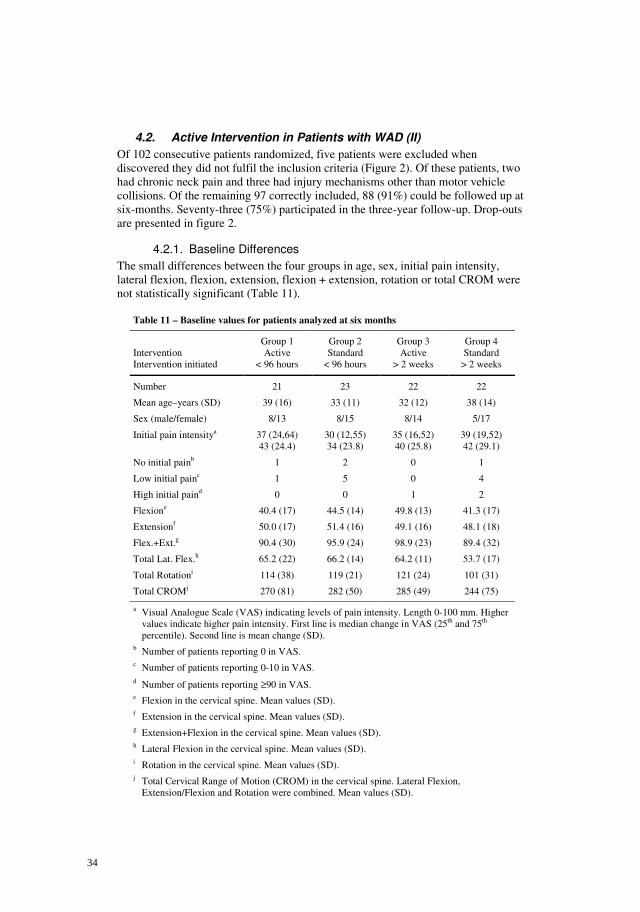
4.2. Active Intervention in Patients with WAD (II)
Of 102 consecutive patients randomized, five patients were excluded when discovered they did not fulfil the inclusion criteria (Figure 2). Of these patients, two had chronic neck pain and three had injury mechanisms other than motor vehicle collisions. Of the remaining 97 correctly included, 88 (91%) could be followed up at six-months. Seventy-three (75%) participated in the three-year follow-up. Drop-outs are presented in figure 2.
4.2.1. Baseline Differences
The small differences between the four groups in age, sex, initial pain intensity, lateral flexion, flexion, extension, flexion + extension, rotation or total CROM were not statistically significant (Table 11).
Table 11 – Baseline values for patients analyzed at six months
Group 1 Group 2 Group 3 Group 4 Intervention Active Standard Active Standard Intervention initiated < 96 hours < 96 hours > 2 weeks > 2 weeks
Number 21 23 22 22
Mean age–years (SD) 39 (16) 33 (11) 32 (12) 38 (14)
Sex (male/female) 8/13 8/15 8/14 5/17
Initial pain intensitya 37 (24,64) 43 (24.4)
30 (12,55) 34 (23.8)
35 (16,52) 40 (25.8)
39 (19,52) 42 (29.1)
No initial painb 1 2 0 1
Low initial painc 1 5 0 4
High initial paind 0 0 1 2
Flexione 40.4 (17) 44.5 (14) 49.8 (13) 41.3 (17)
Extensionf 50.0 (17) 51.4 (16) 49.1 (16) 48.1 (18)
Flex.+Ext.g 90.4 (30) 95.9 (24) 98.9 (23) 89.4 (32)
Total Lat. Flex.h 65.2 (22) 66.2 (14) 64.2 (11) 53.7 (17)
Total Rotationi 114 (38) 119 (21) 121 (24) 101 (31)
Total CROMj 270 (81) 282 (50) 285 (49) 244 (75) a Visual Analogue Scale (VAS) indicating levels of pain intensity. Length 0-100 mm. Higher
values indicate higher pain intensity. First line is median change in VAS (25th and 75th percentile). Second line is mean change (SD).
b Number of patients reporting 0 in VAS.
c Number of patients reporting 0-10 in VAS.
d Number of patients reporting ≥90 in VAS.
e Flexion in the cervical spine. Mean values (SD). f Extension in the cervical spine. Mean values (SD).
g Extension+Flexion in the cervical spine. Mean values (SD).
h Lateral Flexion in the cervical spine. Mean values (SD).
i Rotation in the cervical spine. Mean values (SD).
j Total Cervical Range of Motion (CROM) in the cervical spine. Lateral Flexion, Extension/Flexion and Rotation were combined. Mean values (SD).
34

Avail
ab
le f
or
an
aly
sis
at
6 m
on
ths (
n=
24)
Analy
sed a
t 6 m
on
ths (
n=
22)
Exc
luded f
rom
analy
sis
(n
=2
)E
xclu
de
d d
ue t
o n
on
-tra
ffic
in
jury
(n=
2)
Avail
ab
le f
or
an
aly
sis
at
3 y
ears
(n
=20
)A
naly
sed a
t 3 y
ears
(n=
18)
Exc
luded f
rom
analy
sis
(n
=2
)
E
xclu
de
d d
ue t
o n
on
-tra
ffic
in
jury
(n=
2)
All
oc
ate
d t
o e
arl
y s
tan
dard
in
terv
en
tio
n (
n=
26)
Receiv
ed
allo
cate
d inte
rvention (
n=
26)
Did
not
receiv
e a
llocate
d inte
rventio
n (
n=
0)
All
oc
ate
d t
o e
arl
y a
cti
ve i
nte
rven
tio
n (
n=
25)
Re
ceiv
ed
allo
cate
d inte
rve
ntion (
n=
24
)D
id n
ot
rece
ive a
llocate
d inte
rventio
n (
n=
1)
Exclu
de
d d
ue
to
non
-tra
ffic
inju
ry (
n=
1)
Ex
clu
de
d (
n=
16
)
No
t m
eeting inclu
sio
n c
rite
ria (
n=
7)
Pre
vio
uslly in
jure
d (
n=
1)
Non
-tra
ffic
infjury
(n
=1
)
>9
6ho
urs
aft
er
tra
um
a(n
=5)
Oth
er
reaso
ns (
n=
9)
No
-sh
ow
s (
n=
3)
Refu
se
d p
art
icip
atio
n (
n=
6)
As
se
ss
es
fo
r e
lig
ibil
ity (
n=
11
8)
All
ocate
d t
o d
ela
ye
d s
tan
da
rd i
nte
rven
tio
n (
n=
25)
Receiv
ed a
llocate
d inte
rvention (
n=
25)
Did
not
receiv
e a
llocate
d in
terv
en
tion(n
=0)
Ra
nd
om
ise
d (
n=
10
2)
All
oc
ate
d t
o d
ela
yed
acti
ve i
nte
rven
tio
n (
n=
26)
Receiv
ed
allo
cate
d inte
rve
ntion (
n=
22)
Did
not
rece
ive a
llocate
d inte
rventio
n(n
=4)
Exc
lud
ed
due
to
non
-tra
ffic
inju
ry (
n=
2)
Refu
sed
part
icip
atio
n (
n=
1)
Mo
ve
d a
bro
ad (
n=
1)
Lo
st
to 6
mo
nth
fo
llo
w u
p (
n=
2)
Dis
contin
ued inte
rventio
n (
n=
0)
Oth
er
reasons (
n=
2)
Re
fuse
d p
art
icip
ation
(n=
1)
No
t re
ach
ed (
n=
1)
Lo
st
to 6
mo
nth
fo
llo
w u
p (
n=
2)
Dis
contin
ued inte
rventio
n (
n=
1)
Co
uld
not
be r
each
ed (
n=
1)
Oth
er
reasons (
n=
1)
Mo
ve
d a
bro
ad
(n=
1)
Lo
st
to 6
mo
nth
fo
llo
w u
p (
n=
1)
Dis
contin
ued inte
rventio
n(n
=1)
Re
fuse
d p
art
icip
atio
n (
n=
1)
Lo
st
to 6
mo
nth
fo
llo
w u
p (
n=
3)
Dis
con
tinued in
terv
en
tion (
n=
0)
Oth
er
reason
s (
n=
3)
Re
fuse
d p
art
icip
atio
n (
n=
3)
Avail
ab
le f
or
an
aly
sis
at
3 y
ears
(n
=19)
Analy
se
d a
t 3 y
ears
(n=
16)
Exc
lud
ed f
rom
an
aly
sis
(n
=3)
Exclu
de
d d
ue t
o c
hro
nic
ne
ck p
ain
(n
=1
)
Exclu
de
d d
ue t
o r
ein
jury
(n=
2)
Avail
ab
le f
or
an
aly
sis
at
3 y
ears
(n
=21
)A
naly
sed a
t 3 y
ears
(n
=21
)E
xclu
ded f
rom
analy
sis
(n=
0)
Ava
ilab
le f
or
an
aly
sis
(n
=2
1)
Ana
lysed a
t 3
years
(n=
18)
Exclu
ded
fro
m a
naly
sis
(n=
3)
Exc
lud
ed
due
to
non
-tra
ffic
inju
ry (
n=
1)
Exc
lud
ed
due
to
chro
nic
ne
ck p
ain
(n
=1
)E
xclu
ded
due
to
rein
jury
(n
=1
)
Lo
st
to 3
year
foll
ow
up
(n
=3)
Oth
er
reasons (
n=
3)
Un
ab
le t
o fin
d t
ime
fo
r fo
llow
-up (
n=
1)
No
t re
ache
d f
or
follow
-up
(n=
2)
Lo
st
to 3
year
follo
w u
p (
n=
4)
Dis
contin
ued inte
rventio
n (
n=
0)
Oth
er
reasons (
n=
4)
Refu
se
d s
ec
on
d e
va
lua
tio
n (
n=
1)
No
t re
ac
hed
(n
=3
)
Lo
st
to 3
year
foll
ow
up
(n
=2)
Dis
con
tinued in
terv
en
tion (
n=
0)
Oth
er
reason
s (
n=
2)
Tra
ve
ling
(n
=1
)N
ot
reache
d (
n=
1)
Avail
ab
le f
or
an
aly
sis
at
6 m
on
ths (
n=
23
)A
naly
se
d a
t 6 m
onth
s (
n=
22)
Exc
lud
ed f
rom
an
aly
sis
(n
=1)
Exclu
de
d d
ue t
o c
hro
nic
ne
ck p
ain
(n
=1
)
Avail
ab
le f
or
an
aly
sis
at
6 m
on
ths (
n=
24)
Analy
sed a
t 6 m
on
ths (
n=
21)
Exc
luded f
rom
analy
sis
(n
=3
)E
xclu
de
d d
ue t
o n
on
-tra
ffic
in
jury
(n=
1)
Exclu
de
d d
ue t
o c
hro
nic
neck p
ain
(n=
1)
Exclu
de
d d
ue t
o r
ein
jury
(n
=1)
Avail
ab
le f
or
an
aly
sis
at
6 m
on
ths
(n
=2
3)
Analy
sed a
t 6 m
on
ths (
n=
23)
Exc
luded f
rom
analy
sis
(n=
0)
Lo
st
to 3
year
foll
ow
up
(n
=4)
Dis
contin
ued inte
rventio
n (
n=
2)
Tra
ve
ling
(n
=)
Lo
st
co
nta
ct (n
=1)
Oth
er
reaso
ns (
n=
2)
Re
inju
ry (
n=
1)
Un
ab
le t
o fin
d t
ime
fo
r fo
llow
-up (
n=
1)
F
igu
re 2
– P
arti
cip
ants
flo
w-c
har
t
35

4.2.2. Treatment Sessions
Of the patients receiving active intervention two received one instruction/treatment session, 13 received two sessions, and 10 received three sessions. The remaining patients received more than three sessions. The mean number of instruction/treatment sessions in the active intervention groups was 3.95. Symptoms persisting more than 20 days were seen in 63% (27/43) of patients in the active intervention group. They were re-examined and treated as described previously.
The number of patients receiving interventions from sources outside the control of this study did not differ statistically between the groups (Table 12).
4.2.3. Active versus Standard Intervention
Evaluation of the two interventions showed a reduction in pain intensity after six months (Table 12) and three years (Table 13) in all patients. However, the reduction of pain intensity was greater and the need for sick leave was lower for patients receiving active intervention compared to standard (Table 14).
The short-term effect of active intervention on CROM was not significant (Table 12 and 14). However, the three-year follow-up showed a trend (P=0.06-0.08) favoring active intervention over standard (Table 13 and 14). Patients receiving an early active intervention (Group 1) had a total CROM similar to matched controls at the three-year follow-up (Table 15). All other groups (Group 2-4) had decreased CROM compared to matched controls (Table 15). Active intervention significantly increases the chances for regaining/retaining CROM measured by comparing patients to unexposed healthy individuals (Table 16).
36

Table 12 – Six-month follow-up in patients exposed to whiplash trauma
Group 1 Group 2 Group 3 Group 4 Intervention Active Standard Active Standard Intervention initiated < 96 hours < 96 hours > 2 weeks > 2 weeks
Number 21 23 22 22
Mean days to follow-up (SD)
213 (41) 244 (100) 219 (48) 256 (77)
Change in pain intensitya -27 (-14,-46) -29.6 (24)
-6 (+24,-16) +0.74 (30)
-11 (-5,-27) -15 (19)
-8.5 (-2,-13) -7.1 (22)
No pain at follow-upb 38% (8/21) 17% (4/23) 23% (5/22) 5% (1/22)
Low pain at follow-upc 52% (11/21) 30% (7/23) 36% (8/22) 9% (2/22)
Sick leave days for all patientsd
15.1 (42) 10.3 (22) 11.5 (38) 28.9 (51)
Sick leave days for patients 20-65 yearsd
17.7 (46) 10.7 (23) 13.8 (42) 31.8 (52)
Sick leave ≥30 days for all patientse
2/21 3/23 1/22 6/22
Sick leave ≥30 days for patients 20-65 yearse
2/18 3/22 1/18 6/20
Change in Flexionf +9.8 (18) -1.1 (16) +0.3 (17) +8.0 (18)
Change in Extensiong +8.4 (15) +7.1 (14) +8.2 (15) +3.7 (16)
Change in Flex.+Ext.h +18.2 (27) +6.0 (22) +8.5 (23) +11.7 (28)
Change in total Lat. Flex.i
+10.1 (18) +4.7 (16) +7.3 (12) +10.1 (18)
Change in total Rotationj +23.6 (37) +14.4 (37) +7.5 (21) +22.8 (25)
Change in total CROMk +51.9 (70) +25.2 (62) +23.3 (47) +44.6 (59)
Received interventions from sources outside the control of this studyl
3/21 9/23 5/22 9/21
a First line is median change in Visual Analogue Scale (VAS) (25th and 75th percentile). Second line is
mean change (SD). Negative values indicate a decrease in pain level.
b Proportion of patients reporting 0 in VAS.
c Proportion of patients reporting ≤10 in VAS (including those reporting 0).
d Sick leave during the preceding six months as estimated by the patient. Mean number of working days (SD).
e Number of patients reporting sick leave ≥30 days during the preceding six months due to whiplash injury. (after / no. of patients with sufficient data)
f The mean change (SD) in flexion in the cervical spine. Positive values indicate increased range of motion.
g The mean change (SD) in extension in the cervical spine. Positive values indicate increased range of motion.
h The mean change (SD) in flexion + extension in the cervical spine. Positive values indicate increased range of motion.
i The mean change (SD) in lateral flexion in the cervical spine. Positive values indicate increased range of motion.
j The mean change (SD) in rotation in the cervical spine. Positive values indicate increased range of motion.
k The mean change (SD) in total cervical range of motion (CROM). Positive values indicate increased range of motion.
l The number of patients who received interventions from sources outside the control of this study. Data is missing from one patient in group 4.
37

Table 13 – Three-year follow-up in patients exposed to whiplash trauma
Group 1 Group 2 Group 3 Group 4 Intervention Active Standard Active Standard Intervention initiated < 96 hours < 96 hours > 2 weeks > 2 weeks
Number 18 21 18 16
Mean days to follow-up (SD)
1213 (110) 1240 (121) 1227 (129) 1234 (126)
Change in pain intensitya -17 (-2,-28) -21 (27.6)
-5 (+18,-23) -1.8 (29.7)
-15.5 (-8,-28) -15.8 (22.4)
-10 (+11,-20) -5.2 (27.3)
No pain at follow-upb 33% (6/18) 33% (7/21) 44% (8/18) 31% (5/16)
Low pain at follow-upc 39% (7/18) 43% (9/21) 61% (11/18) 31% (5/16)
Sick leave days for all patientsd
11.2 (44) 40.2 (71) 10.0 (42) 20.5 (50)
Sick leave days for patients 20-65 yearsd
13.6 (48) 42.2 (72) 12.9 (48) 21.9 (52)
Sick leave ≥30 days for all patientse
1/17 6/21 1/18 3/15
Sick leave ≥30 days for patients 20-65 yearse
1/14 6/20 1/14 3/14
Change in Flexionf +17.7 (18) +6.2 (19) +3.8 (21) +5.9 (15)
Change in Extensiong +8.9 (15) +1.4 (15) +6.9 (16) +3.8 (15)
Change in Flex.+Ext.h +26.7 (27) +7.6 (27) +10.7 (30) +9.7 (22)
Change in total Lat. Flex.i
+8.8 (19) -3.2 (18) +4.2 (15) +3.9 (13)
Change in total Rotationj +25.6 (34) +11.8 (32) +10.7 (22) +9.8 (17)
Change in total CROMk +61.1 (61) +16.2 (67) +25.6 (60) +23.4 (43)
Received interventions from sources outside the control of this studyl
(Not asked for at three year follow-up)
a First line is median change in Visual Analogue Scale (VAS) (25th and 75th percentile). Second line is
mean change (SD). Negative values indicate a decrease in pain level.
b Proportion of patients reporting 0 in VAS.
c Proportion of patients reporting ≤10 in VAS (including those reporting 0).
d Sick leave during the preceding six months as estimated by the patient. Mean number of working days (SD).
e Number of patients reporting sick leave ≥30 days during the preceding six months due to whiplash injury. (after / no. of patients with sufficient data)
f The mean change (SD) in flexion in the cervical spine. Positive values indicate increased range of motion.
g The mean change (SD) in extension in the cervical spine. Positive values indicate increased range of motion.
h The mean change (SD) in flexion + extension in the cervical spine. Positive values indicate increased range of motion.
i The mean change (SD) in lateral flexion in the cervical spine. Positive values indicate increased range of motion.
j The mean change (SD) in rotation in the cervical spine. Positive values indicate increased range of motion.
k The mean change (SD) in total cervical range of motion (CROM). Positive values indicate increased range of motion.
l The number of patients who received additional treatments from sources outside the control of this study.
38

Table 14 – Differences in outcome (P-values) active versus standard intervention
Six-month follow-up Three-year follow-up Anovaa Friedmannb Anova Friedmann
Change in pain intensityc 0.0004 0.0009 (0.019)
0.020 0.026 (0.028)
Sick leave days for all patientsd ----- NSe ----- 0.030
Sick leave days for patients 20-65 yearsd
----- NS ----- NS (0.063)
Change in Flexionf NS NS NS NS
Change in Extensiong NS NS NS NS
Change in Flex.+Ext.h NS NS NS NS (0.081)
Change in total Lat. Flex.i NS NS NS NS
Change in total Rotationj NS NS NS NS
Change in total CROMk NS NS NS (0.092) NS (0.062)
a In case of skewed data, the non-parametric test described by Friedmann was used. In such cases this is noted by -----
b Friedmann’s test is usually performed on raw data. In a visual analogue scale (VAS) it may also be performed on transformed data where improvement is coded as +1, worsening as –1 and unchanged as 0. The outcome of Friedmann’s test used on transformed data is given within parentheses.
c The mean change in VAS
d Mean number of days on sick leave during the preceding six months as estimated by the patient.
e NS = Non-significant (p>0.05). P-values 0.05-0.1 presented in paranthesis.
f The mean change in flexion in the cervical spine.
g The mean change in extension in the cervical spine.
h The mean change in flexion + extension in the cervical spine.
i The mean change in lateral flexion in the cervical spine.
j The mean change in rotation in the cervical spine.
k The mean change in total total cervical range of motion (CROM).
39

T
able
15
– M
ean
dif
fere
nce
s in
cer
vica
l ran
ge o
f m
otio
n (
CR
OM
) b
etw
een
pat
ien
ts a
nd
un
exp
osed
ind
ivid
ual
s
G
roup
1
Gro
up 2
G
roup
3
Gro
up 4
Ma
SD
b pc
M
SD
p
M
SD
p
M
SD
p
Fle
xion
-3
.3
15.4
N
Sd
-12.
7 24
.4
0.02
7 -1
0.0
14.4
0.
009
-6.9
17
.9
NS
Ext
ensi
on
-7.2
13
.6
0.03
8 -2
0.8
20.5
0.
0002
-1
0.8
22.7
N
S
-6.3
20
.0
NS
Fle
xion
+E
xten
sion
-1
0.6
25.4
N
S
-33.
5 38
.6
0.00
07
-20.
8 33
.1
0.01
6 -1
3.1
33.4
N
S
Tot
al L
ater
al F
lexi
on
+13
.6
26.7
0.
045
-12.
7 26
.5
0.04
0 -1
.4
18.3
N
S
-7.3
17
.8
NS
Tot
al R
otat
ion
+4.
6 20
.9
NS
-1
7.1
34.6
0.
034
-6.7
31
.4
NS
-2
1.2
27.6
0.
008
Tot
al C
ervi
cal R
ange
of
Mot
ion
+7.
7 60
.6
NS
-1
8.1
44.3
N
S
+0.
4 31
.0
NS
-2
4.2
56.1
N
S
a M
ean
valu
e of
all
dif
fere
nces
, neg
ativ
e va
lues
ind
icat
e th
at t
he p
atie
nts
have
low
er C
RO
M t
han
the
unex
pose
d in
divi
dual
s.
b S
tand
ard
Dev
iati
on
c P
-val
ue o
btai
ned
by S
tude
nt’s
one
way
t-t
est
d N
o si
gnif
ican
ce
40

Table 16 – Effect of intervention, time factor and their combined effect (p-values) on retaining/regaining cervical range of motion when comparing patients at three year follow-up to unexposed individuals
Interventiona Timeb Interactionc
Flexion NSd NS NS
Extension NS NS NS (0,053)
Flexion + Extension NS NS NS (0.054)
Total Lateral Flexion 0.0042 NS NS (0.063)
Total Rotation 0.011 NS NS
Total CROM 0.032 NS NS
a Intervention = Active intervention versus standard intervention
b Time = <94 hours versus versus >two weeks
c Interaction = Interaction between intervention and time
d NS = Non-significant (P>0.05). P-values 0.05-0.1 presented in paranthesis.
4.2.4. The Importance of the Time Factor
The time factor, defined as initiating intervention immediately or with a delay of 14 days, did not by itself affect the outcome at either the six-month or three-year follow-up.
Combining the intervention and time factor in a two-way factorial design showed, at six months, an interaction between type of intervention and timing on the reduction of pain intensity (P=0.04) and on the improvement of cervical flexion (P=0.01). When active intervention was applied it was better to receive it early and if standard intervention was given it was better to receive it late. No interaction effect was found at the three-year follow-up.
Comparing the patients with individually matched unexposed controls showed a combined effect of timing and intervention on retaining/regaining CROM at the three-year follow-up (Table 16). CROM was greater retained/regained when active intervention was received early and when standard intervention was received late.
4.2.5. No Initial Pain
Four of 97 patients (4.1%, 95% confidence interval 0.15-8.0%) had no initial pain (WAD 0, VAS 0 mm of maximum 100 mm). One patient in group 1 had no pain at six months or at three years. The other three patients in group 2 and 4 had pain at six months (8-64/100) and two of them at three years (50-51/100).
41

4.3. Basic body awareness therapy compared to exercise therapy for patients with chronic whiplash associated disorders (III)
4.3.1. Recruitment and follow-up of participants
Details of patient recruitment and dropouts are shown in Figure 3. 3570 patients were identified in the electronic medical journals of all 30 primary health units in Southern Älvsborg County. A random sample of 1573 was selected to be contacted by mail. 1546 had an address in the Swedish personal address registry (SPAR) and received the mailed survey with PROs and consent information. 996 responded to the survey and 373 stated having chronic WAD and accepted participation. After baseline assessment 113 were randomly allocated to ET (n=57) or BBAT (n=56).
All patients that underwent random allocation were analyzed according to group assignment. Thus, we attempted to follow up all 113 patients regardless of compliance. One patient in the ET group and three patients in BBAT group never came to any treatment appointment.
4.3.2. Baseline characteristics
The groups were similar at baseline (Table 17). Patients had been suffering from chronic WAD for several years, had high levels of pain, were moderately disabled, and had high fear of movement.
42

Primary care visits coded whiplash (n=5613)
Individuals in primary care coded whiplash (n=3570)
Random sample (n=1573)
Individuals with address in SPAR (n=1546)
Individuals missing in SPAR (n=27)
Survey responders (n=996)
No reply (n=529), Deceased (n=2), Living abroad (n=2), Unknown addressee (n=17)
Assessed (n=372)
Declined participation (n=567) No whiplash trauma (n=17) Reply after deadline (n=11) Pain never/almost never (n=28) Failure to correctly include (n=1)
Randomized (n=113)
Unable to contact the patient (n=73) I have no time for this (n=62) Comorbidity that hinders participation (n=38) Can’t because of work (n=21) Recovered (n=15) Not interested (n=11) Treatment too intensive (n=10) Had trauma but no WAD (n=7) Declined (n=5) Other reasons (n=17)
Exercise Therapy (n=57) Received treatment (n=56)
Did not receive treatment (n=1) (Patient never came to treatment
sessions)
Basic Body Awareness Therapy (n=56) Received treatment (n=53)
Did not receive treatment (n=3) (Patients never came to treatment sessions)
Follow-up after treatment Complete evaluation (n=54)
No examination (n=1) Neither examination nor questionnaires
(n=2)
Follow-up after treatment Complete evaluation (n=51)
No examination (n=1) Neither examination nor questionnaires (n=4)
3-month follow-up Complete evaluation (n=52)
No examination (n=3) Neither examination nor questionnaires
(n=2)
3-month follow-up Complete evaluation (n=50)
No examination (n=2) Neither examination nor questionnaires (n=4)
Figure 3 – Details of patient recruitment and drop-outs
43

Table 17 – Baseline characteristics for exercise therapy (ET) and basic body awareness therapy (BBAT) groups [mean (SD) and median (interquartile range) or n (%)]
ET
(n=57) BBAT (n=56)
Age, years 49 (11) 47 (15)
47 (13) 49 (21)
Female Male
44 (77%) 13 (23%)
37 (66%) 19 (34%)
Duration of symptoms, years 9.3 (7.7) 7 (8)
10 (9.5) 7 (9.3)
WAD classification 1 2 3
1 (2%) 16 (28%) 40 (70%)
0 (0%) 13 (23%) 43 (77%)
Pain intensitya 3.6 (0.71) 3 (1)
3.6 (0.78) 3 (1)
Pain frequencyb 4.2 (1.07) 5 (2)
4.2 (1.08) 5 (2)
Number of whiplash traumas 1.7 (1.2) 1 (1)
1.8 (1.4) 1 (1)
Days on sick-leave during past 6 months 62 (75.3) 18.1 (137)
69.7 (80) 22.8 (182.5)
Neck Disability Indexc 19 (7.6) 18 (12)
20 (8.9) 20 (17)
SF-36 v2 Physical functioningd 70 (17) 75 (25)
67 (21) 75 (35)
SF-36 v2 Role - physical 25 (37) 0 (50)
34 (39) 13 (75)
SF-36 v2 Bodily pain 35 (18) 41 (19)
34 (20) 41 (20)
SF-36 v2 General health 49 (19) 50 (23)
55 (22) 57 (36)
SF-36 v2 Vitality 35 (22) 30 (38)
39 (24) 43 (44)
SF-36 v2 Social functioning 59 (27) 50 (44)
60 (27) 63 (25)
SF-36 v2 Role - emotional 52 (45) 33 (100)
55 (42) 67 (100)
SF-36 v2 Mental health 63 (24) 68 (44)
66 (22) 72 (31)
Tampa Scale of Kinesiophobiae 35 (7.6) 36 (10)
37 (10) 35 (13)
Body Awareness Scale - observationf 30 (6.5) 31 (9.4)
30 (7.5) 29 (9.8)
Body Awareness Scale - interviewg 16 (7.3) 17 (13)
16 (6.3) 15 (7.8)
a “how often do you have pain in your shoulder, head or neck nowadays?” (1 representing
never/almost never, 2 representing few times/month, 3 representing few times a week, 4 representing few times a day, 5 representing constant pain)
b “if you answered that you have pain in shoulder, head or neck how intensive is this pain?” (0 representing no pain at all, 1 representing barely noticeable, 2 representing weak pain, 3 representing moderate pain, 4 representing strong pain, 5 representing very strong pain)
c Neck Disability Index score (0-50 scale, 10 items, higher scores represent greater disability)
d Short Form 36 version 2 score (0-100 scale, 36 items, higher scores represent a better health state)
e Tampa Scale of Kinesiophobia score (17-68 scale, 17 items, higher scores represent greater pain-related fear of movement)
f Body Awareness Scale observation score (0-63 scale, 21 items, higher scores represent lower
quality of movement)
g Body Awareness Scale interview score (0-51 scale, 17 items, higher scores represent experiencing lower quality of movement)
44

4.3.3. Compliance with treatment
The mean number of treatment sessions attended was 13 (95% CI: 11 to 14) for the ET group and 14 (95% CI: 13 to 16) for the BBAT group. The range was 0 to 20 sessions for both groups. There were no statistically significant differences in compliance between groups.
4.3.4. Additional treatment
At first follow-up directly after the last treatment 25 patients (47%) in the ET group and 21 patients (42%) in the BBAT group reported seeking additional treatment. At three months the corresponding figures were 29 patients (57%) in the ET group and 22 patients (45%) in the BBAT group (Table 18). Table 18 – Additional treatment reported at the post treatment and three month follow-up
Post treatment follow-up 3-month follow-up ET (n=53) BBAT (n=50) ET (n=51) BBAT (n=49)
No additional treatment
28 29 22 27
Physical and manual therapy
6 8 11 9
Massage 12 7 15 9
Non-supervised physical activity
9 12 3 9
Medical device 2 2 6 4
Dietary supplements and natural medicine
1 0 3 0
Surgery 0 2 1 0
Miscellaneous 7 1 2 1
4.3.5. Effectiveness of treatment
The BBAT group had greater improvement than the ET group on most primary and secondary outcomes at the post-treatment follow-up. These differences in improvement were still evident at three months (Table 19a, 19b, 20a and 20b).
45

Tab
le 1
9a –
Ch
ange
s in
ou
tcom
e m
easu
res
from
bas
elin
e to
en
d o
f tr
eatm
ent
for
exer
cise
(E
T)
and
bas
ic b
ody
awar
enes
s gr
oup
(B
BA
T)
Pri
mar
y ou
tcom
e m
easu
res
N
C
hang
e E
ffec
t siz
e P
-val
ues
B
BA
T
ET
B
BA
T
ET
C
ohen
’s d
T
-tes
t M
Wf
MW
-Tg
BA
S -
obs
erva
tion
a 50
52
-1
2 (6
.9)
-11 (
8.3
) -3
.4 (
5.8)
-3
.5 (
7.9
) -1
.35
(-1.
77, -
0.91
) 2.
9x10
-9
2.8x
10-9
5.
6x10
-5
BA
S -
inte
rvie
wb
50
53
-7.2
(6.
3)
-6.8
(7.3
) -2
.6 (
5.4)
-2
.5 (
6.8
) -0
.79
(-1.
18, -
0.38
) 0.
0001
2 0.
0002
7 0.
054
ND
Ic 48
50
-2
.3 (
3.6)
-2
(5)
-0.6
0 (5
.2)
0 (
6)
-0.3
8 (-
0.78
, 0.0
2)
0.07
0 0.
064
0.05
3
TS
Kd
48
48
-2.5
(6.
1)
-1 (
6.8
) -2
.9 (
5.8)
-3
.5 (
6.8
) 0.
07
(-0.
33, 0
.47)
0.
732
0.38
2 0.
225
SF
-36
Phy
sica
l
func
tion
inge
52
54
7.6
(14)
5 (
15)
0.09
3 (1
4)
0 (
15)
0.54
(0
.14,
0.9
2)
0.00
6 0.
032
0.16
4
SF
-36
Rol
e -
phys
ical
52
54
17
(38
) 25 (
25)
12 (
43)
0 (
50)
0.12
(-
0.26
, 0.5
0)
0.56
9 0.
262
0.05
7
SF
-36
Bod
ily
pain
52
54
11
(16
) 10 (
22)
5.4
(18)
9 (
28)
0.33
(-
0.06
, 0.7
1)
0.11
0 0.
077
0.11
4
SF
-36
Gen
eral
hea
lth
52
54
8.3
(17)
10 (
25)
4.0
(15)
2.5
(20)
0.27
(-
0.12
, 0.6
5)
0.16
5 0.
119
0.06
8
SF
-36
Vit
alit
y 52
54
11
(18
) 13 (
24)
5.4
(17)
5 (
22)
0.32
(-
0.07
, 0.7
0)
0.12
0 0.
075
0.16
1
SF
-36
Soc
ial f
unct
ioni
ng
52
54
13 (
20)
13 (
25)
5.6
(24)
0 (
28)
0.33
(-
0.05
, 0.7
2)
0.09
6 0.
092
0.07
5
SF
-36
Rol
e –
emot
iona
l 52
54
14
(47
) 0 (
33)
13 (
43)
0 (
33)
0.02
(-
0.36
, 0.4
0)
0.86
9 0.
401
0.22
6
SF
-36
Men
tal h
ealt
h 52
54
3.
5 (1
8)
4 (
27)
4.1
(17)
4 (
16)
-0.0
3 (-
0.41
, 0.3
5)
0.85
9 0.
713
0.73
3
a
Bod
y A
war
enes
s S
cale
obs
erva
tion
sco
re (
0-63
sca
le, 2
1 it
ems,
hig
her
scor
es r
epre
sent
low
er q
uali
ty o
f m
ovem
ent)
. b
Bod
y A
war
enes
s S
cale
int
ervi
ew s
core
(0-
51 s
cale
, 17
item
s, h
ighe
r sc
ores
rep
rese
nt e
xper
ienc
ing
low
er q
uali
ty o
f m
ovem
ent)
. c
Nec
k D
isab
ilit
y In
dex
scor
e (0
-50
scal
e, 1
0 it
ems,
hig
her
scor
es r
epre
sent
gre
ater
dis
abil
ity)
. d
Tam
pa S
cale
of
Kin
esio
phob
ia s
core
(17
-68
scal
e, 1
7 it
ems,
hig
her
scor
es r
epre
sent
gre
ater
pai
n-re
late
d fe
ar o
f m
ovem
ent)
. e
Sho
rt F
orm
36
vers
ion
2 sc
ore
(0-1
00 s
cale
, 36
item
s, h
ighe
r sc
ores
rep
rese
nt a
bet
ter
heal
th s
tate
).
f M
ann-
Whi
tney
U te
st
g M
ann-
Whi
tney
U te
st o
n tr
ansf
orm
ed v
alue
s (“
impr
ovem
ent”
=+
1, “
wor
seni
ng”?
-1 a
nd “
unch
ange
d”=
0)
46

T
able
19b
– C
han
ges
in o
utc
ome
mea
sure
s fr
om b
asel
ine
to e
nd
of
trea
tmen
t fo
r ex
erci
se (
ET
) an
d b
asic
bod
y aw
aren
ess
grou
p (
BB
AT
) S
econ
dar
y ou
tcom
e m
easu
res
N
C
hang
e E
ffec
t siz
e P
-val
ues
B
BA
T
ET
B
BA
T
ET
C
ohen
’s d
T
-tes
t M
Wd
MW
-Te
Pai
n in
tens
itya
52
53
-0.6
2 (1
.2)
0 (
1)
-0.4
2 (0
.72)
0 (
1)
-0.2
0 (-
0.58
, 0.1
8)
0.30
1 0.
066
0.45
4
Pai
n fr
eque
ncyb
52
53
-0.7
1 (1
.1)
-0.5
0 (
1)
-0.3
6 (1
.2)
0 (1
) -0
.30
(-0.
69, 0
.08)
0.
130
0.32
9 0.
059
Com
plia
nce
to tr
eatm
ent
56
57
14 (
6.2)
17
(8)
13
(5.
7)
14 (
10)
0.17
(-
0.20
, 0.5
4)
0.21
9 0.
068
-
Fle
xion
50
53
7.
4 (1
5)
6 (
17)
4.9
(10)
6 (
14)
0.20
(-
0.19
, 0.5
8)
0.32
8 0.
358
0.92
9
Ext
ensi
on
50
53
2.6
(10)
2 (
14)
5.9
(9.2
) 6 (
10)
-0.3
4 (-
0.73
, 0.0
5)
0.08
3 0.
084
0.07
4
Lef
t la
tera
l fle
xion
50
53
1.
6 (6
.8)
1 (
6)
1.2
(6.3
) 0 (
10)
0.06
(-
0.33
, 0.4
5)
0.76
0 0.
737
0.80
1
Rig
ht l
ater
al f
lexi
on
50
53
1.8
(5.6
) 2 (
8.5
) 0.
45 (
6.5)
0 (
9)
0.22
(-
0.17
, 0.6
1)
0.26
4 0.
215
0.21
9
Lef
t ro
tati
on
50
53
5.8
(9.7
) 4 (
14)
3.6
(12)
2 (
14)
0.20
(-
0.19
, 0.5
9)
0.28
7 0.
183
0.34
7
Rig
ht r
otat
ion
50
53
5.2
(9.5
) 4 (
12)
4.3
(11)
2 (
11)
0.09
(-
0.30
, 0.4
7)
0.67
7 0.
689
0.51
5
Hea
d po
siti
onc
46
50
2 (2
.8)
1 (
4)
-0.5
7 (2
.4)
-1 (
3.3
) 0.
99
(0.5
6, 1
.40)
5.
4x10
-6
3.7x
10-5
0.
0002
7
Sag
itta
l C
RO
M
50
53
10 (
22)
7 (
23)
11 (
15)
10 (
15)
-0.0
5 (-
0.44
, 0.3
3)
0.83
2 0.
866
0.35
5
Fro
ntal
CR
OM
50
53
3.
4 (1
1)
4 (
13)
1.7
(11)
2 (
16)
0.15
(-
0.23
, 0.5
4)
0.42
9 0.
363
0.73
8
Hor
ison
tal C
RO
M
50
53
11 (
17)
8 (
20)
7.9
(21)
2 (
24)
0.16
(-
0.23
, 0.5
5)
0.41
0 0.
198
0.11
4
a “h
ow o
ften
do
you
have
pai
n in
you
r sh
ould
er, h
ead
or n
eck
now
aday
s?”
(1 r
epre
sent
ing
neve
r/al
mos
t ne
ver,
2 r
epre
sent
ing
few
tim
es/m
onth
, 3
repr
esen
ting
few
tim
es a
wee
k, 4
rep
rese
ntin
g fe
w ti
mes
a d
ay, 5
rep
rese
ntin
g co
nsta
nt p
ain)
. b
“if
you
answ
ered
tha
t yo
u ha
ve p
ain
in s
houl
der,
hea
d or
nec
k ho
w i
nten
sive
is t
his
pain
?” (
0 re
pres
enti
ng n
o pa
in a
t all
, 1 r
epre
sent
ing
bare
ly
noti
ceab
le, 2
rep
rese
ntin
g w
eak
pain
, 3 r
epre
sent
ing
mod
erat
e pa
in, 4
rep
rese
ntin
g st
rong
pai
n, 5
rep
rese
ntin
g ve
ry s
tron
g pa
in).
c
Hea
d pr
otra
ctio
n in
sta
ndin
g po
stur
e m
easu
red
in d
egre
es (
high
er v
alue
s re
pres
enti
ng m
ore
upri
ght p
ostu
re)
d M
ann-
Whi
tney
U te
st
e M
ann
Whi
tney
U t
est
on tr
ansf
orm
ed v
alue
s (“
impr
ovem
ent”
=+
1, “
wor
seni
ng”?
-1 a
nd “
unch
ange
d”=
0)
47

T
able
20a
– C
han
ges
in o
utc
ome
mea
sure
s fr
om b
asel
ine
to t
hre
e m
onth
fol
low
-up
for
exe
rcis
e (e
t) a
nd
bas
ic b
ody
awar
enes
s gr
oup
(b
bat
) P
rim
ary
outc
ome
mea
sure
s
N
C
hang
e E
ffec
t siz
e P
-val
ues
B
BA
T
ET
B
BA
T
ET
C
ohen
’s d
T
-tes
t M
W f
MW
-T g
BA
S -
obs
erva
tion
a 49
49
-1
2 (7
.7)
-12 (
8.8
) -4
.9 (
5.4)
-5
(6.8
) -1
.07
(-1.
48, -
0.64
) 2.
1x10
-6
3.2x
10-6
0.
222
BA
S -
inte
rvie
wb
49
51
-5.3
(7)
-4
(8)
-2.7
(5.
7)
-3 (
8)
-0.4
1 (-
0.80
, -0.
01)
0.04
2 0.
087
0.08
4
ND
Ic 48
51
-2
(5.
3)
-2.5
(8.5
) -1
.1 (
5.1)
0
(6)
-0.1
7 (-
0.57
, 0.2
2)
0.36
8 0.
342
0.20
8
TS
Kd
47
51
-3.1
(5.
6)
-3 (
8)
-3.2
(5.
4)
-3 (
6)
0.02
(-
0.38
, 0.4
1)
0.93
6 0.
991
0.75
2
SF
-36
Phy
sica
l
func
tion
inge
50
54
7.1
(14)
5 (
15)
0.46
(13
) 0 (
15)
0.49
(0
.10,
0.8
8)
0.01
5 0.
071
0.22
6
SF
-36
Rol
e -
phys
ical
50
54
18
(41
) 0 (
50)
19 (
35)
0 (
50)
-0.0
3 (-
0.41
, 0.3
6)
0.84
3 0.
962
0.88
0
SF
-36
Bod
ily
pain
50
54
12
(19
) 11 (
22)
4.9
(18)
4.5
(14)
0.38
(-
0.01
, 0.7
7)
0.04
4 0.
025
0.13
1
SF
-36
Gen
eral
hea
lth
50
54
7.5
(18)
8 (
25)
4.5
(17)
5 (
24)
0.17
(-
0.22
, 0.5
6)
0.38
3 0.
279
0.19
0
SF
-36
Vit
alit
y 50
54
7.
3 (2
2)
5 (
30)
5.6
(22)
5 (
30)
0.08
(-
0.31
, 0.4
6)
0.68
8 0.
620
0.56
5
SF
-36
Soc
ial f
unct
ioni
ng
50
54
13 (
24)
13 (
25)
3.5
(24)
0 (
25)
0.40
(0
.00,
0.7
8)
0.03
7 0.
019
0.01
8
SF
-36
Rol
e -
emot
iona
l 50
54
9.
3 (4
1)
0 (
33)
4.0
(45)
0 (
33)
0.12
(-
0.26
, 0.5
1)
0.53
2 0.
224
0.14
4
SF
-36
Men
tal h
ealt
h 51
54
2.
8 (1
7)
4 (
24)
1.2
(17)
0 (
22)
0.09
(-
0.29
, 0.4
8)
0.64
1 0.
591
0.74
0
a
Bod
y A
war
enes
s S
cale
obs
erva
tion
sco
re (
0-63
sca
le, 2
1 it
ems,
hig
her
scor
es r
epre
sent
low
er q
uali
ty o
f m
ovem
ent)
. b
Bod
y A
war
enes
s S
cale
int
ervi
ew s
core
(0-
51 s
cale
, 17
item
s, h
ighe
r sc
ores
rep
rese
nt e
xper
ienc
ing
low
er q
uali
ty o
f m
ovem
ent)
. c
Nec
k D
isab
ilit
y In
dex
scor
e (0
-50
scal
e, 1
0 it
ems,
hig
her
scor
es r
epre
sent
gre
ater
dis
abil
ity)
. d
Tam
pa S
cale
of
Kin
esio
phob
ia s
core
(17
-68
scal
e, 1
7 it
ems,
hig
her
scor
es r
epre
sent
gre
ater
pai
n-re
late
d fe
ar o
f m
ovem
ent)
. e
Sho
rt F
orm
36
vers
ion
2 sc
ore
(0-1
00 s
cale
, 36
item
s, h
ighe
r sc
ores
rep
rese
nt a
bet
ter
heal
th s
tate
).
f M
ann-
Whi
tney
U te
st
g M
ann-
Whi
tney
U te
st o
n tr
ansf
orm
ed v
alue
s (“
impr
ovem
ent”
=+
1, “
wor
seni
ng”?
-1 a
nd “
unch
ange
d”=
0)
48

T
able
20b
– C
han
ges
in o
utc
ome
mea
sure
s fr
om b
asel
ine
to t
hre
e m
onth
fol
low
-up
for
exe
rcis
e (e
t) a
nd
bas
ic b
ody
awar
enes
s gr
oup
(b
bat
) S
econ
dar
y ou
tcom
e m
easu
res
N
C
hang
e E
ffec
t siz
e P
-val
ues
B
BA
T
ET
B
BA
T
ET
C
ohen
’s d
T
-tes
t M
W d
MW
-T e
Pai
n in
tens
itya
49
54
-0.6
1 (1
.2)
0 (
1)
-0.4
1 (0
.77)
0 (
1)
-0.2
0 (-
0.59
, 0.1
9)
0.30
1 0.
437
0.66
0
Pai
n fr
eque
ncyb
49
54
-0.8
2 (1
.2)
-1 (
1.5
) -0
.33
(0.8
2)
0 (
1)
-0.4
8 (-
0.87
, -0.
09)
0.02
0 0.
014
0.01
6
Com
plia
nce
to tr
eatm
ent
41
42
38 (
41)
23 (
91)
32 (
38)
11 (
66)
0.15
(-
0.28
, 0.5
8)
0.51
4 0.
538
-
Fle
xion
50
52
4.
9 (1
2)
4 (
17)
5.3
(10)
6 (
12)
-0.0
4 (-
0.42
, 0.3
5)
0.87
2 0.
765
0.51
2
Ext
ensi
on
50
52
4.9
(9.3
) 4 (
10)
5.8
(9.4
) 5 (
12)
-0.1
0 (-
0.48
, 0.2
9)
0.64
9 0.
675
0.73
7
Lef
t la
tera
l fle
xion
50
52
1.
3 (5
.8)
2 (
8)
1.3
(6.5
) 0 (
9.5
) 0.
00
(-0.
39, 0
.39)
0.
970
0.68
6 0.
434
Rig
ht l
ater
al f
lexi
on
50
52
2.2
(5.5
) 3 (
8)
0.52
(5.
1)
0 (
8)
0.32
(-
0.08
, 0.7
1)
0.10
5 0.
105
0.15
4
Lef
t ro
tati
on
50
52
5.4
(9.3
) 6 (
11)
3.4
(9.9
) 3 (
9.5
) 0.
21
(-0.
18, 0
.60)
0.
294
0.14
1 0.
303
Rig
ht r
otat
ion
50
52
4.7
(9.9
) 5 (
12)
4.3
(10)
2 (
11.5
) 0.
04
(-0.
35, 0
.43)
0.
868
0.49
1 0.
529
Hea
d po
siti
onc
46
48
1 (3
.0)
1 (
4)
-0.5
9 (2
.8)
-1 (
4)
0.55
(0
.13,
0.9
6)
0.00
9 0.
012
0.03
9
Sag
itta
l C
RO
M
50
52
9.8
(18)
9 (
19)
11 (
16)
11 (
19)
-0.0
7 (-
0.46
, 0.3
2)
0.72
2 0.
580
0.19
8
Fro
ntal
CR
OM
50
52
3.
5 (1
0)
5 (
14)
1.9
(10)
0 (
16)
0.16
(-
0.23
, 0.5
5)
0.40
3 0.
295
0.16
6
Hor
ison
tal C
RO
M
50
52
10 (
17)
12 (
21)
7.7
(18)
3 (
14)
0.13
(-
0.26
, 0.5
2)
0.50
8 0.
274
0.89
5
a
“how
oft
en d
o yo
u ha
ve p
ain
in y
our
shou
lder
, hea
d or
nec
k no
wad
ays?
” (1
rep
rese
ntin
g ne
ver/
alm
ost
neve
r, 2
rep
rese
ntin
g fe
w ti
mes
/mon
th, 3
re
pres
enti
ng f
ew ti
mes
a w
eek,
4 r
epre
sent
ing
few
tim
es a
day
, 5 r
epre
sent
ing
cons
tant
pai
n).
b “i
f yo
u an
swer
ed t
hat
you
have
pai
n in
sho
ulde
r, h
ead
or n
eck
how
int
ensi
ve is
thi
s pa
in?”
(0
repr
esen
ting
no
pain
at a
ll, 1
rep
rese
ntin
g ba
rely
no
tice
able
, 2 r
epre
sent
ing
wea
k pa
in, 3
rep
rese
ntin
g m
oder
ate
pain
, 4 r
epre
sent
ing
stro
ng p
ain,
5 r
epre
sent
ing
very
str
ong
pain
).
c H
ead
prot
ract
ion
in s
tand
ing
post
ure
mea
sure
d in
deg
rees
(hi
gher
val
ues
repr
esen
ting
mor
e up
righ
t pos
ture
) d
Man
n-W
hitn
ey U
test
e
Man
n W
hitn
ey U
tes
t on
tran
sfor
med
val
ues
(“im
prov
emen
t”=
+1,
“w
orse
ning
”?-1
and
“un
chan
ged”
=0)
49

Analysis of primary outcome measures showed that BBAT had a large treatment effect on
quality of movement pattern (BAS observation) and the subjective experience of quality of movement (BAS interview) compared to ET. Furthermore a moderate effect was observed on the physical functioning domain of SF-36. Also, another treatment effect on the social functioning and bodily pain domains of SF-36 appeared at three months.
Analysis of secondary outcome measures showed that BBAT had a large treatment effect on improved head posture compared to ET and an additional moderate effect was seen in reduced pain frequency at three months.
4.3.6. Adverse events
One patient suffered a partial calf muscle rupture during assessment at the post-treatment follow-up and was in need of medical attention. This minor injury subsequently healed and symptoms receded completely. No serious adverse events were reported during this trial neither during nor in connection with treatment. 105 of 113 patients were asked about adverse events (4 could not be reached by telephone and 4 did not participate in treatment at all). 21 of 53 patients in the ET group and 14 of 52 patients in the BBAT group reported an adverse event. The main complaint was increased neck pain (28). Other adverse effects were fatigue (2), back pain (1), hip pain (1), increased migraine headache (1), nausea (1) and training soreness (1).
4.4. Applying the fear-avoidance model to patients with chronic whiplash associated disorders (IV)
4.4.1. Participants
3570 patients were identified in the electronic medical journals from all 30 primary health units in Southern Älvsborg County. A random sample of 1573 patients was selected for further contact. 1546 were found in the Swedish personal address registry (SPAR) and received the survey with PROs and consent information. 996 patients responded and 373 stated having chronic WAD and accepted participation. Finally, 113 patients were available for analysis (Figure 3).
The participating patients had suffered from WAD for several years and had high levels of pain, moderate disability and high fear of movement/(re)injury. Most were females (72%) and the mean age was 48 (sd=12; range 18 to 79). Additional descriptive information about scores on other variables is presented in Table 21.
50

Table 21 – Descriptive data of the 113 patients studied
Mean (SD)
Median (interquartile range) N (%)
Age, years 48 (12) 48 (18)
Female Male
81 (72%) 32 (28%)
Duration of symptoms, years 9.7 (8.6) 7 (8.5)
WAD classification 1 2 3
1 (1%) 29 (26%) 83 (73%)
Pain frequencya 4.2 (1.1) 5 (2)
Pain intensityb 3.6 (0.74) 3 (1)
Number of whiplash traumas 1.8 (1.3) 1 (1)
Days on sick-leave during past 6 months 66 (77) 21 (183)
SF-36 v2 Physical Health Composite Score c 35 (9.4) 34 (14)
SF-36 v2 Mental Health Composite Score 40 (14) 41 (24)
Tampa Scale of Kinesiophobiad 36 (9) 36 (12)
Body Awareness Scale - observatione 30 (7.1) 30 (9.4)
a “how often do you have pain in your shoulder, head or neck nowadays?” (1 representing never/almost never, 2 representing few times/month, 3 representing few times a week, 4 representing few times a day, 5 representing constant pain).
b “if you answered that you have pain in shoulder, head or neck how intensive is this
pain?” (0 representing no pain at all, 1 representing barely noticeable, 2 representing weak pain, 3 representing moderate pain, 4 representing strong pain, 5 representing very strong pain).
c Short Form 36 version 2 composite scores (T-distribution with mean=50 and sd=10, higher scores represent a better health state).
d Tampa Scale of Kinesiophobia score (17-68 scale, 17 items, higher scores represent greater pain-related fear of movement).
e Body Awareness Scale observation score (0-63 scale, 21 items, higher scores represent lower quality of movement).
4.4.2. Correlations
A correlation matrix was constructed from the variables pain, guarded movement, fear of movement/(re)injury, mental health and physical health. All variables exhibited significant correlations (Table 22). The negative correlation coefficients in relation to MCS and PCS indicated that higher values of pain, guarded movement and fear of movement/(re)injury were associated with poorer mental and physical health.
51
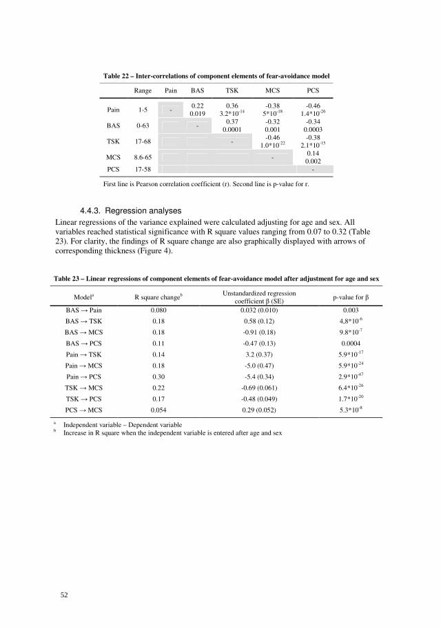
Table 22 – Inter-correlations of component elements of fear-avoidance model
Range Pain BAS TSK MCS PCS
Pain 1-5 - 0.22
0.019 0.36
3.2*10-14 -0.38
5*10-18 -0.46
1.4*10-26
BAS 0-63 - 0.37
0.0001 -0.32 0.001
-0.34 0.0003
TSK 17-68 - -0.46
1.0*10-22 -0.38
2.1*10-15
MCS 8.6-65 - 0.14
0.002 PCS 17-58 -
First line is Pearson correlation coefficient (r). Second line is p-value for r.
4.4.3. Regression analyses
Linear regressions of the variance explained were calculated adjusting for age and sex. All variables reached statistical significance with R square values ranging from 0.07 to 0.32 (Table 23). For clarity, the findings of R square change are also graphically displayed with arrows of corresponding thickness (Figure 4). Table 23 – Linear regressions of component elements of fear-avoidance model after adjustment for age and sex
Modela R square changeb Unstandardized regression
coefficient β (SE) p-value for β
BAS → Pain 0.080 0.032 (0.010) 0.003
BAS → TSK 0.18 0.58 (0.12) 4,8*10-6
BAS → MCS 0.18 -0.91 (0.18) 9.8*10-7
BAS → PCS 0.11 -0.47 (0.13) 0.0004
Pain → TSK 0.14 3.2 (0.37) 5.9*10-17
Pain → MCS 0.18 -5.0 (0.47) 5.9*10-24
Pain → PCS 0.30 -5.4 (0.34) 2.9*10-47
TSK → MCS 0.22 -0.69 (0.061) 6.4*10-26
TSK → PCS 0.17 -0.48 (0.049) 1.7*10-20
PCS → MCS 0.054 0.29 (0.052) 5.3*10-8
a Independent variable – Dependent variable b Increase in R square when the independent variable is entered after age and sex
52

Physical disability
Guarded
movement
Fear of (re)injury
Emotional
disability
Pain
Figure 4 – The fear avoidance model, FAM (arrow thickness represents R square change)
53

5. Discussion
5.1. A review of treatment interventions in WAD (I)
The main finding of this review was the large number of physical therapy articles on the subject. Apparently, this profession is often called upon to treat patients with WAD. The interventions for acute WAD that have the strongest scientific support are: early physical activity [62, 63, 68, 74, 75, 78, 79, 82, 87] (degree 2), electromagnetic field therapy [67, 84] (degree 2) and high-dose Methylprednisolone therapy [76] (degree 2). The interventions for chronic WAD with the strongest scientific support are: radiofrequency neurotomy [72, 86] (degree 1), combined cognitive behavioural therapy with physical therapy interventions [71, 77, 83] (degree 2), melatonin therapy [85] (degree 2) and coordination exercise therapy [66, 70] (degree 2). Although radiofrequency neurotomy is described in a study of fairly high methodological quality score the method is only used in one place in Australia. Furthermore, sample size in that study was small and differences in baseline between groups were not adjusted for. Thus, it is doubtful if radiofrequency neurotomy really has enough evidence to be recommended.
5.1.1. Methodological aspects
The inherent risk of bias must be considered in this review. The selection process was limited to articles published in the English language. Therefore it is possible that relevant articles published in other languages have been missed. Trials with positive outcomes are more likely to be published, however it was noted that articles both in favor and in disfavor of interventions were identified. It is the purpose of systematic reviews to pool, if possible, the results of trials. This can provide valuable information on size of treatment effect and clinical relevance. Nevertheless, intervention trials on WAD display heterogeneity of patient populations. This is, in part, due to differences in the definition of WAD, making pooling of results in practice, impossible.
5.1.2. Scientific shortcomings with some types of interventions
The lists commonly used for measuring methodological quality of RCTs have been developed for drug therapy rather than health sciences. This could lead to bias towards interventions of health sciences. In this review, physical therapy RCTs achieved consistently lower methodological scores compared to drug therapy and orthopedic surgery RCTs (Table 6-8). The interventions used by several health professions often cannot be administered in a double blind fashion. Therefore, they can never achieve full score. The authors recommend that reviews of such RCTs utilize modified methodological quality lists that disregard double blinding.
5.1.3. Acute and chronic WAD
Patients suffering from acute WAD differ from those suffering from chronic WAD. It is currently not possible to identify the injured structures in the acute stage. The available data [63, 82, 88, 89] lead to the suggestion that the combination of the injury with psychological factors such as coping style [90] and explanatory style [91] may lead to chronic WAD. With this in mind and the evidence available, this review recommends that patients suffering from acute WAD be prescribed advice to ‘act-as-usual’ and early, controlled, physical activity to tolerance level.
54

5.1.4. Treatment of acute WAD
Early physical activity reduces pain, increases cervical range of motion and reduces sick leave in patients with acute WAD. This is supported by one high quality study [79] and several low quality studies [62, 63, 73-75, 78, 82].
One high quality RCT suggests that high-dose methylprednisolone therapy within 8 hours of injury should be prescribed [76]. In the authors’ view, however, the practical difficulties of this treatment (8-hour limitation, 23-hour infusion, need for hospitalization, cost) only warrant its use on patients that run higher risk of developing chronic WAD. Findings that are associated with increased risk are neurological signs, initially reduced cervical range of motion [92] and brachial plexus tension signs [93].
The use of magnetic fields is not recommended by this review on the basis that the equipment used in the high quality RCT by Foley-Nolan [67] was collar-mounted, thus conflicting with the advice on early activity and negative effects of collars. It is possible that magnetic field therapy administered by other equipment is equally efficient but the authors did not locate any RCTs on the subject.
5.1.5. Treatment of chronic WAD
Knowledge of the origin of symptoms in the late stage is increasing. Thus, treatment interventions can be directed or developed in a purposeful manner. The identification of cervical zygapophysial joints as the symptom-giving structure in approximately 50% of the patients suffering from chronic WAD [94, 95] is an example. Interventions were evaluated on this condition and intraarticular corticosteroid injection therapy was discarded [61] in favor of radiofrequency neurotomy. This review recommends that patients suffering from chronic WAD be examined for cervical zygapophysial joint pain. In cases of such findings, radiofrequency neurotomy could be considered on the basis of two high quality RCTs [72, 86]. However, the technique is difficult even for experienced personnel [96].
The prescription of combined, cognitive, behavioral therapy and physical therapy interventions in chronic WAD could be recommended on the basis of three low quality RCTs. These interventions have respectively strong and moderate evidence of significant reductions in neck pain and sick leave in patients suffering from chronic WAD.
Abnormalities in sleep quality have been reported in patients suffering from chronic WAD [97]. Melatonin therapy could be considered to improve melatonin onset and sleep/wake rhythm in patients exhibiting delayed melatonin onset and chronic WAD. This recommendation is based on one high quality RCT [85], although one month of treatment did not influence other sleep parameters, pain, quality of life, cognitive processing speed or vigilance.
This review also cautiously recommends including coordination exercises in physical therapy interventions on the basis of two low quality RCTs. There is moderate evidence that coordination exercises significantly reduce neck pain in patients suffering from chronic WAD.
5.2. Active Intervention in Patients with WAD (II)
The main finding in this study was that an active intervention in patients with WAD resulted in a significantly greater reduction in pain intensity, a greater chance to retain or regain CROM and reduced sick leave compared to a standard intervention. These findings could have implications for the management of patients with WAD.
55

5.2.1. Methodological Aspects
It was estimated that the majority of patients exposed to whiplash trauma in this area fulfilling the inclusion criteria were included [78]. It is highly unlikely that patients randomized to standard intervention also received treatment outside the control of this study similar to the active intervention [78].
The last decade has seen a tendency from parametric to non-parametric statistical methods when analyzing VAS. However, opinions differ on comparing changes in VAS over time between groups. Some accept non-parametric methods applied to raw differences. Others state that common mathematical operations such as addition and subtraction are not defined in ordinal scales. Thus, raw changes in VAS were also transformed to –1, 0 or +1 as described previously. There is no international consensus in this matter, therefore changes in VAS are analyzed using the parametric ANOVA and the non-parametric Friedmann’s test applied to both raw and transformed differences, although the authors prefer the latter. When comparing changes in VAS, the results from all three statistical methods favor active intervention (Table 14).
The two-factor design not only divides patients into groups, but also calculates p-values using a two-way-ANOVA or Friedmann’s test. In these statistical tests, all four groups are used simultaneously when calculating any p-value. Thus, the number used to calculate the p-values in table 4 are not 16 versus 21, or 18 versus 18, but simply 73. The two-factor design reduces the large numbers of patients required when several one-factor trials are used.
Evaluating improvement of CROM in patients exposed to a whiplash trauma tells us if the patient is better but also utilizing a control group tells us if the patient is well. An unexposed control group is the only way to estimate pre-injury CROM in injured patients. This comparison has a clinical value and is the motivation for introducing unexposed individuals at the three-year follow-up.
Information to patients before inclusion was not controlled. If some patients randomized to standard intervention prior to inclusion had been given advice similar to that given in active intervention, it would reduce our significant findings. This implies that differences between interventions might be even greater than those found in our study.
5.2.2. Exposed to whiplash trauma and no initial pain
A person exposed to a whiplash trauma initially not reporting symptoms may still bear a minor whiplash injury that might express symptoms later. At present, it is impossible to confirm or rule out an association between the trauma and delayed manifestations of whiplash-associated disorders. Thus, we have chosen to include all patients exposed to a whiplash trauma (caused in a motor vehicle collision) attending the health care system. In our study four patients were initially WAD 0 and three of these later developed manifestations. Interestingly, the only person not developing delayed manifestations belonged to the group receiving early active intervention while the three others received standard.
5.2.3. Possible Mechanisms of the Active Intervention
The most important elements of the active intervention were the high frequency and intensity of self-mobilization and the use of the McKenzie protocol for patients with unresolved symptoms. In short, the standard intervention emphasizes caution, whereas the active intervention encourages active movement.
56

To what extent chronic whiplash syndrome is functional (chronic illness behavior) or organic (persistent tissue injury) is unresolved. It was recently proposed in a clinical practice guideline that psychosocial factors may be present in delayed recovery [98].
Since pain lacks an external convention of reference it allows considerable room for interpretation and thus, the importance of cognitive processes in the pain experience is great [99]. Left unsupervised an exaggerated negative response to pain may develop. The resulting pain related fear is a strong factor in the development of illness behavior [99-101]. Prescribing immediate exercise within comfort limits may alleviate fear of serious injury [46, 102]. Continued supervision during the first weeks would provide ongoing reassurance of a satisfactory outcome, thus promoting wellness behavior. This interaction with a therapist prescribing activity was unavailable in the standard intervention.
The organic aspect of WAD is dealt with by utilizing rotational exercises in the acute stage and repeated movements or positions based on the McKenzie evaluation in the sub-acute stage. Movement encourages regional blood flow and facilitates removal of exudate, thus allowing healing to occur by aiding nutrition of joint structures [103, 104].
It should be pointed out that neither fear-avoidance behavior nor regional blood flow was measured in this study.
5.2.4. Why Cervical Rotation?
Studies indicate that upper limb pain and paraesthesiae in patients with WAD may arise from hyperalgesic cervical or brachial plexus nervous tissue [93, 105]. Cervical spine rotation addresses this involvement by mobilizing the nerve structures on the contralateral side [106] thus preventing scar tissue from forming adhesions causing later dysfunction. Rotation avoids the longitudinal stress on the neural axis caused by flexion and lateral flexion [106]. Mobilisation may also affect possible inhibition of intraneural microvascular blood flow due to compression [107].
5.3. Basic body awareness therapy compared to exercise therapy for patients with chronic WAD (III)
A 10-week treatment regimen of twice-weekly, 90-minute sessions of BBAT for patients suffering from chronic WAD produced better outcomes than ET of equal intensity and duration in this RCT. BBAT produced large effects in posture and quality of movement pattern and moderate effects in the physical functioning domain of SF-36 compared to ET. These effects remained significant at three months while additional moderate effects in the social functioning and bodily pain domains of SF-36 and reduced pain frequency appeared. This is the first RCT, to our knowledge, of the effect of BBAT on chronic WAD.
We employed a trial coordinator responsible for tracking down and maintaining communication with patients. We also provided a great degree of flexibility in booking appointments in order to maximize the number of participating patients. Despite our rigorous efforts to recruit, many patients declined participation. This reflects the difficulty in recruiting patients with long-standing chronic pain to a clinical trial. This limitation must be kept in mind when interpreting the results of this study.
A possible limitation may be the beliefs and attitudes of the treating physical therapist affecting their performance or the mood of the patients and thereby affecting the PROs. Compliance did not differ between groups but the effect of therapist-related variables on outcomes cannot be excluded in the current trial design.
57

New treatments are often evaluated along with a placebo or waiting list. In the current trial the
comparison was made with an effective treatment. This was evident in that the ET group also improved on outcome measures. We presume that greater effects for BBAT would be evident in a comparison to placebo or waiting list.
Although we prefer evaluating comparisons with effect size and t-tests others favour strictly non-parametric testing. Non-parametric testing was also provided in tables 19a, 19b, 20a and 20b. It is important to keep in mind that sample size was calculated for parametric testing. Our sample size may have been too small to power non-parametric testing.
The aim of BBAT is to improve posture and movement pattern. We assume that these improvements are a lifestyle change that will have positive consequences in the long run. Some support for this claim was found in our trial where improvement in additional outcomes was noted at three months. It is possible that improved quality of movement works as a “push in the right direction”.
The effects on the Physical Functioning (PF) domain of SF-36 need to be interpreted in comparison with the findings on NDI. The items comprising PF involve activities of daily living while the NDI covers both symptoms and activities of daily living. BBAT appears to have primarily improved function in activities of daily living and not symptoms.
While reduction in pain intensity did not differ between the groups at three months there was a difference in the Social Functioning (SF) domain of SF-36. SF is the extent and frequency with which physical health or emotional problems interfere with social activities. Being able to participate in social activities despite suffering from chronic WAD is an important finding likely to have bearing on these patients’ quality of life.
The overall trend evident in tables 19a, 19b, 20a and 20b is in favour of BBAT. The data shows greater improvements in favour of BBAT for all outcomes irrespective of significance levels and effect size.
Stewart et al [108] conducted an RCT comparing exercise to advice as treatment for patients with chronic WAD. Patients in that trial experienced clinically significant improvements in pain intensity in response to treatment with exercise compared to advice. These improvements were however no longer statistically significant at the 12-month follow-up as the patients in the advice group improved. This could indicate that the benefits of exercise diminish when exercise is no longer a regular part of a patient’s lifestyle. Their patients initially showed greater improvement in pain intensity and neck disability than we could observe in our trial. However, patients in our trial had a significantly longer duration of symptoms and were older. Thus, our findings support the hypothesis by Stewart et al that patients with a duration of WAD beyond 12 months may be less likely to respond to exercise treatment.
The question whether these effects are worthwhile in terms of treatment and labor costs needs to be investigated in future trials. Further research is also needed to evaluate BBAT as a treatment for WAD of shorter duration (three to 12 months). Another research question arising from the reality of clinical practice is whether an intensive treatment incorporating both BBAT and ET would yield different results.
58

5.4. Applying the fear-avoidance model to patients with chronic WAD (IV)
The FAM of chronic pain appeared valid for patients suffering from chronic WAD in this cross-sectional analysis. Measures of pain, guarded movement, fear of movement/(re)injury, mental health and physical health were significantly correlated and the variables explained each other’s variance to a statistically significant degree.
According to FAM, high levels of fear of movement/(re)injury are related to more guarded movement, greater disability and more pain [109]. Results from our analysis were consistent with this hypothesis. Wideman et al [110] conducted a prospective study of 121 individuals suffering from work-related musculoskeletal injury. Results from that study also corroborate the validity of FAM. However, only 2 individuals in their sample had suffered neck injury, making it difficult to extrapolate their findings to patients suffering from WAD.
Pain catastrophizing is a concept that probably overlaps with fear of movement/(re)injury [46, 109]. Pain catastrophizing contains both a primary appraisal of pain as a threatening experience and a secondary appraisal of perceived inability to cope with pain [111]. We did not include a direct measure of pain catastrophizing in our study. There are conflicting reports concerning the role of pain catastrophizing in FAM. In a study by Leeuw et al [112], fear of movement/(re)injury was not a mediator of pain catastrophizing in a non-clinical sample of people with low-back pain. In another study by Nieto et al [113], the effect of pain catastrophizing on disability was shown to be completely mediated by fear of movement/(re)injury in a study of 147 patients suffering from sub-acute WAD. The different findings might be explained by sample differences.
Studies of FAM have been criticized for using samples of patients with chronic symptoms. This is valid criticism since it is perhaps more important to predict and prevent musculoskeletal injuries from developing into chronic conditions. This could perhaps be better achieved by studying acute or subacute patients or even individuals that have not yet developed symptoms. We must be similarly cautious when interpreting results from cross-sectional studies as they are of limited value in determining causation. The current way of describing FAM in diagrams [109] can be misleading since it is largely based on cross-sectional studies. There is, to our knowledge, no statistical method to guide us when choosing the direction of arrows between the component causes of chronic musculoskeletal disorders; thus, prospective study designs are preferable.
Path analysis and structural equation modelling can provide some guidance in graphically
depicting FAM but are only applicable in acyclic relations between causes. However, the component elements of FAM seem to significantly affect each other and there is no statistical method to study cyclical relations as far as we know. We are therefore sceptical to the notion that the current graphic illustrations of FAM indicates chronological order of the occurrence of the component elements [110]. In fact, Gheldof et al [114] found that fear of movement/(re)injury significantly predicted disability but was best modelled as a consequence of pain severity rather than as an antecedent. Perhaps regarding each component cause as a risk factor for the occurrence of chronicity would suffice. This highlights the need for epidemiological studies of these factors.
The regression analyses in this study were statistically significant which speaks for the validity of FAM in chronic WAD. However the R square values were generally small. We interpret this as an indication that there are still many unknown factors that affect the elements of FAM. This could be partly due to the instruments used to measure each element and partly to other factors not currently included in FAM.
59

The current study is one of few that attempt to include a measure of guarded movement [115] in a model of FAM for chronic WAD. Nederhand et al [45, 47] studied 92 patients with acute WAD and found that both fear of movement/(re)injury and pain intensity were independently associated with a decreased level of muscle activation. Another finding from their study was the inverse relationship between the level of neck pain disability and muscle activation. These findings support FAM as decreased muscle activation is indicative of minimizing the use of painful muscles, which is, in effect, avoidance behavior. Pearson et al [116] found no correlation between maximal voluntary isometric neck strength and pain intensity, disability, pain catastrophizing and fear of movement/(re)injury. However, their study had a sample of only 14 patients making it difficult to draw conclusions on any correlations.
60

6. Summary and conclusions Further prospective studies including acute and subacute patients are warranted when considering the crucial question of transition from acute to chronic symptoms. However, applying the FAM of chronic pain to patients suffering from chronic WAD appears valid.
The active intervention in study II utilizes the ideas of FAM and addresses both organic and functional aspects of acute WAD, reducing cervical pain, need for sick leave and restoring impaired CROM. The main clinical implication is that patients with acute WAD 0, 1 or 2 should be instructed in self-mobilization as soon as possible. The emphasis should be on frequently repeated cervical rotation. Instructions should be repeated until comprehension and compliance is ensured. If symptoms persist more than 20 days after trauma patients should be referred to a health professional educated in mechanical diagnosis and therapy according to the McKenzie system.
Some of the ideas in BBAT are compatible with the FAM. BBAT appears to be a slightly better alternative to ET for patients suffering from chronic WAD with several years duration. However, this should be confirmed in a long-term follow-up.
Based on the degrees of evidence and the practical obstacles the following treatments can be recommended:
→ Early physical activity in acute WAD. → A combination of cognitive behavioral therapy with physical therapy interventions and
coordination exercise therapy in chronic WAD. BBAT has the potential to become a valuable addition to the therapeutic arsenal for patients with chronic WAD.
High quality RCTs are not common in the field of WAD. More research is needed,
particularly on the treatment of chronic WAD.
61

7. Acknowledgements I wish to express my sincere gratitude and appreciation to all those who in various ways assisted, contributed and supported me in this long journey. Greek poet Constantine Kavafis writes: “When you set sail for Ithaca, wish for the road to be long, full of adventures, full of knowledge” and it definitely has, was and definitely will be. Or one can say that it’s only just beginning. To the following people, in no particular order, I would like to dedicate a few lines of text: To my father, professor Konstantin Seferiadis and mother Chrysoula Seferiadi for believing in me, loving me and making everything in my life possible. To my chief supervisor, Ronny Gunnarsson, for teaching, inspiring and guiding me through the jungles of academia. You sir, are a gentleman and a scholar. To my co-supervisor Annika Billhult, for unbridled enthusiasm and useful advice. To Mark Rosenfeld, without you I would never have ended up here. I really enjoy working with you and am smitten by your open-heartedness (is this a word?) and hospitality. You welcomed me to your home so many times and made work into play. To Pernilla Ohlin, for her work ethic, patience (with me and others) and bright smile in the face of troubles. To Lena Nordeman, for stimulating, thought provoking professional talks and endless energy. To my boss Mona Asp, for never doubting, always supporting, maintaining focus, and looking out for me. To all my colleagues, both at work and in the research project: thank you. To all the patients for putting up with us and the examinations and giving us their time. Nothing would have been possible without your help. To Milda Kairyt÷, for everything that we shared and for being there, whenever. To Sally Marshall, our talks meant more to me than I had imagined. To Ermin Catic, for saving the day when the shit hit the fan. To Michalis Seferiadis and Georgia Peta, for infecting me with their passion for physiotherapy. To Giorgos Kitsoulis, probably the best physical therapist I have ever met. You are the reason I am in this country. To all my friends in Sweden for warming the winters: Antreas (διαλιέχτε!), Tomas, Joan, Giorgos H, Apostolos and Panagiota, Petros, Nikos and Peny, Antonis, Marilena (I put your name close to JP), Jean-Pierre, Dimitris and Maria, Giorgos P, Oscar, Anthi, Johan and Petra, Nikos (“I have 2 questions to ask you…”), Yniel, Måns, Axel and Julietta, Hedvig and Jari, Jakob, Markus, Elias, Martina, Alex and Johannes, Janerik, Christian and Martin H.
62

To all my friends in Greece for cooling the summers: Kostantis, Vangelis and Phoebe, Angelos (ok technically London), Giorgos and Popi, Giorgos T, Panagiotis, Dimitris K (Ras), Giannis and Loukas. To Lena, Magdalena and Lisa (Joan I hope you are reading this). Finally, to everyone that I shared a statistically significant part of my life with.
63

8. References [1]. Russell G, Nicol P. 'I've broken my neck or something!' The general practice experience of
whiplash. Fam Pract 2009;26(2):115-20. [2]. Spitzer WO, Skovron ML, Salmi LR et al. Scientific monograph of the Quebec Task Force
on Whiplash-Associated Disorders: redefining "whiplash" and its management. Spine 1995;20(8 SUPPL):1S-73S.
[3]. Barnsley L, Lord S, Bogduk N. Whiplash injury. Pain 1994;58:283-307. [4]. Guzman J, Hurwitz EL, Carroll LJ et al. A new conceptual model of neck pain: linking
onset, course, and care: the Bone and Joint Decade 2000-2010 Task Force on Neck Pain and Its Associated Disorders. Spine 2008;33(4 SUPPL):S14-23.
[5]. Nordin M, Carragee EJ, Hogg-Johnson S et al. Assessment of neck pain and its associated disorders: results of the Bone and Joint Decade 2000-2010 Task Force on Neck Pain and Its Associated Disorders. Spine 2008;33(4 SUPPL):S101-22.
[6]. Kivioja J, Rinaldi L, Ozenci V et al. Chemokines and their receptors in whiplash injury: elevated RANTES and CCR-5. J Clin Immunol 2001;21(4):272-7.
[7]. Kivioja J, Ozenci V, Rinaldi L, Kouwenhoven M, Lindgren U, Link H. Systemic immune response in whiplash injury and ankle sprain: elevated IL-6 and IL-10. Clin Immunol 2001;101(1):106-12.
[8]. Moskovich R, Petrizzo A. Evaluation of the neck. In Musculoskeletal disorders in the workplace: Principles and practice. Nordin M, Andersson G, Pope M. Philadelphia, PA: Mosby Elsevier; 1997, pp 55-72.
[9]. Lindh M, Rosenfeld M, Tenenbaum A, Jansson A. The care of patients with whiplash-associated disorders: guidelines for the Region Västra Götaland. Göteborg: Region Västra Götaland; 2003.
[10]. Stiell IG, Wells GA, Vandemheen KL et al. The Canadian C-spine rule for radiography in alert and stable trauma patients. JAMA 2001;286(15):1841-8.
[11]. Hoffman JR, Mower WR, Wolfson AB, Todd KH, Zucker MI. Validity of a set of clinical criteria to rule out injury to the cervical spine in patients with blunt trauma. National Emergency X-Radiography Utilization Study Group. N Engl J Med 2000;343(2):94-9.
[12]. Holm LW, Carroll LJ, Cassidy JD et al. The burden and determinants of neck pain in whiplash-associated disorders after traffic collisions: results of the Bone and Joint Decade 2000-2010 Task Force on Neck Pain and Its Associated Disorders. Spine 2008;33(4 SUPPL):S52-9.
[13]. Hogg-Johnson S, van der Velde G, Carroll LJ et al. The burden and determinants of neck pain in the general population: results of the Bone and Joint Decade 2000-2010 Task Force on Neck Pain and Its Associated Disorders. Spine 2008;33(4 SUPPL):S39-51.
[14]. Cassidy JD, Carroll LJ, Côté P, Lemstra M, Berglund A, Nygren A. Effect of eliminating compensation for pain and suffering on the outcome of insurance claims for whiplash injury. N Engl J Med 2000;342(16):1179-86.
[15]. Carroll LJ, Holm LW, Hogg-Johnson S et al. Course and prognostic factors for neck pain in whiplash-associated disorders (WAD): results of the Bone and Joint Decade 2000-2010 Task Force on Neck Pain and Its Associated Disorders. Spine 2008;33(4 SUPPL):S83-92.
[16]. Borenstein P, Rosenfeld M, Gunnarsson R. Cognitive symptoms, cervical range of motion and pain as prognostic factors after whiplash trauma. Acta Neurol Scand 2009;.
[17]. Côté P, Hogg-Johnson S, Cassidy JD, Carroll L, Frank JW, Bombardier C. Initial patterns of clinical care and recovery from whiplash injuries: a population-based cohort study. Arch
Intern Med 2005;165(19):2257-63.
64

[18]. Côté P, Hogg-Johnson S, Cassidy JD, Carroll L, Frank JW, Bombardier C. Early aggressive care and delayed recovery from whiplash: isolated finding or reproducible result? Arthritis Rheum 2007;57(5):861-8.
[19]. Forrester-Brown MF. Posture as a factor in health and disease. Br Med J 1926;1:690-3. [20]. Hansson KG. Body mechanics and posture. JAMA 1945;128:947-53. [21]. Griegel-Morris P, Larson K, Mueller-Klaus K, Oatis CA. Incidence of common postural
abnormalities in the cervical, shoulder, and thoracic regions and their association with pain in two age groups of healthy subjects. Phys Ther 1992;72(6):425-31.
[22]. Nilsson BM, Söderlund A. Head posture in patients with whiplash-associated disorders and the measurement method's reliability - A comparison to healthy subjects. Adv Physiother 2005;7(1):13-9.
[23]. Yip CH, Chiu TT, Poon AT. The relationship between head posture and severity and disability of patients with neck pain. Man Ther 2008;13(2):148-54.
[24]. Silva AG, Punt TD, Sharples P, Vilas-Boas JP, Johnson MI. Head posture and neck pain of chronic nontraumatic origin: a comparison between patients and pain-free persons. Arch
Phys Med Rehabil 2009;90(4):669-74. [25]. Falla D, Jull G, Russell T, Vicenzino B, Hodges P. Effect of neck exercise on sitting
posture in patients with chronic neck pain. Phys Ther 2007;87(4):408-17. [26]. Lau KT, Cheung KY, Chan KB, Chan MH, Lo KY, Wing Chiu TT. Relationships between
sagittal postures of thoracic and cervical spine, presence of neck pain, neck pain severity and disability. Man Ther 2010;.
[27]. Heikkilä H, Aström PG. Cervicocephalic kinesthetic sensibility in patients with whiplash injury. Scand J Rehabil Med 1996;28(3):133-8.
[28]. Loudon JK, Ruhl M, Field E. Ability to reproduce head position after whiplash injury. Spine 1997;22(8):865-8.
[29]. Knox JJ, Beilstein DJ, Charles SD et al. Changes in head and neck position have a greater effect on elbow joint position sense in people with whiplash-associated disorders. Clin J
Pain 2006;22(6):512-8. [30]. Sandlund J, Djupsjöbacka M, Ryhed B, Hamberg J, Björklund M. Predictive and
discriminative value of shoulder proprioception tests for patients with whiplash-associated disorders. J Rehabil Med 2006;38(1):44-9.
[31]. Sandlund J, Röijezon U, Björklund M, Djupsjöbacka M. Acuity of goal-directed arm movements to visible targets in chronic neck pain. J Rehabil Med 2008;40(5):366-74.
[32]. Sterling M, Jull G, Vicenzino B, Kenardy J, Darnell R. Development of motor system dysfunction following whiplash injury. Pain 2003;103(1-2):65-73.
[33]. Treleaven J, Jull G, Sterling M. Dizziness and unsteadiness following whiplash injury: characteristic features and relationship with cervical joint position error. J Rehabil Med 2003;35(1):36-43.
[34]. Heikkilä HV, Wenngren BI. Cervicocephalic kinesthetic sensibility, active range of cervical motion, and oculomotor function in patients with whiplash injury. Arch Phys Med
Rehabil 1998;79(9):1089-94. [35]. Falla D. Unravelling the complexity of muscle impairment in chronic neck pain. Man Ther
2004;9(3):125-33. [36]. Jull G, Barrett C, Magee R, Ho P. Further clinical clarification of the muscle dysfunction
in cervical headache. Cephalalgia 1999;19(3):179-85. [37]. Jull G. Deep Cervical Flexor Muscle Dysfunction in Whiplash. J Musculoskel Pain
2000;8(1/2):143-54. [38]. Falla DL, Jull GA, Hodges PW. Patients with neck pain demonstrate reduced
electromyographic activity of the deep cervical flexor muscles during performance of the craniocervical flexion test. Spine 2004;29(19):2108-14.
65

[39]. Falla D, Rainoldi A, Merletti R, Jull G. Myoelectric manifestations of sternocleidomastoid and anterior scalene muscle fatigue in chronic neck pain patients. Clin Neurophysiol 2003;114(3):488-95.
[40]. Uhlig Y, Weber BR, Grob D, Müntener M. Fiber composition and fiber transformations in neck muscles of patients with dysfunction of the cervical spine. J Orthop Res 1995;13(2):240-9.
[41]. Nederhand MJ, IJzerman MJ, Hermens HJ, Baten CT, Zilvold G. Cervical muscle dysfunction in the chronic whiplash associated disorder grade II (WAD-II). Spine 2000;25(15):1938-43.
[42]. Falla D, Bilenkij G, Jull G. Patients with chronic neck pain demonstrate altered patterns of muscle activation during performance of a functional upper limb task. Spine 2004;29(13):1436-40.
[43]. Nederhand MJ, Hermens HJ, IJzerman MJ, Turk DC, Zilvold G. Cervical muscle dysfunction in chronic whiplash-associated disorder grade 2: the relevance of the trauma. Spine 2002;27(10):1056-61.
[44]. van der Hulst M, Vollenbroek-Hutten MM, Rietman JS, Schaake L, Groothuis-Oudshoorn KG, Hermens HJ. Back muscle activation patterns in chronic low back pain during walking: a "guarding" hypothesis. Clin J Pain 2010;26(1):30-7.
[45]. Nederhand MJ, Hermens HJ, IJzerman MJ, Turk DC, Zilvold G. Chronic neck pain disability due to an acute whiplash injury. Pain 2003;102(1-2):63-71.
[46]. Vlaeyen JW, Linton SJ. Fear-avoidance and its consequences in chronic musculoskeletal pain: a state of the art. Pain 2000;85(3):317-32.
[47]. Nederhand MJ, Hermens HJ, Ijzerman MJ, Groothuis KG, Turk DC. The effect of fear of movement on muscle activation in posttraumatic neck pain disability. Clin J Pain 2006;22(6):519-25.
[48]. Elliott J, Jull G, Noteboom JT, Darnell R, Galloway G, Gibbon WW. Fatty infiltration in the cervical extensor muscles in persistent whiplash-associated disorders: a magnetic resonance imaging analysis. Spine 2006;31(22):E847-55.
[49]. Jadad AR, Moore RA, Carroll D et al. Assessing the quality of reports of randomized clinical trials: is blinding necessary? Control Clin Trials 1996;17(1):1-12.
[50]. Verhagen AP, de Vet HC, de Bie RA et al. The Delphi list: a criteria list for quality assessment of randomized clinical trials for conducting systematic reviews developed by Delphi consensus. J Clin Epidemiol 1998;51(12):1235-41.
[51]. Peeters GG, Verhagen AP, de Bie RA, Oostendorp RA. The efficacy of conservative treatment in patients with whiplash injury: a systematic review of clinical trials. Spine 2001;26(4):E64-73.
[52]. Van Tulder MW, Touray T, Furlan AD, Solway S, Bouter LM. Muscle relaxants for nonspecific low back pain: a systematic review within the framework of the cochrane collaboration. Spine 2003;28(17):1978-92.
[53]. Huskisson EC. Measurement of pain. Lancet 1974;2(7889):1127-31. [54]. Carlsson AM. Assessment of chronic pain. I. Aspects of the reliability and validity of the
visual analogue scale. Pain 1983;16(1):87-101. [55]. Peolsson A, Hedlund R, Öberg B. Intra- and inter-tester reliability and range of motion of
the neck. Physiother Can 2000;52:233-42. [56]. Linton S, Halldén K, Hellsing AL. The Reliability of Self-reported Sick Absenteeism: A
Pilot Study. Cogn Behav Ther 1995;24(4):145-50. [57]. Salter RB. The biologic concept of continuous passive motion of synovial joints. The first
18 years of basic research and its clinical application. Clin Orthop Relat Res 1989;242:12-25.
[58]. McKenzie RA. Cervical Trauma. The Cervical and Thoracic spine, Mechanical Diagnosis and Therapy. Waikanae, New Zealand: Spinal Publications Limited; 1990, pp 200-3.
66

[59]. Roxendal G. Body awareness therapy and the body awareness scale, treatment and evaluation in psychiatric physiotherapy. Gothenburg: Department of Psychiatry, University of Gothenburg; 1985.
[60]. Cohen J. Statistical Power Analysis for the Behavioral Sciences. New Jersey: Lawrence Erlbaum Associates Inc.; 1988.
[61]. Barnsley L, Lord SM, Wallis BJ, Bogduk N. Lack of effect of intraarticular corticosteroids for chronic pain in the cervical zygapophyseal joints. N Engl J Med 1994;330(15):1047-50.
[62]. Bonk AD, Ferrari R, Giebel GD, Edelmann M, Huser R. Prospective, randomized, controlled study of activity versus collar, and the natural history for whiplash injury, in Germany. World Congress on Whiplash-Associated Disorders in Vancouver, British Columbia, Canada in February of 1999. J Musculoskel Pain 2000;8(1/2):123-32.
[63]. Borchgrevink GE, Kaasa A, McDonagh D, Stiles TC, Haraldseth O, Lereim I. Acute treatment of whiplash neck sprain injuries. Spine 1998;23(1):25-31.
[64]. Byrn C, Olsson I, Falkheden L et al. Subcutaneous sterile water injections for chronic neck and shoulder pain following whiplash injuries [see comments]. Lancet 1993;341:449-52.
[65]. Cassidy JD, Lopes AA, Yong-Hing K. The immediate effect of manipulation versus mobilization on pain and range of motion in the cervical spine: a randomized controlled trial. J Manipulative Physiol Ther 1992;15(9):570-5.
[66]. Fitz-Ritson D. Phasic exercises for cervical rehabilitation after "whiplash" trauma. J
Manipulative Physiol Ther 1995;18(1):21-4. [67]. Foley-Nolan D, Moore K, Codd M, Barry C, O'Connor P, Coughlan RJ. Low energy high
frequency pulsed electromagnetic therapy for acute whiplash injuries. A double blind randomized controlled study.PG - 51-9. Scand J Rehabil Med 1992;24(1):51-9.
[68]. Gennis P, Miller L, Gallagher EJ, Giglio J, Carter W, Nathanson N. The effect of soft cervical collars on persistent neck pain in patients with whiplash injury. Acad Emerg Med 1996;3(6):568-73.
[69]. Hendriks O, Horgan A. Ultra-reiz current as an adjunct to standard treatment of the acute whiplash patient. Physiother Ireland 1996;17(1):3-7.
[70]. Humphreys BK, Irgens PM. The effect of a rehabilitation exercise program on head-repositioning accuracy and reported levels of pain in chronic neck pain subjects. Journal
Of Whiplash & Related Disorders 2002;1(1):99-112. [71]. Johansson C, Dahl J, Jannert M, Melin L, Andersson G. Effects of a cognitive-behavioral
pain-management program. Behav Res Ther 1998;36(10):915-30. [72]. Lord SM, Barnsley L, Wallis BJ, McDonald GJ, Bogduk N. Percutaneous radio-frequency
neurotomy for chronic cervical zygapophyseal-joint pain. N Engl J Med 1996;335(23):1721-6.
[73]. McKinney LA. Early mobilisation and outcome in acute sprains of the neck. BMJ 1989;299(6706):1006-8.
[74]. Mealy K, Brennan H, Fenelon GC. Early mobilization of acute whiplash injuries. Br Med J
(Clin Res Ed) 1986;292(6521):656-7. [75]. Pennie BH, Agambar LJ. Whiplash injuries. A trial of early management. J Bone Joint
Surg Br 1990;72(2):277-9. [76]. Pettersson K, Toolanen G. High-dose methylprednisolone prevents extensive sick leave
after whiplash injury. A prospective, randomized, double-blind study. Spine 1998;23(9):984-9.
[77]. Provinciali L, Baroni M, Illuminati L, Ceravolo MG. Multimodal treatment to prevent the late whiplash syndrome. Scand J Rehabil Med 1996;28(2):105-11.
[78]. Rosenfeld M, Gunnarsson R, Borenstein P. Early intervention in whiplash-associated disorders: a comparison of two treatment protocols. Spine 2000;25(14):1782-7.
67

[79]. Rosenfeld M, Seferiadis A, Carlsson J, Gunnarsson R. Active intervention in patients with whiplash-associated disorders improves long-term prognosis. A randomized controlled trial. Spine 2003;28(22):2491-8.
[80]. Sand T, Bovim G, Helde G. Intracutaneous sterile water injections do not relieve pain in cervicogenic headache. Acta Neurol Scand 1992;86(5):526-8.
[81]. Schreiber S, Vinokur S, Shavelzon V, Pick CG, Zahavi E, Shir Y. A randomized trial of fluoxetine versus amitriptyline in musculo-skeletal pain. Isr J Psychiatry Relat Sci 2001;38(2):88-94.
[82]. Soderlund A, Olerud C, Lindberg P. Acute whiplash-associated disorders (WAD): the effects of early mobilization and prognostic factors in long-term symptomatology. Clin
Rehabil 2000;14(5):457-67. [83]. Soderlund A, Lindberg P. An integrated physiotherapy/cognitive-behavioural approach to
the analysis and treatment of chronic whiplash associated disorders, WAD. Disabil Rehabil 2001;23(10):436-47.
[84]. Thuile C, Walzl M. Evaluation of electromagnetic fields in the treatment of pain in patients with lumbar radiculopathy or the whiplash syndrome. NeuroRehabilitation 2002;17(1):63-7.
[85]. van Wieringen S, Jansen T, Smits M, Nagtegaal J, Coenen A. Melatonin for chronic whiplash syndrome with delayed melatonin onset: Randomised, placebo-controlled trial. Clin Drug Inv 2001;21(12):813-20.
[86]. Wallis BJ, Lord SM, Bogduk N. Resolution of psychological distress of whiplash patients following treatment by radiofrequency neurotomy: a randomised, double-blind, placebo-controlled trial. Pain 1997;73(1):15-22.
[87]. McKinney LA, Dornan JO, Ryan M. The role of physiotherapy in the management of acute neck sprains following road-traffic accidents. Arch Emerg Med 1989;6(1):27-33.
[88]. Linton SJ. A review of psychological risk factors in back and neck pain. Spine 2000;25(9):1148-56.
[89]. Lee J, Giles K, Drummond PD. Psychological disturbances and an exaggerated response to pain in patients with whiplash injury. J Psychosom Res 1993;37:105-10.
[90]. Buitenhuis J, Spanjer J, Fidler V. Recovery from acute whiplash: the role of coping styles. Spine 2003;28(9):896-901.
[91]. Lin EH, Peterson C. Pessimistic explanatory style and response to illness. Behav Res Ther 1990;28(3):243-8.
[92]. Hartling L, Brison RJ, Ardern C, Pickett W. Prognostic Value of the Quebec Classification of Whiplash-Associated Disorders. Spine 2001;26(1):36-41.
[93]. Ide M, Ide J, Yamaga M, Takagi K. Symptoms and signs of irritation of the brachial plexus in whiplash injuries. J Bone Joint Surg Br 2001;83(2):226-9.
[94]. Barnsley L, Lord SM, Wallis BJ, Bogduk N. The prevalence of chronic cervical zygapophysial joint pain after whiplash. Spine 1995;20:20-5.
[95]. Lord SM, Barnsley L, Wallis BJ, Bogduk N. Chronic cervical zygapophysial joint pain after whiplash. A placebo-controlled prevalence study. Spine 1996;21:1737-44.
[96]. Lord SM, Barnsley L, Bogduk N. Percutaneous radiofrequency neurotomy in the treatment of cervical zygapophysial joint pain: a caution. Neurosurgery 1995;36(4):732-9.
[97]. Schlesinger I, Hering-Hanit R, Dagan Y. Sleep disturbances after whiplash injury: objective and subjective findings. Headache 2001;41(6):586-9.
[98]. Scholten-Peeters GG, Bekkering GE, Verhagen AP et al. Clinical practice guideline for the physiotherapy of patients with whiplash-associated disorders. Spine 2002;27(4):412-22.
[99]. McCracken LM, Gross RT, Aikens J, Carnrike CL. The assessment of anxiety and fear in persons with chronic pain: a comparison of instruments. Behav Res Ther 1996;34(11-12):927-33.
68

[100]. Klenerman L, Slade PD, Stanley IM et al. The prediction of chronicity in patients with an acute attack of low back pain in a general practice setting. Spine 1995;20(4):478-84.
[101]. Linton SJ, Andersson T. Can chronic disability be prevented? A randomized trial of a cognitive-behavior intervention and two forms of information for patients with spinal pain. Spine 2000;25(21):2825-31 DISCUSSION 28.
[102]. Fordyce WE, Brockway JA, Bergman JA, Spengler D. Acute back pain: a control-group comparison of behavioral vs traditional management methods. J Behav Med 1986;9(2):127-40.
[103]. Taylor J, Twomey L. Acute Injuries to Cervical Joints. Spine 1993;18:1115-22. [104]. Buckwalter JA. Effects of early motion on healing of musculoskeletal tissues. Hand
Clin 1996;12(1):13-24. [105]. Quintner JL. A study of upper limb pain and paraesthesiae following neck injury in
motor vehicle accidents: assessment of the brachial plexus tension test of Elvey. Br J
Rheumatol 1989;28(6):528-33. [106]. Harrison DE, Cailliet R, Harrison DD, Troyanovich SJ, Harrison SO. A review of
biomechanics of the central nervous system--Part I: spinal canal deformations resulting from changes in posture. J Manipulative Physiol Ther 1999;22(4):227-34.
[107]. Rempel D, Dahlin L, Lundborg G. Pathophysiology of nerve compression syndromes: response of peripheral nerves to loading. J Bone Joint Surg Am 1999;81(11):1600-10.
[108]. Stewart MJ, Maher CG, Refshauge KM, Herbert RD, Bogduk N, Nicholas M. Randomized controlled trial of exercise for chronic whiplash-associated disorders. Pain 2007;128(1-2):59-68.
[109]. Leeuw M, Goossens ME, Linton SJ, Crombez G, Boersma K, Vlaeyen JW. The fear-avoidance model of musculoskeletal pain: current state of scientific evidence. J Behav
Med 2007;30(1):77-94. [110]. Wideman TH, Adams H, Sullivan MJ. A prospective sequential analysis of the fear-
avoidance model of pain. Pain 2009;145(1-2):45-51. [111]. Severeijns R, Vlaeyen JW, van den Hout MA. Do we need a communal coping model
of pain catastrophizing? An alternative explanation. Pain 2004;111(3):226-9. [112]. Leeuw M, Houben RM, Severeijns R, Picavet HS, Schouten EG, Vlaeyen JW. Pain-
related fear in low back pain: a prospective study in the general population. Eur J Pain 2007;11(3):256-66.
[113]. Nieto R, Miró J, Huguet A. The fear-avoidance model in whiplash injuries. Eur J Pain 2009;13(5):518-23.
[114]. Gheldof EL, Crombez G, Van den Bussche E et al. Pain-related fear predicts disability, but not pain severity: A path analytic approach of the fear-avoidance model. Eur J Pain 2010;.
[115]. van der Hulst M, Vollenbroek-Hutten MM, Rietman JS, Hermens HJ. Lumbar and abdominal muscle activity during walking in subjects with chronic low back pain: support of the "guarding" hypothesis? J Electromyogr Kinesiol 2010;20(1):31-8.
[116]. Pearson I, Reichert A, De Serres SJ, Dumas JP, Côté JN. Maximal voluntary isometric neck strength deficits in adults with whiplash-associated disorders and association with pain and fear of movement. J Orthop Sports Phys Ther 2009;39(3):179-87.
69

Original publications
I. Seferiadis A, Rosenfeld M, Gunnarsson R. A review of treatment interventions in whiplash-associated disorders. Eur Spine J. 2004 Aug;13(5):387-97.
II. Rosenfeld M, Seferiadis A, Carlsson J, Gunnarsson R. Active intervention in patients with whiplash-associated disorders improves long-term prognosis: a randomized controlled clinical trial. Spine (Phila Pa 1976). 2003 Nov 15;28(22):2491-8.
III. Seferiadis A, Ohlin P, Billhult A, Gunnarsson R. Basic body awareness therapy superior to exercise therapy for patients with chronic whiplash-associated disorders: a randomized controlled clinical trial. Submited Manuscript.
IV. Seferiadis A, Ohlin P, Billhult A, Gunnarsson R. Applying the fear-avoidance model to patients with chronic whiplash associated disorders: a cross-sectional study. Manuscript.
The papers have been reprinted with permission of the journals.
70