Treatment Failure after Uterine Artery Embolization …...derwent uterine fibroid treatment as a...
8
Ewha Med J 2012;35(2):102-109 pISSN 2234-3180 / eISSN 2234-2591 Original Article 102 Treatment Failure after Uterine Artery Embolization for Symptomatic Uterine Fibroids: Significance of Ovarian Arterial Collateral Vessels in Predicting the Outcome Byung Chul Kang Department of Radiology, Ewha Womans University School of Medicine, Seoul, Korea Objectives: To evaluate the treatment failure (TF) rate of leiomyoma after uterine artery embolization (UAE) for uterine leiomyomas in cases of the presence of anastomoses between the ovarian arteries (OA) and uterine arteries (UA). Methods: The results of 163 consecutive UAE for uterine fibroid were reviewed. Mean patient age was 42.8 years (range, 25 to 57 years). TF was evaluated according to the anastomoses between OA and UA on pre-embolization angiography. Magnetic resonance images (MRIs) were obtained at 1∼6 months or 12 months after UAE. MRIs were gadolinium (Gd)-enhanced images and/or T2-weighted images. MRIs exhibited focal enhancement portion on fibroid and evaluated the TF rate of the leiomyoma in patients of presence of anastomoses between OA and UA. Results: Fifty six patients had anastomoses between UA and OA on pre-embolization angiography (56/163, 34.4%). Angiographic subtypes were type Ia (n=19), type Ib (n=16), type II (n=11) and type III (n=10). Of all pa- tients, 10 patients showed the focal enhancements of the leiomyomas on follow-up enhanced MRIs (10/163, 6.1%). Three treatments failed in patients demonstrated type Ia (3/19, 15,8%). One had type Ib (1/16, 6.3%). Other 6 had no anastomoses. There was no TF rate difference between patients with communication (4/56, 7.1%) and without communication (6/107, 5.7%). However, TF rate in patients with type Ia communication (15.8%) was higher than that without communication (5.7%; P<0.05). Conclusion: Type Ia utero-ovarian anastomoses communication could be a contraindication for embolization treatment for leiomyoma. (Ewha Med J 2012;35(2):102-109) Key Words: Leiomyoma; Treatment failure; Uterine artery embolization Received: July 30, 2012, Accepted: August 22, 2012 Corresponding author: Byung Chul Kang, Department of Radiology, Ewha Womans University School of Medicine, 1071 Anyangcheon-ro, Yangcheon-gu, Seoul 158-710, Korea Tel: 82-2-2650-5092, Fax: 82-2-2650-5302 E-mail: [email protected] Introduction Uterine leiomyomas affect approximately one fourth to one third of women in the United States, with 140,000 ∼180,000 new diagnoses annually [1]. Patients com- plain of various symptoms including menorrhagia, met- rorrhagia, localized cyclical pelvic pain, and pelvic full- ness [1]. Uterine arterial embolization (UAE) has been accepted as an effective treatment alternative for symp- tomatic uterine fibroid [2-5]. Clinical failure has been also reported from 4% to 19% [2-6] and it has been suggested of technical or anatomical origin [7-10].
Transcript of Treatment Failure after Uterine Artery Embolization …...derwent uterine fibroid treatment as a...

Ewha Med J 2012;35(2):102-109
pISSN 2234-3180 / eISSN 2234-2591
Original Article
102
Treatment Failure after Uterine Artery Embolization for Symptomatic Uterine Fibroids: Significance of Ovarian Arterial Collateral Vessels in Predicting the OutcomeByung Chul Kang Department of Radiology, Ewha Womans University School of Medicine, Seoul, Korea
Objectives: To evaluate the treatment failure (TF) rate of leiomyoma after uterine artery embolization (UAE) for uterine leiomyomas in cases of the presence of anastomoses between the ovarian arteries (OA) and uterine arteries (UA).Methods: The results of 163 consecutive UAE for uterine fibroid were reviewed. Mean patient age was 42.8 years (range, 25 to 57 years). TF was evaluated according to the anastomoses between OA and UA on pre-embolization angiography. Magnetic resonance images (MRIs) were obtained at 1∼6 months or 12 months after UAE. MRIs were gadolinium (Gd)-enhanced images and/or T2-weighted images. MRIs exhibited focal enhancement portion on fibroid and evaluated the TF rate of the leiomyoma in patients of presence of anastomoses between OA and UA. Results: Fifty six patients had anastomoses between UA and OA on pre-embolization angiography (56/163, 34.4%). Angiographic subtypes were type Ia (n=19), type Ib (n=16), type II (n=11) and type III (n=10). Of all pa-tients, 10 patients showed the focal enhancements of the leiomyomas on follow-up enhanced MRIs (10/163, 6.1%). Three treatments failed in patients demonstrated type Ia (3/19, 15,8%). One had type Ib (1/16, 6.3%). Other 6 had no anastomoses. There was no TF rate difference between patients with communication (4/56, 7.1%) and without communication (6/107, 5.7%). However, TF rate in patients with type Ia communication (15.8%) was higher than that without communication (5.7%; P<0.05).Conclusion: Type Ia utero-ovarian anastomoses communication could be a contraindication for embolization treatment for leiomyoma. (Ewha Med J 2012;35(2):102-109)
Key Words: Leiomyoma; Treatment failure; Uterine artery embolization
Received: July 30, 2012, Accepted: August 22, 2012
Corresponding author: Byung Chul Kang, Department of Radiology,Ewha Womans University School of Medicine, 1071 Anyangcheon-ro, Yangcheon-gu, Seoul 158-710, Korea Tel: 82-2-2650-5092, Fax: 82-2-2650-5302E-mail: [email protected]
Introduction
Uterine leiomyomas affect approximately one fourth
to one third of women in the United States, with 140,000∼180,000 new diagnoses annually [1]. Patients com-plain of various symptoms including menorrhagia, met-rorrhagia, localized cyclical pelvic pain, and pelvic full-ness [1]. Uterine arterial embolization (UAE) has been accepted as an effective treatment alternative for symp-tomatic uterine fibroid [2-5]. Clinical failure has been also reported from 4% to 19% [2-6] and it has been suggested of technical or anatomical origin [7-10].

Kang BC: Treatment Failure after Uterine Fibroid Embolization
THE EWHA MEDICAL JOURNAL 103
Anastomoses between the ovarian artery and uterine arteries have been regarded as a cause of early treatment failure as well as premature ovarian failure [9-12]. Magnetic resonance imaging (MRI) feature has been successfully used to assess uterine leiomyomas in symp-tomatic patients [13-15] and a recent report suggests that gadolinium (Gd)-diethylenetriaminepenta acetic acid (DTPA)-enhanced MRI can be used to identify the early treatment failure after UAE [16]. The objective of this study was therefore to assess the early treatment failure rate in symptomatic uterine fibroid patients with or without anastomotic collaterals between ovarian and uterine artery using Gd-DTPA enhanced MRI.
Methods
Anastomoses ovarian artery and uterine artery were recorded after reviewing pelvic angiogram and selective uterine angiogram in 169 consecutive women who un-derwent uterine fibroid treatment as a primary treat-ment for symptomatic uterine fibroids from November 1997 through August 2005. The mean patient age was 42.8 years (range, 25 to 57 years). The patients showed menorrhagia (n=27, 16%), pelvic pain (n=5, 3%), bulk- related symptoms (n=8, 5%), or combination of the above symptoms (n=129, 76%). The mean numbers of leiomyomas were 3.6 (range, 1 to 11). Pre-procedural workup included full gynecologic evaluation by a gyne-cologist, routine history and physical examination, se-rum blood urea nitrogen and creatinine levels. All these women underwent ultrasonogram or pelvic MRI before UAE to confirm the presence of leiomyomas and exclude other pathology. Fibroids were determined to be the cause of symptoms in all patients. Women had other uterine pathology such as adenomyosis, infarcted leio-myomas, or other nonuterine disease were excluded. As a part of conventional follow-up, they were evaluated in clinical setting at 1∼6 and 12 months and annually thereafter.
1. Angiographic procedure and UAE technique
All patients were counseled for the risk, benefits, and alternatives of uterine fibroid embolization, and
written informed consent was obtained. Vascular access was done via the right or left common femoral artery. Initial abdominal aortography was done after placement of a standard 5-French (Fr) introducer sheath and a pigtail catheter at the level of the renal arteries. Then selective uterine angiography was performed by in-jection of 1∼3 mL contrast material (UltravistⓇ, Bayer Healthcare, Wayne, NJ, USA) per second for 5 seconds using either a 4-Fr catheter (Barenstein Glide, Meditech, Natick, MA, USA) or another uterine catheter (Roberts Uterine Curve Catheter, Cook, Bloomington, IN, USA). The operator chose the rate and volume of injection according to the size of the uterine artery. If the catheter was judged to induce vasospasm, a microcatheter (Masstransit, Cordis Endovascular, Miami, FL, USA) was introduced into the uterine artery in a coaxial fashion and the angiographic catheter was pulled back into the internal iliac artery. The uterine artery was embol-ized to complete stasis of flow in the main uterine artery by using either 355∼500 μm polyvinyl alcohol particles (Truefill, Cordis Endovascular, Miami, FL, USA) or 500∼700 μm triacryl gelatin microspheres (Embosphereum, BioSphere Medical, Rockland, MA, USA). Repeat pelvic angiography with the pigtail cathe-ter at the level of the renal arteries was then performed. If the ovarian artery was seen on the completion angio-gram, it was investigated further with selective angio-graphy. The endpoint for embolization was stasis of flow in the proximal uterine artery on angiographic imaging. Pre- and post-embolization arteriography were used to establish successful bilateral uterine artery occlusion.
2. Angiographic evaluation
Angiographic patterns of anastomoses were catego-rized as three main, four subtype according to the fol-lowing classification scheme proposed by Razavi et al. [12] (Fig. 1). Type Ia was determined as that the ovarian artery was a major source of blood supply to the fibroids with anastomoses to intramural uterine artery, which the flow in the tubal artery was toward the uterus, without evidence of retrograde reflux in the direction of the ovary. Type Ib was determined as that the ovarian artery

Ewha Med J Vol. 35, No. 2, 2012
104 THE EWHA MEDICAL JOURNAL
Fig. 1. Types of ovarian artery to uterine artery anastomoses. (A) Type I ovarian artery to uterine artery anastomoses. The ovarianartery connects to the intramural uterine artery before the fibroid supply through the tubo-ovarian segment (arrow). (B) Type IIovarian artery to uterine artery anastomosis. The ovarian artery supplies the fibroid directly, without prior connection to the uterineartery. (C) Type III ovarian artery to uterine artery anastomosis. The ovarian supply is at least in part from the uterine artery,with flow in the tubo-ovarian segment toward the ovary. Arrow indicates the direction of flow. OA, ovarian artery; UA, uterineartery.
supplied the fibroids in a similar manner as that of Ia. But, flow toward the uterus in the tubal artery had reflux into the ovarian artery on the pre-embolization selective uterine angiogram. Type II was determined as that the ovarian artery supplied the fibroids directly and independent of the uterine artery. Type III was determined as that flow in the tubal artery was overall toward the ovary on selective uterine angiogram. All angiograms were evaluated by two experienced inter-ventional radiologists and angiographic anastomotic types were determined by a consensus.
3. Pelvic MRI
Pelvic MRI was performed on GE Signa 1.5-T MRImager (GE Medical Systems, Milwaukee, WI, USA). The routine pelvic MRI protocol for evaluating women with suspected leiomyomas included axial and coronal T2-weighted images (repetition time/echo time [TR/TE] 3,500∼4,000/100∼120 msec; field of view [FOV] 30∼34 cm; slice thickness 9∼10 mm; pixel size 512×192), axial T1-weighted images (TR/TE 300∼950; TE 14/92 msec; FOV 32∼36 cm; slice thickness 10 mm, pixel size 288∼320×160∼224). Follow-up pelvic MRI con-sisted of axial and coronal T2-weighted images, axial T1-weighted images and axial, sagittal T1-weighted
imaging after the administration of 0.1 mmol/kg dose of gadopentetate dimeglumine (MagnevistⓇ, Berlex Pharmaceuticals, Wayne, NJ, USA). The parameters of the enhanced MRI were TR/TE 3.5∼4.6/0.85∼1.2 msec, FOV 30∼40 cm, slice thickness 4 mm, pixel size 288∼320×192. Pelvic MRI was obtained at 1∼6 months after the procedure to document the effects of UAE. Patients underwent another MRI after 6 months if they had unchanged or aggravated pre-procedural symptoms, new symptoms, and/or clinical concern for infection. If MRI exhibits gadolinium enhancement on fibroid with presence of anastomoses between the ovarian and uterine arteries, volume changes of fibroid were docu-mented by MR (magnetic resonance) images.
4. Evaluation of the effectiveness
It was reviewed that the pre-procedural and post-pro-cedural clinical reports if the patient’s symptoms and signs had been changed or aggravated. Post-procedural clinical follow-ups were performed in 12 months and annually thereafter. Clinical failure was defined as per-sistence or aggravation of symptoms at 1∼6 months of follow-up and recurrence as return of symptoms [17]. Treatment failure was defined as peripheral re-

Kang BC: Treatment Failure after Uterine Fibroid Embolization
THE EWHA MEDICAL JOURNAL 105
Table 1. Distribution and outcomes by types of ovarian artery-to-uterine artery anastomoses
Type of anastomoses
No. of patients
No. of observed ovarian collaterals
No. of clinical failure
No. of focal enhancement on follow-up MRI
No. of treatment failure
IaIbIIIIINoneAll
19 16 11 10107163
24211414 073
32308
16
3 1 0 0 610
3 1 0 0 610
sidual enhancement or no volumetric decrease of fibroid on follow-up MRI in clinical failure women.
5. Statistical analysis
Chi-square test was used to compare mean treatment failure difference between patients with communica-tion and without communication. Excel software (Microsoft, Redmond, WA, USA) was used for statistical analysis. Statistical significance was defined as a proba-bility value below 0.05.
Results
Of the all 169 patients, three patients had an asso-ciated disease such as adenomyosis. They did not dem-onstrate the evidence of the focal enhancement on fol-low-up enhanced MRI. These three patients with ad-enomyosis were excluded, even though two of them complained of no symptomatic relief. Three patients demonstrated additional extrauterine collaterals such as inferior epigastric artery, lumbar artery, and um-bilical artery. One of them underwent hysterectomy secondary to recurrence of menorrhagia due to an extra-uterine blood supply. The other two patients had no symptomatic recurrence during the following period. They were excluded taking no account of the treatment failure of this study. Fifty six patients had anastomoses between uterine artery and ovarian artery on pre-embolization angiog-raphy (56/163, 34.4%). Angiographic subtypes were type Ia in 19 patients, type Ib in 16 patients, type II in 11 patients and type III in 10 patients (Table 1). Of all patients, 10 patients showed the peripheral focal
enhancements of the leiomyomas on follow-up en-hanced MR images (10/163, 6.1%), one of them showed enlargement of the leiomyoma. Of the patients with type Ia anastomoses between uterine artery and ovarian artery on pre-embolization angiography, three patients showed the peripheral focal enhancement of the leio-myomas on follow-up enhanced MR imagings (3/19, 15.8%) (Fig. 2). One patient with type Ib (1/16, 6.3%) showed focal enhancements of the leiomyomas on fol-low-up enhanced MR images. Other 6 patients among the one who showed the peripheral focal enhancements of the leiomyomas on follow-up enhanced MR images had no anastomoses between uterine artery and other vessels (6/107, 5.7%) (Fig. 3). Sixteen patients, who did not benefit from the proce-dure owing to persistence of symptoms, were considered as clinical failure (9.8%) (Table 1). Ten of these 16 patients (62.5%) showed persistent gadolinium en-hancement of their dominant treated uterine leiomyo-mas on MR imaging. Four of them had communications between uterine artery and ovarian artery on pre-embo-liation angiography with focal enhancement on fol-low-up enhancement MR images (Table 1, Fig. 2). Six of them had no communications between uteirine artery and ovarian artery without focal enhancement on fol-low-up enhanced MR images. The other patients showed complete tumor ischemia, as evidenced by lack of any gadolinium enhancement in the internal portion of the leiomyomas. These ten women were regarded as treatment failure. Of these treatment failure patients, three patients subsequently chose to undergo myomec-tomies, two patients received hysterectomy. Remaining five women did not make a consensus on additional

Ewha Med J Vol. 35, No. 2, 2012
106 THE EWHA MEDICAL JOURNAL
Fig. 2. Bilateral type Ia anastomoses in a 46-year-old woman with treatmentfailure. (A) Pre-embolizationanteroposterior abdominalaortogram shows opacifi-cation of the both ovarianarteries (arrows). (B, C) Selec-tive right uterine angio-grams in anteroposterior projection show typical myomatous blush without evidence of contrast ma-terial reflux into the tubo-ovarian segment (arrows). (D) MR image 4 months after uterine artery emboli-zation shows focal periph-eral enhancements (white arrows).
uterine fibroid embolization or operation and decided just the clinical follow-up. There was no statistical significance in treatment fail-ure rate difference between patients with communica-tion (4/56, 7.1%) and without communication (6/107, 5.7%) (P>0.05). But, treatment failure rate in patients with type Ia communication (15.8%) was higher than that without communication (5.7%, P<0.05).
Discussion
UAE is a minimally invasive alternative to traditional surgical options for managing symptomatic fibroids. Although UAE has a high success rate and a low compli-cation rate, early treatment failure associated with tem-
porary improvement or absence of clinical relief after UAE is reported in 6∼20% [2,4,5,18-20]. Anastomoses between the ovarian and uterine arteries have been suggested as a cause of early treatment failure [9-12]. In the consecutive 163 symptomatic uterine fibroid cases, it was evaluated to demonstrate if the anastomoses be-tween the ovarian and uterine arteries could be a cause of the treatment failure. Early treatment failures are technical or anatomical origin, or sometimes associated with adenomyosis. Uterine fibroid with adenomyosis has been argued as a wrong indication for UAE because long-term results are not as good as in cases of only uterine fibroids [9,17,21,22] even though patients with adenomyosis re-ported clinical improvement in some papers [23-25].
Kang BC: Treatment Failure after Uterine Fibroid Embolization
THE EWHA MEDICAL JOURNAL 107
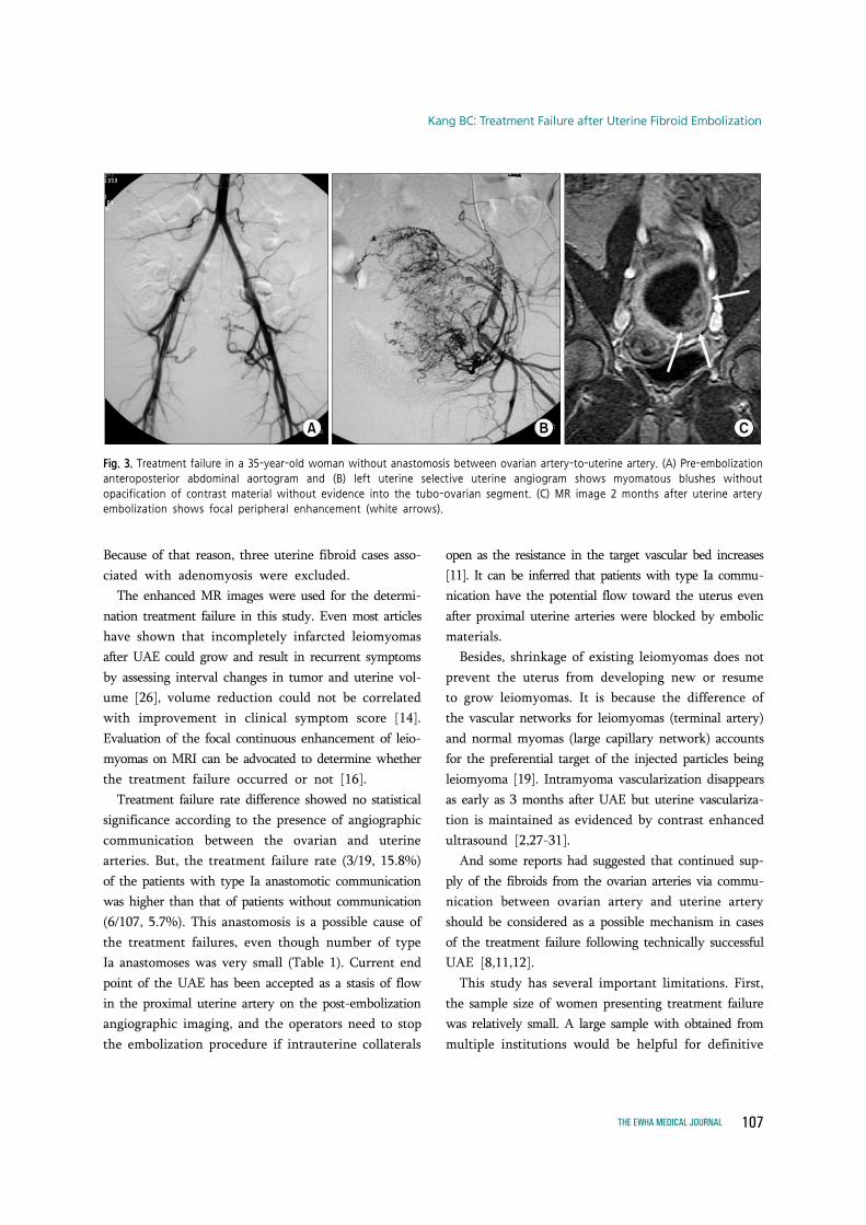
Fig. 3. Treatment failure in a 35-year-old woman without anastomosis between ovarian artery-to-uterine artery. (A) Pre-embolizationanteroposterior abdominal aortogram and (B) left uterine selective uterine angiogram shows myomatous blushes without opacification of contrast material without evidence into the tubo-ovarian segment. (C) MR image 2 months after uterine artery embolization shows focal peripheral enhancement (white arrows).
Because of that reason, three uterine fibroid cases asso-ciated with adenomyosis were excluded. The enhanced MR images were used for the determi-nation treatment failure in this study. Even most articles have shown that incompletely infarcted leiomyomas after UAE could grow and result in recurrent symptoms by assessing interval changes in tumor and uterine vol-ume [26], volume reduction could not be correlated with improvement in clinical symptom score [14]. Evaluation of the focal continuous enhancement of leio-myomas on MRI can be advocated to determine whether the treatment failure occurred or not [16]. Treatment failure rate difference showed no statistical significance according to the presence of angiographic communication between the ovarian and uterine arteries. But, the treatment failure rate (3/19, 15.8%) of the patients with type Ia anastomotic communication was higher than that of patients without communication (6/107, 5.7%). This anastomosis is a possible cause of the treatment failures, even though number of type Ia anastomoses was very small (Table 1). Current end point of the UAE has been accepted as a stasis of flow in the proximal uterine artery on the post-embolization angiographic imaging, and the operators need to stop the embolization procedure if intrauterine collaterals
open as the resistance in the target vascular bed increases [11]. It can be inferred that patients with type Ia commu-nication have the potential flow toward the uterus even after proximal uterine arteries were blocked by embolic materials. Besides, shrinkage of existing leiomyomas does not prevent the uterus from developing new or resume to grow leiomyomas. It is because the difference of the vascular networks for leiomyomas (terminal artery) and normal myomas (large capillary network) accounts for the preferential target of the injected particles being leiomyoma [19]. Intramyoma vascularization disappears as early as 3 months after UAE but uterine vasculariza-tion is maintained as evidenced by contrast enhanced ultrasound [2,27-31]. And some reports had suggested that continued sup-ply of the fibroids from the ovarian arteries via commu-nication between ovarian artery and uterine artery should be considered as a possible mechanism in cases of the treatment failure following technically successful UAE [8,11,12]. This study has several important limitations. First, the sample size of women presenting treatment failure was relatively small. A large sample with obtained from multiple institutions would be helpful for definitive

Ewha Med J Vol. 35, No. 2, 2012
108 THE EWHA MEDICAL JOURNAL
conclusion. Second, review of the MR images after pri-mary UAE was not done in a blinded fashion, potentially introducing observer bias. Third, the same size and same type of embolic materials were not used. The uterine artery was embolized to complete stasis of flow in the main uterine artery by using either 355∼500 μm poly-vinyl alcohol particles or 500∼700 μm triacryl gelatin microspheres. The full clinical significance of collateral flow to fib-roid has been emphasized [8,9,11,12,32,33] and it has been conceivable that many treatment failures from UAE are caused by unrecognized collateral fibroid feed-ers [8,9,11]. Although data presented here are too lim-ited for conclusion, it can suggest the possibility that patients with type Ia anastomoses between the ovarian artery and the uterine artery could have the early treat-ment failure.
Acknowledgments
This work was supported by grant from Ewha Womans University Mokdong Hospital Research Fund.
References
1. Marshall LM, Spiegelman D, Barbieri RL, Goldman MB, Manson JE, Colditz GA, et al. Variation in the incidence of uterine leiomyoma among premenopausal women by age and race. Obstet Gynecol 1997;90:967- 973.
2. Goodwin SC, McLucas B, Lee M, Chen G, Perrella R, Vedantham S, et al. Uterine artery embolization for the treatment of uterine leiomyomata midterm results. J Vasc Interv Radiol 1999;10:1159-1165.
3. Hutchins FL Jr, Worthington-Kirsch R. Embolotherapy for myoma-induced menorrhagia. Obstet Gynecol Clin North Am 2000;27:397-405.
4. Spies JB, Scialli AR, Jha RC, Imaoka I, Ascher SM, Fraga VM, et al. Initial results from uterine fibroid embolization for symptomatic leiomyomata. J Vasc Interv Radiol 1999;10:1149-1157.
5. Worthington-Kirsch RL, Popky GL, Hutchins FL Jr. Uterine arterial embolization for the management of leiomyomas: quality-of-life assessment and clinical response. Radiology 1998;208:625-629.
6. Pelage JP, Le Dref O, Soyer P, Kardache M, Dahan H, Abitbol M, et al. Fibroid-related menorrhagia: treat-
ment with superselective embolization of the uterine arteries and midterm follow-up. Radiology 2000;215: 428-431.
7. Hutchins FL Jr, Worthington-Kirsch R, Berkowitz RP. Selective uterine artery embolization as primary treat-ment for symptomatic leiomyomata uteri. J Am Assoc Gynecol Laparosc 1999;6:279-284.
8. Nikolic B, Spies JB, Abbara S, Goodwin SC. Ovarian artery supply of uterine fibroids as a cause of treatment failure after uterine artery embolization: a case report. J Vasc Interv Radiol 1999;10:1167-1170.
9. Pelage JP, Jacob D, Le Dref O, Laurent A. Re: Leiomyoma recurrence after uterine artery emboli-zation. J Vasc Interv Radiol 2004;15:773.
10. Spies JB. Uterine artery embolization for fibroids: un-derstanding the technical causes of failure. J Vasc Interv Radiol 2003;14:11-14.
11. Matson M, Nicholson A, Belli AM. Anastomoses of the ovarian and uterine arteries: a potential pitfall and cause of failure of uterine embolization. Cardiovasc Intervent Radiol 2000;23:393-396.
12. Razavi MK, Wolanske KA, Hwang GL, Sze DY, Kee ST, Dake MD. Angiographic classification of ovarian artery-to-uterine artery anastomoses: initial ob-servations in uterine fibroid embolization. Radiology 2002;224:707-712.
13. Burn PR, McCall JM, Chinn RJ, Vashisht A, Smith JR, Healy JC. Uterine fibroleiomyoma: MR imaging ap-pearances before and after embolization of uterine arteries. Radiology 2000;214:729-734.
14. deSouza NM, Williams AD. Uterine arterial emboliza-tion for leiomyomas: perfusion and volume changes at MR imaging and relation to clinical outcome. Radiology 2002;222:367-374.
15. Jha RC, Ascher SM, Imaoka I, Spies JB. Symptomatic fibroleiomyomata: MR imaging of the uterus before and after uterine arterial embolization. Radiology 2000;217: 228-235.
16. Chrisman HB, West D, Corpuz B, Rye RK, Salem R, Carr J, et al. Primary failure of uterine artery emboliza-tion: use of magnetic resonance imaging to select pa-tients for repeated embolization. J Vasc Interv Radiol 2005;16:1143-1147.
17. Marret H, Cottier JP, Alonso AM, Giraudeau B, Body G, Herbreteau D. Predictive factors for fibroids re-currence after uterine artery embolisation. Brit J Obstet Gynecol 2005; 112:461-465.
18. Goodwin SC, Vedantham S, McLucas B, Forno AE, Perrella R. Preliminary experience with uterine artery embolization for uterine fibroids. J Vasc Interv Radiol 1997;8:517-526.

Kang BC: Treatment Failure after Uterine Fibroid Embolization
THE EWHA MEDICAL JOURNAL 109
19. Marret H, Alonso AM, Cottier JP, Tranquart F, Herbreteau D, Body G. Leiomyoma recurrence after ute-rine artery embolization. J Vasc Interv Radiol 2003;14: 1395-1399.
20. Walker WJ, Pelage JP. Uterine artery embolisation for symptomatic fibroids: clinical results in 400 women with imaging follow up. Brit J Obstet Gynecol 2002;109:1262- 1272.
21. McLucas B, Adler L, Perrella R. Uterine fibroid emboli-zation: nonsurgical treatment for symptomatic fibroids. J Am Coll Surg 2001;192:95-105.
22. Smith SJ, Sewall LE, Handelsman A. A clinical failure of uterine fibroid embolization due to adenomyosis. J Vasc Interv Radiol 1999;10:1171-1174.
23. Jha RC, Takahama J, Imaoka I, Korangy SJ, Spies JB, Cooper C, et al. Adenomyosis: MRI of the uterus treated with uterine artery embolization. Am J Roentgenol 2003; 181:851-856.
24. Kim MD, Won JW, Lee DY, Ahn CS. Uterine artery embolization for adenomyosis without fibroid. Clin Radiol 2004;59:520-526.
25. Siskin GP, Tublin ME, Stainken BF, Dowling K, Dolen EG. Uterine artery embolization for the treatment of adenomyosis: clinical response and evaluation with MR imaging. Am J Roentgenol 2001;177:297-302.
26. Pelage JP, Guaou NG, Jha RC, Ascher SM, Spies JB.
Uterine fibroid tumors: long-term MR imaging outcome after embolization. Radiology 2004;230:803-809.
27. Marret H, Tranquart F, Sauget S, Alonso AM, Cottier JP, Herbreteau D. Contrast-enhanced sonography dur-ing uterine artery embolization for the treatment of leiomyomas. Ultrasound Obstet Gynecol 2004;23:77-79.
28. Itkin M, Shlansky-Goldberg R. Uterine fibroid emboliza-tion for the treatment of symptomatic leiomyomata. Appl Radiol 2002;31(10):9-17.
29. Worthington-Kirsch RL, Andrews RT, Siskin GP, Shlansky-Goldberg R, Lipman JC, Goodwin SC, et al. II. Uterine fibroid embolization: technical aspects. Tech Vasc Interv Radiol 2002;5:17-34.
30. Sampson JA. The blood supply of uterine myomata. Surg Gynecol Obstet 1912;14:215-234.
31. Lindenbaum E, Brandes JM, Itskovitz J. Ipsi- and con-tralateral anastomosis of the uterine arteries. Acta Anat 1978;102:157-161.
32. Andrews RT, Bromley PJ, Pfister ME. Successful embo-lization of collaterals from the ovarian artery during uterine artery embolization for fibroids: a case report. J Vasc Interv Radiol 2000;11:607-610.
33. Marx M, Wack JP, Baker EL, Stevens SK, Barakos JA. Ovarian protection by occlusion of uteroovarian col-lateral vessels before uterine fibroid embolization. J Vasc Interv Radiol 2003;14:1329-1332.