Trauma Radiology: An Algorithmic Approach · Div. of Emergency Medicine, UCSF Introduction Trauma...
32
Div. of Emergency Medicine, UCSF Trauma Radiology: An Algorithmic Approach Martin Kernberg, MD, Asst. Clinical Professor Steve Polevoi, MD, Assoc. Clinical Professor Division of Emergency Medicine, Dept. of Medicine and Department of Radiology University of California, San Francisco
Transcript of Trauma Radiology: An Algorithmic Approach · Div. of Emergency Medicine, UCSF Introduction Trauma...
Div. of Emergency Medicine, UCSF
Trauma Radiology:An Algorithmic Approach
Martin Kernberg, MD, Asst. Clinical Professor Steve Polevoi, MD, Assoc. Clinical Professor
Division of Emergency Medicine, Dept. of Medicineand Department of Radiology
University of California, San Francisco
Div. of Emergency Medicine, UCSF
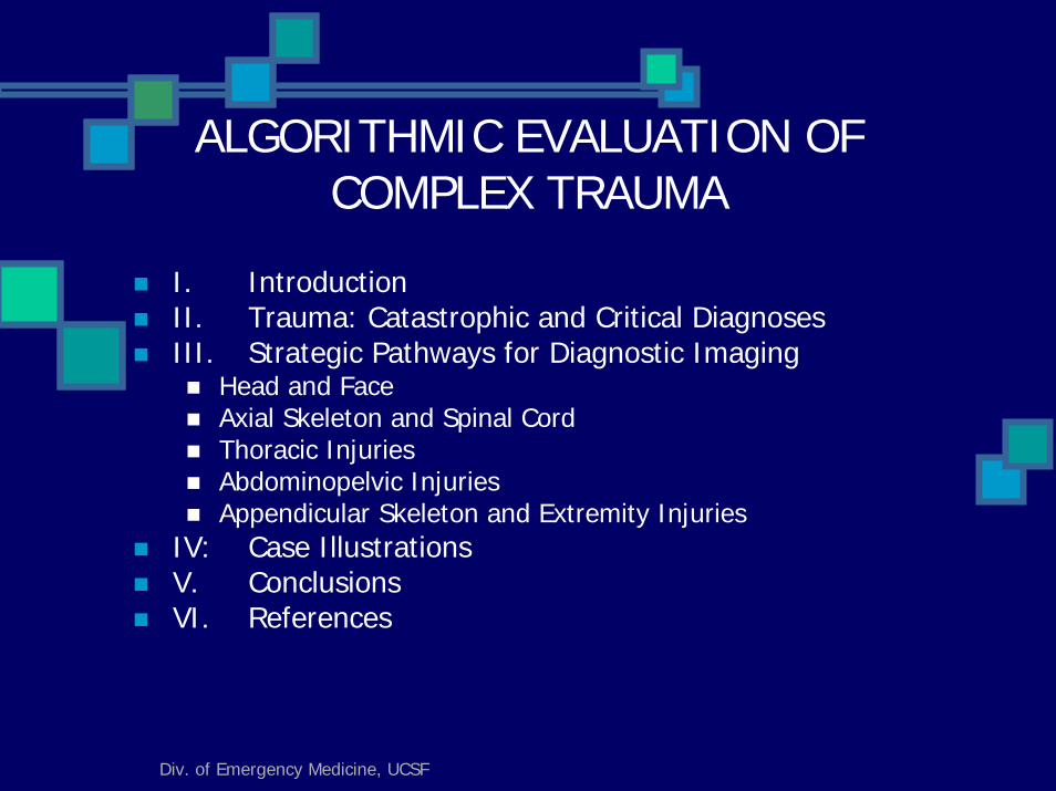
ALGORITHMIC EVALUATION OF COMPLEX TRAUMA
I. IntroductionII. Trauma: Catastrophic and Critical DiagnosesIII. Strategic Pathways for Diagnostic Imaging
Head and FaceAxial Skeleton and Spinal CordThoracic InjuriesAbdominopelvic InjuriesAppendicular Skeleton and Extremity Injuries
IV: Case IllustrationsV. ConclusionsVI. References
Div. of Emergency Medicine, UCSF
Introduction Trauma remains the leading cause of death for children and young adults
under 40 despite recent advances in management. The contemporaryevaluation and management of the trauma patient require parallelefforts to assess the patient clinically and radiologically. The selection of radiological investigations remains a source of controversy. Advancing imaging modalities yield diagnoses previously overlooked; medicolegal concerns influence clinical decisions; decision rules and protocols designed to reduce unnecessary costs, radiation exposure, and clinical delays can seem complex, contradictory, and excessively rigid; resources are progressively limited. In reviewing these issues, a system is described that may prove useful in clinical practice, with a critical review of the advantages and disadvantages of various radiological modalities. While a set of algorithms is advocated, it is underscored that this will vary depending on the facilities available. It is appropriate however to be aware of the limitations of the radiological techniques that are utilized in trauma on a daily basis and to have a knowledge of how selective use of advanced imaging modalities will improve patient care.
Modified from P Jaye, ME Kernberg, and T Green, “Trauma Radiology,” The Lancet, in press, 2007.
Div. of Emergency Medicine, UCSF
Trauma: Parallel Processing of Information
1. Consider the high risk differential diagnosis, on the basis of clinical history, physical examination, and laboratory studies.
2. Concurrently stabilize, initiate imaging sequence, and/or contact appropriate surgical consultants.
3. Confirm benign etiologies directly, or indirectly after formal exclusion of the catastrophic differential diagnosis.
Div. of Emergency Medicine, UCSF
How are traumatic catastrophic conditions defined?
Catastrophic conditions are those which have a significant risk of mortality, if the diagnosis is emergently missed.Critical traumatic conditions are those which have a significant risk of morbidity, if the diagnosis is delayed (e.g., cervical spine injuries, occult fractures, or internal derangements).
Div. of Emergency Medicine, UCSF
7 Catastrophic traumatic conditions
Intracranial hemorrhageAortic transections and vascular injuryMyocardial contusion and lacerationPericardial hemorrhagePneumothoraxSolid organ laceration (liver, spleen, adrenal, renal, and pancreas)Bowel and bladder perforation
Div. of Emergency Medicine, UCSF
7 Critical Injuries: Axial and Extremity TraumaCervical spine fracturesShoulder dislocationsEpiphyseal avulsion fracturesNavicular fracturesPelvic fractures Femoral neck impaction fracturesLisfranc fractures
Div. of Emergency Medicine, UCSF
General Vital Sign Indications for Catastrophic Differential Diagnosis
1. Tachycardia or bradycardia (heart rate <50)2. Tachypnea or bradypnea (respiratory rate <7)3. Significant pyrexia or hypothermia4. Hypotension and hypertension5. Acute hypoxia6. Pain severity
Div. of Emergency Medicine, UCSF
Local Vital Sign Indications for Traumatic Differential Diagnosis
Glasgow Coma ScoreAdultPediatric
Visual acuityInjury site related painPeripheral pulsesPeripheral pulse oximetryPeripheral capillary refill
Div. of Emergency Medicine, UCSF
Clinical Catastrophic CriteriaAcuity, severity, progression, persistence, refractory, atypical or unexplained:
Critical acute chest symptoms (i.e., chest pain, chest pressure, or respiratory distress)Critical abdominal and pelvic clinical symptoms (i.e., pain, nausea, vomiting, diarrhea, distension, bleeding, or irritability)Selective physical findings (absence of breath sounds, cardiac murmurs; pericardial friction rub; altered bowel sounds, masses or peritoneal signs). Aberrant laboratory, electrocardiographic, or plain radiographic abnormalities.
Div. of Emergency Medicine, UCSF
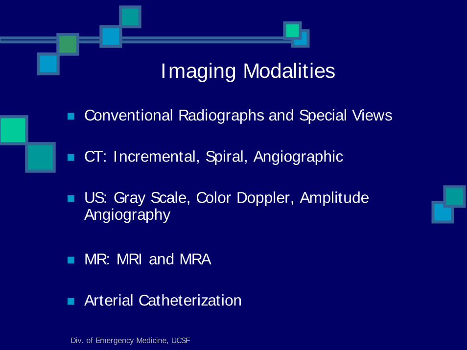
Imaging Modalities
Conventional Radiographs and Special Views
CT: Incremental, Spiral, Angiographic
US: Gray Scale, Color Doppler, Amplitude Angiography
MR: MRI and MRA
Arterial Catheterization

Div. of Emergency Medicine, UCSF
Head and Facial Trauma: Diagnostic Strategy
Catastrophic Craniofacial Findings
Clinical Information Standard Diagnostic Testing
Advanced Imaging Options
1. Laboratory 1. CT/CTA
2. XR 2. MRI
3. Angiography
Vital Signs
History
Neurologic Examination
Div. of Emergency Medicine, UCSF
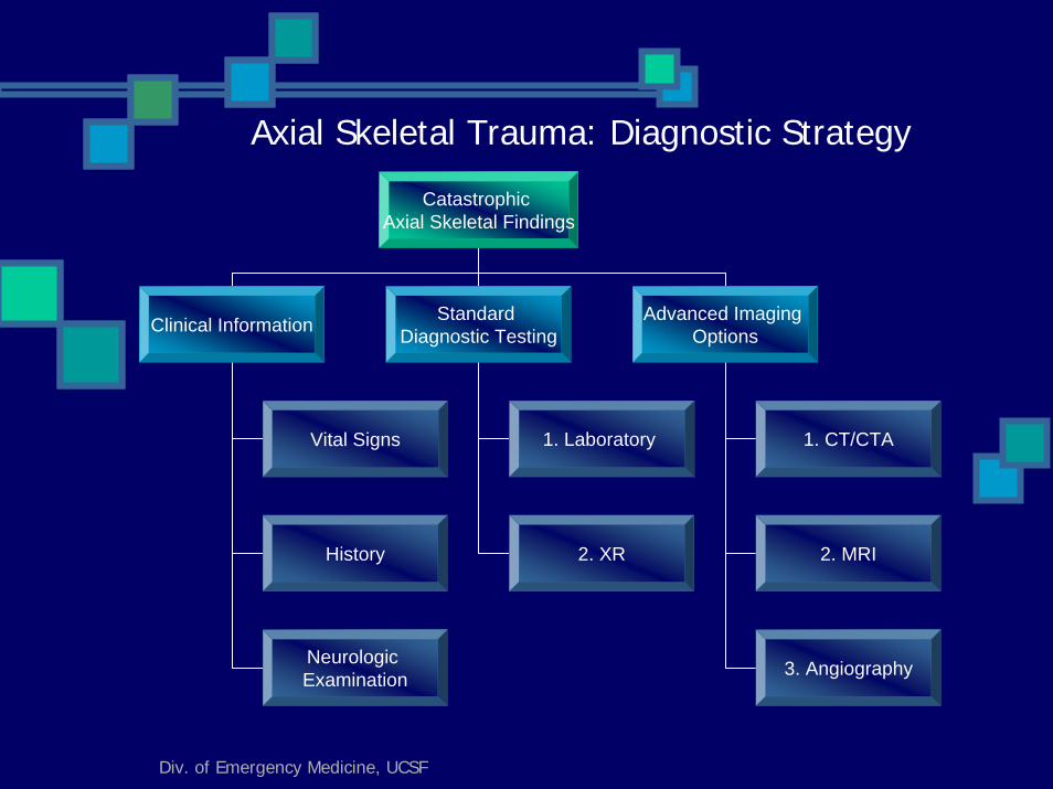
Axial Skeletal Trauma: Diagnostic Strategy
Catastrophic Axial Skeletal Findings
Clinical Information Standard Diagnostic Testing
Advanced Imaging Options
1. Laboratory 1. CT/CTA
2. XR 2. MRI
3. Angiography
Vital Signs
History
Neurologic Examination
Div. of Emergency Medicine, UCSF
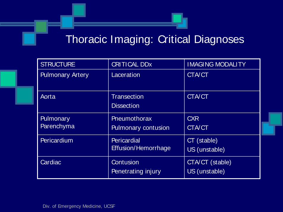
Thoracic Imaging: Critical Diagnoses
STRUCTURE CRITICAL DDx IMAGING MODALITY
Pulmonary Artery Laceration CTA/CT
Aorta TransectionDissection
CTA/CT
Pulmonary Parenchyma
PneumothoraxPulmonary contusion
CXR CTA/CT
Pericardium Pericardial Effusion/Hemorrhage
CT (stable) US (unstable)
Cardiac ContusionPenetrating injury
CTA/CT (stable)US (unstable)
Div. of Emergency Medicine, UCSF
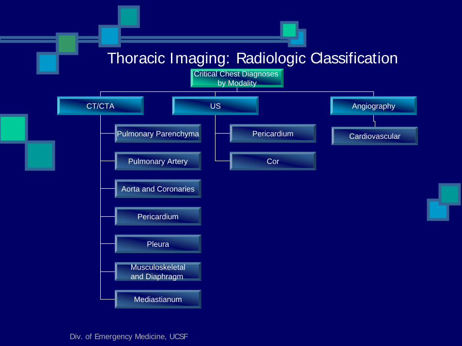
Thoracic Imaging: Radiologic ClassificationCritical Chest Diagnoses
by Modality
CT/CTA US Angiography
Pulmonary Parenchyma Pericardium Cardiovascular
Pulmonary Artery
Aorta and Coronaries
Pericardium
Pleura
Musculoskeletaland Diaphragm
Mediastianum
Cor
Div. of Emergency Medicine, UCSF
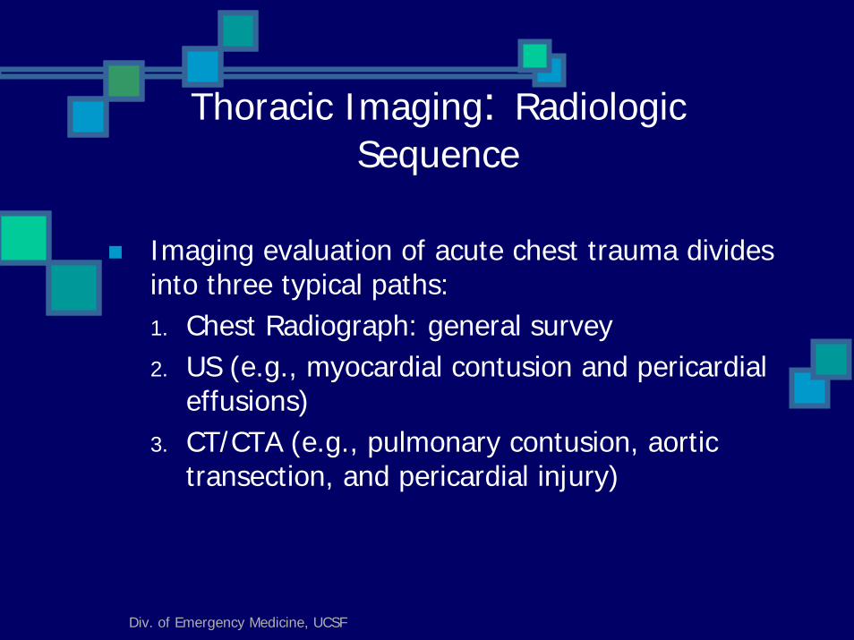
Thoracic Imaging: Radiologic Sequence
Imaging evaluation of acute chest trauma divides into three typical paths:1. Chest Radiograph: general survey2. US (e.g., myocardial contusion and pericardial
effusions) 3. CT/CTA (e.g., pulmonary contusion, aortic
transection, and pericardial injury)
Div. of Emergency Medicine, UCSF
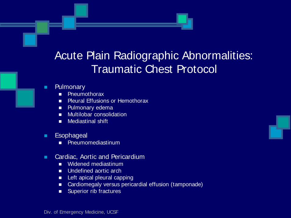
Acute Plain Radiographic Abnormalities: Traumatic Chest Protocol
PulmonaryPneumothoraxPleural Effusions or HemothoraxPulmonary edemaMultilobar consolidation Mediastinal shift
EsophagealPneumomediastinum
Cardiac, Aortic and PericardiumWidened mediastinumUndefined aortic archLeft apical pleural cappingCardiomegaly versus pericardial effusion (tamponade)Superior rib fractures
Div. of Emergency Medicine, UCSF
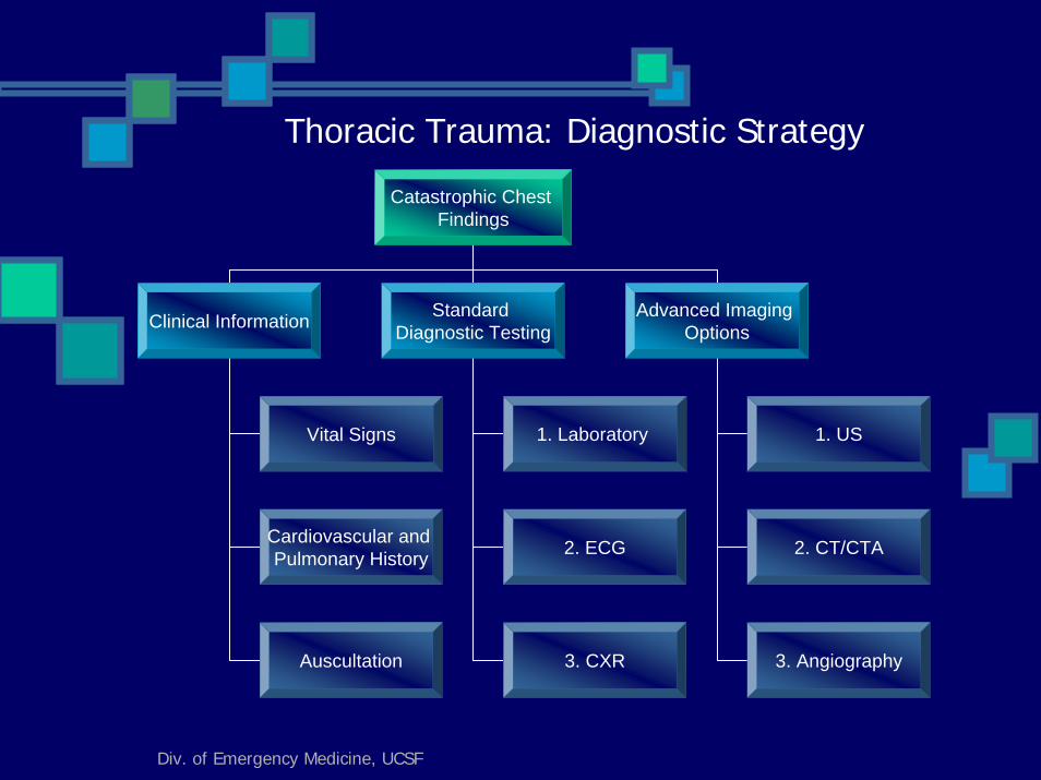
Thoracic Trauma: Diagnostic Strategy
Catastrophic Chest Findings
Clinical Information Standard Diagnostic Testing
Advanced Imaging Options
1. Laboratory 1. US
2. ECG
3. CXR
2. CT/CTA
3. Angiography
Vital Signs
Cardiovascular and Pulmonary History
Auscultation
Div. of Emergency Medicine, UCSF
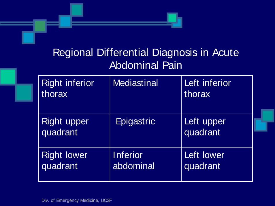
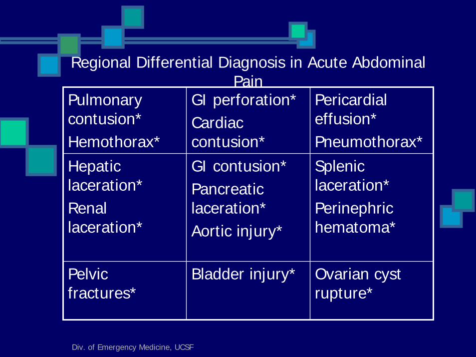
Regional Differential Diagnosis in Acute Abdominal Pain
Right inferior thorax
Mediastinal Left inferior thorax
Right upper quadrant
Epigastric Left upper quadrant
Right lower quadrant
Inferior abdominal
Left lower quadrant
Div. of Emergency Medicine, UCSF
Regional Differential Diagnosis in Acute Abdominal Pain
Pulmonary contusion*Hemothorax*
GI perforation*Cardiac contusion*
Pericardial effusion*Pneumothorax*
Hepatic laceration*Renal laceration*
GI contusion*Pancreatic laceration*Aortic injury*
Splenic laceration*Perinephric hematoma*
Pelvic fractures*
Bladder injury* Ovarian cyst rupture*
Div. of Emergency Medicine, UCSF
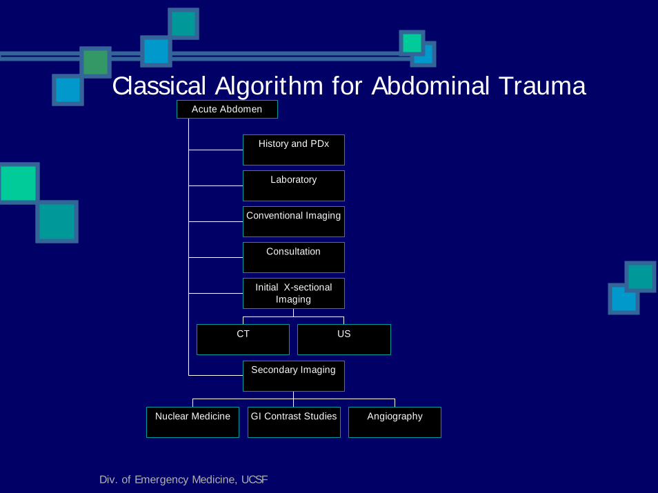
Classical Algorithm for Abdominal Trauma
History and PDx
Laboratory
Conventional Imaging
Consultation
CT US
Initial X-sectionalImaging
Nuclear Medicine GI Contrast Studies Angiography
Secondary Imaging
Acute Abdomen
Div. of Emergency Medicine, UCSF
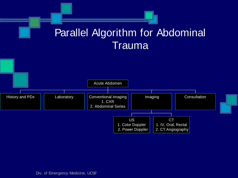
Parallel Algorithm for Abdominal Trauma
History and PDx Laboratory Conventional Imaging1. CXR
2. Abdominal Series
US1. Color Doppler
2. Power Doppler
CT1. IV, Oral, Rectal
2. CT Angiography
Imaging Consultation
Acute Abdomen
Div. of Emergency Medicine, UCSF
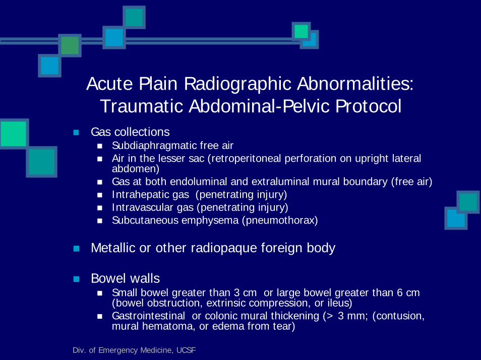
Acute Plain Radiographic Abnormalities: Traumatic Abdominal-Pelvic Protocol
Gas collectionsSubdiaphragmatic free airAir in the lesser sac (retroperitoneal perforation on upright lateral abdomen)Gas at both endoluminal and extraluminal mural boundary (free air)Intrahepatic gas (penetrating injury)Intravascular gas (penetrating injury)Subcutaneous emphysema (pneumothorax)
Metallic or other radiopaque foreign body
Bowel wallsSmall bowel greater than 3 cm or large bowel greater than 6 cm (bowel obstruction, extrinsic compression, or ileus)Gastrointestinal or colonic mural thickening (> 3 mm; (contusion, mural hematoma, or edema from tear)
Div. of Emergency Medicine, UCSF
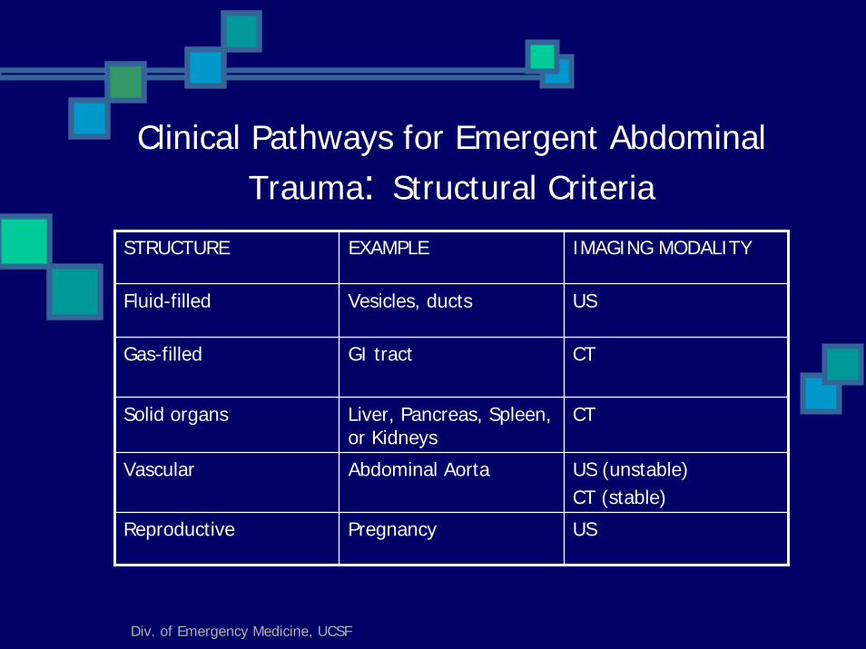
Clinical Pathways for Emergent Abdominal Trauma: Structural Criteria
STRUCTURE EXAMPLE IMAGING MODALITY
Fluid-filled Vesicles, ducts US
Gas-filled GI tract CT
Solid organs Liver, Pancreas, Spleen, or Kidneys
CT
Vascular Abdominal Aorta US (unstable)CT (stable)
Reproductive Pregnancy US
Div. of Emergency Medicine, UCSF
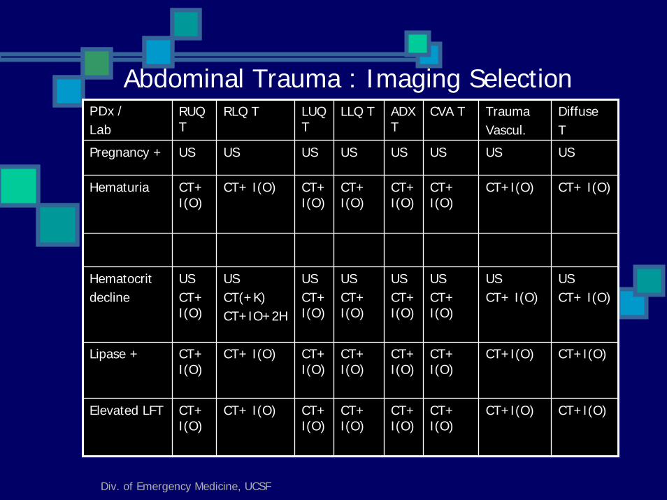
Abdominal Trauma : Imaging SelectionPDx /Lab
RUQ T
RLQ T LUQ T
LLQ T ADX T
CVA T TraumaVascul.
DiffuseT
Pregnancy + US US US US US US US US
Hematuria CT+ I(O)
CT+ I(O) CT+ I(O)
CT+ I(O)
CT+ I(O)
CT+ I(O)
CT+I(O) CT+ I(O)
Hematocritdecline
USCT+ I(O)
USCT(+K)CT+IO+2H
USCT+ I(O)
USCT+ I(O)
USCT+ I(O)
USCT+ I(O)
USCT+ I(O)
USCT+ I(O)
Lipase + CT+ I(O)
CT+ I(O) CT+ I(O)
CT+ I(O)
CT+I(O)
CT+ I(O)
CT+I(O) CT+I(O)
Elevated LFT CT+ I(O)
CT+ I(O) CT+ I(O)
CT+ I(O)
CT+I(O)
CT+ I(O)
CT+I(O) CT+I(O)
Div. of Emergency Medicine, UCSF
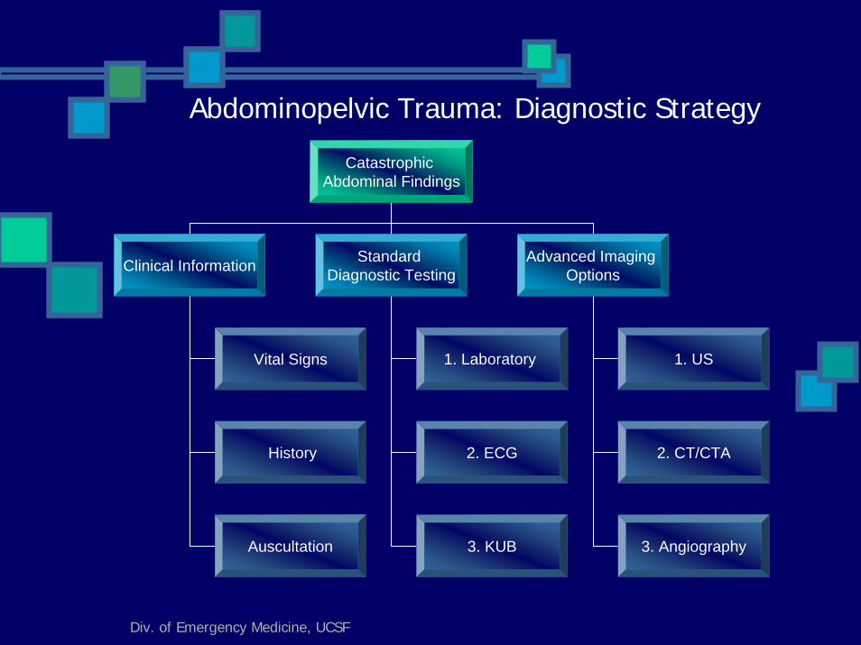
Abdominopelvic Trauma: Diagnostic Strategy
Catastrophic Abdominal Findings
Clinical Information Standard Diagnostic Testing
Advanced Imaging Options
1. Laboratory 1. US
2. ECG
3. KUB
2. CT/CTA
3. Angiography
Vital Signs
History
Auscultation
Div. of Emergency Medicine, UCSF
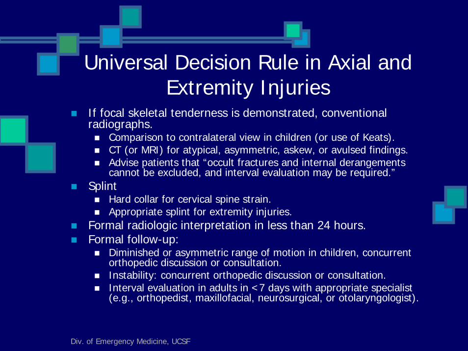
Universal Decision Rule in Axial and Extremity Injuries
If focal skeletal tenderness is demonstrated, conventional radiographs.
Comparison to contralateral view in children (or use of Keats). CT (or MRI) for atypical, asymmetric, askew, or avulsed findings. Advise patients that “occult fractures and internal derangements cannot be excluded, and interval evaluation may be required.”
Splint Hard collar for cervical spine strain.Appropriate splint for extremity injuries.
Formal radiologic interpretation in less than 24 hours.Formal follow-up:
Diminished or asymmetric range of motion in children, concurrentorthopedic discussion or consultation.Instability: concurrent orthopedic discussion or consultation.Interval evaluation in adults in <7 days with appropriate specialist (e.g., orthopedist, maxillofacial, neurosurgical, or otolaryngologist).
Div. of Emergency Medicine, UCSF
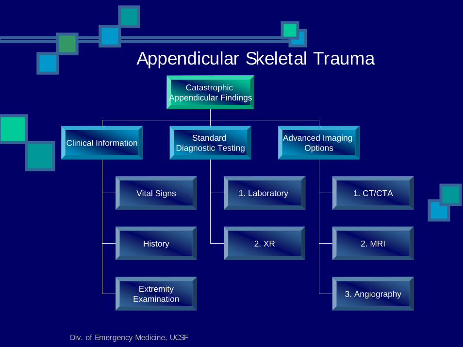
Appendicular Skeletal TraumaCatastrophic
Appendicular Findings
Clinical Information Standard Diagnostic Testing
Advanced Imaging Options
1. Laboratory 1. CT/CTA
2. XR 2. MRI
3. Angiography
Vital Signs
History
ExtremityExamination
Div. of Emergency Medicine, UCSF
Workshop Case Illustrations
Div. of Emergency Medicine, UCSF
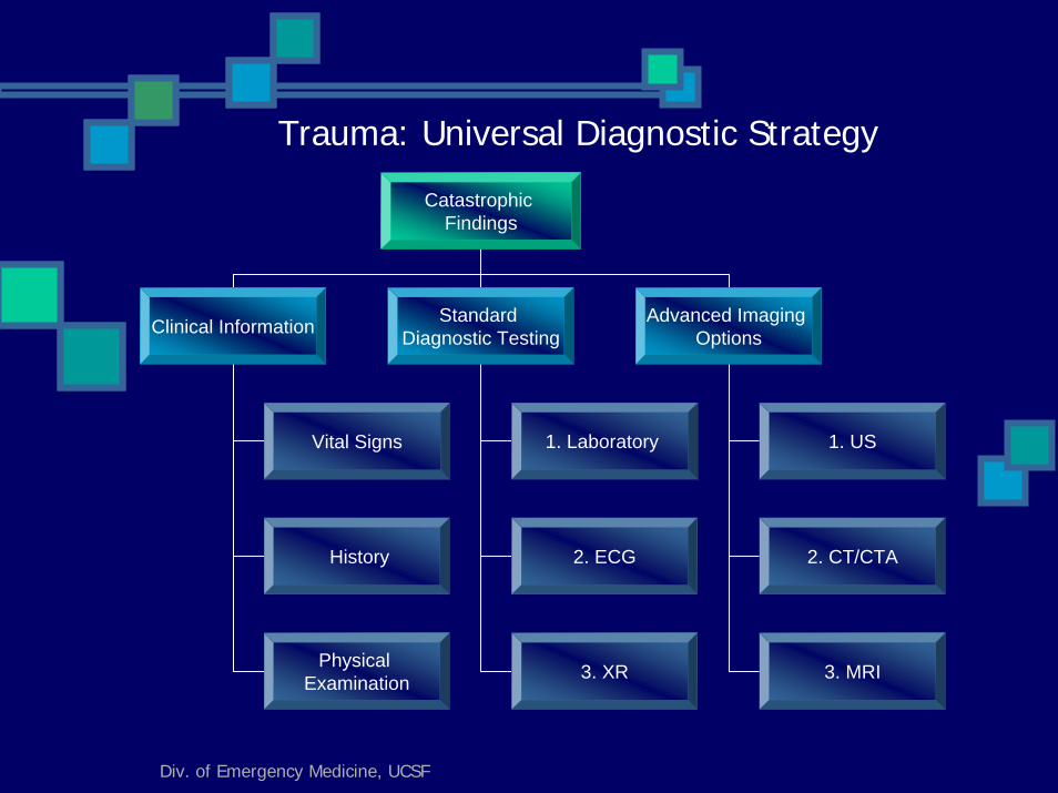
Trauma: Universal Diagnostic Strategy
Catastrophic Findings
Clinical Information Standard Diagnostic Testing
Advanced Imaging Options
1. Laboratory 1. US
2. ECG
3. XR
2. CT/CTA
3. MRI
Vital Signs
History
Physical Examination
Div. of Emergency Medicine, UCSF
References
1. Kernberg ME, Polevoi SK, Lewin M, and Murphy C, Catastrophic errors: algorithmic solutions, 3rd Mediterranean Emergency Medicine Conference, Nice, France, September 4, 2005 (Catastrophic errors evaluated in a consecutive case series of 125,000 emergency room patients).2. P Jaye, ME Kernberg, and T Green, “Trauma Radiology,”The Lancet, in press, 2007.3. Radiation Risks and Pediatric Computed Tomography (CT): A Guide for Health Care Providers, National Cancer Institute (USA) and Society for Pediatric Radiology, 2002 (modified for Table 1).
Div. of Emergency Medicine, UCSF
After a closed head injury, with transient loss of consciousness, a 2 year old female infant has persistent nausea and vomiting. Imaging should include:
1. None 2. Skull films3. Head CT scan4. Head MRI