Trauma-CT - Amazon Web Servicesh24-files.s3.amazonaws.com/110213/287339-kx0Ta.pdf · Trauma...
71
Swedish Radiological Week, Norrköping, Sept 18, 2003 Mats Beckman, Karolinska Sjukhuset, Bertil Leidner, Huddinge Universitetssjukhus Trauma-CT Part I Why? How To?
Transcript of Trauma-CT - Amazon Web Servicesh24-files.s3.amazonaws.com/110213/287339-kx0Ta.pdf · Trauma...

Swedish Radiological Week,Norrköping, Sept 18, 2003
Mats Beckman, Karolinska Sjukhuset, Bertil Leidner, Huddinge Universitetssjukhus
Trauma-CTPart I
Why? How To?
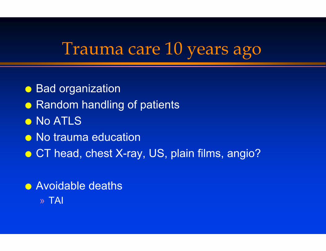
Trauma care 10 years ago
● Bad organization● Random handling of patients● No ATLS● No trauma education● CT head, chest X-ray, US, plain films, angio?
● Avoidable deaths» TAI

Multiple Trauma● Few trauma patients ● Limited personal experience
● Life threatening ● STRESS● Systematic and structured approach clinically and
radiologically
● Trauma CT● Nordic Trauma Radiology Courses
» 1st + 2nd course (2000 + 2002) Stockholm

Trauma
● Background facts● Epidemiology● Trauma Care – History● Role of Trauma -CT

◆Trauma commonest cause of death for all young people
●Young everyone age<45

Trauma causes● Fall ● Traffic- MC, cycling● Suicide● Penetrating
11 september 2001
SAS Milano 011028
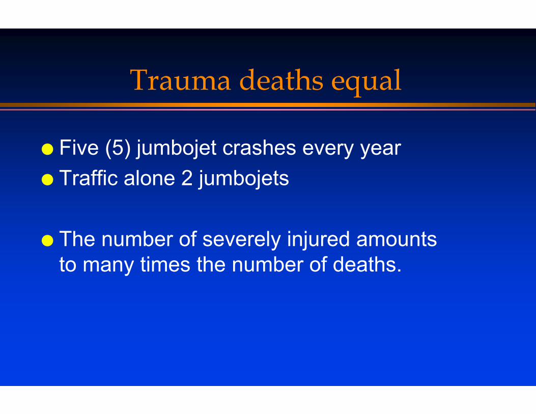
Trauma deaths equal
● Five (5) jumbojet crashes every year● Traffic alone 2 jumbojets
● The number of severely injured amounts to many times the number of deaths.
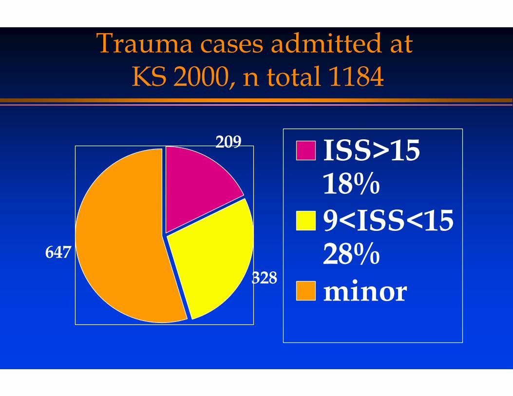
Trauma cases admitted at KS 2000, n total 1184
209
328647
ISS>1518%9<ISS<1528%minor

Causes of trauma KS 2000
Fall 18%
MC 15%
Ped 12%
Penetr 3.1%
Cycling 5%
Other 6.9
MVC 41%
MVC 41%Fall 18%MC 15%Ped 12%Cycling 5%Penetr 3.1%Other 6.9

Statistics KS
● Ca. 90 % CT● 76 % men● 73% 15-54 YO
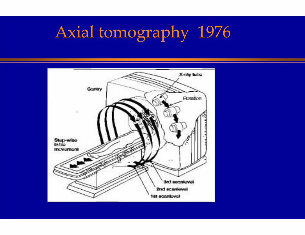
Axial tomography 1976
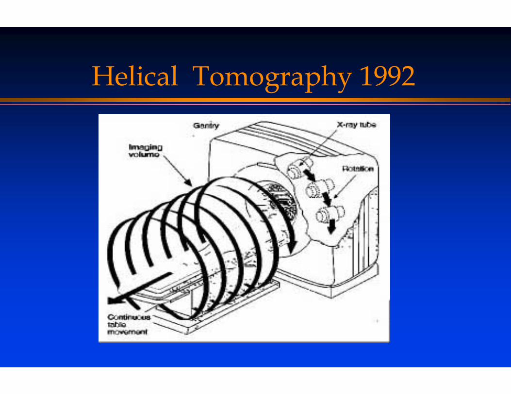
Helical Tomography 1992
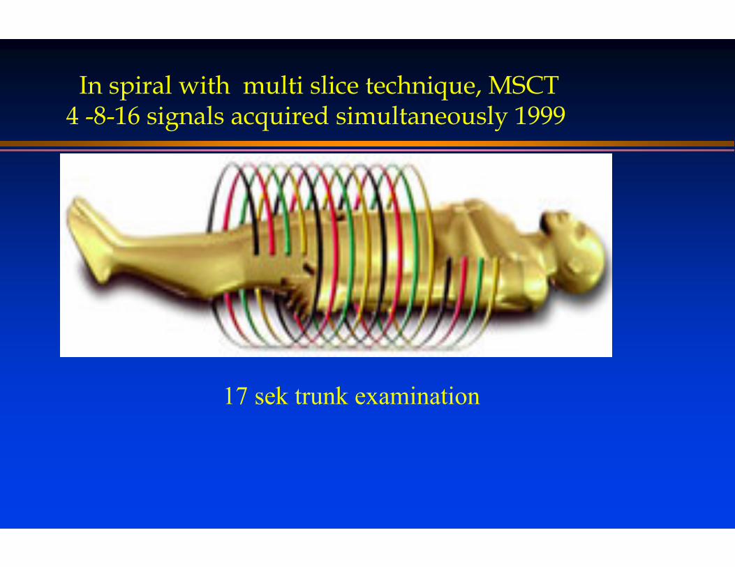
In spiral with multi slice technique, MSCT4 -8-16 signals acquired simultaneously 1999
17 sek trunk examination

Multi-slice (detector)
● 4 channels = 8 x faster scanning compared to single detector spiral-CT
● 1990 non spiral – 5 images/minute● 1992 spiral 60 images/minute● 4 channels 500 images/minute● 16 channels 2400 images/minute
– (x 0,75 mm + 0,42 sec rotation time)
● Speed + Accuracy improves trauma value

International acceptance
● Abdominal CT USA standard since late 1980’s; EAST Level 1 recommendation for blunt abdominal trauma» “physical examination inadequate”
● Trauma - CT concept implemented in» Denmark (Veile)» Finland (Tölöö, Helsinki)» Germany (München, Mainz)

Why CT?PneumothoraxTamponadeHaemothorax
”Flail Chest”Aorta, TAITrachea, bronchiDiaphragmEsofagusLung contusion
&laceration
SpleenLiverKidneysPancreasRetroperitoneumBowel
BrainSkullFaceEars
C-spineT-L spinePelvis, ShouldersHips
Bleeding
Trauma CT
● Team work with many involved clinical specialties
● Trauma Radiology & Trauma CT - a vital link in chain of trauma care
Modern Trauma Management● ATLS -Advanced Trauma Life Support
» Systematic clinical examination/treatment » First survey - ABCDE – fast vital exam + treatment» Secondary survey from top-to toe
● “Trauma – CT” becomes a systematic ATLS adjunct» (multislice)-CT used as a tertiary survey – from “top to toe”» standardized procedure every time
– possible to train for techs– fast– minimizes possible omissions

● “Whole body” examination● head & c-spine
» non-contrast» arms by body side
● thorax & abdomen/pelvis» arms above head» always intravenous contrast
● total time in practice– 5 min + 5 min pre-post exam patient handling table– 12 min exam
What is a Trauma CT?
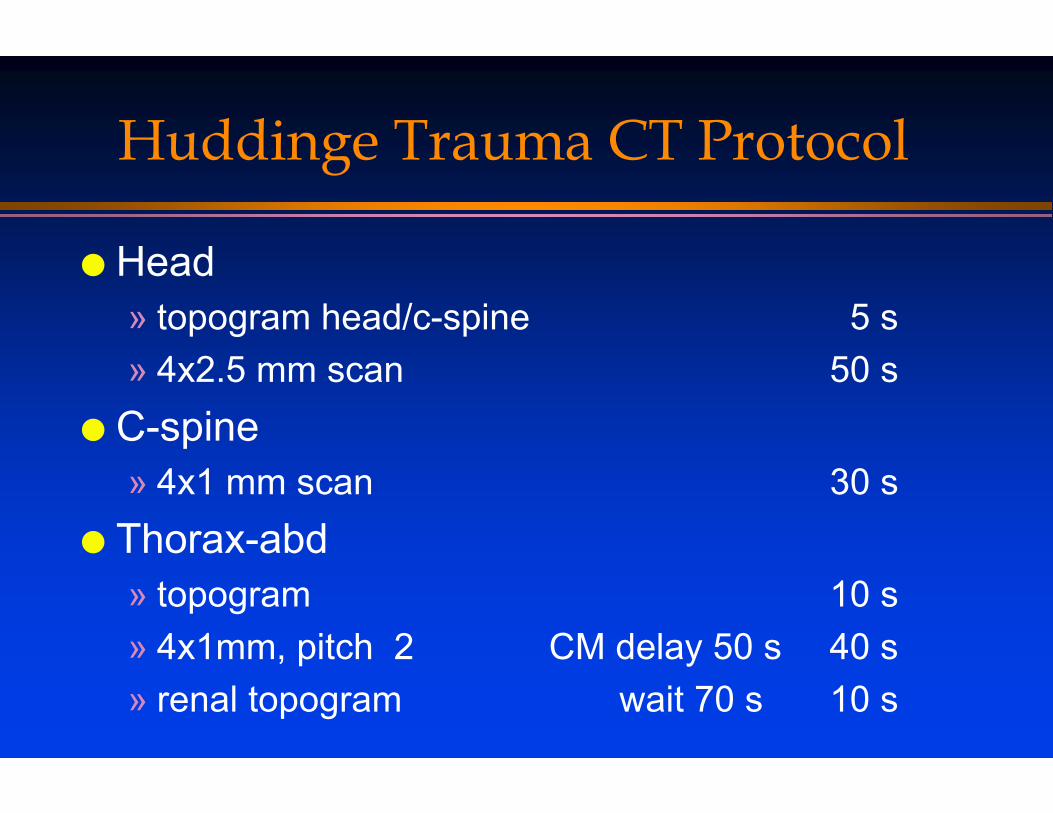
Huddinge Trauma CT Protocol
● Head» topogram head/c-spine 5 s» 4x2.5 mm scan 50 s
● C-spine» 4x1 mm scan 30 s
● Thorax-abd» topogram 10 s» 4x1mm, pitch 2 CM delay 50 s 40 s» renal topogram wait 70 s 10 s

CT- Scanning Parameters
5/5
1 /0.8
5/5 mm
Slice width/incr
90
120
300
Eff mAs
120
140/120
120
kVp
4 x 120.5Thorax-abdomen
4 x 1 1.50.75C-spine
4 x 2.50.651Head
Detectorwidth
PitchRot time
Area

Why ”Whole Body” CT?
● Choice:» Single area CT guided by clinical findings
or» Whole body CT
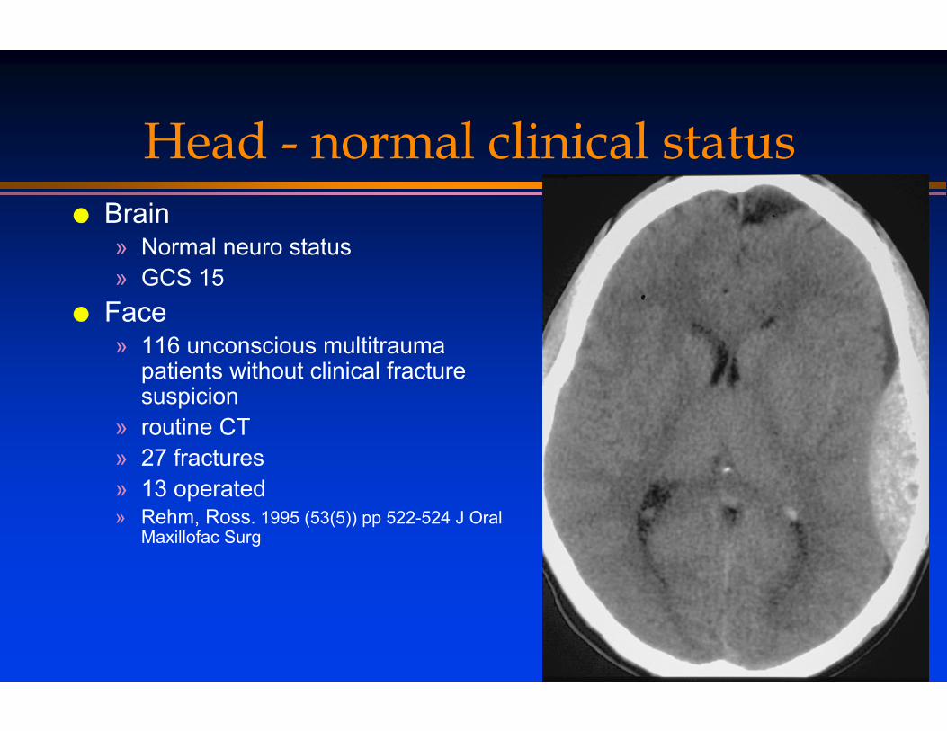
Head - normal clinical status● Brain
» Normal neuro status» GCS 15
● Face» 116 unconscious multitrauma
patients without clinical fracture suspicion
» routine CT» 27 fractures » 13 operated» Rehm, Ross. 1995 (53(5)) pp 522-524 J Oral
Maxillofac Surg

Thorax - normal clinical status
● Thorax– small
pneumothorax/ hemothorax/ aortic injury
» MVA» normal breath
sounds (??!)

Abdomen - normal clinical status● 15 year old MC driver
» CGS 15» Skin bruise of thorax» Normal abdominal palpation
● 35 year old drug addict» Fall» Normal abdominal palpation

Pelvis - normal clinical status● 35 year old drug addict
» clinically stable pelvis
● Karolinska Hospital » 7/10 unstable pelvic fx
undiagnosed clinically

Why “Whole Body” CT ? Clinical status cannot rule out injury!● C-spine – clinical injury exclusion needs
» Alert patient, no drugs» No distracting painful injury in any other body part
● i.e. excludes injured multitrauma patients
● In high energy trauma not possible to clear spine, thorax, abdomen, aorta with only clinical examination » Multiple references» Leidner B, Beckman MO. Standardized whole-body computed
tomography as a screening tool in blunt multitrauma patients. Emergency Radiology 2001; 8:20-28.
Whole Body CT - advantage● Fast - the Golden Hour● Whole body evaluation in one setting● Evaluation of circulation
» hypovolemia » active bleeding, pseudoaneurysms» multislice - angiographic evaluation
● High quality skeletal evaluation ● Intestinal injuries – 75-90% sensitivity

Whole Body CT - advantage● Superior skeletal evaluation
» Spine & pelvis» JTrauma aug 2003; Griffen/Hauser
– Sensitivity fx Plain films Helical CT– C-spine 2946 pat´s 60% 98%– T&L-spine 215 pat´s 58% 97%
» Multi-slice excellent» Little role for plain films

Multiplanar evalutationMIP – 4x1 mm – duodenal hematoma

Hypovolemia● Male 28 y,
Steeringwheel towards abdomen
● Hypovolemia» significant
bleeding» constriction of
aorta» low volume IVC
TF 105
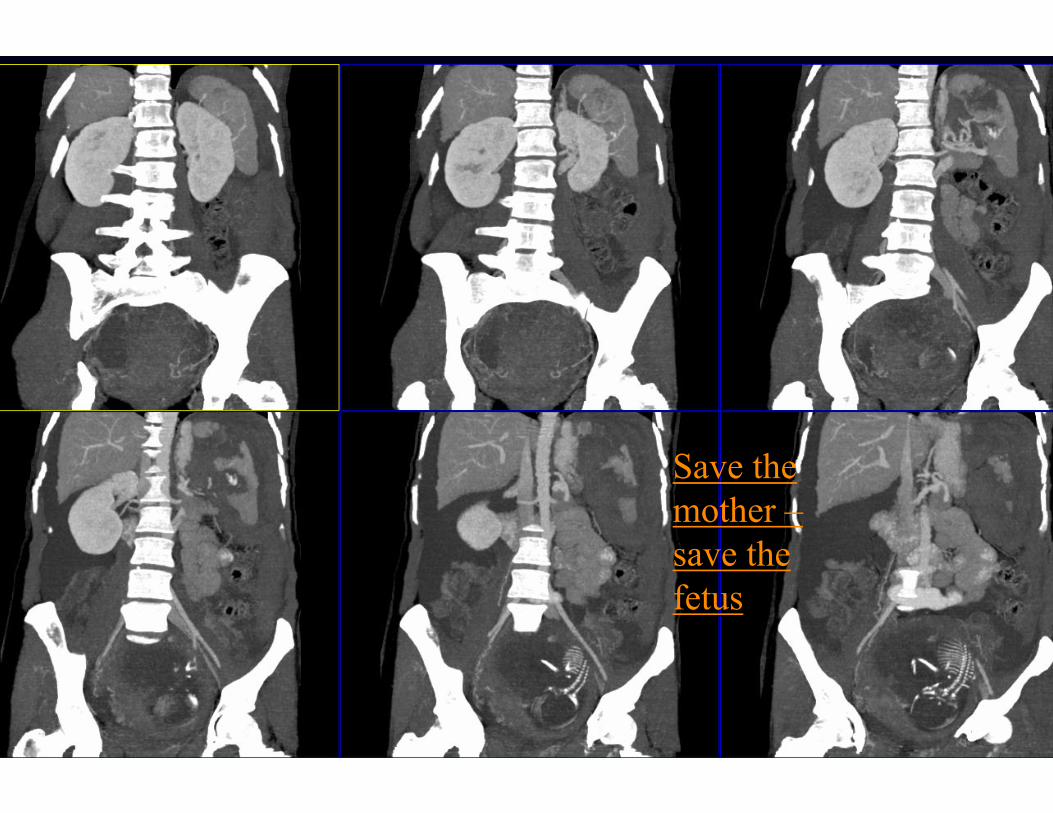
Work in progress
Save the mother –save the fetus

Swedish Trauma Scene
● Hospital size trauma patients radiologist pat/rad. 25% severe injuries
● Small 30 6 5 1● Medium/Large 250 30 8 2● Trauma center 1000 30 33 8
● Few severe cases per radiologist● Clinical standardization – ATLS● Radiological standard – Trauma CT

Problems in Trauma Radiology
● Untrained staff● Untrained radiologist ● Unfamiliar
diagnostics● Need for
standardization and joint training
● Cooperation & training with trauma team, OP & anestesiology.
● ”Train the Trauma Chain”
● Trauma CT

Swedish Radiological Week,Norrköping, Sept 18, 2003
Mats Beckman, Karolinska Sjukhuset, Bertil Leidner, Huddinge Universitetssjukhus
Trauma-CTPart II
Who to scan? - Indications
Trauma alarm -Traumadefinition
● Physiological parameters– Breathing difficulties– BP fall– Lowered
consciousness or spinal cord symtoms

Trauma alarm -Traumadefinition
● Physiological parametersInjury mechanisms – Car crash 50 /70
km/– MC– Pedestrian/bicyclist– Fall from >3m– Compression of
trunk

Trauma alarm -Traumadefinition
● Physiological parametersInjury mechanismsInjury types
– Penetrating neck&torso– Pelvic fractures– Paralyses– Amputations– Trauma and
– Burn– Cooling– Drowning
– Flail Chest
Who to scan?
● Selection difficult because of limited value of clinical examination
● Selection hampered by» costs in money and radiation dose» limited number of observation beds.

Case A
● Male age ca 35● Fall 3 meter● Subjective pain upper thoracic spine● No pain on examination
● Next step?

Case A

Case B
● Male age 43● Fall 6 meters● Admitted on spineboard● toe pain● On examination foot pain, no back pain
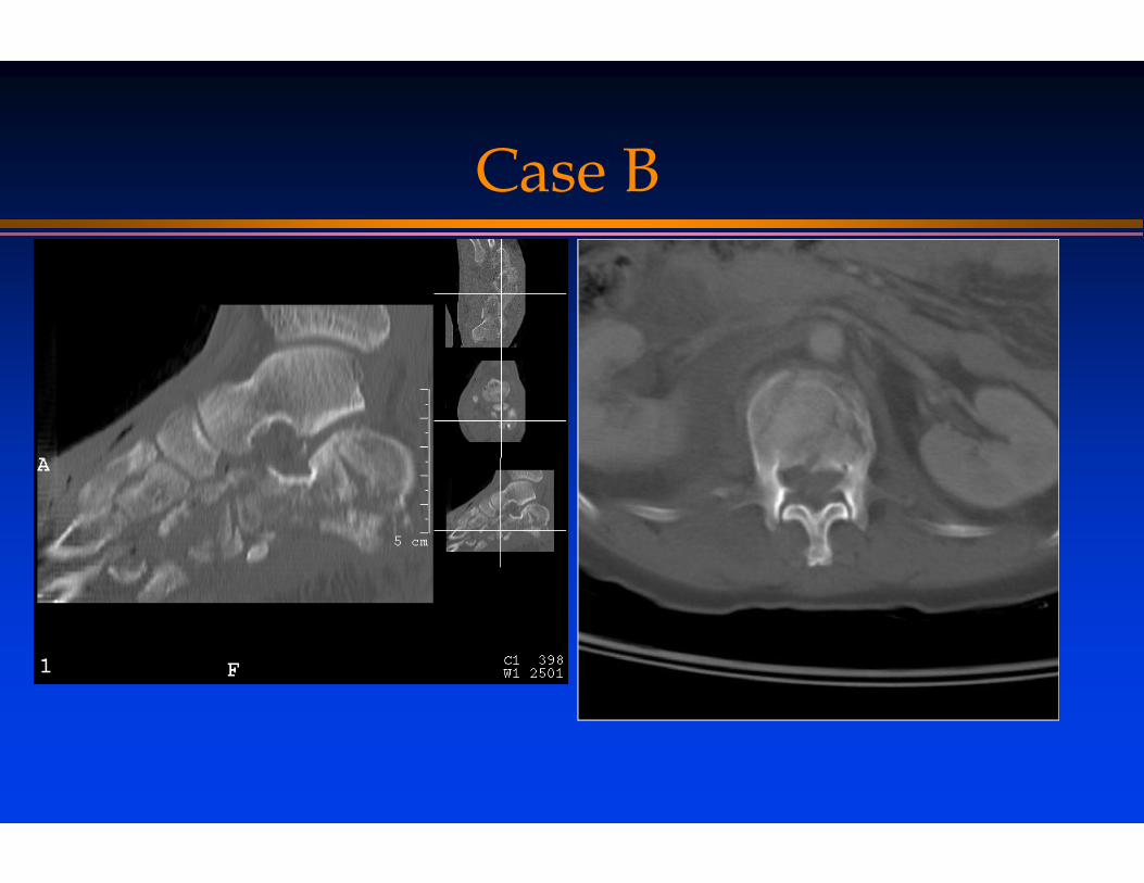
Case B

Case C● Male age 75● Fall on staircase unclear length● Back pain● Admitted as trauma 3- apparently uninjured
● 1st examination CR TH-L- spine is normal

Case C
● CT head normal● CT C-spine normal
but incidental finding

Case C
● CT head normal● CT C-spine normal
but incidental finding
Case D
● 12 year old girl» Fall from 1.5 m tree» Back pain

Case D
● 12 year old girl» Fall from 1.5 m tree» Back pain

Trauma CT in practice -Uppsala WBCT-results
● Siemens Somatom Plus/Plus 4● 304 patients (mid-1990)● Pos CT = significant injury =
» 118 patients - 39%» 195 injuries
Trauma CT in practice -Uppsala WBCT-results
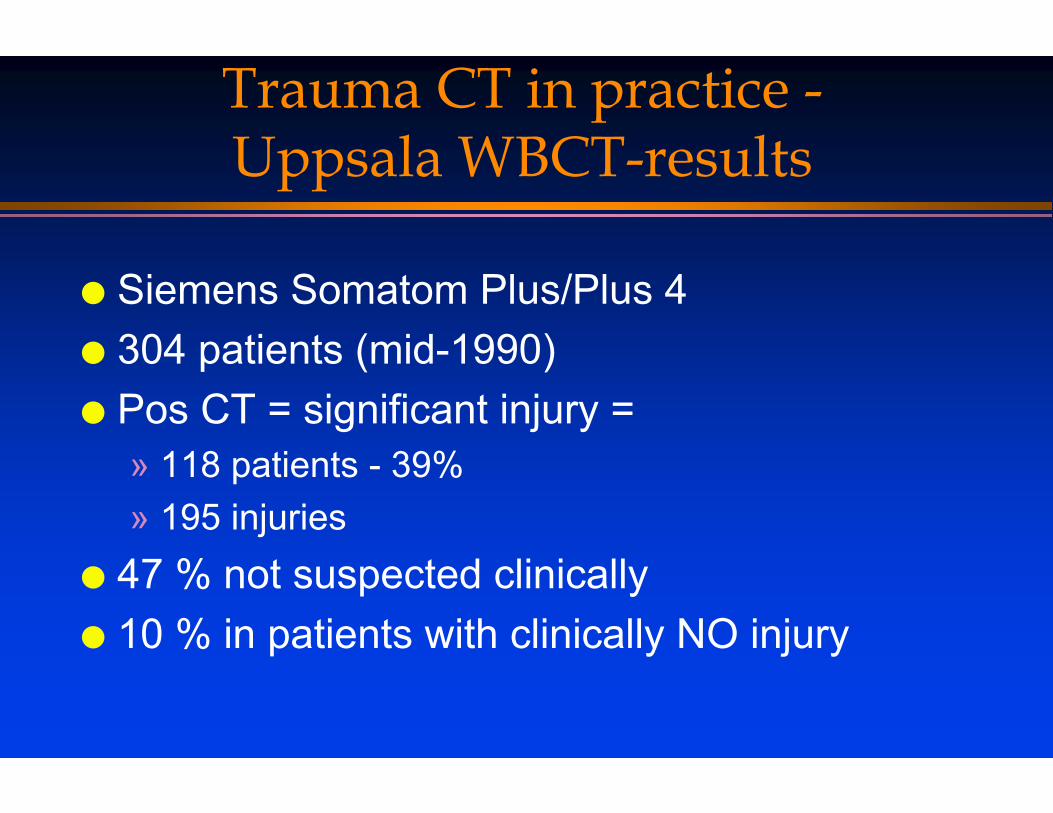
● Siemens Somatom Plus/Plus 4● 304 patients (mid-1990)● Pos CT = significant injury =
» 118 patients - 39%» 195 injuries
● 47 % not suspected clinically● 10 % in patients with clinically NO injury

How do we select patients to Trauma CT?
● Huddinge University Hospital» Active participation in trauma room» Trauma surgeon in CT suite» Stepdown» Trauma team training
● Karolinska Sjukhuset » Central Trauma Room» Level 1,2,3

The Trauma Team
Anesthesiologist
GeneralSurgeon ER Nurse
Radiologist
Radiology Nurse
OR Nurse Assistants
Operation Nurse
TraumaBay area
Anesthetic nurse
PrehospitalStaff
OrtopedicSurgeon
Trauma leaderSurgeon
Trauma care education
− multidisciplinary involvement
− all staff involved in trauma trained
− mortality/problem conferences

Inclusion criterias - discussion
● Cirkulatory stable
● By trauma mechanism● By vital & clinical signs
Trauma mechanism analysis
● Major trauma mechanism» Focal or general trauma
– Hit by bat once vs fall from height– Body parts at risk? – no head/c-spine? No thorax-abd??– Unknown
» How great was the violence?– 90 or 50 km/hour?– Vehicle intrusion? – crash pictures
» Body protection– Vehicle? Seatbelt? Airbag?– Beware of the Unprotected body!
Clinical signs CT
● Decreased level of consciousness ● Non-reliable patient
» Alcohol, drugs, language, culture● Respiratory distress● Cirkulatory distress● Overt signs of injury

Step-down clinical observation
● Limited violence by analysis● Reliable patient● No clinical signs of injury?
● When in doubt – take the patient to the CT● Remember – the patient´s life could be in
immediate danger

The price: Dose & Cancer Induction
● Dose (HS)» Head 2.0 mSv» C-spine 3.5 mSv» Body 9.0 mSv
● Total dose ~14,5 mSv ● KS – 13 mSv● 5 cancers/100 Sv 0.00075 cancers/15 mSV
» 1 lifetime deadly cancer per 1333 patients» 266 spontaneous cancers per 1333 patients
● HS 250 / KS 1000 Trauma CT/year

Dose
● CT Dose» head 2.0 mSv» c-spine 3.5 mSv» body 9.0 mSv
● Total dose ~14.5 mSv
● 4-6 years Swedish background radiation
● CT + US + Convent. X-rays» Head (CT) 2.0 mSv» C-spine 0.5 mSv» Chest 0.02 mSv» T+L-spine 2.0 mSv» Pelvis 0.7 mSv» US 0.0 mSv» Angio 10% x 5 0.5 mSv
● Total dose ~ 5.7 mSv» Incomplete abdominal exam –
US << CT» Substandard spine evaluation
Addditional benefits:Incidental findings at trauma CT
● Tumors 2-6/1000» kidney lung» liver skeletal metastases» lymfoma
● Non- cancers» Aneurysm (cerebral, abdominal » Fatty liver ,Teeth infections» Calculi -gallstones, kidneystones» Myoma, lung granulomas

Conclusions
● High energy impact causes injuries● Clinical evaluation is not enough● CT is the radiological method of choice● Need to limit unnecessary examinations
» Cost» Radiation

Conclusions -recommendations
● Trauma patients – high risk● Intimate cooperation
» The radiologist in the traumaroom
» The surgeon in the CT-suite
● Recognize the risk patient» Trauma mechanism» How great was the force?» Beware: unprotected body» Deranged physiology (ie
tachycardia)
● Clinical step-down
● Whole Body Trauma CTshould be used for general trauma patients.
● With ”focal traumamechanism”» Scanning alternatives
– Whole body– Head + c-spine– Thorax + abdomen
Swedish Radiological Week,Norrköping, Sept 18, 2003
Mats Beckman, Karolinska Sjukhuset, Bertil Leidner, Huddinge Universitetssjukhus
Trauma-CTPart III
Discussion
What about ultrasound?
● Ultrasound not for diagnosing organ injury
● Helpful to select patients for immediate intervention- splenectomy, Bűlau.
● Only use if CT is sceduled soon● Trust result ca 10 min
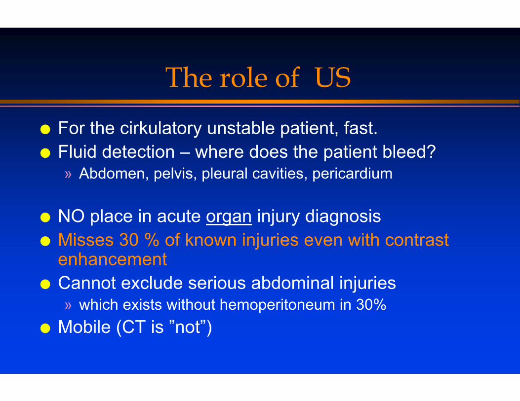
The role of US● For the cirkulatory unstable patient, fast.● Fluid detection – where does the patient bleed?
» Abdomen, pelvis, pleural cavities, pericardium
● NO place in acute organ injury diagnosis● Misses 30 % of known injuries even with contrast
enhancement● Cannot exclude serious abdominal injuries
» which exists without hemoperitoneum in 30%● Mobile (CT is ”not”)


Work in progress
Save the mother –save the fetus

Pelvimetry &cancer
● 25344 pelvimetries● identical controls● foetal marrow dose ca 1 mSv ● 42 cancers in study group, 41 in control
group● Beckman et al unp.

The Trauma Care Chain

”Missed patients”
● No trauma alarm» By ”own” car
● Bypassed trauma room» Fall from ladder – back pain – to ortopedics» Fall – ear bleed - ENT» Pregnant patients – Gynecology
● Incomplete trauma mechanism analysis» Fall from low height to hard pointed objects
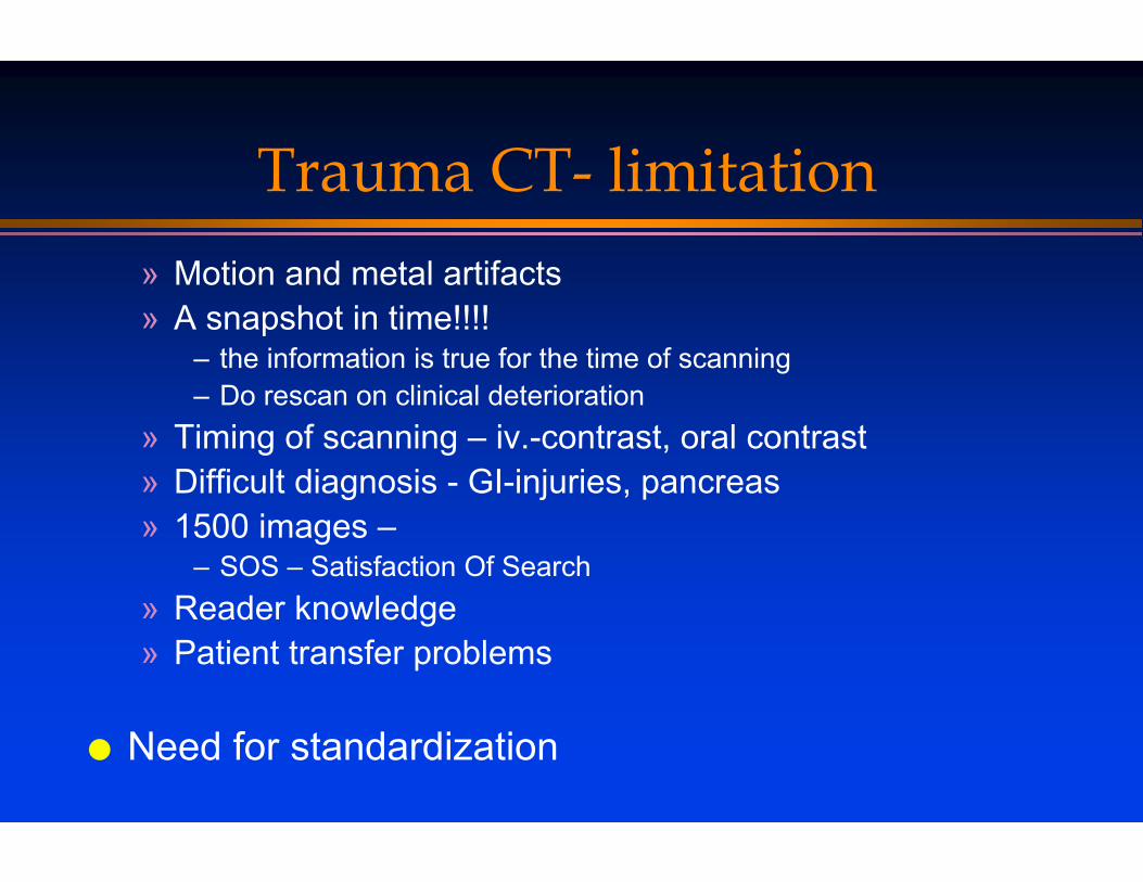
Trauma CT- limitation» Motion and metal artifacts» A snapshot in time!!!!
– the information is true for the time of scanning– Do rescan on clinical deterioration
» Timing of scanning – iv.-contrast, oral contrast» Difficult diagnosis - GI-injuries, pancreas» 1500 images –
– SOS – Satisfaction Of Search» Reader knowledge» Patient transfer problems
● Need for standardization