
Transoral Removal of aGiant Submandibular Sialolith: A Case … · Sialolithiasis is the most...
16
International Journal of Scientific & Engineering Research Volume 9, Issue 1, January-2018 32 ISSN 2229-5518 IJSER © 2018 http://www.ijser.org Transoral Removal of aGiant Submandibular Sialolith: A Case Report Hesham Alowaimer, Tarek Kasem, Daij AL Daiji Oral Surgery Department, Al-Rass General Hospital, Al-Rass, Saudi Arabia ([email protected] ) Abstract: Submandibular salivary glands are commonly affected by sialolith (calculus),with patients suffering pain and swelling, especially at mealtime. Sialoliths and giant calculi larger than 15 mm are rarely reported. This case report presents a 28 mm sialolith which was removed successfully intraorally under local anesthesia (transoral approach; sialolithotomy). There were no complications either during the operation or postoperatively, and the salivary function of the gland remained normal. Keywords:Sialolithiasis; Submandibular swelling; Sialoliths; Salivary gland obstruction;Minimally invasive surgery —————————— —————————— INTRODUCTION Sialolithiasis is one of the most frequent salivary gland disorders, with the submandibular gland being the most affected gland (80–87%). 1–4 Salivary gland stones, or sialoliths, are calcified structures in the salivary glands or their ducts, resulting in the obstruction of salivary secretion and retention of saliva. 5,6 This is usually accompanied by symptoms of swelling and pain, especially during mealtime. 7 The etiology of salivary stones is not completely understood, and various hypotheses related to local and systemic predisposing factors have been discussed in the literature. 5,6 IJSER
Transcript of Transoral Removal of aGiant Submandibular Sialolith: A Case … · Sialolithiasis is the most...
International Journal of Scientific & Engineering Research Volume 9, Issue 1, January-2018 32 ISSN 2229-5518
IJSER © 2018 http://www.ijser.org
Transoral Removal of aGiant Submandibular Sialolith: A Case Report
Hesham Alowaimer, Tarek Kasem, Daij AL Daiji Oral Surgery Department, Al-Rass General Hospital, Al-Rass, Saudi Arabia
Abstract: Submandibular salivary glands are commonly affected by sialolith (calculus),with patients suffering pain and swelling,
especially at mealtime. Sialoliths and giant calculi larger than 15 mm are rarely reported. This case report presents a 28 mm sialolith
which was removed successfully intraorally under local anesthesia (transoral approach; sialolithotomy). There were no
complications either during the operation or postoperatively, and the salivary function of the gland remained normal.
Keywords:Sialolithiasis; Submandibular swelling; Sialoliths; Salivary gland obstruction;Minimally invasive surgery
—————————— —————————— INTRODUCTION
Sialolithiasis is one of the most frequent salivary gland disorders, with the submandibular
gland being the most affected gland (80–87%).1–4
Salivary gland stones, or sialoliths, are calcified structures in the salivary glands or their
ducts, resulting in the obstruction of salivary secretion and retention of saliva.5,6This is usually
accompanied by symptoms of swelling and pain, especially during mealtime.7
The etiology of salivary stones is not completely understood, and various hypotheses related
to local and systemic predisposing factors have been discussed in the literature.5,6
IJSER
International Journal of Scientific & Engineering Research Volume 9, Issue 1, January-2018 33 ISSN 2229-5518
IJSER © 2018 http://www.ijser.org
In addition to clinical examination, several imaging techniques can be applied for the
diagnosis of sialoliths, such as intra- and extra-oral radiographs, computed tomography
(CT), cone beam computer tomography (CB-CT), ultrasonography, and sialography.6,8
Calculi commonly measure less than 10 mm in size, and calculi surpassing 15 mm are
termed giant salivary gland calculi (Sialoliths).3,9The giant sialoliths (>15 mm) in the
submandibular gland have rarely been reported.10
The management of salivary stones depends on the size and location of the stone. The
primary objective in the treatment of sialolithiasis should be focused on removing the salivary
stones and preserving gland function in combination with a low level of complication and
discomfort for the patient.6
Various procedures have been proposed for the management of sialolithiasis, which range
from conservative minimally-invasive procedures, to more invasive procedures.6,11
IJSER
International Journal of Scientific & Engineering Research Volume 9, Issue 1, January-2018 34 ISSN 2229-5518
IJSER © 2018 http://www.ijser.org
This case report presented a 28 mm sialolith which was removed intraorally under local
anesthesia (transoral approach; sialolithotomy).
CASE REPORT
A 39-year-old male patient was referred by a private clinic to our Oral Surgery Department.
The patient presented with complaints of pain and swelling in the right submandibular region
for 6 months, with episodes of discomfort, especially during mealtime.
A clinical examination revealed a mild to moderate tender and firm right submandibular
swelling. Bimanual palpation of the right posterior floor of the mouth revealed a palpable and
tender mass measuring approximately 3 cm. In comparison to the contralateral gland region,
decreased salivary flow was observed at the opening of the Wharton’s duct.
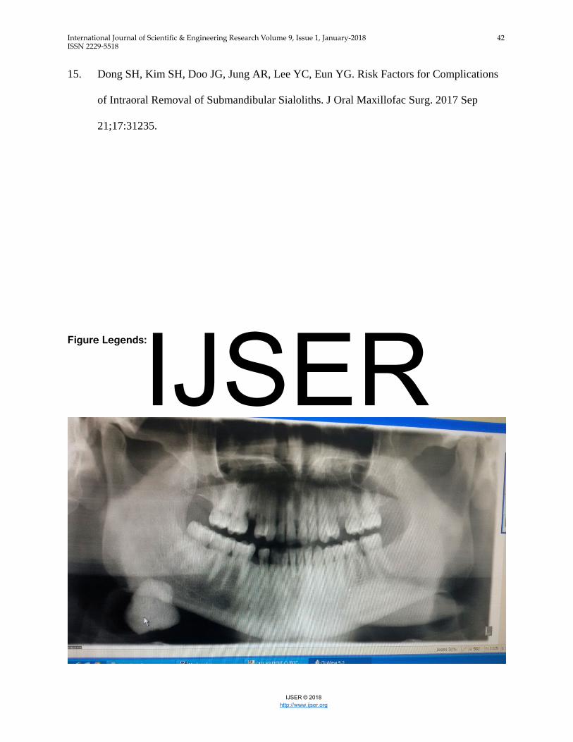
The Orthopantomogram (OPG) confirmed the presence of a radiopaque lesion of around 3
cm in the right submandibular area (Figure 1). A diagnosis of sialolithiasis was given on the
basis of the clinical and radiographic examination.
IJSER
International Journal of Scientific & Engineering Research Volume 9, Issue 1, January-2018 35 ISSN 2229-5518
IJSER © 2018 http://www.ijser.org
Under local anesthesia, transoral removal of the sialolith was performed. A mucosal incision
of 3–4 cm was made directly overthe stone in the floor of the mouth in the region ofthe
rightfirst premolar to thesecond molar. The depth of the incision was superficial in order to
prevent damage of the lingual nerve. This was followed by a deep dissection of the gland
and duct without widening the duct.
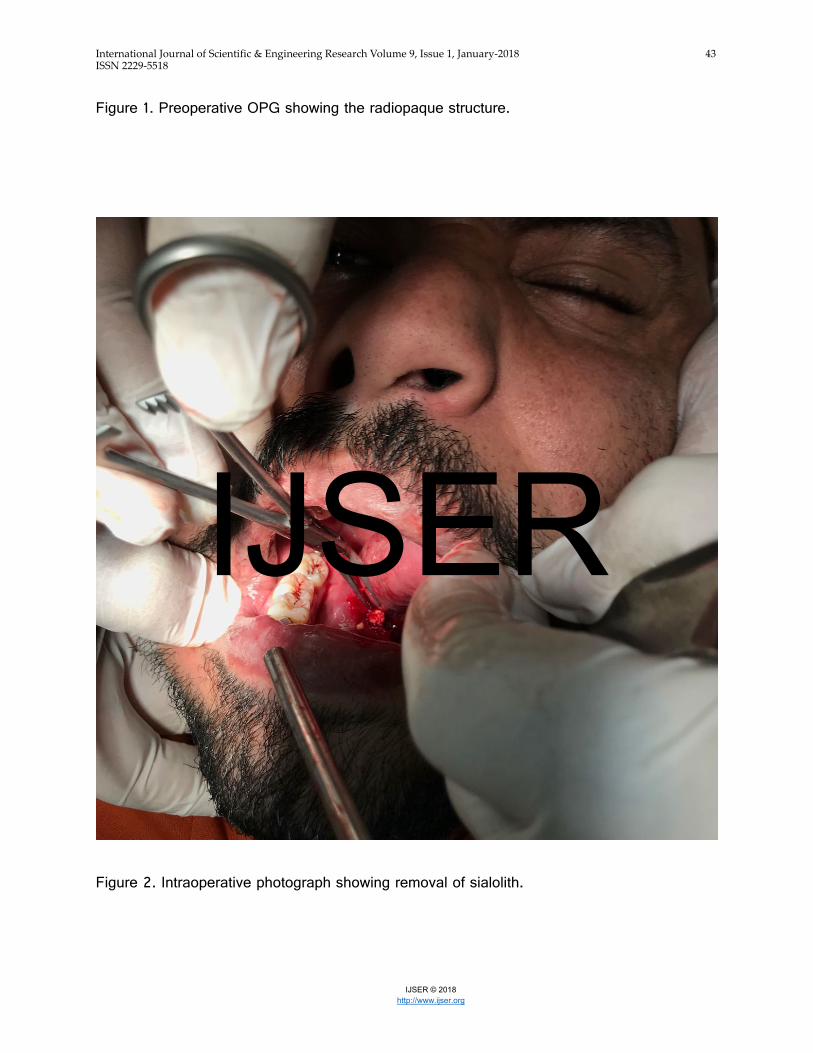
The sialolith was in the hilum of the submandibular salivary gland, while part was in the
proximal duct (Hilar submandibular stone).The sialolith was exposed, fragmented and then
retrieved using Kocher forceps (Figure 2); this was followed by irrigation with normal saline
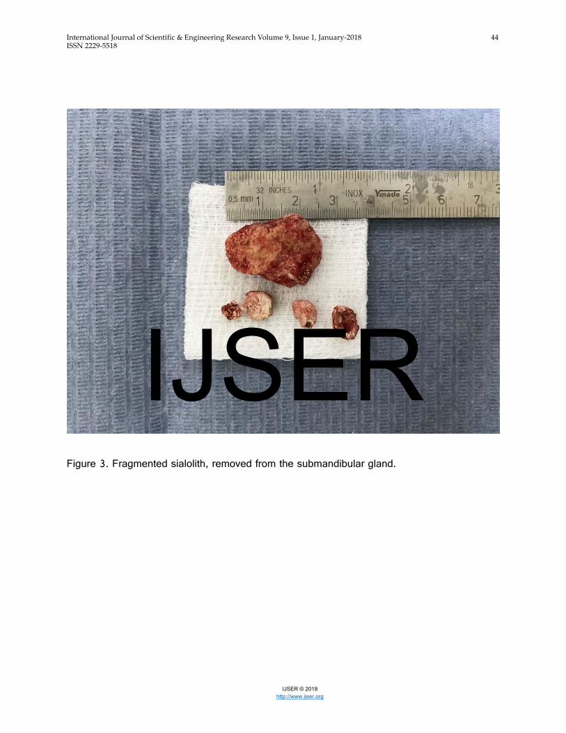
and suturing. The size of the stone was 28 mm, while there were also four small
pieces/fragments (Figure 3).
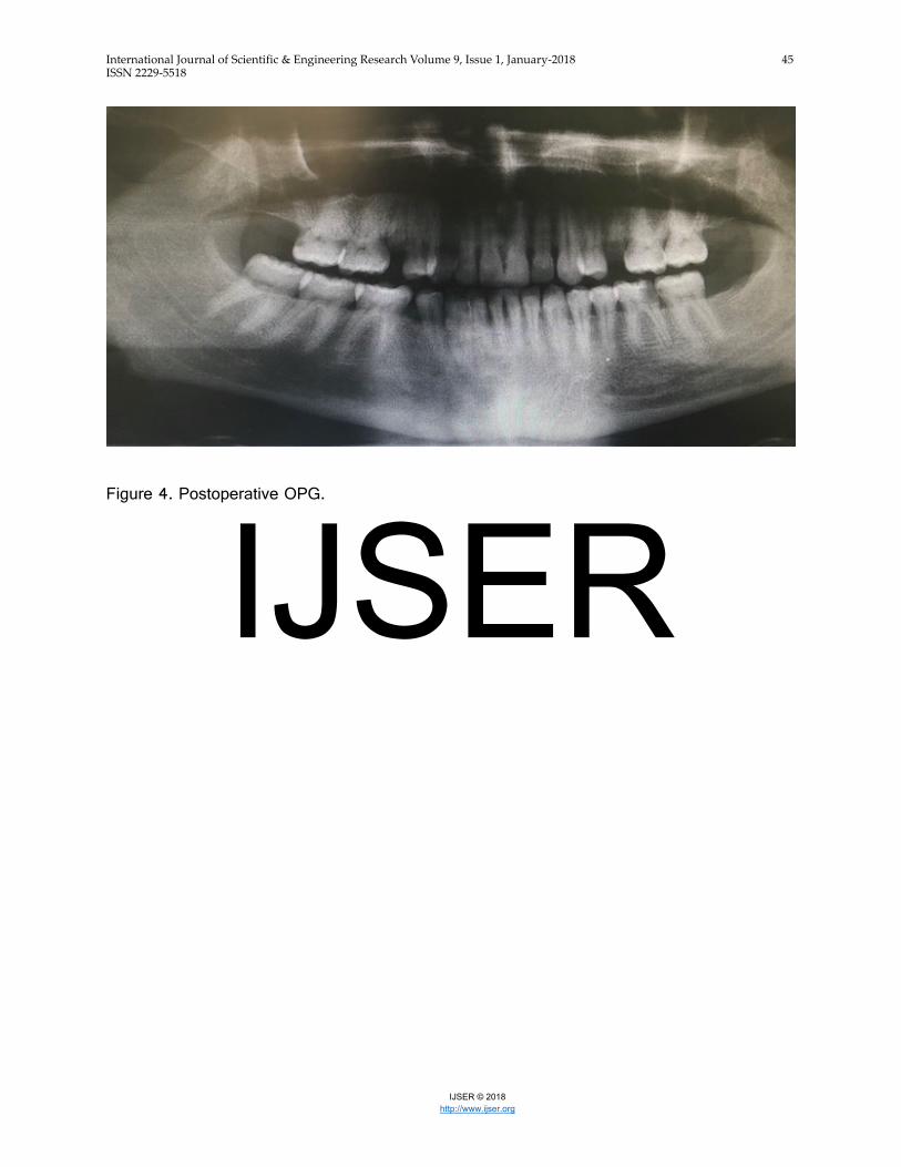
The postoperative OPG was clear (Figure 4), indicating no remaining sialoliths. After one
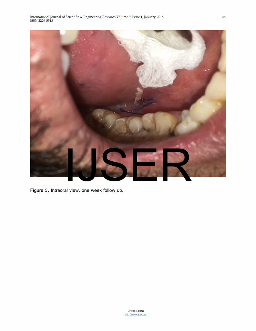
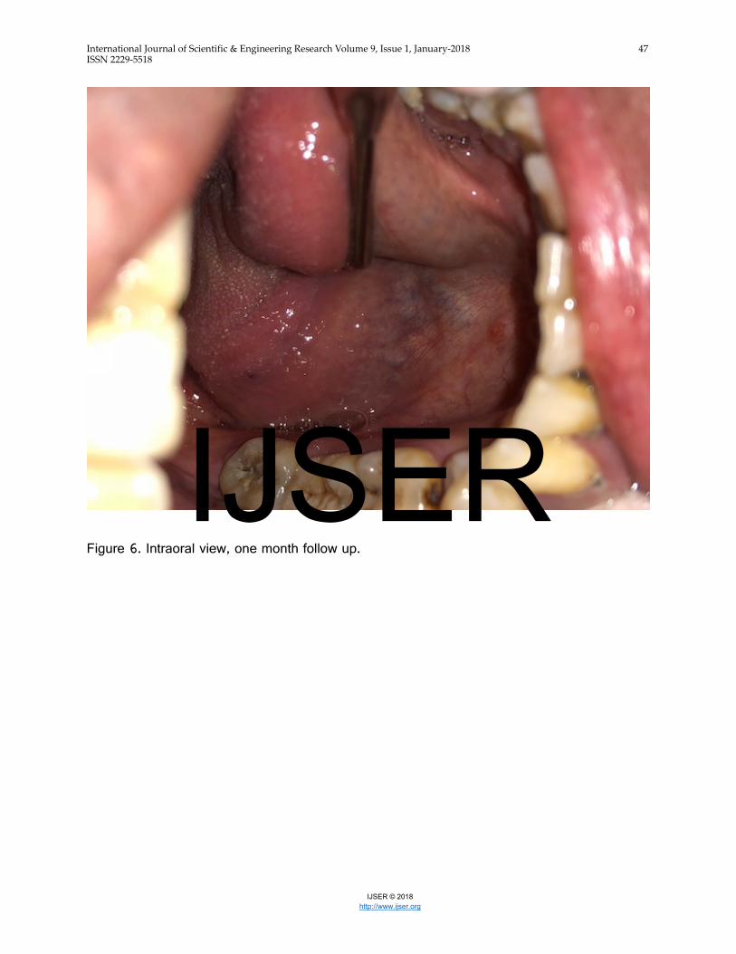
week (Figure 5), one month (Figure 6), and then again at two months follow up, the patient
was asymptomatic, and the salivary gland recovered normal function.
DISCUSSION
IJSER
International Journal of Scientific & Engineering Research Volume 9, Issue 1, January-2018 36 ISSN 2229-5518
IJSER © 2018 http://www.ijser.org
Sialolithiasis is the most common pathology of the salivary glands.3,5,6This case report
discussed a sialolith which presented in the submandibular gland.
The diagnosis of salivary calculi is mainly based on clinical symptoms and imaging. In
addition to the physical examination, several imaging techniques can be applied. Intra- and
extra-oral radiographs are effective in most cases. Extra-oral OPGswould detect fewer
salivary stones assome calculi could be superimposed on bony structures or teeth.6Hence, it
has been suggested that intra-oral occlusal radiographs represent amore useful method for
the detection of a submandibular sialolith.5 In this case, the diagnosis was clearly obtained by
an OPG examination alone, and the need for further investigation was not indicated.
In more challenging cases, more advanced techniques can be used for imaging and
examining the salivary stones.12These diagnostic tools include ultrasonography, computed
tomograms (CT), cone beam computer tomography (CB-CT), sialography and
sonography.4,12
IJSER
International Journal of Scientific & Engineering Research Volume 9, Issue 1, January-2018 37 ISSN 2229-5518
IJSER © 2018 http://www.ijser.org
The management of salivary stones is focused on removing the salivary stones and
preserving salivary gland function based on the size and location of the stone.6,13
The minimally invasive treatment options are numerous and significantly improve the
prognosis of sialolithiasis.13The conservative therapeutic options include hydration of the
patient, prescription of sialogogues,and gland massage.3,4,8
While patients with small sialoliths can undergo conservative treatment, those with standard
size or larger sialoliths require sialendoscopy, or a transoral approach (sialolithotomy).14The
use of these minimally-invasive and conservative techniques makes it possible to preserve
the function of the affected submandibular gland in most cases.7
At present, management through surgical treatment is the mainstay treatment modality. The
surgical removal via a direct incision in the stone (sialolithotomy) is relatively simple to
perform; it is alsowell-tolerated, can be carried out under local anesthesia,and does not
often result in complications.6
IJSER
International Journal of Scientific & Engineering Research Volume 9, Issue 1, January-2018 38 ISSN 2229-5518
IJSER © 2018 http://www.ijser.org
This traditional management of submandibular stones based on sialolithotomy is usually for
stones located in the distal and mid-third of the Wharton's duct, while sialadenectomy
(removal of the gland) is used for deeper stones.7
The surgical intraoral removal of submandibular sialoliths is a technique for the treatment of
sialolithiasis and is reported to have excellent outcomes.15Carelessness during this treatment,
however, can cause rupturing of the duct.3
After sialolith therapy that does not include surgical removal of the gland, most patients
experience no complaints or discomfort, and are left with a normal functioning gland. Minor
complications include a postoperative infection, postoperative ranulas, and lingual nerve
dysfunction.8,15In the presented case, there were no complications and the gland recovered
normal function quickly following removal of the stoneat up to two months follow up.
Recurrence of sialoliths is rather uncommon, and is estimated to occur in 1–10% of patients
during the first 6 months postoperatively.14 Long-term follow up was planned for this case.
IJSER
International Journal of Scientific & Engineering Research Volume 9, Issue 1, January-2018 39 ISSN 2229-5518
IJSER © 2018 http://www.ijser.org
CONCLUSION
Sialolithiasis is common and should be considered in patients experiencing submandibular
swelling and pain, especially at mealtime.
This case report draws attention to the feasibility of a harmless therapeutic approach to such
a huge sialolith. No submandibular function impairment resulted after the therapy.
Funding: None
Conflict of interest: None
IJSER
International Journal of Scientific & Engineering Research Volume 9, Issue 1, January-2018 40 ISSN 2229-5518
IJSER © 2018 http://www.ijser.org
References
1. Bilahari N, Kumari B, James B, Kuruvila V. Submandibular sialolithiasis: Report of six
cases. J Pharm Bioallied Sci. 2013 Jul;5(3):240.
2. Batori M, Mariotta H, Chatelou H, Casella G, Casella MC. Diagnostic and surgical
management of submandibular gland sialolithiasis: Report of a stone of unusual size. Eur
Rev Med Pharmacol Sci. 2005;9(1):67–8.
3. Cho S-H, Han J-D, Kim J-H, Lee S-H, Jo J-B, Kim C-H, et al. Removal of submandibular
calculi by surgical method and hydraulic power with curved needle: a case report. J
Korean Assoc Oral Maxillofac Surg. 2017 Jun;43(3):182.
4. Kopeć T, Wierzbicka M, Szyfter W, Leszczyńska M. Algorithm changes in treatment of
submandibular gland sialolithiasis. Eur Arch Oto-Rhino-Laryngology. 2013 Jul
9;270(7):2089–93.
5. Delli K, Spijkervet FKL, Vissink A. Salivary Gland Diseases: Infections, Sialolithiasis
and Mucoceles. In: Monographs in oral science. 2014. p. 135–48.
IJSER
International Journal of Scientific & Engineering Research Volume 9, Issue 1, January-2018 41 ISSN 2229-5518
IJSER © 2018 http://www.ijser.org
6. Kraaij S, Karagozoglu KH, Forouzanfar T, Veerman ECI, Brand HS. Salivary stones:
symptoms, aetiology, biochemical composition and treatment. Br Dent J. 2014 Dec
5;217(11):E23.
7. Capaccio P, Marciante G, Gaffuri M, Spadari F. Submandibular swelling: Tooth or
salivary stone? Indian J Dent Res. 2013;24(3):381.
8. Ardekian L, Klein HH, Araydy S, Marchal F. The Use of Sialendoscopy for the Treatment
of Multiple Salivary Gland Stones. J Oral Maxillofac Surg. 2014 Jan;72(1):89–95.
9. Lim EH, Nadarajah S, Mohamad I. Giant Submandibular Calculus Eroding Oral Cavity
Mucosa. Oman Med J. 2017 Sep 27;32(5):432–5.
10. Rodrigues GHC, Carvalho VJG, Alves FA, Domaneschi C. Giant submandibular sialolith
conservatively treated. Autops case reports. 2017;7(1):9–11.
11. Erkul E, Gillespie MB. Sialendoscopy for non-stone disorders: The current evidence.
Laryngoscope Investig Otolaryngol. 2016 Oct;1(5):140–5.
12. Pabst G, Strobel K, Zehnder J. The value of the twinkling artefact for the diagnosis of
sialolithiasis of the large salivary glands. J Laryngol Otol. 2017 Dec 18;18:1–6.
13. Foletti JM, Graillon N, Avignon S, Guyot L, Chossegros C. Salivary Calculi Removal by
Minimally Invasive Techniques: A Decision Tree Based on the Diameter of the Calculi
and Their Position in the Excretory Duct. J Oral Maxillofac Surg. 2018 Jan;76(1):112–8.
14. Ruiz R, Brygo A, Nicot R, Ferri J. Sialolithiasis Removal under general anesthesia: a
descriptive retrospective study in the maxillo-facial surgery department in Lille University
Hospital. J Stomatol Oral Maxillofac Surg. 2017 Nov 10;17:30203.
IJSER
International Journal of Scientific & Engineering Research Volume 9, Issue 1, January-2018 42 ISSN 2229-5518
IJSER © 2018 http://www.ijser.org
15. Dong SH, Kim SH, Doo JG, Jung AR, Lee YC, Eun YG. Risk Factors for Complications
of Intraoral Removal of Submandibular Sialoliths. J Oral Maxillofac Surg. 2017 Sep
21;17:31235.
Figure Legends:
IJSER
International Journal of Scientific & Engineering Research Volume 9, Issue 1, January-2018 43 ISSN 2229-5518
IJSER © 2018 http://www.ijser.org
Figure 1. Preoperative OPG showing the radiopaque structure.
Figure 2. Intraoperative photograph showing removal of sialolith.
IJSER
International Journal of Scientific & Engineering Research Volume 9, Issue 1, January-2018 44 ISSN 2229-5518
IJSER © 2018 http://www.ijser.org
Figure 3. Fragmented sialolith, removed from the submandibular gland.
IJSER
International Journal of Scientific & Engineering Research Volume 9, Issue 1, January-2018 45 ISSN 2229-5518
IJSER © 2018 http://www.ijser.org
Figure 4. Postoperative OPG.
IJSER
International Journal of Scientific & Engineering Research Volume 9, Issue 1, January-2018 46 ISSN 2229-5518
IJSER © 2018 http://www.ijser.org
Figure 5. Intraoral view, one week follow up.
IJSER
International Journal of Scientific & Engineering Research Volume 9, Issue 1, January-2018 47 ISSN 2229-5518
IJSER © 2018 http://www.ijser.org
Figure 6. Intraoral view, one month follow up.
IJSER


![Application of Multicategory Exposure Marginal Structural ... · basis of disease severity and response to previous treatment [3,5,6]. Consequently, it is important to consider time-dependent](https://static.fdocuments.net/doc/165x107/5e209b30a41ada3827003102/application-of-multicategory-exposure-marginal-structural-basis-of-disease-severity.jpg)


![Bacteriophages in the Dairy Environment: From Enemies to Allies · 2018. 5. 3. · of dairy products by the inclusion of fruits and cereals [3,5,6]. Moreover, the creation of new](https://static.fdocuments.net/doc/165x107/60cd8378d7b33d01814d5289/bacteriophages-in-the-dairy-environment-from-enemies-to-allies-2018-5-3-of.jpg)

