Translational research in transcranial direct current stimulation (tDCS): a systematic review of...
11
Rev. Neurosci., Vol. 22(4): 471–481, 2011 • Copyright © by Walter de Gruyter • Berlin • Boston. DOI 10.1515/RNS.2011.042 Translational research in transcranial direct current stimulation (tDCS): a systematic review of studies in animals Andre Russowsky Brunoni 1,2, *, Felipe Fregni 3, * and Rosana Lima Pagano 4 1 Department of Neurosciences and Behavior, Institute of Psychology, University of São Paulo, Cidade Universitária, 05508-000 Butantã, São Paulo, Brazil 2 University Hospital, University of São Paulo, Brazil 3 Laboratory of Neuromodulation, Department of Physical Medicine and Rehabilitation, Spaulding Rehabilitation Hospital and Massachusetts General Hospital, Harvard Medical School, Boston, MA 02114, USA 4 Laboratory of Neuromodulation and Experimental Pain, Hospital Sírio-Libanês, São Paulo 01308-050, Brazil *Corresponding authors e-mail: [email protected]; [email protected] Abstract Recent therapeutic human studies testing transcranial direct current stimulation (tDCS) has shown promising results, although many questions remain unanswered. Translational research with experimental animals is an appropriate frame- work for investigating its mechanisms of action that are still undetermined. Nevertheless, animal and human studies are often discordant. Our aim was to review tDCS animal stud- ies, examining and comparing their main findings with human studies. We performed a systematic review in Medline and other databases, screening for animal studies in vivo that deliv- ered tDCS. Studies in vitro and using other neuromodulatory techniques were excluded. We extracted data according to Animal Research: Reporting In Vivo Experiments (ARRIVE) guidelines for reporting in vivo animal research. Thus, we collected data on sample characteristics (size, gender, weight and specimen) and methodology (experimental procedures, experimental animals, housing and husbandry, as well as anal- ysis). We also collected data on methods for delivering tDCS (location, size, current and current density of electrodes and electrode montage), experimental effects (polarity-, intensity- and after-effects) and safety. Only 12 of 48 potentially eligible studies met our inclusion criteria and were reviewed. Quality assessment reporting was only moderate and studies were het- erogeneous regarding tDCS montage methodology, position of active and reference electrodes, and current density used. Nonetheless, almost all studies demonstrated that tDCS had positive immediate and long-lasting effects. Vis-à-vis human trials, animal studies applied higher current densities (34.2 vs. 0.4 A/m 2 , respectively), preferred extra-cephalic positions for reference electrodes (60% vs. 10%, respectively) and used electrodes with different sizes more often. Potential implica- tions for translational tDCS research are discussed. Keywords: animal models; brain; direct current; electrical stimulation; neuroplasticity; systematic review; translational research. Introduction Transcranial direct current stimulation (tDCS) is a non- invasive neuromodulation technique that uses weak, direct electric currents applied through electrodes placed over the scalp to induce changes in cortical excitability (Stagg and Nitsche, 2011). tDCS has been widely researched over the past decade and recent therapeutic studies in neuropsychi- atric disorders have shown promising results in epilepsy (Fregni et al., 2006; Nitsche and Paulus, 2009), chronic pain (Williams et al., 2009) and major depression (Boggio et al., 2008; Ferrucci et al., 2009; Brunoni et al., 2011b), for exam- ple. Compared to other therapies, tDCS is safer than invasive brain stimulation (e.g., deep brain stimulation, motor cortex stimulation) that is associated with surgical risks (infection, hemorrhage, seizures) and has increased costs (Zaghi et al., 2009). It also seems to have additional advantages when compared with transcranial magnetic stimulation (TMS): in particular, tDCS is relatively more portable, affordable, tolerable and, perhaps, also safer (Priori et al., 2009; Zaghi et al., 2009; Brunoni et al., 2011a). Furthermore, tDCS can be combined with pharmacotherapy to enhance its clinical effects and/or reduce adverse effects by avoiding drug dos- age increases (Brunoni et al., 2011c). Although single sessions of tDCS are mostly associated with short-lived effects (Nitsche et al., 2008), emerging evidence shows that daily, repeated tDCS sessions can induce effects that persist for several weeks (Reis et al., 2008; Rigonatti et al., 2008). Hence, current clinical research has focused on investigating how tDCS can be delivered to achieve the most effective clinical gains, testing several parameters such as time course of stimulation, time period between stimulation sessions and optimal dosage. Nonetheless, the mechanisms by which tDCS changes cortical excitability and induces long-lasting changes beyond the period of stimulation are not fully understood. Along these lines, studies in animals are useful for exploring the electrophysiological properties of tDCS mechanisms. For instance, these studies allow direct inves- tigation of the cellular and histologic modifications induced by tDCS. Moreover, animal studies are especially useful for testing different combinations of parameters of stimulation (e.g., safety range, threshold for brain damage and other parameters), identifying those that are more advantageous for clinical research. Brought to you by | Bibliotheque de l'Universite Laval Authenticated Download Date | 11/27/14 12:22 AM
-
Upload
rosana-lima -
Category
Documents
-
view
215 -
download
2
Transcript of Translational research in transcranial direct current stimulation (tDCS): a systematic review of...

Rev. Neurosci., Vol. 22(4): 471–481, 2011 • Copyright © by Walter de Gruyter • Berlin • Boston. DOI 10.1515/RNS.2011.042
Translational research in transcranial direct current stimulation (tDCS): a systematic review of studies in animals
Andre Russowsky Brunoni 1,2, *, Felipe Fregni 3, * and Rosana Lima Pagano 4
1 Department of Neurosciences and Behavior , Institute of Psychology, University of S ã o Paulo, Cidade Universit á ria, 05508-000 Butant ã , S ã o Paulo , Brazil 2 University Hospital , University of S ã o Paulo , Brazil 3 Laboratory of Neuromodulation , Department of Physical Medicine and Rehabilitation, Spaulding Rehabilitation Hospital and Massachusetts General Hospital, Harvard Medical School, Boston, MA 02114 , USA 4 Laboratory of Neuromodulation and Experimental Pain , Hospital S í rio-Liban ê s, S ã o Paulo 01308-050 , Brazil
* Corresponding authors e-mail: [email protected] ; [email protected]
Abstract
Recent therapeutic human studies testing transcranial direct current stimulation (tDCS) has shown promising results, although many questions remain unanswered. Translational research with experimental animals is an appropriate frame-work for investigating its mechanisms of action that are still undetermined. Nevertheless, animal and human studies are often discordant. Our aim was to review tDCS animal stud-ies, examining and comparing their main fi ndings with human studies. We performed a systematic review in Medline and other databases, screening for animal studies in vivo that deliv-ered tDCS. Studies in vitro and using other neuromodulatory techniques were excluded. We extracted data according to Animal Research: Reporting In Vivo Experiments (ARRIVE) guidelines for reporting in vivo animal research. Thus, we collected data on sample characteristics (size, gender, weight and specimen) and methodology (experimental procedures, experimental animals, housing and husbandry, as well as anal-ysis). We also collected data on methods for delivering tDCS (location, size, current and current density of electrodes and electrode montage), experimental effects (polarity-, intensity- and after-effects) and safety. Only 12 of 48 potentially eligible studies met our inclusion criteria and were reviewed. Quality assessment reporting was only moderate and studies were het-erogeneous regarding tDCS montage methodology, position of active and reference electrodes, and current density used. Nonetheless, almost all studies demonstrated that tDCS had positive immediate and long-lasting effects. Vis- à -vis human trials, animal studies applied higher current densities (34.2 vs. 0.4 A/m 2 , respectively), preferred extra-cephalic positions for reference electrodes (60 % vs. 10 % , respectively) and used electrodes with different sizes more often. Potential implica-tions for translational tDCS research are discussed.
Keywords: animal models; brain; direct current; electrical stimulation; neuroplasticity; systematic review; translational research.
Introduction
Transcranial direct current stimulation (tDCS) is a non-invasive neuromodulation technique that uses weak, direct electric currents applied through electrodes placed over the scalp to induce changes in cortical excitability (Stagg and Nitsche , 2011 ). tDCS has been widely researched over the past decade and recent therapeutic studies in neuropsychi-atric disorders have shown promising results in epilepsy (Fregni et al. , 2006 ; Nitsche and Paulus , 2009 ), chronic pain (Williams et al. , 2009 ) and major depression (Boggio et al. , 2008 ; Ferrucci et al. , 2009 ; Brunoni et al. , 2011b ), for exam-ple. Compared to other therapies, tDCS is safer than invasive brain stimulation (e.g., deep brain stimulation, motor cortex stimulation) that is associated with surgical risks (infection, hemorrhage, seizures) and has increased costs (Zaghi et al. , 2009 ). It also seems to have additional advantages when compared with transcranial magnetic stimulation (TMS): in particular, tDCS is relatively more portable, affordable, tolerable and, perhaps, also safer (Priori et al. , 2009 ; Zaghi et al. , 2009 ; Brunoni et al. , 2011a ). Furthermore, tDCS can be combined with pharmacotherapy to enhance its clinical effects and/or reduce adverse effects by avoiding drug dos-age increases (Brunoni et al. , 2011c ).
Although single sessions of tDCS are mostly associated with short-lived effects (Nitsche et al. , 2008 ), emerging evidence shows that daily, repeated tDCS sessions can induce effects that persist for several weeks (Reis et al. , 2008 ; Rigonatti et al. , 2008 ). Hence, current clinical research has focused on investigating how tDCS can be delivered to achieve the most effective clinical gains, testing several parameters such as time course of stimulation, time period between stimulation sessions and optimal dosage. Nonetheless, the mechanisms by which tDCS changes cortical excitability and induces long-lasting changes beyond the period of stimulation are not fully understood.
Along these lines, studies in animals are useful for exploring the electrophysiological properties of tDCS mechanisms. For instance, these studies allow direct inves-tigation of the cellular and histologic modifi cations induced by tDCS. Moreover, animal studies are especially useful for testing different combinations of parameters of stimulation (e.g., safety range, threshold for brain damage and other parameters), identifying those that are more advantageous for clinical research.
Brought to you by | Bibliotheque de l'Universite LavalAuthenticated
Download Date | 11/27/14 12:22 AM

472 A.R. Brunoni et al.
Although pre-clinical studies, including experiments with animals, are critical in developing novel human therapies, translational research also has several challenging aspects, as animal and human studies can differ in characteristics of dis-ease (i.e., ‘ human disease ’ vs. ‘ experimental animal model ’ ), defi nition of outcomes (especially for neurological research that often rely on behavioral measurements) and assumptions of optimal dosages for safety and effi cacy. For instance, a review found that the treatment effects observed in animal and human studies are often discordant (Perel et al. , 2007 ), possibly owing to the lack of similarity between the model used in animals from clinical practice.
Therefore, our aim was to systematically review all tDCS studies performed in experimental animals. Our objectives were: (1) to describe the different methodologic approaches used for delivering direct currents (DC) transcranially in ani-mals; (2) to collect data regarding dosage, current density, period of stimulation and duration of after-effects; (3) spe-cifi cally, to explore whether the effects observed in animal tDCS studies were polarity-dependent, intensity-dependent and induced changes in brain activity after the stimulation period; and (4) to synthesize all adverse effects described. Our secondary aim was to compare these fi ndings with the parameters and results used in tDCS clinical research and, by assessing their differences and similarities, contribute to the development of translational research in the fi eld.
Methods
Eligibility criteria
We included studies according to the following inclusion criteria: (1) articles published from the fi rst data available to February 2011; (2) experimental studies in animals; (3) studies delivering DC trans-cranially – i.e., positioning electrodes over/onto the skull; and (4) studies in English. We excluded studies if they were: (1) experiments applying electric currents invasively (i.e., cortical and epidural stim-ulation); (2) experiments using other forms of electric stimulation [i.e., TMS and alternating current (AC) stimulation and other forms of non-constant current stimulation]; (3) studies in humans; (4) stud-ies in vitro ; and (5) articles that were reviews, editorials or reporting duplicate data or data extracted from original articles.
Literature search
Two authors (ARB, RLP) independently browsed through the fol-lowing databases: Medline, Embase and Scopus. The following key words were used: ‘ transcranial direct current stimulation ’ OR ‘tDCS ’ OR ‘brain polarization ’ AND ‘animal(s)’ OR ‘rat(s) ’ OR ‘rodent(s) ’ . We also looked for articles in reference lists and surveyed experts in the fi eld. The discrepancies were resolved by consensus by all authors.
Data extraction
For each study, we extracted the following data:
1. sample characteristics, i.e., sample size, gender, weight and ani-mal specimen;
2. methods for delivering tDCS, i.e., location, size, current and current density of electrodes, as well as description of surgery/approach applied to position the electrodes; 3. experimental effects of tDCS including safety , especially regard-ing whether the intensity, polarity and time period of discharging electric currents induced physiologic effects, after-effects, behav-ioral changes and lesions.
Qualitative analysis
In order to assess quality of reporting, we developed a semi-struc-tured form based on the criteria used from ARRIVE guidelines for reporting of animals (Kilkenny et al. , 2010 ; Hooijmans et al. , 2011 ). We therefore analyzed the following aspects for each study: (a) methodology – ethical statement, study design (randomization and blinding), description of experimental procedures, experimental animals (sample, gender, weight), housing (type of facility, type of cage, bedding material and number of cage companions), husband-ry (light/dark cycle, temperature, type of food, access to food and water), sample size; (b) results – outcomes, attrition, adverse events, post-mortem analysis; and (c) discussion – interpretation of fi ndings and scientifi c implications.
Quantitative analysis
Foreseeing that the number of tDCS studies in animals would be low and heterogeneous, our initial purpose was not to perform a meta-analysis. Thus, the quantitative analysis was limited to the descrip-tion of mean, frequencies and other descriptive statistics. Also, when one article described several experiments using different parameters, we considered the highest value to calculate the statistics.
Comparison with human studies
We compared the fi ndings of this present study with a systematic re-view recently published by our group (Brunoni et al. , 2011a ), where we identifi ed 209 human tDCS studies which were categorized ac-cording to clinical, demographic and methodologic aspects. The aim of this comparison was to identify the main differences between ani-mal and human studies, and to explore to which extent such differ-ences would impact translational research.
Results
The initial literature search yielded 48, 45 and 52 potentially eligible articles on Medline, Embase and Scopus databases, respectively. Most articles were excluded after screening the title and abstract, as they were reviews, editorials, humans or in vitro studies or because of other reasons (Figure 1 ). Sixteen articles were further retrieved for full-text assessment. In this phase, four studies were excluded: one study used tDCS in mouse slices, thus not characterizing in vivo assessment (Fritsch et al. , 2010 ); three studies delivered non-constant currents (Kabalak et al. , 2005 ; Nekhendzy et al. , 2006 ; Ozen et al. , 2010 ). The study by Nekhendzy et al. (2004) tested var-ious types of electric stimulation, but it was included because, in one group of experiments, it tested different types of DC stimulation. Thus, 12 articles were included. The experiments consisted of testing the physiologic effects of tDCS, either in
Brought to you by | Bibliotheque de l'Universite LavalAuthenticated
Download Date | 11/27/14 12:22 AM

tDCS research in animals 473
healthy animals, measuring its effects on cerebral blood fl ow (Wachter et al. , 2011 ), motor excitability (Cambiaghi et al. , 2010 ), neuroimaging (Takano et al. , 2011 ), safety limits of electric charges (Liebetanz et al. , 2009 ) and visual attention task (Schweid et al. , 2008 ), or in pathologic models of stroke (Kim et al. , 2010 ), epilepsy (Liebetanz et al. , 2006b ; Kamida et al. , 2011 ), cortical spreading depression (Liebetanz et al. , 2006a ; Fregni et al. , 2007 ), cerebellar lesion (Ben Taib and Manto , 2009 ) and pain (Nekhendzy et al. , 2004 ) (Table 1 ).
These studies tested small animals. Eight out of 12 stud-ies included Sprague-Dawley rats, two studies (Ben Taib and Manto , 2009 ; Kamida et al. , 2011 ) included Wistar rats, one study (Cambiaghi et al. , 2010 ) included C5TBL/6 mice, and one study (Schweid et al. , 2008 ) tested domestic cats. The mean sample size in rodent studies was 30.8 animals (SD = 22.2, ranging from eight to 65 rodents) having a mean weight of 323 g (SD = 46). The only study in cats had a sample of three cats (Schweid et al., 2008 ). Most studies had only male specimens, although one study (Cambiaghi et al. , 2010 ) employed only female mice and three (Liebetanz et al. , 2009 ; Kim et al. , 2010 ; Wachter et al. , 2011 ) had rodents of both genders (Table 1 ).
Among the methods used for delivering DC transcranially, Schweid et al. (2008) used a thick paste to fi x 4 cm 2 elec-trodes over the scalp of cats. In rodents, we found that most authors employed a montage similar to that fi rst described by Liebetanz et al. (2006b) that fi xes the electrode directly onto the skull. In this approach, the electrode is a small (3.5 mm 2 ), cylindrical plastic jacket fi xed with non-toxic cement and fi lled with saline before stimulation. Other authors used varia-tions of this technique, mainly regarding the size of the active electrode, being 3.5 mm 2 (Kamida et al. , 2011 ; Wachter et al. , 2011 ), 4.5 mm 2 (Cambiaghi et al. , 2010 ), 7 mm 2 (Liebetanz
et al. , 2006a ; Fregni et al. , 2007 ) and 7.1 mm 2 (Ben Taib and Manto 2009 ). On the other hand, Nekhendzy et al. (2004) fi xed ‘ small ’ (size not described) electrodes onto the skin of the rat ’ s shaved head using an industrial adhesive, Kim et al. (2010) used a ‘molded plastic cup ’ to transcranially attach the active electrode (contact area of 0.785 cm 2 ) and Takano et al. (2011) placed carbon fi ber electrodes coated with conductive gel onto the animals ’ head (contact area of 5 mm 2 ). In addition, excluding one study (Liebetanz et al. , 2009 ) that purposely applied several high electric currents to assess safety limits, the current intensity varied from 100 to 400 µ A (mean = 227, SD = 110) and the current density from 1.27 to 57.14 A/m 2 (mean = 36.5, SD = 20.8). In their study with cats, Schweid et al. (2008) applied a 2 mA current over an electrode area of 400 mm 2 , thus eliciting a current density of 5 A/m 2 .
The studies also presented different reference electrode positions: seven placed it over the ventral thorax or trunk, two over the neck (Kamida et al. , 2011 ; Takano et al. , 2011 ), one on the mastoid area (Nekhendzy et al. , 2004 ), one over the contralateral forehead (Schweid et al. , 2008 ) and one over the supraorbital region (Ben Taib and Manto , 2009 ). Regarding polarity, two studies (Ben Taib and Manto , 2009 ; Takano et al. , 2011 ) delivered only anodal (positive) currents; three studies (Nekhendzy et al. , 2004 ; Schweid et al. , 2008 ; Kamida et al. , 2011 ), only cathodal (negative) currents; seven studies tested both polarities (Liebetanz et al., 2006a,b, 2009; Fregni et al., 2007; Cambiaghi et al., 2010; Kim et al., 2010; Wachter et al., 2011). The mean time of stimulation was 26 min (SD = 17.6). Finally, two studies tested the cumulative effects of daily, repeated DC stimulation (both for two weeks), in models of stroke (Kim et al. , 2010 ) and epilepsy (Kamida et al. , 2011 ), while 10 investigated the effects of a single stimulation ses-sion (Table 1 ).
Medline terms: ‘transcranial direct current stimulation’ OR ‘tDCS’ OR ‘brainpolarization’
AND‘animals(s)’ OR ‘rat(s)’ OR ‘rodent(s)’
&Period: 1st date available to February 2011
32 Records screened and excluded:1) Reviews;
2) Studies in humans;3) Studies in vitro;4) Other reasons
4 Full-text articles assessed and excluded:5) One study in vitro;
6) Three studies using non-constantelectric currents
48 Results
10 Studies with rats
1 Study with mice
1 Study with cats
Inclusion criteria
12References
included
(1) Written in English;(2) Studies in animals;(3) DC stimulation applied above the skull (NOT cortical/epidural stimulation)
Figure 1 Flow chart used to identify studies in this review.
Brought to you by | Bibliotheque de l'Universite LavalAuthenticated
Download Date | 11/27/14 12:22 AM
474 A.R. Brunoni et al.
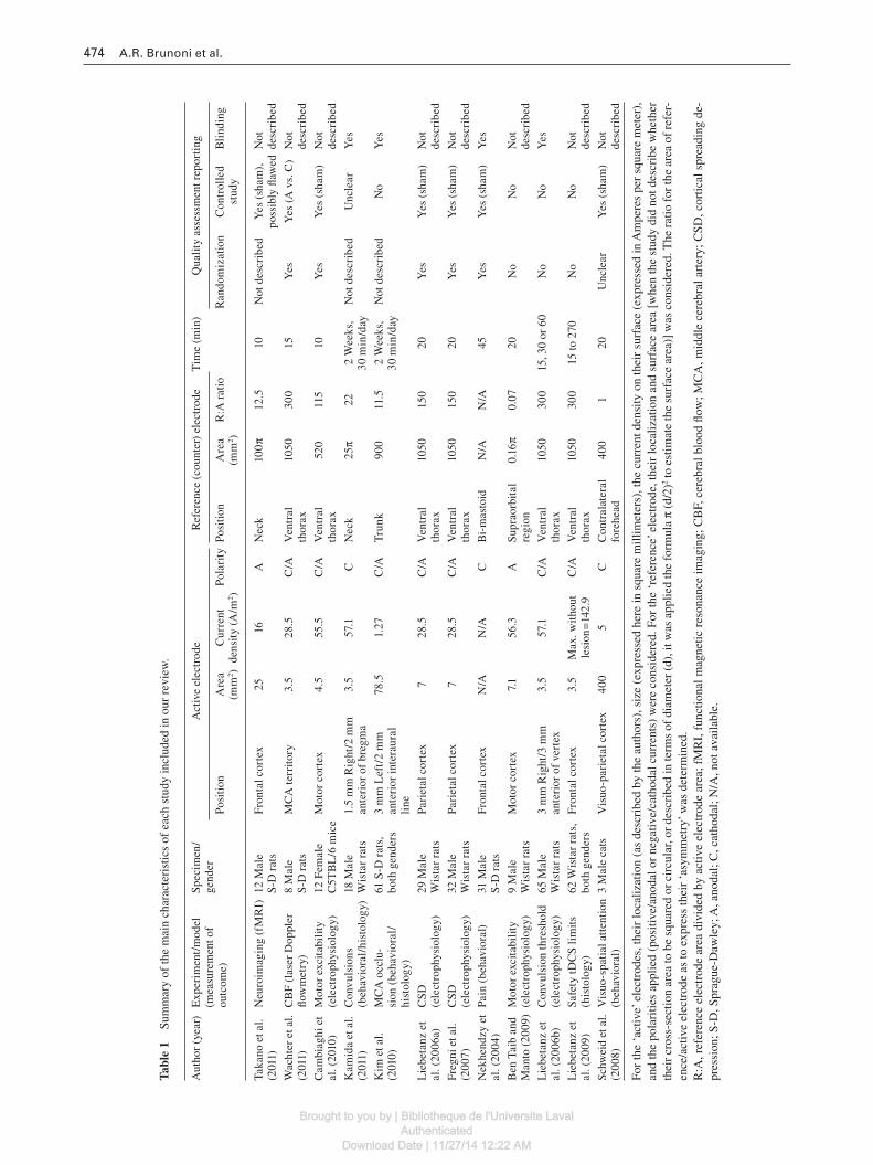
Tabl
e 1
Sum
mar
y of
the
mai
n ch
arac
teri
stic
s of
eac
h st
udy
incl
uded
in o
ur r
evie
w.
Aut
hor
(yea
r)E
xper
imen
t/m
odel
(m
easu
rem
ent o
f ou
tcom
e)
Spec
imen
/ge
nder
Act
ive
elec
trod
eR
efer
ence
(co
unte
r) e
lect
rode
Tim
e (m
in)
Qua
lity
ass
essm
ent r
epor
ting
Posi
tion
Are
a (m
m 2 )
Cur
rent
de
nsit
y (A
/m 2 )
Pola
rity
Posi
tion
Are
a (m
m 2 )
R:A
rat
ioR
ando
miz
atio
nC
ontr
olle
d st
udy
Bli
ndin
g
Taka
no e
t al.
(201
1)
Neu
roim
agin
g (f
MR
I)12
Mal
e S
-D r
ats
Fro
ntal
cor
tex
2516
AN
eck
100 π
12
.510
Not
des
crib
edY
es (
sham
),
poss
ibly
fl aw
edN
ot
desc
ribe
d
Wac
hter
et a
l. (2
011)
C
BF
(las
er D
oppl
er
fl ow
met
ry)
8 M
ale
S-D
rat
sM
CA
terr
itory
3.5
28.5
C/A
Ven
tral
th
orax
1050
300
15Y
esY
es (A
vs.
C)
Not
de
scri
bed
Cam
biag
hi e
t al
. (20
10)
Mot
or e
xcit
abil
ity
(ele
ctro
phys
iolo
gy)
12 F
emal
e C
5TB
L/6
mic
eM
otor
cor
tex
4.5
55.5
C/A
Ven
tral
th
orax
520
115
10Y
esY
es (
sham
)N
ot
desc
ribe
d
Kam
ida
et a
l. (2
011)
C
onvu
lsio
ns
(beh
avio
ral/
hist
olog
y)18
Mal
e W
ista
r ra
ts1.
5 m
m R
ight
/2 m
m
ante
rior
of
breg
ma
3.5
57.1
CN
eck
25 π
222
Wee
ks,
30 m
in/d
ayN
ot d
escr
ibed
Unc
lear
Yes
Kim
et a
l. (2
010)
M
CA
occ
lu-
sion
(be
havi
oral
/ hi
stol
ogy)
61 S
-D r
ats,
bo
th g
ende
rs3
mm
Lef
t/2
mm
an
teri
or in
tera
ural
li
ne
78.5
1.27
C/A
Tru
nk90
011
.52
Wee
ks,
30 m
in/d
ayN
ot d
escr
ibed
No
Yes
Lie
beta
nz e
t al
. (20
06a)
CSD
(e
lect
roph
ysio
logy
)29
Mal
e W
ista
r ra
tsPa
riet
al c
orte
x 7
28.5
C/A
Ven
tral
th
orax
1050
150
20Y
esY
es (
sham
)N
ot
desc
ribe
d
Fre
gni e
t al.
(200
7)
CSD
(e
lect
roph
ysio
logy
)32
Mal
e W
ista
r ra
tsPa
riet
al c
orte
x 7
28.5
C/A
Ven
tral
th
orax
1050
150
20Y
esY
es (
sham
)N
ot
desc
ribe
d
Nek
hend
zy e
t al
. (20
04)
Pain
(be
havi
oral
)31
Mal
e S
-D r
ats
Fro
ntal
cor
tex
N/A
N/A
CB
i-m
asto
idN
/AN
/A45
Yes
Yes
(sh
am)
Yes
Ben
Tai
b an
d M
anto
(20
09)
Mot
or e
xcit
abil
ity
(ele
ctro
phys
iolo
gy)
9 M
ale
Wis
tar
rats
Mot
or c
orte
x 7.
156
.3A
Supr
aorb
ital
re
gion
0.16
π 0.
0720
No
No
Not
de
scri
bed
Lie
beta
nz e
t al
. (20
06b)
Con
vuls
ion
thre
shol
d (e
lect
roph
ysio
logy
)65
Mal
e W
ista
r ra
ts3
mm
Rig
ht/3
mm
an
teri
or o
f ve
rtex
3.5
57.1
C/A
Ven
tral
th
orax
1050
300
15, 3
0 or
60
No
No
Yes
Lie
beta
nz e
t al
. (20
09)
Safe
ty tD
CS
lim
its
(his
tolo
gy)
62 W
ista
r ra
ts,
both
gen
ders
Fro
ntal
cor
tex
3.5
Max
. wit
hout
le
sion
= 14
2.9
C/A
Ven
tral
th
orax
1050
300
15 to
270
No
No
Not
de
scri
bed
Schw
eid
et a
l. (2
008)
V
isuo
-spa
tial
att
enti
on
(beh
avio
ral)
3 M
ale
cats
Vis
uo-p
arie
tal c
orte
x40
05
CC
ontr
alat
eral
fo
rehe
ad40
01
20U
ncle
arY
es (
sham
)N
ot
desc
ribe
d
For
the
‘ act
ive ’
ele
ctro
des,
thei
r lo
caliz
atio
n (a
s de
scri
bed
by th
e au
thor
s), s
ize
(exp
ress
ed h
ere
in s
quar
e m
illim
eter
s), t
he c
urre
nt d
ensi
ty o
n th
eir
surf
ace
(exp
ress
ed in
Am
pere
s pe
r sq
uare
met
er),
an
d th
e po
lari
ties
appl
ied
(pos
itive
/ano
dal o
r ne
gativ
e/ca
thod
al c
urre
nts)
wer
e co
nsid
ered
. For
the
‘ ref
eren
ce ’ e
lect
rode
, the
ir lo
caliz
atio
n an
d su
rfac
e ar
ea [
whe
n th
e st
udy
did
not d
escr
ibe
whe
ther
th
eir
cros
s-se
ctio
n ar
ea to
be
squa
red
or c
ircu
lar,
or d
escr
ibed
in te
rms
of d
iam
eter
(d)
, it w
as a
pplie
d th
e fo
rmul
a π
(d/2
) 2 to
estim
ate
the
surf
ace
area
)] w
as c
onsi
dere
d. T
he r
atio
for
the
area
of
refe
r-en
ce/a
ctiv
e el
ectr
ode
as to
exp
ress
thei
r ‘ a
sym
met
ry ’ w
as d
eter
min
ed.
R:A
, ref
eren
ce e
lect
rode
are
a di
vide
d by
act
ive
elec
trod
e ar
ea; f
MR
I, f
unct
iona
l mag
netic
res
onan
ce im
agin
g; C
BF,
cer
ebra
l blo
od fl
ow; M
CA
, mid
dle
cere
bral
art
ery;
CSD
, cor
tical
spr
eadi
ng d
e-pr
essi
on; S
-D, S
prag
ue-D
awle
y; A
, ano
dal;
C, c
atho
dal;
N/A
, not
ava
ilabl
e.
Brought to you by | Bibliotheque de l'Universite LavalAuthenticated
Download Date | 11/27/14 12:22 AM

tDCS research in animals 475
Regarding polarity-dependent tDCS effects, anodal stimu-lation was signifi cantly different than sham and/or cathode in increasing brain activity [measured by functional magnetic resonance imaging (fMRI); Takano et al. , 2011 ], increasing cerebral blood fl ow (Wachter et al. , 2011 ), increasing motor cortex excitability (Ben Taib and Manto , 2009 ; Cambiaghi et al. , 2010 ) and increasing cortical spreading depression veloc-ity (Liebetanz et al. , 2006a ; Fregni et al. , 2007 ). Interestingly, however, anodal stimulation did not change the threshold for seizure in one study (Liebetanz et al. , 2006b ), while Kim et al. (2010) obtained mixed fi ndings for a putative neuropro-tective effect of anodal stimulation for stroke. In addition, cathodal stimulation, compared to sham and/or anodal, was shown to decrease the rate (Kamida et al. , 2011 ) and thresh-old (Liebetanz et al. , 2006b ) of seizures in two epilepsy mod-els, have analgesic effects in a pain model (Nekhendzy et al. , 2004 ), decrease attention (Schweid et al. , 2008 ), decrease cerebral blood fl ow (Wachter et al. , 2011 ) and decrease motor cortex excitability (Cambiaghi et al. , 2010 ). However, cath-odal stimulation did not infl uence the velocity of cortical spreading depression in one study (Liebetanz et al. , 2006a ), while it induced a slight increase in another study by the same group (Fregni et al. , 2007 ). Also, Kim et al. (2010) did not observe differences between cathodal vs. sham stimulation
for stroke. Thus, most experiments supported the concept of polarity-specifi c tDCS effects (Table 2 ).
In addition, four studies (Nekhendzy et al. , 2004 ; Liebetanz et al. , 2006b , 2009 ; Kamida et al. , 2011) applied tDCS at dif-ferent intensities, showing that higher current intensities elic-ited larger effects. Also, fi ve studies addressed after-effects within the fi rst hour of tDCS. Nekhendzy et al. (2004) and Liebetanz et al. (2006b) reported after-effects lasting up to 50 and 60 min, respectively, but only for the highest charge delivered; Cambiaghi et al. (2010) and Wachter et al. (2011) reported after-effects lasting up to 10 and 30 min, respec-tively, regardless of polarity; Liebetanz et al. (2006a) reported after-effects fading away after 5 min. Thus, only one of these fi ve studies showed tDCS effects that did not last beyond the stimulation period (Table 2 ).
Finally, nine of 12 studies performed histologic analysis of the specimen studied. In three of them, the post-mortem analysis was also an outcome, as these studies assessed tDCS-induced effects in the safety current intensity limits on the tissue damage (Liebetanz et al. , 2009 ), integrity of white mat-ter axon and infarct lesion size in a stroke model (Kim et al. , 2010 ), and cell loss and mossy fi ber sprouting in an epilepsy model (Kamida et al. , 2011 ). Although another six studies (Liebetanz et al. , 2006a, b ; Fregni et al. , 2007 ; Ben Taib and
Table 2 Main results according to polarity, intensity and long-lasting effects.
Polarity effectsAnodal stimulation = pro-depolarization effects Cathodal stimulation = pro-
hyperpolarization effectsDiverging/mixed effects
↑ Brain activity (Takano et al. , 2011 ) ↓ Pain (Nekhendzy et al. , 2004 )
(A) No effects in seizure threshold (Liebetanz et al. , 2006b )
↑ Cerebral blood fl ow (Wachter et al. , 2011 )
↓ Cerebral blood fl ow (Wachter et al. , 2011 )
(C) No effects in CSD velocity (Liebetanz et al. , 2006a )
↑ CSD velocity (Liebetanz et al. , 2006a ; Fregni et al. , 2007 )
↓ Seizure threshold (Liebetanz et al. , 2006b )
(C) Increased CSD velocity (Fregni et al. , 2007 )
↑ Motor cortex excitability (Ben Taib and Manto , 2009 ; Cambiaghi et al. , 2010 )
↓ Motor cortex excitability (Cambiaghi et al. , 2010 )
(A, C) Mixed effects in stroke healing (Kim et al. , 2010 )
↓ Frequency of seizures (Kamida et al. , 2011 )
↓ Attention (Schweid et al. , 2008 )
Intensity effectsInteraction between higher current densities and observed effects No current-effect interaction ↑ Seizure threshold (Liebetanz et al. , 2006b ) N/A ↑ Brain damage (Liebetanz et al. , 2009 ) ↑ Antinociceptive effects (Nekhendzy et al. , 2004 ) ↑ Cerebral blood fl ow (Wachter et al. , 2011 )After-effectsLong-lasting effects were observed Not observedAntinociceptive effects lasted for 50 min after stimulation (Nekhendzy et al. , 2004 ) Sustained decrease in seizure threshold lasting for 60 min after stimulation (Liebetanz et al. , 2006b )
CSD velocity returned to baseline levels after 5 min (Liebetanz et al. , 2006a )
Cerebral blood fl ow changes evident for 30 min after stimulation (Wachter et al. , 2011 )Motor excitability changes lasted for 10 min after stimulation (Cambiaghi et al. , 2010 )
CSD, cortical spreading depression; A, anodal; C, cathodal; N/A, not available.
Brought to you by | Bibliotheque de l'Universite LavalAuthenticated
Download Date | 11/27/14 12:22 AM

476 A.R. Brunoni et al.
Manto , 2009 ; Cambiaghi et al. , 2010 ; Wachter et al. , 2011 ) assessed, in parts of their samples, histology to address haz-ardous tDCS effects, only Wachter et al. (2011) reported, in one specimen, a lesion in the parieto-occipital cortex near the electrode site. The authors described that histologic analysis showed loss of the cortical layer I, proliferation of the lep-tomeningeal tissue and enhancement of vasculature, which was compatible with necrosis. According to them, such unexpected adverse effects [as another study (Liebetanz et al. , 2009 ) showed much higher safety limits] could be as a result of: (1) a combination of anodal and cathodal stimula-tion on the same point; (2) pro-coagulant effects of DC stimu-lation; (3) a stimulation point just above the middle cerebral artery that could have been injured; or, (4) random variation of current distribution and effects between different animals. Nonetheless, these adverse effects highlight the relevance of further experimental animal studies on safety.
Quality assessment
We assessed quality according to some of the criteria of the ARRIVE guidelines for reporting of animal experiments. Five out of 12 studies reported randomization methods: in between-group designs, for the allocation group (Liebetanz et al. , 2006a ; Fregni et al. , 2007 ) and in within-group designs, for the order of stimulation (Nekhendzy et al. , 2004 ; Cambiaghi et al. , 2010 ; Wachter et al. , 2011 ). In one study (Schweid et al. , 2008 ), we could not defi ne whether randomization meth-ods were used. The other six studies did not describe random-ization methods (Table 1 ).
Regarding control comparisons, fi ve studies described sham methods, either consisting of turning off the tDCS device after 10 – 30 s of stimulation (Liebetanz et al. , 2006a ; Fregni et al. , 2007 ; Schweid et al. , 2008 ) or delivering no current at all (Nekhendzy et al. , 2004 ; Cambiaghi et al. , 2010 ). We considered one method, described as ‘ sham ’ , to be a potentially active control condition, as a small current (40 uA, 1.6 A/m 2 ) was applied during the entire period of stimulation (Takano et al. , 2011 ). In fact, the sham current, as defi ned by these authors, had a higher current density than most human studies (Nitsche et al. , 2008 ). Two stud-ies compared anodal vs. cathodal stimulation (Liebetanz et al. , 2006b ; Wachter et al. , 2011 ), two studies had control groups which received no intervention (including no mon-tage of tDCS; Kim et al. , 2010 ; Kamida et al. , 2011 ) and two did not use sham methods (Ben Taib and Manto , 2009 ; Liebetanz et al. , 2009 ; Table 1 ). Regarding blinding, only four (Nekhendzy et al. , 2004 ; Liebetanz et al. , 2006b ; Kim et al. , 2010 ; Kamida et al. , 2011 ) of the 12 studies described methods of blinding the assessment of outcomes, assuring group concealment (Table 1 ).
For housing and husbandry, only three studies suffi ciently described housing (two only partially; Nekhendzy et al. , 2004 ; Takano et al. , 2011 ; Wachter et al. , 2011 ) and only half of the studies suffi ciently reported husbandry, while two stud-ies did not (Ben Taib and Manto , 2009 ; Kim et al. , 2010 ) and four reported incompletely (Liebetanz et al. , 2006a , b, 2009 ; Kamida et al. , 2011 ).
Discussion
We reviewed 12 studies that applied DC transcranially over the skulls of 326 rats, 12 mice and three cats (a total of 341 animals). These studies tested healthy and also disease model animals and used different outcomes. One fi nding that can be generalized across these studies is that most of them found polarity-specifi c DC effects, i.e., that anodal stimulation has an opposite effect when compared with cathodal stimulation as indexed by different methods such as motor excitability, cerebral blood fl ow, velocity of cortical spreading depression and frequency and threshold for seizures, thus supporting specifi c effects of tDCS on brain physiology. There were two studies that did not fi nd polarity specifi c effects. Fregni et al. (2007) showed that cathodal stimulation, similarly to anodal tDCS, slightly increased the velocity of cortical spreading depression (CSD). One potential explanation by authors of this study is that stimulation could have still induced post-synaptic hyperpolarization, which could have paradoxically increased CSD velocity by diminishing long-term depression plasticity that also requires post-synaptic depolarization. This hypothesis could also explain the lack of cathodal stimulation effects on CSD velocity in another study (Liebetanz et al. , 2006a ). Likewise, Liebetanz et al. (2006b) theorized that the lack of pro-convulsive effects after anodal stimulation was related to tDCS increasing spontaneous neuronal activity in a non-synchronous way, while seizure activity involves syn-chronous activity of neuronal networks.
Also of relevance was whether tDCS effects last beyond the initial period of stimulation. We observed that tDCS after-effects varied according to current intensity and were cumu-lative over time. Most studies showed that tDCS induces after-effects (Table 2 ), supporting the hypothesis of tDCS inducing long-term potentiation (LTP), and not just changing electric neuronal membrane potential (Nitsche et al. , 2003 , 2004 ; Stagg et al. , 2009 ). All studies also showed that higher current densities were associated with larger neurophysi-ologic responses (and also that low densities may induce no response at all), an observation that is similar to some human studies (Iyer et al. , 2005 ; Boggio et al. , 2006 ). However, only two animal studies tested a sequential daily stimulation pro-tocol, with positive (Kamida et al. , 2011 ) and mixed (Kim et al. , 2010 ) fi ndings. Such an approach may be clinically use-ful, as repetitive tDCS is being successfully applied for treat-ing various neurologic and psychiatric disorders (Boggio et al. , 2007 ; Valle et al. , 2009 ; Antal et al. , 2010 ; Mori et al. , 2010 ; Vanneste et al. , 2010 ; Brunoni et al. , 2011b ). Thus, con-sidering that daily, repetitive tDCS is being consolidated as a treatment modality, further animal experiments using this approach may be an interesting focus for future research.
Safety
Pre-clinical animal studies are critical for exploring adverse effects and safety limits. Interestingly, no study described behaviors suggesting pain or discomfort during stimulation, which were performed in much higher levels than humans, although most studies anesthetized the specimens during
Brought to you by | Bibliotheque de l'Universite LavalAuthenticated
Download Date | 11/27/14 12:22 AM
tDCS research in animals 477
experimentation. Also, scalp burn and skin scars were not observed, although, again, most studies implanted electrodes directly on the skull. Regarding safety limits, one study (Liebetanz et al. , 2009 ) specifi cally assessed current density and charge threshold for brain damage, which only occurred after cathodal stimulation above 100 A/m 2 or, on average, two orders of magnitude higher than what is used in humans. In addition, most studies performed post-mortem analysis in parts of their samples, not observing brain damage, with a single exception (Wachter et al. , 2011 ) being observed in one animal that received six trains (half anodal, half cathodal) of tDCS using large current densities from 7 to 28 A/m 2 . One point of note here is that some inherent conditions, such as stimula-tion over the middle cerebral artery, alternated cathodal/anodal stimulation and random effects between subjects should have contributed to this lesion, as this current density is still lower than the limit for lesion observed in the study by Liebetanz et al. (2006a) . Thus, future tDCS animal studies must still include post-mortem analysis, especially when higher current intensities or cumulative protocols are researched.
Reporting quality
We observed that studies have limited quality on report-ing of methodology according to some of the criteria of the ARRIVE guidelines. Although we only focused on some aspects (ethical statement, randomization, blinding and sham-control, experimental procedures, sample size, gender, hous-ing, husbandry conditions, outcomes, attrition and adverse events), it was observed that each one was not fulfi lled by most studies. Therefore, the internal validity of these studies can be considered only moderate at best, as full quality could not be assessed as a result of limited reporting. In fact, ani-mal studies usually present low quality standards for report-ing, as observed in a review of 271 animal studies that found that 87 % and 86 % did not report randomization and blinding, respectively, along with other important fl aws (Kilkenny et al., 2010). Thus, although the studies composing our review had similar reporting shortcomings, they were at least comparable with the current standards of animal research reporting. Also, as we did not employ meta-analytic techniques we could not measure the impact of publication bias (i.e., unpublished data, often caused by negative fi ndings) in our results, which can be important, according to a review of animal stroke studies that estimated a non-publication rate of 14 % (Sena et al. , 2010 ).
Comparison with human studies
In order to explore the implications for translational research of the present review, we compared the results here found with another review (Brunoni et al. , 2011a ) that assessed tDCS parameters of 209 human studies. In fact, there are important methodologic differences between animal and human studies (Table 3 and Figure 2 ). For instance, the mean current density applied in humans (0.4 A/m 2 ) is 85 times lower than the applied in rats (34.2 A/m 2 ). Almost 90 % of human studies favor reference electrodes positioned over the scalp, while studies in animals are mixed, placing them over
the head, neck and trunk. In addition, almost all human stud-ies had symmetrical or slightly asymmetrical electrode sizes whereas in rodents the reference electrode was, on average, almost one hundred times larger than the active electrode. Moreover, the electrodes in humans are positioned over the skin, forcing the electric current throughout four anatomical layers (skin, skull, meninges and cerebrospinal fl uid), while almost half of rat studies applied the montage initially used in Liebetanz ’ study (2006a) which effectively removes the out-ermost layer. Interestingly, human and animal studies were akin to the duration of stimulation and the number of subjects per experiment.
What do these differences represent for translational research ? One critical issue is that tDCS effects depend on the current distribution as shown by a recent study (Mendonca et al. , 2011 ). Although modeling studies would be necessary, one possible effect of parameters of tDCS in animals would be that current applied in animal studies is probably more focal than humans because (1) active and ref-erence electrodes are very asymmetrical in animals, a condi-tion that increases focality on the active electrode (Nitsche et al. , 2007 ; Parazzini et al. , 2011 ) and (2) active electrodes were nearly punctiform – smaller electrodes also increase focality (Nitsche et al. , 2007 ; Parazzini et al. , 2011 ). In fact a recent modeling study using smaller electrodes showed focal effects of tDCS; however, in this study four reference elec-trodes were placed around the stimulating electrode (Datta et al. , 2009 ).
Animal studies may also have elicited ‘ stronger ’ brain modulation (i.e., greater current injection in neuronal tissue) given that: (1) even though previous modeling studies show that there is no linear relation between current density and brain modulation (Miranda et al. , 2006 ; Datta et al. , 2008 ), current densities applied in animals were almost one hundred times higher; and, (2) by placing directly over the skull, the tDCS montage in animals reduces tissue shunting and cur-rent dispersion and the areas just beneath the electrodes also receive higher currents (Miranda et al. , 2009 ; Sadleir et al. , 2010 ). Regarding extra-cephalic vs. cephalic reference elec-trodes, three studies showed that the clinical effects using extra-cephalic references are smaller (Mahmoudi et al. , 2011 ; Moliadze et al. , 2010 ; Mendonca et al. , 2011 ), although it is complicated to determine whether these fi ndings apply for small animals as rodents as one of the studies (Moliadze et al. , 2010 ) showed that the effects were negatively correlated to the distance between the electrodes.
Nonetheless, such dissimilarities are expected in basic research, which serves as proof-of-principle and safety stud-ies and therefore bolder parameters are applied [for instance, pharmacologic animal studies also use higher dosages than in humans (Sharma and McNeill , 2009 )]. On the other hand, the above-mentioned differences reveal that the clinical and pre-clinical protocols differ in several, and important, aspects. To ascertain whether the results are interchangeable, one possi-bility would be to perform modeling studies with animal head models to simulate to what extent such differences in proto-col refl ect in changes in inter-species brain modulation, for example, similar to a modeling mouse TMS study exploring
Brought to you by | Bibliotheque de l'Universite LavalAuthenticated
Download Date | 11/27/14 12:22 AM

478 A.R. Brunoni et al.
38 cm
OA
B
38 cm
1050 mm2
3.5 mm2
(III)
(IV)
L
B
N
N
15-18 cm
1 cm
2 cm
7 cm
5 cm
5 cm
7 cm
B
(I)
(II)
Figure 2 Comparative illustration of transcranial direct current stimulation ( tDCS) montages in (A) human scalp and (B) the head and body of a Wistar rat weighing 300 g. (A) This drawing represents two symmetrical electrodes that are positioned over the supra-orbital area (I) and contra-lateral motor cortex (II). This is a typical setup for clinical human studies investigating pain and stroke rehabilitation. In (B), it shows the ‘ Liebetanz montage ’ , which employs a punctiform, cephalic active electrode (III) and a large, extra-cephalic reference electrode (IV). While most human trials utilize the described setup (or with minor variations), animal montages vary, although asymmetrical electrodes with small active electrodes are usually the rule. Skull references: O, occipicum; N, nasum; L, lambda and B, bregma .
Table 3 Comparison of the current intensity and electrode positioning in different specimens.
Specimen Rat Mouse Cat Human a
Studies (number) 10 1 1 209 Healthy (number) 5 1 1 159 Pathological model/disease (number) 5 N/A N/A 50 Studies using control comparisons (number) 4 1 1 138 Daily stimulation (number) 2 N/A N/A 35Sample (total) 327 12 3 3836 Active electrode (mean values) Electric current (A) 0.22 × 10 -3 0.25 × 10 -3 2 × 10 -3 1.25 × 10 -3 Area (m 2 ) 0.17 × 10 -4 0.04 × 10 -4 4 × 10 -4 31.5 × 10 -4 Current density (A/m 2 ) 34.2 55.5 5 0.4 Reference electrode (mean values) Area (m 2 ) 6.8 × 10 -4 5.2 × 10 -4 4 × 10 -4 � Similar size of
active Position 60 % Extra-cephalic Extra-cephalic Cephalic � 90 % Cephalic R:A ratio 118.3 115.5 1 � 1 Duration (min) 25.6 10 20 13.5
a Human column is highlighted as it contains data from another study (Brunoni et al., 2011a).R:A, reference electrode area divided by active electrode area; N/A, not available; �, represents approximate values.
Brought to you by | Bibliotheque de l'Universite LavalAuthenticated
Download Date | 11/27/14 12:22 AM
tDCS research in animals 479
the infl uence of coil aspects in brain stimulation (Salvador and Miranda , 2009 ).
One interesting effect that was confi rmed in human studies, on the other hand, was the after-effects of tDCS. Several of the animal studies we analyzed showed that tDCS effects last beyond the period of stimulation. This supports the hypothesis that tDCS induces changes in cellular mechanisms resulting in LTP, a form of long-lasting synaptic strengthening. Recent evidence from pharmacologic studies showed that anodal tDCS modifi es N-methyl-D-aspartate (NMDA) receptor-dependent LTP, e.g., tDCS combination with d-cycloserine (a NMDA receptor agonist) increased its after-effects, while with dextromethorphane (an NMDA receptor antagonist) the after-effects were abolished (Stagg and Nitsche , 2011 ). Also, another study showed that brain-derived neurotrophic fac-tor (BDNF) [a neurotrophin that plays a critical role in late-phase LTP (Lu et al. , 2005 )] is necessary for the after-effects of direct cortical stimulation (Fritsch et al. , 2010 ). Cathodal tDCS after-effects are also modifi ed according to the phar-macologic modulation of glutamatergic activity (Stagg and Nitsche , 2011 ).
A fi nal issue is whether higher current densities, as used in rodents, could be applied in clinical studies. However, in humans, smaller electrodes and higher currents are limited because these parameters have been associated with discom-fort and skin burn (Nitsche et al. , 2008 ). Nonetheless, recent clinical studies showed that smaller, round shaped electrodes are as comfortable as conventional, squared electrodes (Ambrus et al. , 2010 ), Ag/AgCl pellets are more comfort-able than rubber pellets (Minhas et al. , 2010 ) and that topi-cally applied anesthetics reduce procedural pain (McFadden et al. , 2011) . Further studies need to be conducted to confi rm these initial fi ndings. These recent advancements may pro-vide evidence for the application of higher and more focal currents.
Concluding remarks
In summary, tDCS studies in animals demonstrate that this technique induces effects that are polarity- and intensity- dependent, which lasts beyond the period of stimulation, indicating that LTP- and LTP-like phenomena are likely to be involved. Studies in animals have also generally shown that the technique is safe, regardless of one unexpected fi nding of histologic lesion. When translating the fi ndings of these studies to clinical practice, it should be considered, however, that the tDCS montages are different in animal vs. human studies. Nonetheless, human studies could benefi t in applying protocols with higher currents/smaller electrodes, as initial data using such parameters in animals have shown promising fi ndings.
tDCS appears to be a promising therapy for clinical use in neurologic and psychiatric disorders as the technique has only mild, transient side effects and is easier applied than other brain stimulation interventions. However, a better understanding of its mechanisms of action is still critical and, for this purpose, experimental animal studies in vivo can be useful. Thus fur-ther studies to test different tDCS montages and also to explore
other outcomes not readily accessible in clinical practice, such as the expression of neurotransmitter receptors and activation of neuronal pathways would provide critical mechanistic insights. Such studies would foment translational tDCS research and thus contribute to the development of the fi eld.
References
Ambrus, G.G., Paulus, W., and Antal, A. (2010). Cutaneous percep-tion thresholds of electrical stimulation methods: comparison of tDCS and tRNS. Clin. Neurophysiol. y 121 , 1908 – 1914.
Antal, A., Terney, D., Kuhnl, S., and Paulus, W. (2010). Anodal trans-cranial direct current stimulation of the motor cortex amelio-rates chronic pain and reduces short intracortical inhibition. J. Pain Symptom Manag. 39 , 890 – 903.
Ben Taib, N.O. and Manto, M. (2009). Trains of transcranial direct current stimulation antagonize motor cortex hypoexcitabil-ity induced by acute hemicerebellectomy. J. Neurosurg. 111, 796 – 806.
Boggio, P.S., Ferrucci, R., Rigonatti, S.P., Covre, P., Nitsche, M., Pascual-Leone, A., and Fregni, F. (2006). Effects of transcranial direct current stimulation on working memory in patients with Parkinson ’ s disease. J. Neurol. Sci. 249 , 31 – 38.
Boggio, P.S., Nunes, A., Rigonatti, S.P., Nitsche, M.A., Pascual-Leone, A., and Fregni, F. (2007). Repeated sessions of nonin-vasive brain DC stimulation is associated with motor function improvement in stroke patients. Restor. Neurol. Neuros. 25 , 123 – 129.
Boggio, P.S., Rigonatti, S.P., Ribeiro, R.B., Myczkowski, M.L. , Nitsche, M.A., Pascual-Leone, A., and Fregni, F. (2008). A ran-domized, double-blind clinical trial on the effi cacy of cortical direct current stimulation for the treatment of major depression. Int. J. Neuropsychop. 11 , 249 – 254.
Brunoni, A.R., Amadera, J., Berbel, B., Volz, M.S. , Rizzerio, B.G., and Fregni, F. (2011a). A systematic review on reporting and assess-ment of adverse effects associated with transcranial direct cur-rent stimulation. Int. J. Neuropsychop. [Epub ahead of print].
Brunoni, A.R., Ferrucci, R., Bortolomasi, M., Vergari, M. , Tadini, L., Boggio, P.S., Giacopuzzi, M., Barbieri, S., and Priori, A. (2011b). Transcranial direct current stimulation (tDCS) in uni-polar vs. bipolar depressive disorder. Prog. Neuro. Psychoph. 35 , 96 – 101.
Brunoni, A.R., Valim, C., and Fregni, F. (2011c). Combination of noninvasive brain stimulation with pharmacotherapy. Expert Rev. Med. Devices 8 , 31 – 39.
Cambiaghi, M., Velikova, S., Gonzalez-Rosa, J.J., Cursi, M. , Comi, G., and Leocani, L. (2010). Brain transcranial direct cur-rent stimulation modulates motor excitability in mice. Eur. J. Neurosci. 31 , 704 – 709.
Datta, A., Elwassif, M., Battaglia, F., and Bikson, M. (2008). Transcranial current stimulation focality using disc and ring electrode confi gurations: FEM analysis. J. Neural. Eng. 5 , 163 – 174.
Datta, A., Bansal, V., Diaz, J., and Patel, J. (2009). Gyri-precise head model of transcranial direct current stimulation: improved spa-tial focality using a ring electrode versus conventional rectangu-lar pad. Brain Stimul. 2 , 201 – 207.
Ferrucci, R., Bortolomasi, M., Vergari, M., Tadini, L ., Salvoro, B., Giacopuzzi, M., Barbieri, S., and Priori, A. (2009). Transcranial direct current stimulation in severe, drug-resistant major depres-sion. J. Affect Disord. 118 , 215 – 219.
Brought to you by | Bibliotheque de l'Universite LavalAuthenticated
Download Date | 11/27/14 12:22 AM
480 A.R. Brunoni et al.
Fregni, F., Thome-Souza, S., Nitsche, M.A., Freedman, S.D ., Valente, K.D., and Pascual-Leone, A. (2006). A controlled clinical trial of cathodal DC polarization in patients with refractory epilepsy. Epilepsia 47 , 335 – 342.
Fregni, F., Liebetanz, D., Monte-Silva, K.K., Oliveira, M.B. , Santos, A.A., Nitsche, M.A., Pascual-Leone, A., and Guedes, R.C.A. (2007). Effects of transcranial direct current stimulation cou-pled with repetitive electrical stimulation on cortical spreading depression. Exp. Neurol. 204 , 462 – 466.
Fritsch, B., Reis, J., Martinowich, K., Schambra, H.M. , Ji, Y., Cohen, L.G., and Lu, B. (2010). Direct current stimulation promotes BDNF-dependent synaptic plasticity: potential implications for motor learning. Neuron 66 , 198 – 204.
Hooijmans, C.R., Leenaars, M., and Ritskes-Hoitinga, M. (2011). A gold standard publication checklist to improve the quality of ani-mal studies, to fully integrate the three rs, and to make system-atic reviews more feasible. Altern. Lab. Anim. 38 , 167 – 182.
Iyer, M.B., Mattu, U., Grafman, J., Lomarev, M., Sato, S., and Wassermann, E.M. (2005). Safety and cognitive effect of fron-tal DC brain polarization in healthy individuals. Neurology 64 , 872 – 875.
Kabalak, A.A., Akcay, M., Ceylan, A., Senel, O.O. , and Gogus, N. (2005). The effects of transcranial electrical stimulation on anaesthesia and analgesia in rats. J. Vet. Med. Sci. 67 , 433 – 436.
Kamida, T., Kong, S., Eshima, N., Abe, T. , Fujiki, M., and Kobayashi, H. (2011). Transcranial direct current stimulation decreases convulsions and spatial memory defi cits following pilocarpine-induced status epilepticus in immature rats. Behav. Brain Rese. 217 , 99 – 103.
Kilkenny, C., Browne, W.J., Cuthill, I.C., Emerson, M. , and Douglas G. (2010). Improving bioscience research reporting: the ARRIVE guidelines for reporting animal research. PLoS Biol. 8 , e1000412.
Kim, S.J., Kim, B.K., Ko, Y.J., Bang, M.S. , Kim, M.H., and Han, T.R. (2010). Functional and histologic changes after repeated transcranial direct current stimulation in rat stroke model. J. Korean Med. Sci. 25 , 1499 – 1505.
Liebetanz, D., Fregni, F., Monte-Silva, K.K., Oliveira, M.B., Am â ncio-dos-Santos, A., Nitsche, M.A., and Guedes, R.C. (2006a). After-effects of transcranial direct current stimulation (tDCS) on cortical spreading depression. Neurosci. Lett. 398 , 85 – 90.
Liebetanz, D., Klinker, F., Hering, D., Koch, R. , Nitsche, M.A., Potschka, H., L ö scher, W., Paulus, W., and Tergau, F. (2006b). Anticonvulsant effects of transcranial direct-current stimula-tion (tDCS) in the rat cortical ramp model of focal epilepsy. Epilepsia 47 , 1216 – 1224.
Liebetanz, D., Koch, R., Mayenfels, S., K ö nig, F., Paulus, W., and Nitsche, M.A. (2009). Safety limits of cathodal transcranial direct current stimulation in rats. Clin. Neurophysiol. 120 , 1161 – 1167.
Lu, B., Pang, P.T., and Woo, N.H. (2005). The yin and yang of neu-rotrophin action. Nat. Rev. Neurosci. 6 , 603 – 614.
Mahmoudi, H., Haghighi, A.B., Petramfar, P., Jahanshahi, S., Salehi, Z., and Fregni, F. (2011). Transcranial direct current stimulation: electrode montage in stroke. Disabil. Rehabil. 33, 1383 – 1388.
McFadden, J.L., Borckardt, J.J., George, M.S., and Beam, W. (2011). Reducing procedural pain and discomfort associated with trans-cranial direct current stimulation. Brain Stimul. 4 , 38 – 42.
Mendonca, M.E., Santana, M.B., Baptista, A.F., Datta, A., Bikson, M., Freqni, F., and Araujo, C.P. (2011). Transcranial DC stimu-lation in fi bromyalgia: optimized cortical target supported by high-resolution computational models. J. Pain 12 , 610 – 617.
Minhas, P., Bansal, V., Patel, J., Ho, J.S., Diaz, J., Datta, A., and Bikson, M. (2010). Electrodes for high-defi nition transcutane-ous DC stimulation for applications in drug delivery and elec-trotherapy, including tDCS. J. Neurosci. Meth. 190 , 188 – 197.
Miranda, P.C., Lomarev, M., and Hallett, M. (2006). Modeling the current distribution during transcranial direct current stimula-tion. Clin. Neurophysiol. 117 , 1623 – 1629.
Miranda, P.C., Faria, P., and Hallett, M. (2009). What does the ratio of injected current to electrode area tell us about current density in the brain during tDCS ? Clin. Neurophysiol. 120 , 1183 – 1187.
Moliadze, V., Antal, A., and Paulus, W. (2010). Electrode-distance dependent after-effects of transcranial direct and random noise stimulation with extracephalic reference electrodes. Clin. Neurophysiol. 121 , 2165 – 2171.
Mori, F., Codeca, C., Kusayanagi, H., Monteleone, F. , Buttari, F., Fiore, S., Bernardi, G., Koch, G., and Centonze, D. (2010). Effects of anodal transcranial direct current stimulation on chronic neuropathic pain in patients with multiple sclerosis. J. Pain 11 , 436 – 442.
Nekhendzy, V., Fender, C.P., Davies, M.F., Lemmens, H.J.M., Kim, M.S., Bouley, D.M., and Maze, M. (2004). The antinociceptive effect of transcranial electrostimulation with combined direct and alternating current in freely moving rats. Anesth. Analg. 98 , 730 – 737.
Nekhendzy, V., Davies, M.F., Lemmens, H.J., and Maze, M. (2006). The role of the craniospinal nerves in mediating the antinocice-ptive effect of transcranial electrostimulation in the rat. Anesth. Analg. 102 , 1775 – 1780.
Nitsche, M.A. and Paulus, W. (2009). Noninvasive brain stimulation protocols in the treatment of epilepsy: current state and perspec-tives. Neurotherapeutics 6 , 244 – 250.
Nitsche, M.A., Fricke, K., Henschke, U., Schlitterlau, A., Liebetanz, D., Lang, D., Henning, S., Tergau, F., and Paulus, W. (2003). Pharmacological modulation of cortical excitability shifts induced by transcranial direct current stimulation in humans. J. Physiol. 553 (Pt 1), 293 – 301.
Nitsche, M.A., Jaussi, W., Liebetanz, D., Lang, N., Tergau, F., and Paulus, W. (2004). Consolidation of human motor cortical neuroplasticity by D-cycloserine. Neuropsychopharmacol. 29 , 1573 – 1578.
Nitsche, M.A., Doemkes, S., Karaköse, T., Antal, A., Liebetanz, D., Lang, N., Tergau, F., and Paulus, W. (2007). Shaping the effects of transcranial direct current stimulation of the human motor cortex. J. Neurophysiol. 97 , 3109 – 3117.
Nitsche, M.A., Cohen, L.G., Wassermann, E.M., Priori, A., Lang, N., Antal, A., Paulus, W., Hummel, F., Boggio, P.S., Fregni, F., et al. (2008). Transcranial direct current stimulation: state of the art 2008. Brain Stimul. 1 , 206 – 223.
Ozen, S., Sirota, A., Belluscio, M.A., Anastassiou, C.A. , Stark, E., Koch, C., and Buzs á ki, G. (2010). Transcranial electric stimula-tion entrains cortical neuronal populations in rats. J. Neurosci. 30 , 11476 – 11485.
Parazzini, M., Fiocchi, S., Rossi, E., Paglialonga, A., and Ravazzani, P. (2011). Transcranial direct current stimulation: estimation of the electric fi eld and of the current density in an anatomical human head model. IEEE Trans. Biomed. Eng 58 , 1773 – 1780.
Perel, P., Roberts, I., Sena, E., Wheble, P., Sandercock, P., Macleod, M., Mignini, L.E., Jayaram, P., and Khan, K.K. (2007). Comparison of treatment effects between animal experiments and clinical trials: systematic review. BMJ 334 , 197.
Priori, A., Hallett, M., and Rothwell, J.C. (2009). Repetitive transcra-nial magnetic stimulation or transcranial direct current stimula-tion ? Brain Stimul. 2 , 241 – 245.
Brought to you by | Bibliotheque de l'Universite LavalAuthenticated
Download Date | 11/27/14 12:22 AM

tDCS research in animals 481
Reis, J., Robertson, E., Krakauer, J.W., Rothwell, J., Marshall, L., Gerloff, C., Wassermann, E., Pascual-Leone, A., Hummel, F., Celnik, P.A. et al. (2008). Consensus: “Can tDCS and TMS enhance motor learning and memory formation ? ”. Brain Stimul. 1 , 363 – 369.
Rigonatti, S.P., Boggio, P.S., Myczkowski, M.L., Otta, E., Fiquer, J.T., Ribeiro, R.B., Nitsche, M.A., Pascual-Leone, A., and Fregni, F. (2008). Transcranial direct stimulation and fl uoxetine for the treatment of depression. Eur. Psychiat. 23 , 74 – 76.
Sadleir, R.J., Vannorsdall, T.D., Schretlen, D.J., and Gordon, B. (2010). Transcranial direct current stimulation (tDCS) in a real-istic head model. Neuroimage 51 , 1310 – 1318.
Salvador, R. and Miranda, P.C. (2009). Transcranial magnetic stimu-lation of small animals: a modeling study of the infl uence of coil geometry, size and orientation. Conf. Proc. IEEE Eng. Med. Biol. Soc. 674 – 677.
Schweid, L., Rushmore, R.J., and Valero-Cabre, A. (2008). Cathodal transcranial direct current stimulation on posterior parietal cor-tex disrupts visuo-spatial processing in the contralateral visual fi eld. Exploratory Brain Research 186 , 409 – 417.
Sena, E.S., van der Worp, H.B., Bath, P.M.W., Howells, D.W., and Macleod, M.R. (2010). Publication bias in reports of animal stroke studies leads to major overstatement of effi cacy. PLoS Biol. 8 , e1000344.
Sharma, V. and McNeill, J.H. (2009). To scale or not to scale: the prin-ciples of dose extrapolation. Br. J. Pharmacol. 157 , 907 – 921.
Stagg, C.J. and Nitsche, M.A. (2011). Physiological basis of tran-scranial direct current stimulation. Neuroscientist 17 , 37 – 53.
Stagg, C.J., Best, J.G., Stephenson, M.C., O ’ Shea, J., Wylezinska, M., Kincses, Z.T., Morris, P.G., Matthews, P.M., and Johansen-Berg, H. (2009). Polarity-sensitive modulation of cortical
neurotransmitters by transcranial stimulation. J. Neurosci. 29 , 5202 – 5206.
Takano, Y., Yokawa, T., Masuda, A., Niimi, J., Tanaka, S., and Hironaka, N. (2011). A rat model for measuring the effective-ness of transcranial direct current stimulation using fMRI. Neurosci. Lett. 491 , 40 – 43.
Valle, A., Roizenblatt, S., Botte, S., Zaghi, S. , Riberto, M., Tufi k, S., Boggio, P.S., and Fregni, F. (2009). Effi cacy of anodal transcra-nial direct current stimulation (tDCS) for the treatment of fi bro-myalgia: results of a randomized, sham-controlled longitudinal clinical trial. J. Pain Manag. 2 , 353 – 361.
Vanneste, S., Plazier, M., Ost, J., van der Loo, E., Van de Heyning, P., and De Ridder, D. (2010). Bilateral dorsolateral prefrontal cor-tex modulation for tinnitus by transcranial direct current stimu-lation: a preliminary clinical study. Exploratory Brain Research 202 , 779 – 785.
Wachter, D., Wrede, A., Schulz-Schaeffer, W., Taghizadeh-Waghefi , A. , Nitsche, M.A., Kutschenko, A., Rohde, V., and Liebetanz, D. (2011). Transcranial direct current stimulation induces polar-ity-specifi c changes of cortical blood perfusion in the rat. Exp. Neurol. 227 , 322 – 327.
Williams, J.A., Imamura, M., and Fregni, F. (2009). Updates on the use of non-invasive brain stimulation in physical and rehabilita-tion medicine. J. Rehabil. Med. 41 , 305 – 311.
Zaghi, S., Heine, N., and Fregni, F. (2009). Brain stimulation for the treatment of pain: a review of costs, clinical effects, and mecha-nisms of treatment for three different central neuromodulatory approaches. J. Pain Manag. 2 , 339 – 352.
Received April 27, 2011; accepted June 6, 2011
Brought to you by | Bibliotheque de l'Universite LavalAuthenticated
Download Date | 11/27/14 12:22 AM