
Transfusion Reaction1351504464
11
-
Upload
cristol-myers -
Category
Documents
-
view
217 -
download
0
Transcript of Transfusion Reaction1351504464
8/12/2019 Transfusion Reaction1351504464
http://slidepdf.com/reader/full/transfusion-reaction1351504464 1/11

8/12/2019 Transfusion Reaction1351504464
http://slidepdf.com/reader/full/transfusion-reaction1351504464 2/11
TRANSFUSION REACTION (BY DNB CET REVIEW TEAM)
Introduction:
Any adverse reaction that occurs during the administration of blood and blood component must be considered as
transfusion reaction unless proved otherwise. Transfusion reactions occur in 7% to 10% of all recipients of blood or
blood components, fortunately majority of them are minor reactions. It may be immediate or delayed and may have
immune or non-immune mechanism. 10% of these reactions are hemolytic and 90% of these are non-hemolytic.
Incidence of ABO mismatch blood being infused is 1: 30, 000 blood bags. 1 out of 10, ABO mismatch transfusion is
fatal and 81% of fatality is due to clerical human errors which can be minimised and prevented by adhering to strict
identification procedure. Early recognition and initiation of treatment could further reduce mortality. This is added
responsibility of the transfusionist. The initial presenting symptom of a serious hemolytic transfusion reaction is
similar to febrile non-hemolytic transfusion reaction.
Any adverse reaction should be treated as potentially life threatening. Transfusion reactions may be divided as
follows:
ACUTE ADVERSE REACTIONS (<24 HRS):
8/12/2019 Transfusion Reaction1351504464
http://slidepdf.com/reader/full/transfusion-reaction1351504464 3/11
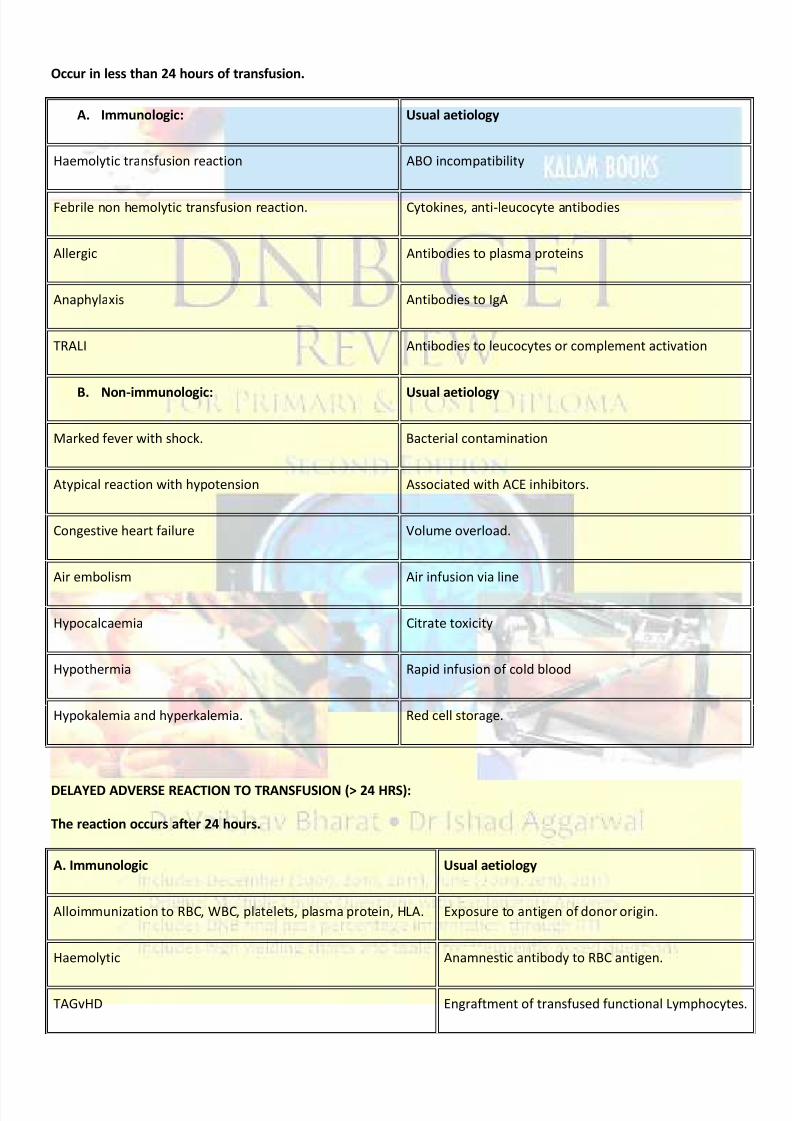
Occur in less than 24 hours of transfusion.
A. Immunologic: Usual aetiology
Haemolytic transfusion reaction ABO incompatibility
Febrile non hemolytic transfusion reaction. Cytokines, anti-leucocyte antibodies
Allergic Antibodies to plasma proteins
Anaphylaxis Antibodies to IgA
TRALI Antibodies to leucocytes or complement activation
B. Non-immunologic: Usual aetiology
Marked fever with shock. Bacterial contamination
Atypical reaction with hypotension Associated with ACE inhibitors.
Congestive heart failure Volume overload.
Air embolism Air infusion via line
Hypocalcaemia Citrate toxicity
Hypothermia Rapid infusion of cold blood
Hypokalemia and hyperkalemia. Red cell storage.
DELAYED ADVERSE REACTION TO TRANSFUSION (> 24 HRS):
The reaction occurs after 24 hours.
A. Immunologic Usual aetiology
Alloimmunization to RBC, WBC, platelets, plasma protein, HLA. Exposure to antigen of donor origin.
Haemolytic Anamnestic antibody to RBC antigen.
TAGvHD Engraftment of transfused functional Lymphocytes.
8/12/2019 Transfusion Reaction1351504464
http://slidepdf.com/reader/full/transfusion-reaction1351504464 4/11
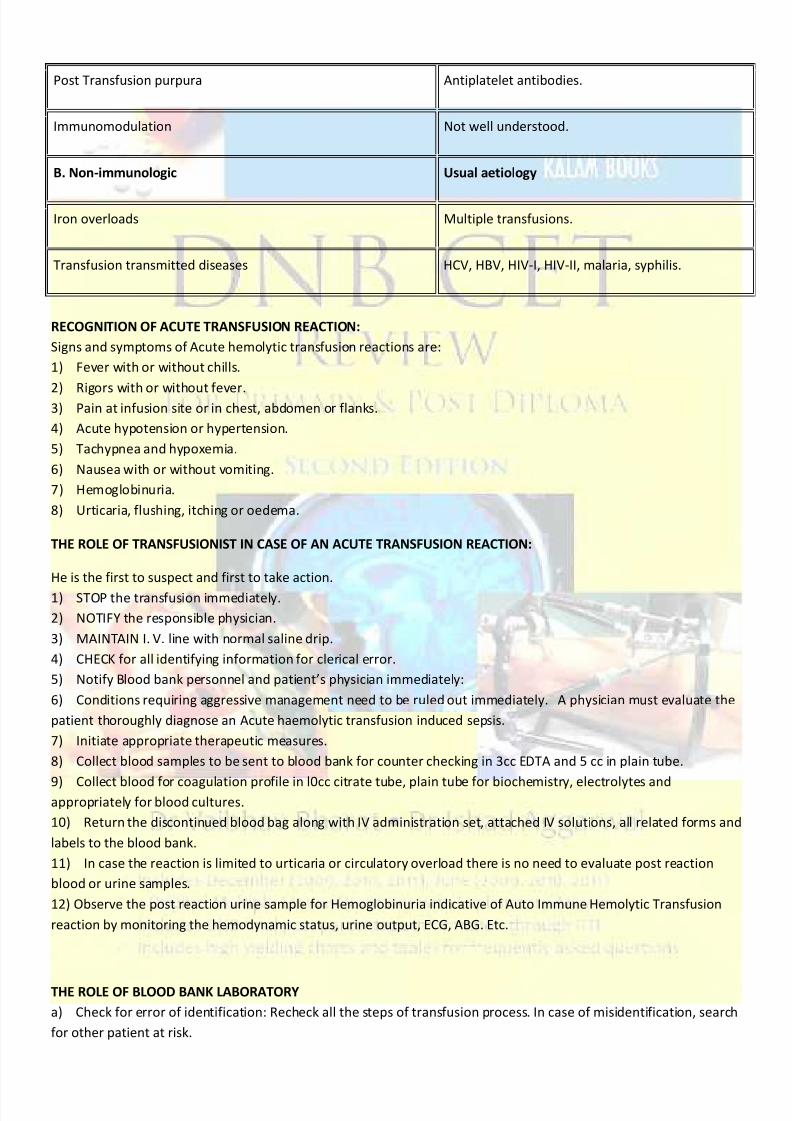
Post Transfusion purpura Antiplatelet antibodies.
Immunomodulation Not well understood.
B. Non-immunologic Usual aetiology
Iron overloads Multiple transfusions.
Transfusion transmitted diseases HCV, HBV, HIV-I, HIV-II, malaria, syphilis.
RECOGNITION OF ACUTE TRANSFUSION REACTION:
Signs and symptoms of Acute hemolytic transfusion reactions are:
1) Fever with or without chills.
2) Rigors with or without fever.3) Pain at infusion site or in chest, abdomen or flanks.
4) Acute hypotension or hypertension.
5) Tachypnea and hypoxemia.
6) Nausea with or without vomiting.
7) Hemoglobinuria.
8) Urticaria, flushing, itching or oedema.
THE ROLE OF TRANSFUSIONIST IN CASE OF AN ACUTE TRANSFUSION REACTION:
He is the first to suspect and first to take action.
1) STOP the transfusion immediately.
2) NOTIFY the responsible physician.
3) MAINTAIN I. V. line with normal saline drip.
4) CHECK for all identifying information for clerical error.
5) Notify Blood bank personnel and patient’s physician immediately:
6) Conditions requiring aggressive management need to be ruled out immediately. A physician must evaluate the
patient thoroughly diagnose an Acute haemolytic transfusion induced sepsis.
7) Initiate appropriate therapeutic measures.
8) Collect blood samples to be sent to blood bank for counter checking in 3cc EDTA and 5 cc in plain tube.
9) Collect blood for coagulation profile in l0cc citrate tube, plain tube for biochemistry, electrolytes andappropriately for blood cultures.
10) Return the discontinued blood bag along with IV administration set, attached IV solutions, all related forms and
labels to the blood bank.
11) In case the reaction is limited to urticaria or circulatory overload there is no need to evaluate post reaction
blood or urine samples.
12) Observe the post reaction urine sample for Hemoglobinuria indicative of Auto Immune Hemolytic Transfusion
reaction by monitoring the hemodynamic status, urine output, ECG, ABG. Etc.
THE ROLE OF BLOOD BANK LABORATORY
a) Check for error of identification: Recheck all the steps of transfusion process. In case of misidentification, search
for other patient at risk.

8/12/2019 Transfusion Reaction1351504464
http://slidepdf.com/reader/full/transfusion-reaction1351504464 5/11
b) Visual check for hemolysis:
Post reaction plasma for hemoglobinemia.
Elevate bilirubin by 5-7 hrs.
Post reaction urine sample for hemoglobinuria
c) Serological check for incompatibility Direct Agglutination testing.
2. FEBRILE NON HEMOLYTIC TRANSFUSION REACTION (FNHTR):
is defined as rise in temperature of 1°C with or without rigors.
It occurs early during transfusion or 1-2hoursafter completion.
Incidence: It is common. Incidence is 0. 5 to 1%. Higher incidence is seen with multiple transfusions and in
multiparous females.
Aetiology: Antibodies to leucocytes; Cytokine release. Clinical picture: Fever with or without chills, mild rise in
temperature; responsive to antipyretic.
Warning sign: Severe rigors, temperature more than 40°C suggest bacterial sepsis.
Recurrence:1 out of 7 with previous Febrile Non Hemolytic Transfusion Reaction.
Management: It is a diagnosis of exclusion. Stop transfusion till hemolytic transfusion reaction ruled out. Blood can
be restarted if hemolytic reaction is ruled out. Restart blood transfusion slowly. If hemolytic reaction is ruled out,
Chlorpheniramine 50 mg & paracetamol may be administered as a supportive treatment.
Prevention: a) Use Leucodepletion filters if history of more than 2 FNHTR. b) Saline washed RBC. c) Premedication
with paracetamol.
3. ALLE RGIC URTICARIAL REACTION:
Incidence: 1 to 3%.
Aetiology: Antibodies to donor plasma proteins.
Clinical picture: Allergic urticarial reaction occurs towards
the end of transfusion or immediately after it and is
characterized by itching, urticaria, flushing rash, rarely
laryngeal oedema, bronchospasm.
Management:
- No Need to stop transfusion.
- IV chlorpheniramine 25mg.
- IV hydrocortisone 100 mg.
- Subcutaneous adrenaline 1: 1000, ONLY if laryngeal oedema.
Prevention:
a. Prophylactic chlorpheniramine before transfusion.
b. Saline washed RBC.
ACUTE IMMUNE HEMOLYTIC TRANSFUSION REACTION (AIHTR):
Incidence;
1: 30, 000 blood bags transfused in Britain.
1: 33, 000 to 1: 12, 000 in USA. Fatality rate: Commonest cause of transfusion related mortality (10% in Britain, 6% in
USA)
Aetiology: It is almost always due to ABO mismatch blood transfusion. Rarely is it due to anti Lewis, anti P and anti H
blood groups. It occurs in emergency, ICU setting or in operation theatre, wherever blood transfusion is being given.
It is a clerical error in labelling or mix-up of samples or a HUMAN ERROR is the cause of this serious event. Technical

8/12/2019 Transfusion Reaction1351504464
http://slidepdf.com/reader/full/transfusion-reaction1351504464 6/11
error in grouping and compatibility testing.
Pathophysiology: It is a catastrophic event following hemolytic reaction when transfused RBCs interact with
preformed antibodies in recipient. Severity is related to amount of transfused blood. Reaction may occur with as
little as 10 to 15 ml of blood. Most reactions occur in the first 1/2 hr of initiation of transfusion.
a) Neuroendocrine response.
Ag + Ab + Xlla factor leads to activation of kinin bradykinin pathway—* Increased capillary leakvasodilatation hypotension Shock.
b) Complement activation:
C3a-C5a leads to shock, hypotension, and bronchospasm
C5-9: hemolysis
c) Coagulation activation:
Disseminated Intravascular Coagulation
d) Cytokines release:
IL6, IL8, TNF alpha fever, hypotension.
Activation of coagulation pathway DIC
e) Renal failure
Clinical features:
1) Symptoms: Transfusion recipient complains of chills, flushing, sweating, chest pain, pain at the infusion site,
back pain, abdominal discomfort, nausea, vomiting and restlessness.
2) Signs: Fever with rigors, tachycardia, dyspnoea, hypotension, tachypnea, pallor, cyanosis, anuria / oliguria,
shock, DIC and hemoglobinuria.
3) In an Unconscious patient: Hypotension, haemoglobin- uria, uncontrolled bleeding (DIC)
“Any febrile reaction — treat as AIHTR unless proved otherwise”
TREATMENT and work up of AIHTR:
Stop the blood component transfusion immediately.
Maintain IV access with crystalloid.
Maintain blood pressure and pulse (vital parameters).
Maintain adequate ventilation.
Give a diuretic and/or institute fluid diuresis.
Obtain blood and urine samples for transfusion reaction workup.
If intravascular hemolysis is confirmed then.
a. Monitor renal status (BUN, Creatinine).
b. Monitor coagulation status (PTA, PTT, fibrinogen, FDP).c. Monitor for signs of hemolysis (bilirubin, LDH, haptoglobin).
d. If sepsis is suspected send appropriate blood cultures &start appropriate antibiotics.
Prevention:
All the precaution to be taken to minimize human error.
a. Well drawn protocols are to be followed.
b. The duties of phlebotomist to medical technologist to transfusionist are to be delineated and followed strictly.
c. Education of the transfusionist is a must as he has the last opportunity to prevent misidentification and the first
one to identify transfusion reaction.
d. It is desirable to have a well-defined transfusion team.
NON IMMUNE HEMOLYSIS - BACTERIAL CONTAMINATION:

8/12/2019 Transfusion Reaction1351504464
http://slidepdf.com/reader/full/transfusion-reaction1351504464 7/11
Aetiology:
1) Healthy donor with transient bacteraemia, asymptomatic carrier of bacterial infection, contamination of
anticoagulant in blood bag, defect in plastic bag, improper handling of needles, inadequate sterilization of skin, all
can contribute to bacterial contamination
2) Bacterial contamination may occur during component preparation
3) It can also occur during storage; platelets stored at 20- 22°C.
4) Fluctuations in temperature can favour bacterial growth.
Organisms:
The common bacterial contaminants are psychrophilic gram negative bacteria; pseudomonas, Citrobacter Freundii,
E. coli, Yersinia enterocolitica, Bartonella, Brucella, Staphylococci, Diphtheroids.
Reaction: The adverse reaction is due to endotoxin.
Risk of bacterial contamination:
(1) 1: 500000 Red blood cells, 1: 10200 platelets, 1: 19 500 platelet pheresis.
(2) Improper storage conditions increase the risk; therefore blood bag should never be stored in unmonitored
nursing station refrigerator.
(3) Blood warming prior to transfusion favours bacterial growth.
(4) Following conditions also favour bacterial contamination; therefore these errors should be avoided.
a. Giving blood over prolonged duration > 4 hrs.
b. Using blood transfusion set for more than one bag.
c. Entry port contamination while thawing blood component.
d. Insertion of medication. Fatality rate is 26%.
Clinical features: Clinical features are like endotoxemia progressing to endotoxic shock with MODS. Patients present
with fever of 40° C with rigors, abdominal cramps, diarrhaea, vomiting, hemoglobinuria, shock, DIC, renal failure.
Treatment and workup:
- STOP TRANSFUSION.
- Inspect blood bag for signs of bacterial overgrowth, cells or plasma brownish or purple, clots, plasma opaque or
muddy, peculiar odour, hemolysis.
- Examine the blood from bag by smear preparation and Gram stain for bacteria and cocci.
- Send cultures from blood bag at 4° C, 20-24° C, 35-37° C.
- Treat patient with proper and adequate antibiotics.
- Treatment similar to AIHTR.
Prevention:
- Attention to the storage conditions of blood.
- Proper sterilisation of phlebotomy site.
- Sterile connecting devices while component preparations.
- Inspection of blood prior to issue.
- Education of transfusionist regarding proper administration of blood.
ANAPHYLACTIC REACTION:
Incidence: 1: 18, 000 to 1: 170, 000
Aetiology: Antibody to donor plasma protein. Most commonly Anti IgA
Clinical features: It occurs after infusion of few ml. of blood.
Patient complains of:
1. Cough, bronchospasm,
8/12/2019 Transfusion Reaction1351504464
http://slidepdf.com/reader/full/transfusion-reaction1351504464 8/11
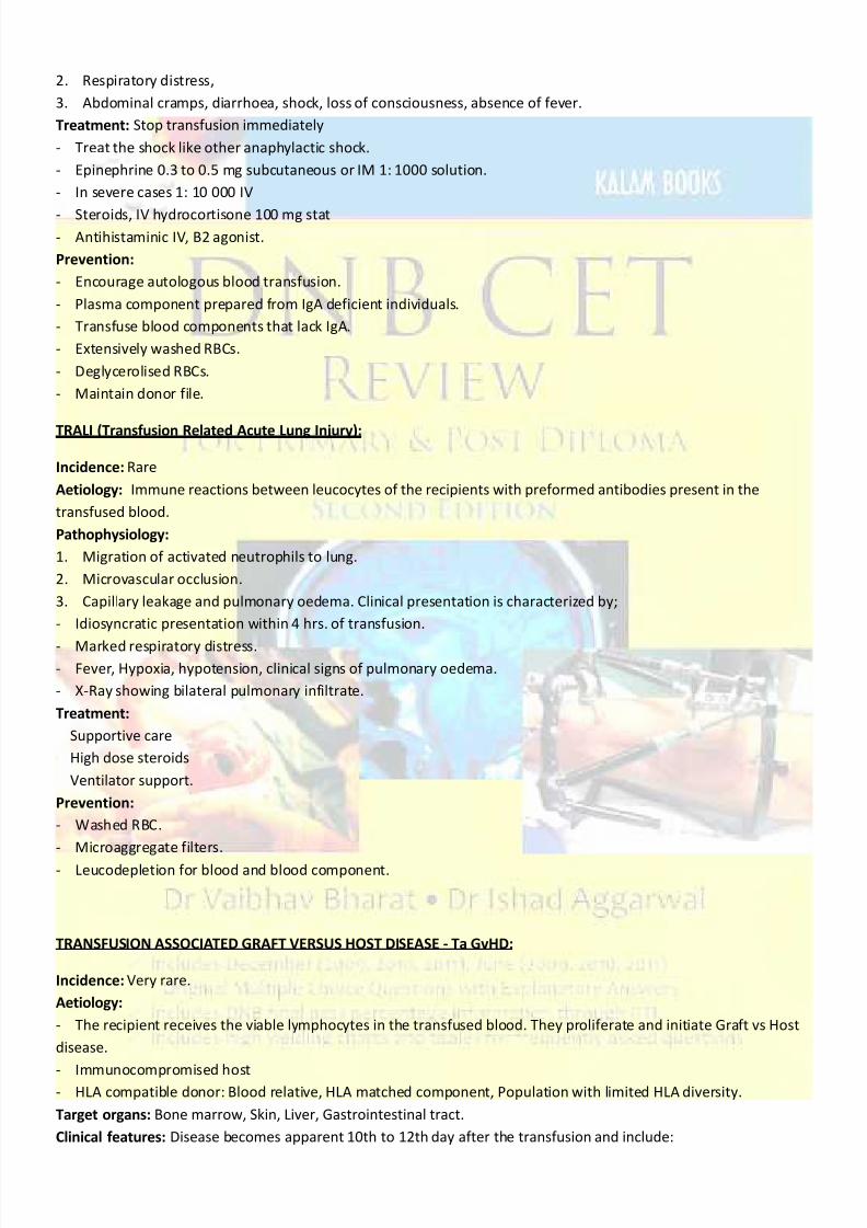
2. Respiratory distress,
3. Abdominal cramps, diarrhoea, shock, loss of consciousness, absence of fever.
Treatment: Stop transfusion immediately
- Treat the shock like other anaphylactic shock.
- Epinephrine 0.3 to 0.5 mg subcutaneous or IM 1: 1000 solution.
- In severe cases 1: 10 000 IV
- Steroids, IV hydrocortisone 100 mg stat- Antihistaminic IV, B2 agonist.
Prevention:
- Encourage autologous blood transfusion.
- Plasma component prepared from IgA deficient individuals.
- Transfuse blood components that lack IgA.
- Extensively washed RBCs.
- Deglycerolised RBCs.
- Maintain donor file.
TRALI (Transfusion Related Acute Lung Injury):
Incidence: Rare
Aetiology: Immune reactions between leucocytes of the recipients with preformed antibodies present in the
transfused blood.
Pathophysiology:
1. Migration of activated neutrophils to lung.
2. Microvascular occlusion.
3. Capillary leakage and pulmonary oedema. Clinical presentation is characterized by;
- Idiosyncratic presentation within 4 hrs. of transfusion.
- Marked respiratory distress.- Fever, Hypoxia, hypotension, clinical signs of pulmonary oedema.
- X-Ray showing bilateral pulmonary infiltrate.
Treatment:
Supportive care
High dose steroids
Ventilator support.
Prevention:
- Washed RBC.
- Microaggregate filters.
- Leucodepletion for blood and blood component.
TRANSFUSION ASSOCIATED GRAFT VERSUS HOST DISEASE - Ta GvHD:
Incidence: Very rare.
Aetiology:
- The recipient receives the viable lymphocytes in the transfused blood. They proliferate and initiate Graft vs Host
disease.
- Immunocompromised host- HLA compatible donor: Blood relative, HLA matched component, Population with limited HLA diversity.
Target organs: Bone marrow, Skin, Liver, Gastrointestinal tract.
Clinical features: Disease becomes apparent 10th to 12th day after the transfusion and include:

8/12/2019 Transfusion Reaction1351504464
http://slidepdf.com/reader/full/transfusion-reaction1351504464 9/11
1. Erythroderma.
2. Jaundice and liver enzyme abnormalities.
3. Diarrhoea, 3 to 4 litters of watery diarrhoea.
4. Pancytopenia.
Diagnosis: is by a) HLA typing, b) Skin biopsy.
Patient at risk:
1. a. Neonate,b. Premature babies,
c. Premature babies with Hemolytic disease of new born (HDN),
d. Intrauterine transfusion,
e. Full term and HDN requiring Exchange transfusion,
f. Full term with neonatal alloimmune, thrombocytopenia requiring mothers’ platelets.
2. a. Severe Combined Immune Deficiency,
b. Wiskott Aldrich Syndrome,
c. Purine nucleotide,d. Nezelof syndrome,
e. Phosphorylase deficiency,
f. Congenital immune deficiency
3. Fresh maternal and paternal plasma.
4. Transfusion from blood relative.
5. HLA matched blood component.
6. Donor of the same ethnic background with limited HLA diversity.
7. In Leukaemia estimated risk is 0. 1 to 1%.8. In Lymphoma estimated risk is 2%.
9. HLA matched allogenic BMT.
10. Post BMT.
11. Solid tumours on intensive chemotherapy.
Components implicated in TaGvHD: Whole blood, packed cells, Granulocyte packs, Platelets, Fresh plasma.
Components not implicated in TaGvHD: FFP & Cryoprecipitate.
Comparison of TaGvHD and GvHD in BMT:
TaGvHD BMT GvHD
Incidence 0. 1 to 1. 0% 30 to 70%
Onset 2 - 47 days 35 to 70 days
Pancytopenia Frequent Rare
BM Hypocellular Not affected
Duration of illness <54 days 5 months
Mortality 87-100% 5 - 10%
Prevention: Irradiation of blood and blood component for patients at risk, Dose: 2500 rads Irradiation of:
a) Cellular component intrauterine transfusion
b) Identified 'at risk' cases for GvHDc) Transfusion of cellular components between blood relatives.
d) Transfusion of HLA selected products

8/12/2019 Transfusion Reaction1351504464
http://slidepdf.com/reader/full/transfusion-reaction1351504464 10/11
Mortality: 87 to 100%
Anaphylactoid reaction to ACE inhibition.
Reaction: Episode of flushing and hypotension in patients on
ACE inhibitors
Mechanism: Prekallikrein present in blood and blood product is converted to vasoactive bradykinin whose
metabolism is inhibited by ACE inhibitors resulting in hypotension.
Procedures associated.- Therapeutic plasma exchange with albumin replacement
- Contact of plasma with dialysis membrane
- Leucocyte reduction filters
- Low-density lipoprotein adsorption column
- Staphylococcal A adsorption column.
DELAYED HEMOLYTIC TRANSFUSION REACTION:
Two types: a) Anamnestic response to transfused RBCs
b) Primary alloimmunization.
Incidence 1: 11000 to 1: 5000 0, 05% to 0. 07% of transfusion recipient
Clinical presentation: More common in multiple transfused & Muciparous women
- Occurs 3-7 days post transfusion.
- Extravascular hemolysis.
- Absence of anticipated Hb or HCT rises following blood transfusion.
- Jaundice
- Fever
- Rarely hemoglobinuria
- Primary alloimmunization not delayed
Investigations:
a) Draw fresh blood to test for alloantibodies.
b) Compare with previous sample.
Treatment: Rarely necessary.
Observe urine output.
Blood transfusion that lack the corresponding antigen.
Prevention: Blood that lacks the responsible antigen.
Issue medical alert card to these patients.
Maintain record of the offending antibodies.
POST TRANSFUSION PURPURA:
Incidence: Uncommon
Presentation:
Acute severe thrombocytopenia 5 - 10 days after transfusion in a previously pregnant female or multiple transfused
individual. Typically perimenopausal or menopausal women. Rare in males.
Pathophysiology:
Patients platelet lack HPA -1 a (PLAl - platelet specific antigen system). 2% of population. Antibodies destroy both
HPA- Al positive and HPA-Al negative platelets.
Course:
Self-limited, recovery in 21 days.
Treatment:
8/12/2019 Transfusion Reaction1351504464
http://slidepdf.com/reader/full/transfusion-reaction1351504464 11/11
-Steroids controversial.
-Plasma exchange,
-IVIgG,
-RDP patient completely refractory hence contraindicated,
-HPA-Al antigen negative platelet of benefit but difficult to arrange.
Prognosis: Good. Recurrence is rare.
Prevention:-Washed platelet concentrate.
-Blood from Human Platelet Antigen- la negative patients.
PLATELET REFRACTORINESS:
Incidence: 20 to 70 % cases requiring multiple platelet transfusion.
Criteria:
-Lack of accepted corrected count increment of two platelet transfusion,
-Poor response to three platelets in 2 weeks.
CCI = Post transfusion count - Pre Transfusion Count x BSA (M2)* No. of Platelet administered x 1011Causes: Platelet alloimmunization Non immune causes
Immune causes:
ABO mismatch
Anti HLA antibodies
Platelet specific allo and autoantibodies Drug dependent antibodies.
Most common cause: Anti HLA antibodies Produced by passenger lymphocytes. May disappear.
Strategies for prevention:
-ABO matched single donor apheresis platelets
-Leucodepletion using leucocyte filters
-HLA matched platelets difficult. Need a donor pool of 2000 to 3000 donor
-Irradiation of HLA matched platelets is a must.
* BSA = Body surface area (M2)


