Transforming growth factor beta 1 induces Snail ... · Transforming growth factor beta 1 induces...
45
TGFβ induces Snail transcription factor Transforming growth factor beta 1 induces Snail transcription factor in epithelial cell lines. Mechanisms for Epithelial Mesenchymal Transitions. Hector Peinado, Miguel Quintanilla and Amparo Cano# Instituto de Investigaciones Biomédicas “Alberto Sols” (CSIC-UAM) and Departamento de Bioquímica (UAM), Arturo Duperier 4, 28029 Madrid. Spain. # Corresponding Author: Dr. Amparo Cano Tel. 34-91-585.4597. Fax. 34-91-585.45.87. E-mail: [email protected] Running title: TGFβ induces Snail transcription factor. Key words: Snail, TGFβ, H-Ras, EMT, E-cadherin. by guest on July 13, 2018 http://www.jbc.org/ Downloaded from
Transcript of Transforming growth factor beta 1 induces Snail ... · Transforming growth factor beta 1 induces...

TGFβ induces Snail transcription factor
Transforming growth factor beta 1 induces Snail transcription factor in epithelial cell lines.
Mechanisms for Epithelial Mesenchymal Transitions.
Hector Peinado, Miguel Quintanilla and Amparo Cano#
Instituto de Investigaciones Biomédicas “Alberto Sols” (CSIC-UAM) and Departamento de
Bioquímica (UAM), Arturo Duperier 4, 28029 Madrid. Spain.
# Corresponding Author:
Dr. Amparo Cano
Tel. 34-91-585.4597.
Fax. 34-91-585.45.87.
E-mail: [email protected]
Running title: TGFβ induces Snail transcription factor.
Key words: Snail, TGFβ, H-Ras, EMT, E-cadherin.
by guest on July 13, 2018http://w
ww
.jbc.org/D
ownloaded from

TGFβ induces Snail transcription factor
2
SUMMARY
The Snail transcription factor has been recently described as a strong repressor of E-
cadherin in epithelial cell lines, where its stable expression leads to the loss of E-cadherin
expression and induces Epithelial-Mesenchymal-Transitions (EMTs) and an invasive phenotype.
The mechanisms regulating Snail expression in development and tumor progression are not yet
known. We show here that transforming growth factor beta-1 (TGFβ1) induces Snail expression in
MDCK cells and triggers EMT, by a mechanisms dependent of the MAPK signaling pathway.
Furthermore, TGFβ1 induces the activity of Snail promoter, while fibroblast growth factor-2
(FGF2) has a milder effect but cooperates with TGFβ1 in the induction of Snail promoter.
Interestingly, TGFβ1-mediated induction of Snail promoter is blocked by a dominant negative form
of H-Ras (N17Ras), whereas oncogenic H-Ras (V12Ras) induces Snail promoter activity and
synergistically cooperates with TGFβ1. The effects of TGFβ1 on Snail promoter are dependent of
MEK1/2 activity but are apparently independent of Smad4 activity. In addition, H-Ras-mediated
induction of Snail promoter, alone or in the presence of TGFβ1, depends on both MAPK and PI3K
activities. These data support that MAPK and PI3K signaling pathways are implicated in TGFβ1-
mediated induction of Snail promoter, probably through Ras activation and its down-stream
effectors.
by guest on July 13, 2018http://w
ww
.jbc.org/D
ownloaded from

TGFβ induces Snail transcription factor
3
INTRODUCTION
The molecular mechanisms underlying local invasion and metastasis are still poorly
understood, but evidence accumulated in the last years indicates the existence of common cellular
mechanisms for the local invasive process which represent the first stage into the metastatic
cascade of carcinomas (1, 2). Among those, loss of expression or function of the E-cadherin cell-
cell adhesion molecule has emerged as an important event for local invasion of epithelial tumor
cells, leading to the consideration of E-cadherin as an invasion-suppressor gene (3-5). The process
of invasion is frequently associated with the loss of other epithelial markers, and the acquisition of
mesenchymal markers and a migratory and motility behavior, collectively known as epithelial-
mesenchymal transitions (EMTs)1 (see ref. 6, for a recent review). EMTs also occur during normal
embryonic development in a strict spatio-temporal control and they are required at specific stages,
such as during gastrulation, formation of the neural crest cells and other morphogenetic processes
(6-8). These developmental EMTs are always accompanied by the loss of functional E-cadherin
mediated cell-cell adhesion (9, 10).
The molecular mechanisms underlying downregulation of E-cadherin during EMTs and
tumor progression are starting to be uncovered. Genetic alterations of the E-cadherin loci have been
found in a scarce number of tumors, particularly, in lobular breast carcinomas and diffuse gastric
carcinomas (3, 11, 12), while the majority of carcinomas with downregulated E-cadherin maintain
an intact E-cadherin locus. Hypermethylation of the E-cadherin promoter and transcriptional
alterations have emerged as the main mechanisms responsible for E-cadherin downregulation in
most carcinomas (5, 13). Several transcriptional repressors of E-cadherin have been recently
isolated, including the zinc finger factors Snail (14, 15) and Slug (16, 17), the two-handed zinc
factor ZEB-1 and SIP-1 (18, 19) and the bHLH factor E12/E47 (20). Snail family factors are in fact
involved in EMTs when overexpressed in epithelial cell lines (14, 15, 17) as well as in embryonic
development (reviewed in ref. 21) and proposed to act as inducers of the invasion process (14, 22).
by guest on July 13, 2018http://w
ww
.jbc.org/D
ownloaded from

TGFβ induces Snail transcription factor
4
Generation of Snail knock out mice has further established the role of this factor in EMT and as E-
cadherin gene repressor. The null Snail embryos die at gastrulation as they fail to undergo a
complete EMT process, forming an altered mesodermal layer which maintains the expression of E-
cadherin (23). Nevertheless, the mechanisms that regulate the expression of Snail factors are still
poorly understood (6, 21).
Different growth factors and cytokines have also been implicated in the process of EMTs
both in epithelial cell systems and in embryonic development. Studies on development have
indicated the participation of several members of the transforming growth factor/bone
morphogenetic (TGFβ/BMP) family of growth factors in specific EMT processes in different
species (24, 25), while fibroblast growth factor (FGF) signaling has been recently reported as a
determinant for mesoderm cell fate specification in the mouse embryo (26). Several studies have
also indicated that a multiple cross talk between TGFβ/BMPs, FGF, and Wnt signals could be
required for some EMTs in development (26-28). In epithelial cell systems, several growth factors
have been widely studied and reported to induce a scattering phenotype or a complete EMT
depending of the specific cell system analyzed (reviewed in refs. 6, 29). Among them, TGFβ has
been identified as an important molecular player of EMT both in vitro and in vivo (30-34). In some
cell systems, a synergistic cooperation between H-Ras activation and TGFβ signaling appears to be
required for induction of a complete EMT (33, 35, 36). Recently, TGFβ has been reported to induce
the expression of Snail in fetal and in immortalized murine hepatocytes and in human mesothelial
cells (37-39), but whether this is a direct or indirect effect has not yet been established.
The participation of specific signaling pathways activated by TGFβ and/or H-Ras-activation
in EMTs has been previously analyzed with somewhat contradictory results as regard to the
specific implication of Smad, mitogen activated protein kinase (MAPK) and/or
phosphatidylinositide 3-OH kinase (PI3K) pathways (36, 40-43). The issue has been recently
unraveled in the EpRas model with the implication of MAPK in TGFβ-induced EMT,
by guest on July 13, 2018http://w
ww
.jbc.org/D
ownloaded from
TGFβ induces Snail transcription factor
5
tumorigenesis and metastasis, while PI3K is involved in cell scattering and resistance to TGF-β
induced apoptosis (36). It remains to be established, however, if the same situation applies to other
systems and, more importantly, the identification of the target genes involved in the specific growth
factor signaling leading to EMTs.
We have used the prototypic epithelial MDCK cells to further analyze the process of EMT
induced by TGFβ and FGF. We have previously used this cell system to show that Snail
overexpression leads to the full repression of E-cadherin expression and induction of a complete
EMT (14). In the present work we have investigated the ability of TGFβ1 and FGF2 to induce an
EMT in MDCK cells and ask whether Snail is a target gene of this process. We present evidence
that TGFβ1 treatment induces an EMT process linked to Snail induction in MDCK cells. Analysis
of the mouse Snail promoter indicates that it is directly induced by TGFβ1, and that FGF2 and
activated H-Ras cooperate with TGFβ1 in induction of the Snail promoter. Our results also indicate
that the MAPK and PI3K pathways are involved in the TGFβ1- and H-Ras-mediated induction of
Snail promoter. These results strongly support that Snail is a direct target of TGFβ1 and oncogenic
H-Ras, and open the way for future studies on the molecular mechanisms and targets of EMTs and
the invasion process.
by guest on July 13, 2018http://w
ww
.jbc.org/D
ownloaded from

TGFβ induces Snail transcription factor
6
EXPERIMENTAL PROCEDURES
Cell culture and treatments. MDCK-II cells were grown in DMEM medium and MCA3D and
PDV cells in Ham´s F12 medium, in the presence of 10% FBS, 10 mM glutamine (Gibco BRL) and
100 µg/ml ampicilin, 32 µg/ml gentamicin (Sigma Chemical Co). Cells were grown at 37ºC in a
humidified CO2 atmosphere. All the transfections and treatments were done in FBS-free culture
medium. For the indicated treatments, 10 µg/ml stocks of recombinant TGFβ1 (BioNova Corp.)
and 100 µg/ml of FGF2 (Peprotech) were prepared according to manufacturer’s instructions and
added to the indicated concentrations. The PI3K and MEK1/2 inhibitors, LY294002 and PD98059
(Calbiochem), respectively, were kept as 30-10 mM stocks in DMSO, that was used as vehicle
control in all the inhibitor treatments.
RT-PCR analyses. Total RNA was isolated from the different cell lines and RT-PCR analyses
were carried out as previously described (14, 17, 20). Canine PCR products, were obtained after
30-35 cycles of amplification with an annealing temperature of 60-65ºC. Primer sequences were as
follows. For canine E-cadherin (sequence kindly provided by Y. Chen, Harvard Medical School,
USA), forward: 5’ GGAATCCTTGGAGGGATCCTC 3’; reverse: 5’ GTCGTCCTCGC-
CACCGCCGTACAT 3’ (amplifies a fragment of 560 bp). For canine Snail, forward: 5’
CCCAAGCCCAGCCGATGAG 3’; reverse: 5’ CTTGGCCACGGAGAGCCC 3’ (amplifies a
fragment of 200 bp). For canine glyceraldehyde-3-phosphatedehydrogenase (GADPH), forward:
5’TGAAGGTCGGT-GTGAACGGATTTGGC3’; reverse: 5’ CATGTAGGCCATGAGGTCCA
CCAC 3’ (amplifies a fragment of 900 bp).
3TP-Lux, E-cadherin and Snail promoter analyses.
For 3TP-Lux assays a reporter construct containing the TPA and TGFβ response elements fused to
the Luciferase reporter gene (44) was used. The generation of mouse E-cadherin promoter
constructs containing -178/+92 sequences in its wild type or mutant Epal (mE-pal) fused to
Luciferase, has been previously reported (17). Generation of full length mouse Snail promoter
by guest on July 13, 2018http://w
ww
.jbc.org/D
ownloaded from

TGFβ induces Snail transcription factor
7
construct (-900 bp) has been also recently described (17). Deletion constructs of the Snail promoter
mutants were obtained by PCR amplification from the full length –900 bp promoter using
appropriate primers containing BamHI and KpnI restriction sites, and the corresponding PCR
products cloned into the same restriction sites in the pXP1-Luciferase vector.
To determine the activity of 3TP-Lux and the Snail promoter 2x10⁵ cells grown in twenty-
four well plates were transiently transfected with 200 to 500 ng of the indicated reporter constructs
and 20 ng of TK-Renilla construct (Promega) as a control of transfection efficiency. Luciferase and
renilla activities were measured using the Dual-Luciferase Reporter assay kit (Promega) and, after
normalization, the results were referred to the wild type promoter activity detected in mock-
transfected cells. Results represent the mean ± s.d. of at least two independent experiments
performed in duplicate samples.
For the cotransfection experiments 500 ng of the following plasmids were used: pSmad4
DN (1-514) in pCMV5 vector (provided by J. Massagué, Sloan-Kettering Center, USA)(44);
pLXSNHRasV12, pLXSNHRasN17 and the different mutants of HRasV12 (pLXSNHRasV12S35,
pLXSNHRasV12C40, pHrasLXSNRasV12G37) in the pLXSN vector (a gift of P. Rodriguez-
Viciana, USCF, USA) (45); β-catenin S33Y (provided by A. Ben-Ze’ev, Weizmann Institute,
Israel) and Lef-1 (provided by H. Clevers, Utrecht University Hospital, The Netherlands) cloned in
pcDNA3. The corresponding empty vectors, pLXSN, pCMV5 or pcDNA3 were used in control
transfections and for normalization of the total amount of DNA.
Immunofluorescence and western blot analyses. For immunofluorescence staining cells grown
on coverslips were fixed in methanol (-20º C, 30 sec) and stained for E-cadherin, vimentin,
cytokeratin-8 and fibronectin, as previously described (14, 17, 20). For F-actin stain, cells were
fixed in 3.7% formaldehyde, 0.5% Triton X-100 for 30 min at room temperature, stained with
TRICT-conjugated phalloidin (Sigma Chemical Co.) and washed (4X) in PBS. The cells were
mounted on Mowiol and the preparations were visualized using a Leica confocal TCS SP2
by guest on July 13, 2018http://w
ww
.jbc.org/D
ownloaded from
TGFβ induces Snail transcription factor
8
microscope. For western blot, whole cell extracts of control and treated cells were obtained in
RIPA buffer (50 mM Tris-HCl pH 7.5, 150 mM NaCl, 1% NP-40, 5% Deoxycholate, 0,1% SDS)
and analyzed for the indicated molecules by western blot and ECL detection as previously
described (14, 17, 20). Primary antibodies included: rat monoclonal anti-E-cadherin ECCD-2
(1:100) (provided by M. Takeichi, Kyoto University, Japan); mouse monoclonal anti-vimentin
(1:200) (Dako); mouse monoclonal anti-cytokeratin 8 (1:200) (Progen); rabbit polyclonal anti-
fibronectin (1:100) and mouse monoclonal anti-α-tubulin (1:1000) (Sigma Chemical Co.). For cell
signaling analysis, western blots were carried out on cell extracts obtained by lysis in Buffer A (20
mM Hepes pH 7.5, 10 mM EGTA, 40 mM β-glycerolphosphate, 2.5 mM MgCl2, 1% NP-40, 1 mM
DTT), containing the apropiate protease and phosphatase inhibitors, during 30 min. at 4ºC. Primary
antisera included: goat anti-AKT (1:1000) (Santa Cruz Biotech.); rabbit anti-phospho (Ser473)-
AKT (1:500); rabbit anti-ERK1/2 and anti-phospho (Thr202/Tyr204)-ERK1/2 (1:1000) (Cell
Signalling Tech.); rabbit anti-Smad2/3 and rabbit anti-phospho (Ser-465/467)-Smad2/3 (1:500)
(Upstate). Secondary antibodies were BODIPY-conjugated goat anti-rat; anti-mouse and anti-rabbit
IgG (Molecular Probes), and HRP-conjugated sheep anti-mouse (1:1000) (Amersham); donkey
anti-goat (1:1000) (Santa Cruz Biotech); goat anti-rat (1:10.000) (Pierce); and goat anti-rabbit
(1:4000) (Nordic) IgG.
Cell proliferation assays. The indicated number of cells (2.5x10⁵ or 5x10⁵) were seeded in
triplicate samples in ninety-two plates and grown in complete medium for 3h. After washing in
PBS, TGFβ1 (10 ng/ml) in FBS-free medium was added and the cells were grown for additional
24h. 3H-thymidine was added during the last 5h of treatment. The cells were collected using a cell
harvester device and 3H-thymidine incorporation was determined in a scintillation counter. The
values, representing the mean+ s.d., were normalized to those obtained in control untreated cells.
Migration assays. The migratory/motility behaviour of MDCK cells was analysed in in vitro
wound healing assays as previously described (14, 17). Monolayers of confluent cultures were
by guest on July 13, 2018http://w
ww
.jbc.org/D
ownloaded from
TGFβ induces Snail transcription factor
9
lightly scratched with a Gilson pipette tip and, after washing to remove detached cells, treated with
TGFβ1 (10 ng/ml) and/or PD98059 (10 µM), as indicated. Cultures were observed at timely
intervals for up to 36h post-incision
by guest on July 13, 2018http://w
ww
.jbc.org/D
ownloaded from

TGFβ induces Snail transcription factor
10
RESULTS
TGFβ1 induces cell scattering and increased motility in MDCK cells dependent on MEK1/2
activity.
Some previous studies have related the TGFβ/BMP signaling pathway to the regulation of
EMT both during embryonic development (24, 25) and in some epithelial cell lines (30, 32, 33, 37),
while others have potentially implicated a similar function for FGFs (46-48). To get further insights
into the regulation of EMT by both kinds of growth factors, we choose the prototypic epithelial
MDCK cell line. Twenty-four hours treatment with TGFβ1 (10 ng/ml) (Fig. 1A, b) or a
combination of TGFβ1 (10 ng/ml) and FGF2 (100 ng/ml) (Fig. 1A, c) induced a dramatic change of
the cellular phenotype: MDCK cells became dissociated with reduced cell-cell contacts and
acquired a more spindle phenotype. Lower concentrations of TGFβ1 (1-5 ng) induced a milder
effect and treatment with FGF2 alone did not affect the phenotype of MDCK cells (data not
shown). The phenotypic changes induced by TGFβ1 were also associated to increased cell motility,
as ascertained by in vitro wound healing assays (Fig. 1B). Eight hours after incision of the wound,
MDCK cells growing in the presence of TGFβ1 started to colonize the wound surface, while
control untreated cells hardly started to migrate (data not shown). The differences in cell motility
were evident 24h after incision when TGFβ1-treated cells colonized about 70-80% of the wound
surface in a random fashion (Fig. 1B, g), in contrast to untreated cells that had only colonized 20-
30% of the wound surface by unidirectional migration (Fig. 1B, e). The increased motility induced
by TGFβ1 treatment is not due to increased cell proliferation. Analysis of 3H-thymidine
incorporation showed that MDCK cells treated with TGFβ1 exhibited an 80% reduction of their
proliferation potential, as compared with control untreated cells (Fig. 2 A). After 3-4 days of
TGFβ1 treatment MDCK cells started to show signs of apoptosis, and most cells died after 7 days
of treatment (data not shown). The sensitivity of MDCK cells to TGFβ1 was also evidenced by the
quick induction of the responsive 3TP-Lux promoter in the presence of the growth factor (Fig. 2B).
by guest on July 13, 2018http://w
ww
.jbc.org/D
ownloaded from
TGFβ induces Snail transcription factor
11
We then analysed the implication of MAPK and PI3K pathways in the phenotypic effects
induced by TGFβ1 in MDCK cells, since they have been previously implicated in epithelial cell
scattering induced in MDCK and in other cell systems by several growth factors (33, 35, 36, 40, 42,
43). One-hour pretreatment with the specific MEK1/2 inhibitor PD98059 (10 µM) abolished the
cell dissociation and scattering induced by TGFβ1 (Fig. 1A, e) or by the combination of TGFβ1
and FGF2 (Fig. 1A, f) treatments. No significant effect of PD98059 on the cell phenotype was
observed in control untreated (Fig. 1A, d) or FGF2-treated cells (data not shown), although
increased intracellular vacuolization was observed in all PD98059 treated samples. In agreement
with those observations, PD98059 pretreatment also blocked the TGFβ1-induced migration of
MDCK cells (Fig. 1B, h), but did not have any effect on the migration of untreated cells (Fig. 1B,
f). Pretreatment with the PI3K inhibitor LY294002 (30 µM) followed by TGFβ1 treatment caused
cell disintegration (data not shown), thus precluding further studies on the implication of PI3K in
the phenotypic or migratory effects of TGFβ1.
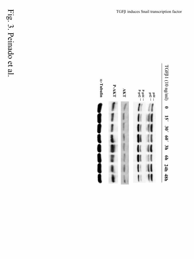
Activation of the MAPK and PI3K pathways following TGFβ1 treatment of MDCK cells
was confirmed by western blot analyses of phosphorylated ERK1/2 and AKT, respectively, using
phospho-specific antibodies to both effectors. As shown in Fig. 3, increased levels of P-ERK2 (Pp-
42) were detected after 30 min. of TGFβ1 treatment, peaking after 1h and slowly decreasing
thereafter (Fig. 3, upper panels). A similar kinetics was observed in the levels of P-ERK1 (Pp-44),
although of a lesser extent. Interestingly, increased levels of P-ERK1/2 were detected even after 6h
of TGFβ1 treatment, indicating a sustained response of the MAPK pathway. Activation of PI3K
followed a slower kinetics in response to TGFβ1, increased levels of P-AKT were firstly detected
after 1h of TGFβ1 treatment, peaked by 3h and decreased thereafter (Fig. 3, middle panels). TGFβ1
treatment also induced a fast and sustained activation of the Smad pathway in MDCK cells, since
P-Smad2 was detected after 15 min. and was maintained up to 3h of TGFβ1 treatment (data not
shown).
by guest on July 13, 2018http://w
ww
.jbc.org/D
ownloaded from
TGFβ induces Snail transcription factor
12
These results indicate that TGFβ1 induces a scattering and motile phenotype in MDCK
cells, apparently depending on MAPK signaling, and suggest that activation of the PI3K pathway
might be required for survival in the presence of TGFβ1. FGF2 by itself does not have a significant
effect on the MDCK phenotype, although it can potentially collaborate with TGFβ1.
TGFβ1 treatment induces EMT associated to Snail induction and E-cadherin repression in
MDCK cells.
The phenotypic changes and increased motility observed in MDCK cells after TGFβ1 treatment
were reminiscent of those observed after stable transfection of MDCK cells with the Snail repressor
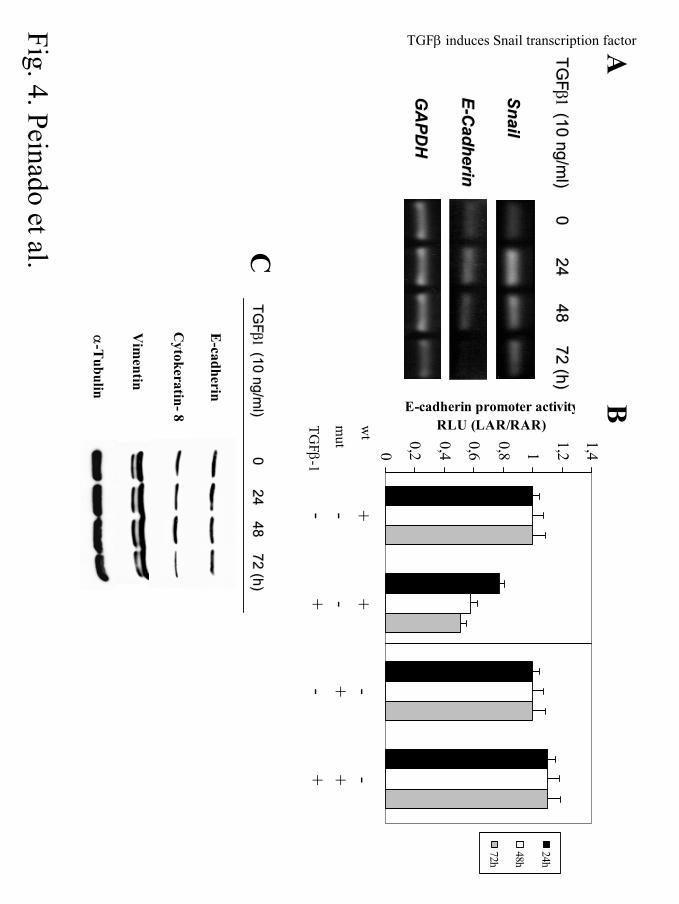
(14), and suggested that Snail might be induced by TGFβ1. To analyze this hypothesis, the
endogenous levels of Snail transcripts in MDCK cells following TGFβ1 treatment were analyzed
by RT-PCR (Fig. 4A). Twenty-four hours treatment with TGFβ1 led to a 2 to 3-fold induction of
Snail mRNA over the basal levels; the level of Snail transcripts decreased thereafter but remained
above the basal levels, at least up to 72h of treatment. Analysis of E-cadherin mRNA levels
showed no significant changes after 24h of TGFβ1 treatment, but a marked decrease was detected
after 48h and 72h of TGFβ1 treatment, when E-cadherin mRNA was almost undetectable (Fig.
4A). Densitometric analyses showed that by 72h of TGFβ1 treatment the Snail and E-cadherin
transcripts were present at levels representing 150% and 20%, respectively, of those detected in
control untreated cells. In agreement with those data, analysis of an exogenous mouse E-cadherin
promoter (17) by transient transfection showed a 50-60 % inhibition after 48 to 72h of TGFβ1
treatment treatment (Fig. 4B, left panel). Furthermore, E-cadherin promoter inhibition by TGFβ1
depends on the presence of Snail-binding site, the E-pal element (14, 17), since its mutation fully
abolished the TGFβ1 effect (Fig. 4B, right panel). These data support that the TGFβ1-induced
repression of E-cadherin can be mediated by Snail expression. Western blot analyses showed a
by guest on July 13, 2018http://w
ww
.jbc.org/D
ownloaded from

TGFβ induces Snail transcription factor
13
moderate decrease (around 35% of control cells) in the total level of E-cadherin, but strong
reduction of other epithelial markers, such as cytokeratin 8 (more than 50% of control levels) after
72h of TGFβ1 treatment (Fig. 4C). The inhibition of E-cadherin promoter activity and decreased
mRNA levels detected between 48-72 h of TGFβ1 treatment contrast with the levels of E-cadherin
protein detected at this time point. This apparent discrepancy has also been observed in other cell
systems (47) and can be explained by the long half-live of the E-cadherin protein, estimated in
more than 40h in other cell systems (49). The above described results suggest that Snail induction,
even at moderate levels, could be required to trigger the repression of E-cadherin and, potentially,
of other epithelial genes that eventually lead to the EMT induced by TGFβ1 treatment in MDCK
cells.
To further investigate if TGFβ1 indeed induces a full EMT in MDCK cells, we analyzed the
expression and localization pattern of E-cadherin, cytokeratin 8, as well as that of vimentin and
fibronectin as prototypic markers of epithelial and mesenchymal cells, respectively. Confocal
immunofluorescence analysis showed that 24 to 48h treatment with TGFβ1 led to a redistribution
of E-cadherin from the cell-cell contacts to the cytoplasm (data not shown). By 72h of TGFβ1
treatment almost complete disappearance of E-cadherin at cell-cell interactions was observed (Fig
5A, b), as compared with control untreated cells (Fig. 5A, a). Cotreatment with TGFβ1 and FGF2
induced a similar redistribution of E-cadherin (Fig. 5A, d). In agreement with the lack of
phenotypic effects, FGF2 treatment alone did not produce redistribution of the E-cadherin
molecules (Fig. 5A, c). The TGFβ1-induced redistribution of E-cadherin was fully abolished by
pretreatment of MDCK cells with PD98059 (Fig 5A, e), which showed a similar E-cadherin stain
as control cells pretreated with PD98059 (Fig. 5A, f). Forty-eight hours treatment of MDCK cells
with TGFβ1 also induced a marked decrease and disorganization of cytokeratin 8 stain (Fig. 5B, b)
also confirmed by western blot (Fig. 4C), and increased staining of vimentin (Fig.5B, e) and
fibronectin (Fig.5B, h) which were organized in clear intermediate filaments and apparently
by guest on July 13, 2018http://w
ww
.jbc.org/D
ownloaded from

TGFβ induces Snail transcription factor
14
secreted matrix respectively, although no changes in total vimentin levels were detected (Fig. 4C).
Staining for F-actin also showed a marked reorganization of the microfilament network with
appearance of stress fibres and membrane protrusions, resembling lamellipodia and filopodia, in
TGFβ1-treated MDCK cells (Fig. 5B, k, arrows), in contrast to untreated control cells that showed a
more defined cortical actin filaments (Fig. 5B, j). These results, together with those shown in Fig.
1, indicate that TGFβ1 induces a full EMT in MDCK cells. Furthermore, the multiple changes
detected in the different markers and in cytoskeleton organization after TGFβ1 treatment were fully
abolished by pre-treatment of MDCK cells with PD98059 (Fig. 5B, c, f, i, l), as well as E-cadherin
redistribution (Fig. 5A, e), indicating that the MAPK activity is necessary for TGFβ1-induced EMT
in this cell line.
TGFβ1 and FGF2 signaling pathways collaborate in Snail promoter induction and
depend on MAPK activity.
To investigate if the observed induction of Snail expression is a direct effect of TGFβ1, we
analyzed the effect of the growth factor on the mouse Snail promoter (17) by transient transfection
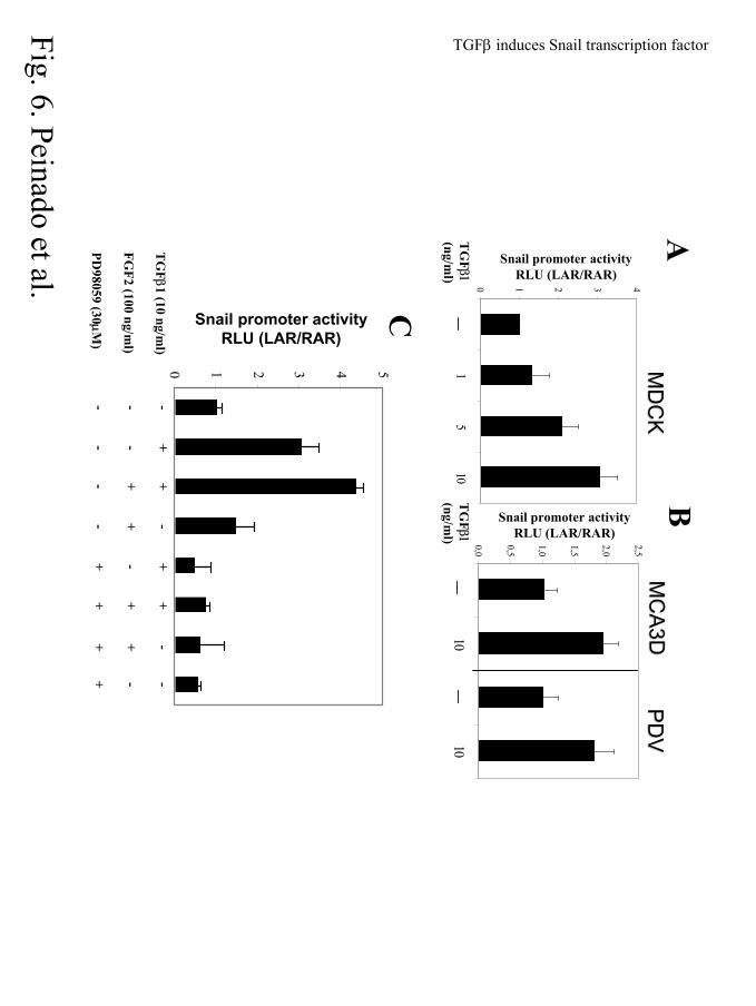
assays. As shown in Fig. 6A, TGFβ1 treatment of MDCK cells induced the Snail promoter activity
in a dose dependent manner. TGFβ1 at 10 ng/ml was able to induce the promoter activity by 3-fold,
while treatments with lower concentrations of 1 and 5 ng/ml induced Snail promoter activity by
1.3- and 2-fold, respectively. To determine if this effect was restricted to MDCK cells, we analyzed
two other epithelial cell lines, the mouse epidermal keratinocyte MCA3D and PDV cells,
representing immortalized and transformed stages of the mouse skin carcinogenesis model,
respectively (49, 50). TGFβ1 (10 ng/ml) treatment induced the Snail promoter activity about 2-fold
in both MCA3D and PDV cell lines (Fig. 6B). These results indicate that Snail promoter could in
fact be controlled by signals downstream of TGFβ1 in epithelial cell lines. Although the level of
by guest on July 13, 2018http://w
ww
.jbc.org/D
ownloaded from
TGFβ induces Snail transcription factor
15
Snail promoter induction by TGFβ1 in the analyzed cell lines is only moderate, it is consistent with
the induction of Snail mRNA level detected in MDCK cells (see Fig. 4A).
As previously indicated, FGF signaling has been recently implicated in the regulation of
Snail expression during embryonic development (26), and previous work in epithelial NBT-II cells
also suggested its involvement in the regulation of Slug (a closely related homolog of Snail) (51).
We, therefore, analyzed the effect of FGF2, alone or in combination with TGFβ1, on the Snail
promoter activity in MDCK cells (Fig. 6C). FGF2 (100 ng/ml) treatment induced a slight activation
of the Snail promoter (1.5-fold), lower than that induced by TGFβ1 at 10 ng/ml (3-fold activation),
but an additive effect on the Snail promoter activity (4.5-fold induction) was observed by the
combination of both FGF2 and TGFβ1 (Fig. 6C). The collaboration between both factors has also
been observed in other contexts, such as in embryonic development where this synergism is
necessary for the subsequent correct development of the EMTs areas, together with others signals,
such as Wnt (28). However, the canonical Wnt signaling pathway seems not to play a significant
role in the regulation of Snail expression in MDCK cells, as no effect on the Snail promoter activity
was observed by the treatment with TGFβ1 in the presence of activated β-catenin and Lef-1 factor
(data not shown). These latter results are also in agreement with a previous report showing that
ILK-induced activation of the human Snail promoter in colon cancer cells is independent of β-
catenin/Tcf complex (52).
We next investigated the TGFβ1 and FGF2 signaling pathways involved in the regulation of
Snail promoter. Cotransfection of a dominant negative version of Smad4 (1-514) that blocks the
classical TGFβ-Smad signaling pathway (44), as confirmed here by its action on the responsive
3TP-lux promoter (Fig. 6E), did not significantly change the TGFβ1-mediated induction of the
Snail promoter activity, and even increased the combined effect of TGFβ1 and FGF2 on the
promoter activity (Fig. 6D). These results indicated that Smad4 signaling is not directly involved in
the regulation of Snail promoter activity by TGFβ1. We then analyzed the participation of the
by guest on July 13, 2018http://w
ww
.jbc.org/D
ownloaded from

TGFβ induces Snail transcription factor
16
MAPK pathway in the regulation of Snail promoter by TGFβ1 and FGF2, since it has been recently
implicated in TGFβ signaling in other contexts (36, 53). To that end, the activity of the Snail
promoter was analyzed in MDCK cells pretreated with the MEK1/2 inhibitor PD98059 (10 µM)
before treatment with TGFβ1 and/or FGF2. Pre-treatment with PD98059 decreased the basal
activity of Snail promoter to about 60% (Figs. 6C, 7A). More significantly, PD98059 pretreatment
fully blocked the Snail promoter induction observed by treatment with TGFβ1, FGF2 or the
combination of both factors (Fig. 6C). These results strongly suggest that MAPK signaling is one
of the pathways implicated in the TGFβ1-mediated regulation of Snail expression in MDCK cells.
TGFβ1 and Ras pathways collaborate in Snail induction.
Several recent works have shown the requirement of Ras downstream signaling in the
process of EMT in different epithelial cell systems, in some cases in cooperation with TGFβ (33,
35, 36). It was, therefore, important to determine the potential contribution of Ras, either by itself
or in cooperation with TGFβ1, to the regulation of the Snail promoter. Cotransfection of a
dominant active version of Ras (HRasV12) induced a 3- to 4-fold activation of the Snail promoter
activity (Fig. 7A, C), similar to that observed in the presence of TGFβ1 (Fig. 7B, D), while a
dominant negative version of Ras (HRasN17) did not have any significant effect on the Snail
promoter (Fig. 7A). Pretreatment with the MEK1/2 inhibitor PD98059 or the PI3K inhibitor
LY294002 resulted in the total blockade of Snail promoter induction after HRasV12 cotransfection
(Fig. 7A), indicating that both MAPK and PI3K signaling pathways are involved in Snail promoter
induction by activated H-Ras. In contrast to PD98059, the LY294002 inhibitor did not have a
significant effect on the basal non-induced Snail promoter (Fig. 7A). Interestingly, activated H-Ras
seems to be required for, and synergistically cooperates with, TGFβ1-mediated Snail induction.
Cotransfection with HRasV12 and TGFβ1 treatment induced a much stronger activation of the
Snail promoter (about 8 to12-fold) than that induced separately by the growth factor or HRasV12
by guest on July 13, 2018http://w
ww
.jbc.org/D
ownloaded from

TGFβ induces Snail transcription factor
17
(Fig. 7B, D). Furthermore, cotransfection with the dominant negative HRasN17 resulted in a 60%
reduction of the TGFβ1-mediated induction of Snail promoter (Fig. 7B).
The above results indicated the participation of activated H-Ras and its cooperation with
TGFβ1 in the regulation of Snail induction, with the involvement of both MAPK and PI3K
signaling pathways. To confirm these results we used different mutants of activated HRasV12 that
are able to transduce signals by specific pathways (45). We cotransfected the mutants RasV12C40
(activated PI3K pathway), RasV12S35 (activated MAPK pathway) and RasV12G37 (activated Ral-
GDS) and analyzed the induction of the Snail promoter in the absence or presence of TGFβ1
treatment. Results indicate that both V12S35 and V12C40 mutants maintain high levels of Snail
promoter activity both in the absence (Fig. 7C) and presence of TGFβ1 (Fig. 7D), accounting for
about 70% of the level obtained by HRasV12 in both situations. In contrast, the V12G37 mutant
had a lower activity, accounting for only about 50% of the level obtained with HRasV12 mutant.
Of note, under TGFβ1 treatment, the V12G37 mutant did not show any significant Snail promoter
induction as compared with the TGFβ1 treatment alone (Fig. 7D).
Taken together, these results indicate that both MAPK and PI3K pathways are required for
the H-Ras and TGFβ1/H-Ras mediated induction of Snail promoter, while the Ral-GDS pathway
might play a more modest role in Snail induction by activated H-Ras alone.
Characterization of the Snail promoter regulatory elements.
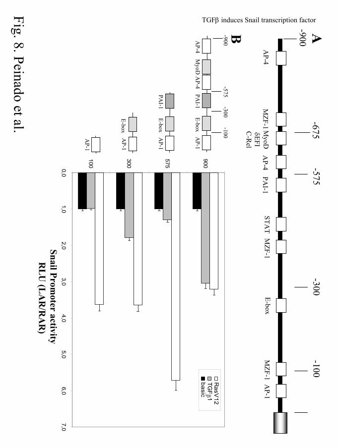
Finally, to get further insights into the regulation of Snail promoter activity by TGFβ1 and
H-Ras signals, we have performed initial studies on the putative regulatory elements implicated. In
silico analysis of the cloned mouse Snail promoter region (–900 bp) indicated the presence of
several putative interaction sites for different transcriptional regulators, including AP-4, AP-1,
STAT, MZF-1 or MyoD consensus sites (Fig. 8A). The organization of this promoter led us to
generate several deletion mutants containing the different control elements, as indicated in the
by guest on July 13, 2018http://w
ww
.jbc.org/D
ownloaded from
TGFβ induces Snail transcription factor
18
schematics of Fig. 8B. Particularly, we were interested in the AP-1 site located at the –23/-33
position (from the ATG start codon), since AP-1 sites are highly sensitive to downstream signals
generated in response to TGFβ and RasV12 pathways (54).
Transfection of MDCK cells with the different Snail promoter mutants showed that the –
900 bp construct exhibited the highest activity, and decreased activities (40-25%) were detected in
most of the other constructs (results not shown). The –100 bp construction has not significant
activity as compared with the other mutants or the full length –900 bp construct, and could,
therefore, be considered as a minimal basal promoter region (data not shown). The effect of TGFβ1
and HRasV12 was analyzed on the different Snail promoter constructs, and the activities were
normalized to that of the basal activity of each promoter construct (Fig. 8B). Surprisingly, the –100
bp promoter region was enough to respond to HRasV12 cotransfection which induced a 3.6-fold
activation of this basic Snail promoter (Fig. 8B). The other Snail promoter constructions showed a
similar sensitivity to HRasV12 cotransfection, with exception of the –575 bp construct that
exhibited a stronger activation (5.8-fold) (Fig. 8B). These results suggested that the proximal AP-1
site could be the main regulatory element implicated in H-Ras induction of Snail promoter, and
point to the potential involvement of negative regulatory elements for H-Ras signals located
between –900 and –575 position of the Snail promoter.
Analyses of the various Snail promoter constructs in response to TGFβ1 treatment showed
that the –900 bp construct exhibited the stronger induction (3-fold activation over basal non-
stimulated control) (Fig. 8B). Deletion of sequences from –575 bp position greatly reduced TGFβ1
activation, and no response to TGFβ1 was achieved with the –100 bp construct (Fig. 8B). Two
additional constructs containing –675 and –200 bp sequences showed the same induction by
TGFβ1 as the –575 and –300 bp constructs, respectively (data not shown). These results suggest
that the TGFβ1 response elements are located between –675 and –900 bp positions, correlating
with all the experiments done with the full-length construction. Several putative binding regions for
by guest on July 13, 2018http://w
ww
.jbc.org/D
ownloaded from
TGFβ induces Snail transcription factor
19
different regulatory transcription factors (AP-4, MZF-1) are present between –675 and –900 bp
region that could be responsible of the TGFβ1 activation of the Snail promoter.
by guest on July 13, 2018http://w
ww
.jbc.org/D
ownloaded from
TGFβ induces Snail transcription factor
20
DISCUSSION
The process of EMT is essential for certain morphogenetic movements within the embryo
and is strongly associated with the pathological process of tumor invasion (6, 7). The molecular
mechanisms and signals involved in EMTs have been previously studied by different groups, with
particular interest in the implication of several growth factors, such as FGF and TGFβ family
factors (6, 29) and transcription factors of the Snail family (21). One of the hallmarks of EMTs in
both normal and pathological situations is the lost of expression or function of the E-cadherin
molecule. In this context, the identification of Snail as a strong E-cadherin repressor in normal and
epithelial tumor cells (14, 15, 22), has reinforced the essential role of Snail family factors in EMTs
(21, 23, 55). Despite this increased understanding, the link between the signals required for EMT
and the direct target genes is still missing, although some recent studies in mouse development
have started to address this important issue (26). In the current work, we present evidence for a
direct link between TGFβ1 signaling and induction of Snail expression during EMT in MDCK
cells.
TGFβ1 induces a scattering phenotype in MDCK cells characterized by the quick
internalization and further loss of E-cadherin from the cell surface, decreased expression of
cytokeratins, induction/reorganization of mesenchymal markers, reorganization of the actin
cytoskeleton and increased cell motility. The phenotypic and differentiation markers changes
observed here are consistent with some of the operational criteria recently proposed for the
definition of a complete EMT process (36). Nevertheless, it should be kept in mind that the EMT
process induced by TGFβ1 in MDCK cells occurs concomitantly to a growth inhibitory response,
in agreement with previous reports on MDCK cells and other non-transformed epithelial cell types
(30, 35, 50, 53). The phenotypic changes induced by TGFβ1 in MDCK cells are similar to those
observed in other epithelial cell systems (37, 40, 56), but differ in the extent of E-cadherin
repression observed in the different systems. They also differ from those observed in the mammary
by guest on July 13, 2018http://w
ww
.jbc.org/D
ownloaded from
TGFβ induces Snail transcription factor
21
EpH4 cells in which TGFβ is not able to induce by itself an EMT process (33), indicating that the
sensitivity and phenotypic response to TFGβ can be modulated by the specific epithelial cell type.
Our present results provide several evidences in support that Snail is mediating the EMT-
triggered by TGFβ1 in MDCK cells. a) Twenty-four hours treatment of MDCK cells with TGFβ1
leads to a 2 to 3-fold induction of Snail mRNA level and the Snail transcripts are maintained above
the basal levels after 72h of treatment. b) The Snail promoter activity is induced to a similar level
after 24h of TGFβ1 treatment. c) The increase and maintenance of Snail transcripts after TGFβ-1
treatment, is correlated with the reduction of E-cadherin mRNA levels and promoter activity
detected between 48h and 72h of treatment and with the overall changes in the cell phenotype. The
partial repression of the exogenous mouse E-cadherin promoter and the fact that total E-cadherin
protein levels are only slightly decreased after 72h of TGFβ1 treatment might argue against a direct
repression of E-cadherin by Snail. However, the repression of the exogenous E-cadherin promoter
activity after TGFβ-1 treatment is dependent on the Snail-interacting promoter sequences (Fig 4B)
located in the E-pal element (14, 17). Furthermore, a strong repression of E-cadherin mRNA is
indeed observed after 72h of TGFβ-1 treatment. The partial repression of the E-cadherin promoter
observed here might reflect intrinsic differences between endogenous and exogenous E-cadherin
promoter regulation in MDCK cells, or be explained by the moderate levels of Snail induction
under those conditions. On the other hand, the slow turnover of E-cadherin protein (49) might well
explain the moderate decrease in E-cadherin protein levels observed after 72h of TGFβ1 treatment.
Despite this fact, the strong redistribution of E-cadherin induced by TGFβ1 in MDCK cells
suggests that perturbation of the functional localization of E-cadherin at cell-cell contacts should be
enough to initiate the EMT, that could be latter sustained by effective repression of E-cadherin
mRNA following TGFβ1 treatment. Furthermore, Snail might regulate other genes required, in
conjunction with E-cadherin downregulation, for the EMT process. Indeed, Snail-mediated
repression of cytokeratin 18 has been recently reported in colon carcinoma HT29 cells (57), and our
by guest on July 13, 2018http://w
ww
.jbc.org/D
ownloaded from
TGFβ induces Snail transcription factor
22
ongoing studies on Snail target genes indicate that besides E-cadherin, expression of genes coding
for several cytokeratins, desmogleins and desmoplakins are strongly repressed in Snail-expressing
MDCK cells (H. Peinado, A. Fabra, J. Palacios, and A. Cano, manuscript in preparation).
A direct effect of TGFβ1 signaling in Snail expression is supported by our analysis of the
mouse Snail promoter, since the growth factor consistently induced the promoter activity by 3-5-
fold over the basal levels in MDCK cells, and also induced the Snail promoter in other epithelial
cell lines. In contrast, FGF2 had a milder effect on the Snail promoter activity, but a cooperation
between FGF2 and TGFβ1 was clearly detected (Fig. 6C). These results are also in agreement with
the phenotypic effects observed in MDCK cells in the presence of these two factors, since FGF2
alone was unable to induce significant phenotypic changes or decreased E-cadherin organization at
the cell-cell contacts (Figs. 1 and 5). Interestingly, activated H-Ras is also able to induce the Snail
promoter activity and, more significantly, synergistically cooperates with TGFβ1 (Fig. 7) . These
results might explain the apparently increased induction of Snail mRNA levels observed by TGFβ
treatment in murine hepatocytes after H-Ras transformation (35). The cooperation between TGFβ
and activated H-Ras has been previously reported to be required for a complete EMT in some cell
systems, such as in EpRas cells where indeed both signals participate into the invasive and
metastatic phenotype (33, 36).
The specific signaling pathways involved in EMT mediated by TGFβ and activated H-Ras
have been also addressed in the present study. Our results do not support a direct involvement of
the Smad pathway in Snail promoter regulation, although an indirect involvement can not be
presently discarded, since the Smad pathway is activated by TGFβ1 in MDCK cells (data not
shown). In fact, the cooperation between FGF2 and TGFβ1 in Snail promoter induction is
magnified by a dominant negative version of Smad4 (Fig. 6D), suggesting a potential cross talk
between Smad and growth factor signals, as reported in other cell systems (50). In contrast, the
MAPK pathway appears to be directly involved in the EMT process driven by TGFβ1 in MDCK
by guest on July 13, 2018http://w
ww
.jbc.org/D
ownloaded from

TGFβ induces Snail transcription factor
23
cells. This conclusion is supported by the strong and sustained activation of ERK1/2 after TGFβ1
treatment, the blockade of the phenotypic effects of the growth factor by the MEK1/2 inhibitor
PD98059 and, more significantly, from the studies on the Snail promoter. Even the basal activity of
the Snail promoter is inhibited by PD98059, suggesting the requirement of active MAPK for
expression of Snail promoter in MDCK cells. An active MAPK pathway is also required for the
induction of Snail promoter by activated H-Ras, as deduced from the studies with PD98059 and
specific RasV12 mutants (Figs. 6 and 7). The PI3K pathway, although apparently not required for
the activity of the basal Snail promoter, it is needed for Snail promoter activation by oncogenic H-
Ras, alone or in cooperation with TGFβ1(Figs. 6 and 7). These results are in agreement with the
observed activation of the PI3K pathway after TGFβ1 treatment (Fig.3), and with recent findings
indicating that PI3K activity is necessary for cell scattering and survival after TGFβ1 treatment in
other cell systems (36, 38), and for the maintenance of the fibroblastic phenotype in H-Ras
transformed murine hepatocytes (35). Taken together, our results support a major role for the
MAPK pathway in TGFβ1-mediated induction of Snail promoter, and the cooperation between
MAPK and PI3K pathways in the synergistic induction of Snail mediated by TGFβ1 and activated
H-Ras. The participation of MAPK pathway into the EMT and invasive phenotype of MDCK cells
has been previously reported either in stable transfectants with an activated MEK1 version (42) or
by using an inducible form of c-Raf (Raf-ER) which also led to the autocrine production of TGFβ
(53). This latter report established a strong link and synergism between TGFβ and the Raf-MAPK
pathway in the promotion of invasiveness and in vivo malignancy. The requirement of TGFβ
signaling for invasiveness and metastasis has also been previously established in the EpRas cell
system (58).
A large body of evidence strongly supports that TGFβ acts as stimulator of malignant
progression in late stages of carcinogenesis (reviewed in refs. 59, 60). The results presented here
provide the first evidence to link TGFβ1 signaling to Snail repressor and EMTs, further reinforcing
by guest on July 13, 2018http://w
ww
.jbc.org/D
ownloaded from

TGFβ induces Snail transcription factor
24
the important role of this growth factor into the malignant progression. Furthermore, the
cooperation between H-Ras and TGFβ1 in Snail promoter induction reported here can be of
biological significance, since activating mutations of H-Ras are present in a high number of tumors
and can eventually contribute, together with acquired resistance to the anti-proliferative effects of
TGFβ, to the malignant conversion. Interestingly, H-Ras activation can led to the autocrine
production of TGFβ in various cell systems (35, 53). These findings, together with the
overproduction of TGFβ observed in a high percentage of human tumors (61) and the fact that most
tumors maintain a functional TGFβ signaling system (59, 60), further reinforce the cooperation
between H-Ras and TGFβ signals in malignancy. Our present results add a further step into the
mechanisms of tumor progression, linking TGFβ signaling and oncogenic Ras activation to
induction of the promoter of invasion Snail.
The promoter region of Snail transcription factor contains several potential control elements
for H-Ras and TGFβ downstream signals. The signals that are highly induced by H-Ras seem to
activate the minimal promoter region of Snail near the initiation site. In contrast, this proximal
region is not sensitive for TGFβ1 signals, indicating that transduction of the different signals could
require the coordination of several response elements in the Snail promoter. On other hand, the
central region of Snail promoter (from –900 to –575 bp) appears to negatively regulate its basal
expression and the signal-mediated induction, suggesting the presence of negative regulators in this
region. It is tempting to speculate that those putative control elements can be involved in the fine
regulation of Snail expression in normal biological process. Although further studies are clearly
required to characterize the specific control elements and transcription factors responsible of Snail
expression in different biological situations, the results reported here can contribute to a better
understanding of the molecular mechanisms of malignant progression, involving some relevant
regulators, such as H-Ras, TGFβ, and Snail. They also open the way to future studies in which
positive regulators of EMT should be considered as promising targets for new anti-tumor therapies.
by guest on July 13, 2018http://w
ww
.jbc.org/D
ownloaded from
TGFβ induces Snail transcription factor
25
REFERENCES
1. Hanahan, D., and Weinberg, R. A. (2000) Cell. 100, 57-70
2. Stetler-Stevenson, W. G., Aznavoorian, S., and Liotta, L. A. (1993) Annu. Rev. Cell Biol. 9,
541-573
3. Berx, G., Cleton-Jansen, A. M., Nollet, F., de Leeuw, W. J., van de Vijver, M., Cornelisse,
C., and van Roy, F. (1995) EMBO J. 14, 6107-6115
4. Birchmeier, W., and Behrens, J. (1994) Biochim. Biophys Acta. 1198, 11-26
5. Christofori, G., and Semb, H. (1999) Trends Biochem. Sci. 24, 73-76
6. Thiery, J. P. (2002) Nat. Rev. Cancer. 2, 442-454
7. Hay, E. D. (1995) Acta Anat. 154, 8-20
8. Sun, D., Baur, S., and Hay, E. D. (2000) Dev. Biol. 228, 337-349
9. Huber, O., Bierkamp, C., and Kemler, R. (1996) Curr. Opin. Cell Biol. 8, 685-691
10. Takeichi, M. (1995) Curr. Opin. Cell Biol. 7, 619-627
11. Becker, K. F., Atkinson, M. J., Reich, U., Becker, I., Nekarda, H., Siewert, J. R., and
Hofler, H. (1994) Cancer Res. 54, 3845-3852
12. Guilford, P., Hopkins, J., Harraway, J., McLeod, M., McLeod, N., Harawira, P., Taite, H.,
Scoular, R., Miller, A., and Reeve, A. E. (1998) Nature. 392, 402-405
13. Cheng, C. W., Wu, P. E., Yu, J. C., Huang, C. S., Yue, C. T., Wu, C. W., and Shen, C. Y.
(2001) Oncogene. 20, 3814-3823
14. Cano, A., Pérez-Moreno, M. A., Rodrigo, I., Locascio, A., Blanco, M. J., del Barrio, M. G.,
Portillo, F., and Nieto, M. A. (2000) Nat. Cell Biol. 2, 76-83
15. Batlle, E., Sancho, E., Franci, C., Dominguez, D., Monfar, M., Baulida, J., and Garcia De
Herreros, A. (2000) Nat. Cell Biol. 2, 84-89.
16. Hajra, K. M., Chen, D. Y., and Fearon, E. R. (2002) Cancer Res. 62, 1613-1618
by guest on July 13, 2018http://w
ww
.jbc.org/D
ownloaded from
TGFβ induces Snail transcription factor
26
17. Bolós,V. Peinado, H., Pérez Moreno, M.A., Fraga, M.F., Esteller, M., and Cano, A. (2003)
J. Cell. Sci. 116, 499-511
18. Comijn, J., Berx, G., Vermassen, P., Verschueren, K., van Grunsven, L., Bruyneel, E.,
Mareel, M., Huylebroeck, D., and van Roy, F. (2001) Mol. Cell. 7, 1267-1278
19. Grooteclaes, M. L., and Frisch, S. M. (2000) Oncogene. 19, 3823-3828
20. Pérez-Moreno, M. A., Locascio, A., Rodrigo, I., Dhondt, G., Portillo, F., Nieto, M. A., and
Cano, A. (2001) J. Biol. Chem. 276, 27424-27431
21. Nieto, M. A. (2002) Nat. Rev. Mol. Cell Biol. 3, 155-166
22. Blanco, M. J., Moreno-Bueno, G., Sarrio, D., Locascio, A., Cano, A., Palacios, J., and
Nieto, M. A. (2002) Oncogene. 21, 3241-3246
23. Carver, E. A., Jiang, R., Lan, Y., Oram, K. F., and Gridley, T. (2001) Mol. Cell Biol. 21,
8184-8188
24. Liem, K. F., Jr., Tremml, G., Roelink, H., and Jessell, T. M. (1995) Cell. 82, 969-979
25. Romano, L. A., and Runyan, R. B. (2000) Dev. Biol. 223, 91-102
26. Ciruna, B., and Rossant, J. (2001) Dev. Cell. 1, 37-49
27. Dorsky, R. I., Moon, R. T., and Raible, D. W. (1998) Nature. 396, 370-373
28. LaBonne, C., and Bronner-Fraser, M. (1998) Development. 125, 2403-2414
29. Boyer, B., Valles, A. M., and Edme, N. (2000) Biochem. Pharmacol. 60, 1091-1099
30. Caulín, C., Scholl, F. G., Frontelo, P., Gamallo, C., and Quintanilla, M. (1995) Cell Growth
Differ. 6, 1027-1035
31. Cui, W., Fowlis, D. J., Bryson, S., Duffie, E., Ireland, H., Balmain, A., and Akhurst, R. J.
(1996) Cell. 86, 531-542
32. Miettinen, P. J., Ebner, R., Lopez, A. R., and Derynck, R. (1994) J. Cell Biol. 127, 2021-
2036
by guest on July 13, 2018http://w
ww
.jbc.org/D
ownloaded from

TGFβ induces Snail transcription factor
27
33. Oft, M., Peli, J., Rudaz, C., Schwarz, H., Beug, H., and Reichmann, E. (1996) Genes Dev.
10, 2462-2477
34. Portella, G., Cumming, S. A., Liddell, J., Cui, W., Ireland, H., Akhurst, R. J., and Balmain,
A. (1998) Cell Growth Differ. 9, 393-404
35. Gotzmann, J., Huber, H., Thallinger, C., Wolschek, M., Jansen, B., Schulte-Hermann, R.,
Beug, H., and Mikulits, W. (2002) J. Cell Sci. 115, 1189-1202
36. Janda, E., Lehmann, K., Killisch, I., Jechlinger, M., Herzig, M., Downward, J., Beug, H.,
and Grunert, S. (2002) J. Cell Biol. 156, 299-313
37. Spagnoli, F. M., Cicchini, C., Tripodi, M., and Weiss, M. C. (2000) J. Cell Sci. 113, 3639-
3647
38. Valdes, F., Alvarez, A. M., Locascio, A., Vega S., Herrera, B., Fernandez, M., Benito, M.,
Nieto, M. A., and Fabregat, I. (2002) Mol. Cancer Res. 1, 68-78
39. Yanez-Mo, M., Lara-Pezzi E., Selgas, R., Ramirez-Huesca, M., Dominguez-Jimenez, C.,
Jimenez-Heffernan, J.A., Aguilera, A., Sanchez-Tomero, J.A., Bajo, M.A., Alvarez, V.,
Castro, M.A., del Peso, G., Cirujeda, A., Gamallo, C., Sanchez-Madrid, F., and Lopez-
Cabrera, M. (2003) N. Engl. J. Med. 348, 403-413
40. Bakin, A. V., Tomlinson, A. K., Bhowmick, N. A., Moses, H. L., and Arteaga, C. L. (2000)
J. Biol. Chem. 275, 36803-36810
41. Chen, Y., Lu, Q., Schneeberger, E. E., and Goodenough, D. A. (2000) Mol. Biol. Cell. 11,
849-862
42. Montesano, R., Soriano, J. V., Hosseini, G., Pepper, M. S., and Schramek, H. (1999) Cell
Growth Differ. 10, 317-332
43. Potempa, S., and Ridley, A. J. (1998) Mol. Biol. Cell. 9, 2185-2200
44. Lagna, G., Hata, A., Hemmati-Brivanlou, A., and Massague, J. (1996) Nature. 383, 832-836
by guest on July 13, 2018http://w
ww
.jbc.org/D
ownloaded from
TGFβ induces Snail transcription factor
28
45. Rodriguez-Viciana, P., Warne, P. H., Khwaja, A., Marte, B. M., Pappin, D., Das, P.,
Waterfield, M. D., Ridley, A., and Downward, J. (1997) Cell. 89, 457-467
46. Migdal, M., Soker, S., Yarden, Y., and Neufeld, G. (1995) J. Cell. Physiol. 162, 266-276
47. Strutz, F., Zeisberg, M., Ziyadeh, F. N., Yang, C. Q., Kalluri, R., Muller, G. A., and
Neilson, E. G. (2002) Kidney Int. 61, 1714-1728
48. Valles, A. M., Boyer, B., Badet, J., Tucker, G. C., Barritault, D., and Thiery, J. P. (1990)
Proc. Natl. Acad. Sci U S A. 87, 1124-1128
49. Lozano, E. and Cano, A. (1998) Cell Adh. Commun. 6, 51-67
50. Iglesias, M., Frontelo, P., Gamallo, C., and Quintanilla, M. (2000) Oncogene. 19, 4134-
4145
51. Savagner, P., Yamada, K. M., and Thiery, J. P. (1997) J. Cell Biol. 137, 1403-1419
52. Tan, C., Costello, P., Sanghera, J., Dominguez, D., Baulida, J., de Herreros, A. G., and
Dedhar, S. (2001) Oncogene. 20, 133-140
53. Lehmann, K., Janda, E., Pierreux, C. E., Rytomaa, M., Schulze, A., McMahon, M., Hill, C.
S., Beug, H., and Downward, J. (2000) Genes Dev. 14, 2610-2622
54. Mulder, K. M. (2000) Cytokine Growth Factor Rev. 11, 23-35
55. Hemavathy, K., Ashraf, S. I., and Ip, Y. T. (2000) Gene. 257, 1-12
56. Piek, E., Moustakas, A., Kurisaki, A., Heldin, C. H., and ten Dijke, P. (1999) J. Cell Sci.
112, 4557-4568
57. Guaita, S., Puig, I., Garrido, M., Domínguez, D., Batlle, E., Sancho, E., Dedhar, S., de
Herreros, A.G., and Baulida, J. (2002). J. Biol. Chem. 277, 39209-39216
58. Oft, M., Heider, K. H., and Beug, H. (1998) Curr. Biol. 8, 1243-1252
59. Akhurst, R. J., and Derynck, R. (2001) Trends Cell Biol. 11, S44-51
60. Massagué, J., Blain, S. W., and Lo, R. S. (2000) Cell. 103, 295-309.
by guest on July 13, 2018http://w
ww
.jbc.org/D
ownloaded from
TGFβ induces Snail transcription factor
29
61. Derynck, R., Goeddel, D. V., Ullrich, A., Gutterman, J. U., Williams, R. D., Bringman, T.
S., and Berger, W. H. (1987) Cancer Res. 47, 707-712
by guest on July 13, 2018http://w
ww
.jbc.org/D
ownloaded from

TGFβ induces Snail transcription factor
30
ABBREVIATIONS
1 The abbreviations used are: AP-1, activator protein 1; EMTs, epithelial-mesenchymal transitions;
ERK, extracellular regulated kinase; FGF, fibroblast growth factor; GADPH, glyceraldehyde-3-
phosphate dehydrogenase; H-Ras, Harvey-ras; MAPK, mitogen-activated protein kinase; MDCK,
Mardin-Darby canine kidney; MEK, mitogen-activated protein kinase kinase; mRNA, messenger
ribonucleic acid; PI3K, phosphatidylinositide 3OH-kinase; RT-PCR, reverse transcription-
polymerase chain reaction; TGFβ, Transforming growth factor beta; Wnt, wing signalling pathway.
ACKNOWLEDGEMENTS
We thank A. Ben-Ze’ev, H. Clevers, T. Gridley, J. Massagué, P. Rodriguez-Viciana and M.
Takeichi for providing vectors, genomic clones and antibodies; and M. A. Nieto and M. C. Iglesias
for helpful suggestions and critical reading of the manuscript. This work was supported by the
Spanish Ministry of Science and Technology (SAF2001-2819 to AC, SAF2001-2361 to MQ), and
by Instituto de Salud Carlos III (FIS01/1174) and the Comunidad Autónoma de Madrid
(08.1/0055./2000) to AC. HP is supported by a predoctoral fellowship of the Spanish Ministry of
Education, Culture and Sports.
by guest on July 13, 2018http://w
ww
.jbc.org/D
ownloaded from
TGFβ induces Snail transcription factor
31
FIGURE LEGENDS
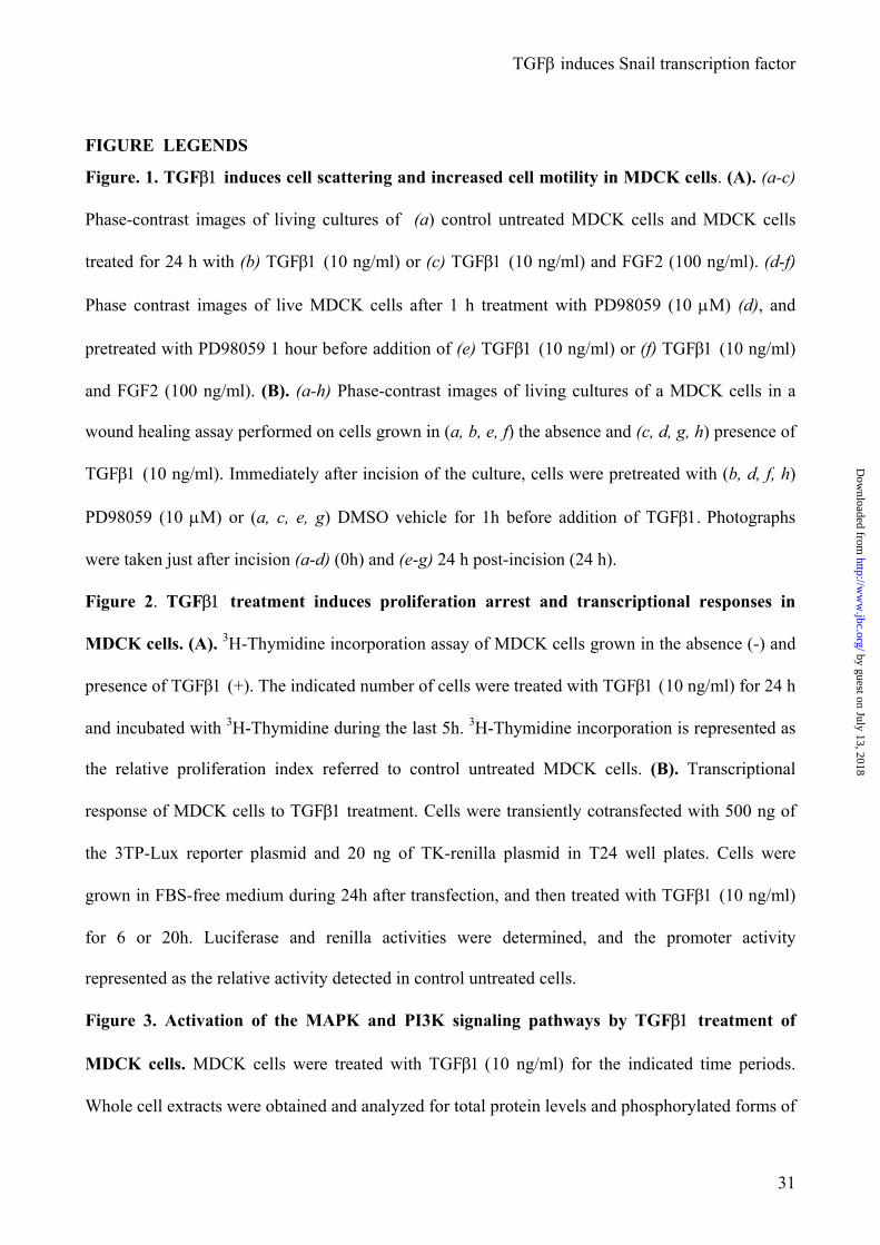
Figure. 1. TGFβ1 induces cell scattering and increased cell motility in MDCK cells. (A). (a-c)
Phase-contrast images of living cultures of (a) control untreated MDCK cells and MDCK cells
treated for 24 h with (b) TGFβ1 (10 ng/ml) or (c) TGFβ1 (10 ng/ml) and FGF2 (100 ng/ml). (d-f)
Phase contrast images of live MDCK cells after 1 h treatment with PD98059 (10 µM) (d), and
pretreated with PD98059 1 hour before addition of (e) TGFβ1 (10 ng/ml) or (f) TGFβ1 (10 ng/ml)
and FGF2 (100 ng/ml). (B). (a-h) Phase-contrast images of living cultures of a MDCK cells in a
wound healing assay performed on cells grown in (a, b, e, f) the absence and (c, d, g, h) presence of
TGFβ1 (10 ng/ml). Immediately after incision of the culture, cells were pretreated with (b, d, f, h)
PD98059 (10 µM) or (a, c, e, g) DMSO vehicle for 1h before addition of TGFβ1. Photographs
were taken just after incision (a-d) (0h) and (e-g) 24 h post-incision (24 h).
Figure 2. TGFβ1 treatment induces proliferation arrest and transcriptional responses in
MDCK cells. (A). 3H-Thymidine incorporation assay of MDCK cells grown in the absence (-) and
presence of TGFβ1 (+). The indicated number of cells were treated with TGFβ1 (10 ng/ml) for 24 h
and incubated with 3H-Thymidine during the last 5h. 3H-Thymidine incorporation is represented as
the relative proliferation index referred to control untreated MDCK cells. (B). Transcriptional
response of MDCK cells to TGFβ1 treatment. Cells were transiently cotransfected with 500 ng of
the 3TP-Lux reporter plasmid and 20 ng of TK-renilla plasmid in T24 well plates. Cells were
grown in FBS-free medium during 24h after transfection, and then treated with TGFβ1 (10 ng/ml)
for 6 or 20h. Luciferase and renilla activities were determined, and the promoter activity
represented as the relative activity detected in control untreated cells.
Figure 3. Activation of the MAPK and PI3K signaling pathways by TGFβ1 treatment of
MDCK cells. MDCK cells were treated with TGFβ1 (10 ng/ml) for the indicated time periods.
Whole cell extracts were obtained and analyzed for total protein levels and phosphorylated forms of
by guest on July 13, 2018http://w
ww
.jbc.org/D
ownloaded from

TGFβ induces Snail transcription factor
32
ERK1/2 and AKT using appropriated specific antibodies. α-tubulin levels were also determined as
a loading control. p44 and p42, ERK1 and ERK2, respectively; P-p44 and P-p42, phospho-ERK1
and phospho-ERK2, respectively; P-AKT, phospho-AKT.
Figure 4. TGFβ1 induces Snail expression and represses E-cadherin in MDCK cells. (A). RT-
PCR analysis of the levels of endogenous canine Snail and E-cadherin transcripts in untreated
MDCK cells (0h) and after the indicated time points of TGFβ1 treatment. The expression of
GAPDH transcripts was analyzed in the same samples as a control for the amount of cDNA present
in each sample. (B). MDCK cells were transiently cotransfected with 200 ng of the proximal mouse
E-cadherin promoter (-178 bp) wt (left panel) or mutated (mut) in E-pal element (right panel) fused
to the Luciferase reporter gene (17) and 20 ng of TK-renilla plasmid in T24 well plates. Cells were
grown in FBS-free medium during 24h after transfection and were treated with 10 ng/ml TGFβ1 for
24, 48, and 72h. Luciferase and renilla activities were determined and the promoter activity was
normalized to that obtained in the absence of treatment. (C). Western blot analysis of epithelial and
mesenchymal protein markers in MDCK-treated cells with TGFβ1 (10 ng/ml) at the indicated time
points. α-tubulin was used as loading control.
Figure 5. TGFβ1 induces EMT in MDCK cells concomitantly with the loss of epithelial
markers, and expression of mesenchymal markers. (A). (a-d), Immunofluorescence images of
MDCK cells showing the localization and organization of E-cadherin in (a) control untreated cells,
and (b-e) cells treated for 72h with (b) TGFβ1 (10 ng/ml), (c) FGF2 (100 ng/ml), (d) TGFβ1 (10
ng/ml) and FGF2 (100 ng/ml), (e) pretreated with PD98059 (10 µM) 1 h before addition of TGFβ1
(10 ng/ml), and (f) treated with PD98059. (B). (a-l), Immunofluorescence images of MDCK cells
showing the localization and organization of the indicated markers, before (a, d, g, j), and after (b,
e, h, k) 48 h treatment with TGFβ1 (10 ng/ml). Cells shown in panels c, f, i and l were pretreated 1h
with PD98059 before TGFβ1 addition. CK8; cytokeratin-8, Vim; vimentin, Fn; fibronectin and F-
by guest on July 13, 2018http://w
ww
.jbc.org/D
ownloaded from
TGFβ induces Snail transcription factor
33
act; Fibrillar-actin. Arrows in panel k indicate reorganization of F-actin at appatent stress fibres and
lamelipodia.
Figure. 6. Growth factor-mediated induction of Snail promoter is blocked by MEK 1/2
inhibitor but not by a negative dominant mutant of Smad4. (A) MDCK, or (B) PDV and
MCA3D cells were transiently cotransfected with 200 ng of the full length Snail promoter construct
(-900 bp) fused to the Luciferase reporter gene (17) and 20 ng of TK-Renilla plasmid in T24 well
plates. Cells grown in FBS-free medium during 24 h after transfection were treated with the
indicated amounts of TGFβ1 for additional 24 h. Luciferase and renilla activities were determined
and the promoter activity was normalized to that obtained in the absence of treatment.
(C). MDCK cells were transiently transfected with the –900 bp Snail promoter construct and
treated with TGFβ1 (10 ng/ml) and/or FGF2 (100 ng/ml) for additional 24h; when indicated cells
were pretreated for 1h with PD98059 (10 µM). (D). MDCK cells cotransfected with the -900 bp
Snail promoter construct and 500 ng of Smad4DN (1-514) expression vector, and treated with
TGFβ1 (10 ng/ml), or TGFβ1 (10 ng/ml) plus FGF2 (100 ng/ml), as indicated. (E). Cells were
cotransfected with 500 ng of the 3TP-Lux reporter plasmid and 500 ng of Smad4DN (1-514)
expression vector, and treated with TGFβ1 (10 ng/ml), as indicated. Luciferase and Renilla
activities were determined 24h after growth factor treatment. The activity of the promoter is
expressed relative to that obtained in the presence of empty control plasmid and/or in the absence
of treatment.
Figure. 7. HRasV12 and TGFβ1 cooperate in the induction of the Snail promoter via PI3K
and MAPK pathways. (A, B) The activity of the -900 bp Snail promoter was measured in MDCK
cells after cotransfection with 500 ng of HRasV12 or H-RasN17 expression vectors without (A),
and with (B) cotreatment with TGFβ1 for 24h. When indicated, cells were pretreated for 1h with
PD98059 (10 µM) or LY294002 (30 µM) inhibitors before TGFβ1 treatment. (C, D) The activity
of the -900 bp Snail promoter was measured in MDCK cells after cotransfection of 500 ng of
by guest on July 13, 2018http://w
ww
.jbc.org/D
ownloaded from

TGFβ induces Snail transcription factor
34
HRasV12 or the indicated HRasV12 effector mutants without (C) and with (D) cotreatment with
TGFβ1 (10 ng/ml). Cells were transiently cotransfected in FBS-free medium with the indicated
HRasV12 mutants; 24h after transfection and when indicated were treated with TGFβ1 (10 ng/ml)
for additional 24h. Luciferase and renilla activities were determined 24h after the growth factor
treatment. The activity of the promoter is expressed relative to that obtained in the presence of
empty control plasmid and in the absence of treatment (Mock).
Figure. 8. HRasV12 and TGFβ1 response elements in Snail promoter. (A). Schematic
representation of the mouse Snail promoter indicating the position of potential regulatory control
elements. (B). Left side, schematics of the deletion mutant constructs generated; right side, diagram
showing the relative promoter activity of the different constructs detected in control non-stimulated
cells (basal, black bars), in the presence of TGFβ1 treatment (TGFβ, light grey bars) or after
HRasV12 cotransfection (RasV12, white bars). Cells were transiently cotransfected with 200 ng of
the indicated Snail promoter constructs and 20 ng of pTK-renilla. When indicated cells were either
treated with TGFβ1 (10 ng/ml) for 24h, or cotransfected with 500 ng of pLXSNHRasV12 vector.
Luciferase and renilla activities were determined as in Figs. 6 and 7. The activity of each promoter
construct is represented relative to that obtained in the presence of empty control plasmid and in the
absence of TGFβ1 treatment.
by guest on July 13, 2018http://w
ww
.jbc.org/D
ownloaded from

A
B
ab
c
de
f
+ TG
Fβ-1+ T
GFβ-1 / FG
F-2-
-PD98059
+ PD98059
Fig. 1. Peinado etal.
TGFβ induces Snail transcription factor
cb
ad
ef
gh
PD98059
++
-
+ TG
Fβ-1
-
-TG
Fβ-1
0 h
24 h
by
gues
t on
July
13,
201
8ht
tp://
ww
w.jb
c.or
g/D
ownl
oade
d fr
om

0.0
0.2
0.4
0.6
1.00.8
1.4
1.2
Proliferation index(3H-thymidine incorporation)
5x10³
2.5x10³
3TP-lux promoter
-6 20
TG
Fβ1(10 ng/ml)
(hours)
1,0
3,0
5,0
7,0
9,0
RLU (LAR/RAR)RLU (LAR/RAR)
TG
Fβ110 (ng/m
l)-
+-
+
AB
TGFβ induces Snail transcription factor
Fig. 2. Peinado etal.
by
gues
t on
July
13,
201
8ht
tp://
ww
w.jb
c.or
g/D
ownl
oade
d fr
om
TGFβ induces Snail transcription factor
030´
60´3h
6h15´
24h48h
TGF
TGFβ β1 (10 ng/
1 (10 ng/ml
ml) )
AK
T
P-AK
T p44p42
P-p44P-p42
α-Tubulin
Fig. 3. Peinado etal.
by
gues
t on
July
13,
201
8ht
tp://
ww
w.jb
c.or
g/D
ownl
oade
d fr
om
0
0,2
0,4
0,6
0,8 1
1,2
1,4
E-cadherin promoter activityRLU (LAR/RAR)
24h
48h
72h
TGF
TGFβ1 β1
(10 ng/ml) 0 24 48 72 (h)
(10 ng/ml) 0 24 48 72 (h)
GAPDH
GAPDH
E E- -Cadherin
Cadherin
SnailSnail
AB
wt
mut
TGFβ-1
+--
+-+
-+-
-++
TGFβ induces Snail transcription factor
TGF
TGFβ1 β1
(10 ng/ml) 0 24 48 72 (h)
(10 ng/ml) 0 24 48 72 (h)
E-cadherin
Cytokeratin-8
Vim
entin
α-Tubulin
C
Fig. 4. Peinado etal.
by
gues
t on
July
13,
201
8ht
tp://
ww
w.jb
c.or
g/D
ownl
oade
d fr
om

Aabd
c
-TG
Fβ1+ T
GFβ1
B
ab
de
gh
jk
TGFβ induces Snail transcription factor
e
TG
Fβ-1
TG
Fβ-1 + FGF-2
FGF-2
PD + T
GFβ-1
PD + T
GFβ1
cfil
PD98059
f
CK
-8
Vim
FnF-act.
Fig. 5. Peinado etal.
by
gues
t on
July
13,
201
8ht
tp://
ww
w.jb
c.or
g/D
ownl
oade
d fr
om
0 1 2 3 4 5
TG
Fβ1 (10 ng/ml)
FGF2 (100 ng/m
l)
PD98059 (30µM
)
---
+--
++-
-+-
+-+
+++
-++
--+
Snail promoter activityRLU (LAR/RAR)
TGFβ induces Snail transcription factor
C
0 1 2 3 4
15
100,0
0,5
1,0
1,5
2,0
2,5
1010
AB
MC
A3D
MC
A3D
PD
VP
DV
MD
CK
MD
CK
Snail promoter activityRLU (LAR/RAR)
Snail promoter activityRLU (LAR/RAR)
TG
Fβ1(ng/m
l)T
GFβ1
(ng/ml)
Fig. 6. Peinado etal.
by
gues
t on
July
13,
201
8ht
tp://
ww
w.jb
c.or
g/D
ownl
oade
d fr
om

0 1 2 3 4 5 6 7
0 10 20 30 40 50 60 70 80
Snail promoter activityRLU (LAR/RAR)
TG
Fβ1 (10 ng/ml)
FGF2 (100 ng/m
l)
Smad-4 D
N
---
+-+
+++
3TP-LUX promoter activityRLU (LAR/RAR)
TG
Fβ1 (10 ng/ml)
Smad-4 D
N
--
+-
-+
++
+--
++-
DE
Fig. 6. Peinado etal.
by
gues
t on
July
13,
201
8ht
tp://
ww
w.jb
c.or
g/D
ownl
oade
d fr
om

0 1 2 3 4 5
AB
Snail promoter activityRLU (LAR/RAR)
Mock
TG
Fβ1T
GFβ1+
HR
asV
12
TG
Fβ1+
HR
asN
17
HR
asV
12H
Ras
N17
HR
asV
12+PD
HR
asV
12+LY
PDL
Y
TGFβ induces Snail transcription factor
0 1 2 3 4 5 6 7 8 9
Mock
Snail promoter activityRLU (LAR/RAR)
Fig. 7. Peinado etal.
by
gues
t on
July
13,
201
8ht
tp://
ww
w.jb
c.or
g/D
ownl
oade
d fr
om

0,0 0,5 1,0 1,5 2,0 2,5 3,0 3,5 4,0 4,5
0 2 4 6 8 10 12 14C
D
+TGFβ
Snail promoter activityRLU (LAR/RAR)
Snail promoter activityRLU (LAR/RAR)
Mock
HR
asV12
TG
Fβ1H
RasV
12H
Ras
V12S35
HR
asV
12G37
HR
asV
12C40
HR
asV12
HR
asV
12S35H
Ras
V12G
37H
Ras
V12C
40M
ock
TGFβ induces Snail transcription factor
Fig. 7. Peinado etal.
by
gues
t on
July
13,
201
8ht
tp://
ww
w.jb
c.or
g/D
ownl
oade
d fr
om
0,01,0
2,03,0
4,05,0
6,07,0
100
300
575
900
RasV
12TG
F 1
basic
-900
AP-1
MZF-1
E-boxM
ZF-1STA
TPA
I-1A
P-4M
yoDδEFIC
-Rel
MZF-1
AP-4
-100-300
-575AB
AP-4
-900-575
-300-100
MyoD
AP-4
PAI-1
E-boxA
P-1
PAI-1
E-boxA
P-1
E-boxA
P-1
AP-1
-675
SnailPromoter
activityR
LU
(LA
R/R
AR
)
β
TGFβ induces Snail transcription factor
Fig. 8. Peinado etal.
by
gues
t on
July
13,
201
8ht
tp://
ww
w.jb
c.or
g/D
ownl
oade
d fr
om
Hector Peinado, Miguel Quintanilla and Amparo Canocell lines. Mechanisms for Epithelial-Mesenchymal transitions
Transforming growth factor beta 1 induces snail transcription factor in epithelial
published online March 28, 2003J. Biol. Chem.
10.1074/jbc.M211304200Access the most updated version of this article at doi:
Alerts:
When a correction for this article is posted•
When this article is cited•
to choose from all of JBC's e-mail alertsClick here
by guest on July 13, 2018http://w
ww
.jbc.org/D
ownloaded from