Transformation Viruses Supports Permissiveness …jvi.asm.org/content/63/5/2152.full.pdf ·...
7
JOURNAL OF VIROLOGY, May 1989, p. 2152-2158 0022-538X/89/052152-07$02.00/0 Copyright © 1989, American Society for Microbiology Transformation of Human Cells by Oncogenic Viruses Supports Permissiveness for Parvovirus H-1 Propagation STEFFEN FAISST, JORG R. SCHLEHOFER,* AND HARALD ZUR HAUSEN Institut fiir Virusforschung, Deut(sches Krebsfrrschungszentrum, Im Neiuenheimer Feld 506, D-6900 Heidelberg, Federal Republic of Germany Received 21 November 1988/Accepted 30 January 1989 Parvovirus H-1 has been shown to suppress spontaneous and chemically or virally induced tumorigenesis in hamsters. In human cell culture systems propagation of H-1 is restricted to transformed cells, which are killed by H-1 infection, in contrast to normal diploid cells, which are nonpermissive for H-1. By analyzing the permissiveness of a variety of human cells for H-1, it was determined that the majority of tested transformed or immortalized cells which were permissive for H-1 contained the DNA of oncogenic viruses (human papillomavirus, simian virus 40, adenovirus, hepatitis B virus, Epstein-Barr virus, and human T-cell lymphotropic virus type I). Of six transformed cell lines negative for persisting tumor virus DNA, only two were permissive for H-1, while two were semipermissive and two were nonpermissive. Thus, persistence and expression of tumor virus functions appear to promote full permissiveness for I--1 in human cells. However, neither expression of genes of specific viral genomes nor the transformed state of apparently virus-free cells alone was sufficient to render human cells permissive for H- I. Therefore, the effect of tumor virus functions on H-1 in transformed cells seems to be indirect, probably mediated by cellular factors which are induced or switched off during the transformation process. It appears that similar factors are induced or switched off by 5-azacytidine or calcium phosphate, both known inducers of cellular gene expression. Mammalian parvoviruses are divided into two groups, the autonomous parvoviruses and the dependoviruses (adeno- associated viruses). There is recent evidence that suggests that this taxonomy does not reflect the properties of parvo- virus replication. Under certain conditions dependoviruses have been shown to replicate without helper viruses (23, 24, 31, 32), whereas the autonomous parvoviruses H-1 and minute virus of mice could be induced in nonpermissive human cells by transformation with simian virus 40 (4, 6). Parvovirus H-1 is a small DNA virus which replicates in the nucleus of infected cells and apparently depends on host cell functions for its own replication (18). It contains a linear single-stranded 5.17-kilobase (kb) genome with two overlap- ping transcription units and two promoters at map positions 4 and 38 (20). The early promoter, P4, regulates the expres- sion of the two noncapsid proteins, NS1 and NS2; the late promoter, P38, regulates the expression of the two capsid proteins, VP1 and VP2. Parvovirus H-1 is capable of efficiently suppressing spon- taneous and chemically or virally induced carcinogenesis in hamsters in vivo: the incidence of spontaneous tumors is reduced from 5 to 0.23% (27), of adenovirus type 12-induced solid tumors from 67 to 28% (29), and of 7,12-dimethyl- benz(a)anthracene-induced tumors from 95 to 38% (30) by prior infection with H-1. The mechanism of tumor suppression by parvoviruses is not understood. A possible insight into this mechanism is that transformed rodent and human cells in vitro show an enhanced susceptibility to parvovirus-induced cell killing (5). This implies that H-1 virus proliferates not in normal but in transformed human cells (28), which are subsequently killed during parvovirus replication (4). The killing appears to be mediated by expression of the parvovirus noncapsid proteins NS1 and NS2 in H-1-permissive cells (19). Since the expression of these proteins is regulated by the H-1 early * Corresponding author. promoter, cells not able to support the comnplete replicative cycle of H-1 may be killed after H-1 infection. Here we ask whether the transformation event renders human cells permissive for H-1. Spontaneous and chemi- cally induced transformation of human cells increases the susceptibility to H-1-induced cell killing but is not sufficient for permissiveness. We provide evidence that a helper function for the replicative cycle of the autonomous parvo- virus H-1 in transformed cells is mediated by a common cellular mechanism, which may be supported by specific types of papillomaviruses, adenoviruses, hepatitis B virus, Epstein-Barr virus, and retroviruses persisting in the same cells. Permissive human cells seem to express cellular H-1 activating factor(s), which are induced by oncogenic viruses in the course of the transformation event. It appears that at least some of these factors are also induced by 5-azacytidine and calcium phosphate, both known to induce cellular genes (11, 16). MATERIALS AND METHODS Abbreviations. The following abbreviations are used: AdS and Ad12, adenovirus types 5 and 12, respectively; mAMSA, 4'-(9-acridinylamino)-methanesulfon-m-aniside; CPE, cytopathic effect; DMBA, 7,12-dimethylbenz(a)an- thracene; DMSO, ditnethyl sulfoxide; EBV, Epstein-Barr virus; HBV, hepatitis B virus; HEFs, primary human embryonal fibroblasts; HTLV, human T-cell lymphotropic virus; HPV, human papillomavirus; MNNG, N-methyl-N'- nitro-N-nitrosoguanidine; NC, nitrocellulose; p.i., postinfec- tion; SDS, sodium dodecyl sulfate; SV40, simian virus 40. Cell lines and virus stocks. SV40-transformed newborn human kidney cells (NB-E; 25), HeLa cells, HeLa x human fibroblast hybrid cells (444, nontumorigenic; CGL3, tumor- igenic) (26), and human diploid fibroblasts were grown in Eagle minimal essential medium (Boehringer GmbH, Mann- heim, Federal Republic of Germany). 2152 Vol. 63, No. 5 on September 1, 2018 by guest http://jvi.asm.org/ Downloaded from
Transcript of Transformation Viruses Supports Permissiveness …jvi.asm.org/content/63/5/2152.full.pdf ·...

JOURNAL OF VIROLOGY, May 1989, p. 2152-21580022-538X/89/052152-07$02.00/0Copyright © 1989, American Society for Microbiology
Transformation of Human Cells by Oncogenic Viruses SupportsPermissiveness for Parvovirus H-1 PropagationSTEFFEN FAISST, JORG R. SCHLEHOFER,* AND HARALD ZUR HAUSEN
Institut fiir Virusforschung, Deut(sches Krebsfrrschungszentrum, Im Neiuenheimer Feld 506,D-6900 Heidelberg, Federal Republic of Germany
Received 21 November 1988/Accepted 30 January 1989
Parvovirus H-1 has been shown to suppress spontaneous and chemically or virally induced tumorigenesis inhamsters. In human cell culture systems propagation of H-1 is restricted to transformed cells, which are killedby H-1 infection, in contrast to normal diploid cells, which are nonpermissive for H-1. By analyzing thepermissiveness of a variety of human cells for H-1, it was determined that the majority of tested transformedor immortalized cells which were permissive for H-1 contained the DNA of oncogenic viruses (humanpapillomavirus, simian virus 40, adenovirus, hepatitis B virus, Epstein-Barr virus, and human T-celllymphotropic virus type I). Of six transformed cell lines negative for persisting tumor virus DNA, only two werepermissive for H-1, while two were semipermissive and two were nonpermissive. Thus, persistence andexpression of tumor virus functions appear to promote full permissiveness for I--1 in human cells. However,neither expression of genes of specific viral genomes nor the transformed state of apparently virus-free cellsalone was sufficient to render human cells permissive for H- I. Therefore, the effect of tumor virus functions onH-1 in transformed cells seems to be indirect, probably mediated by cellular factors which are induced orswitched off during the transformation process. It appears that similar factors are induced or switched off by5-azacytidine or calcium phosphate, both known inducers of cellular gene expression.
Mammalian parvoviruses are divided into two groups, theautonomous parvoviruses and the dependoviruses (adeno-associated viruses). There is recent evidence that suggeststhat this taxonomy does not reflect the properties of parvo-virus replication. Under certain conditions dependoviruseshave been shown to replicate without helper viruses (23, 24,31, 32), whereas the autonomous parvoviruses H-1 andminute virus of mice could be induced in nonpermissivehuman cells by transformation with simian virus 40 (4, 6).
Parvovirus H-1 is a small DNA virus which replicates inthe nucleus of infected cells and apparently depends on hostcell functions for its own replication (18). It contains a linearsingle-stranded 5.17-kilobase (kb) genome with two overlap-ping transcription units and two promoters at map positions4 and 38 (20). The early promoter, P4, regulates the expres-sion of the two noncapsid proteins, NS1 and NS2; the latepromoter, P38, regulates the expression of the two capsidproteins, VP1 and VP2.
Parvovirus H-1 is capable of efficiently suppressing spon-taneous and chemically or virally induced carcinogenesis inhamsters in vivo: the incidence of spontaneous tumors isreduced from 5 to 0.23% (27), of adenovirus type 12-inducedsolid tumors from 67 to 28% (29), and of 7,12-dimethyl-benz(a)anthracene-induced tumors from 95 to 38% (30) byprior infection with H-1.The mechanism of tumor suppression by parvoviruses is
not understood. A possible insight into this mechanism isthat transformed rodent and human cells in vitro show anenhanced susceptibility to parvovirus-induced cell killing(5). This implies that H-1 virus proliferates not in normal butin transformed human cells (28), which are subsequentlykilled during parvovirus replication (4). The killing appearsto be mediated by expression of the parvovirus noncapsidproteins NS1 and NS2 in H-1-permissive cells (19). Since theexpression of these proteins is regulated by the H-1 early
* Corresponding author.
promoter, cells not able to support the comnplete replicativecycle of H-1 may be killed after H-1 infection.Here we ask whether the transformation event renders
human cells permissive for H-1. Spontaneous and chemi-cally induced transformation of human cells increases thesusceptibility to H-1-induced cell killing but is not sufficientfor permissiveness. We provide evidence that a helperfunction for the replicative cycle of the autonomous parvo-virus H-1 in transformed cells is mediated by a commoncellular mechanism, which may be supported by specifictypes of papillomaviruses, adenoviruses, hepatitis B virus,Epstein-Barr virus, and retroviruses persisting in the samecells.
Permissive human cells seem to express cellular H-1activating factor(s), which are induced by oncogenic virusesin the course of the transformation event. It appears that atleast some of these factors are also induced by 5-azacytidineand calcium phosphate, both known to induce cellular genes(11, 16).
MATERIALS AND METHODS
Abbreviations. The following abbreviations are used: AdSand Ad12, adenovirus types 5 and 12, respectively;mAMSA, 4'-(9-acridinylamino)-methanesulfon-m-aniside;CPE, cytopathic effect; DMBA, 7,12-dimethylbenz(a)an-thracene; DMSO, ditnethyl sulfoxide; EBV, Epstein-Barrvirus; HBV, hepatitis B virus; HEFs, primary humanembryonal fibroblasts; HTLV, human T-cell lymphotropicvirus; HPV, human papillomavirus; MNNG, N-methyl-N'-nitro-N-nitrosoguanidine; NC, nitrocellulose; p.i., postinfec-tion; SDS, sodium dodecyl sulfate; SV40, simian virus 40.
Cell lines and virus stocks. SV40-transformed newbornhuman kidney cells (NB-E; 25), HeLa cells, HeLa x humanfibroblast hybrid cells (444, nontumorigenic; CGL3, tumor-igenic) (26), and human diploid fibroblasts were grown inEagle minimal essential medium (Boehringer GmbH, Mann-heim, Federal Republic of Germany).
2152
Vol. 63, No. 5
on Septem
ber 1, 2018 by guesthttp://jvi.asm
.org/D
ownloaded from

TRANSFORMATION SUPPORTS PERMISSIVENESS FOR H-1 VIRUS 2153
Spontaneously immortalized human keratinocytes(HaCaT) (1), cervical carcinoma cell lines SiHa and Caski, asubclone of the vulvar carcinoma cell line A431 (A431-Cl3),AdS Ela gene-transformed human fibroblasts (293 cells),SV40-transformed human fibroblasts (SV80), HBV-containing human liver cells (PLC/PRF/5 line), simian celllines CV-1 and Vero, and SV40-transformed simian cell lineCos were cultivated in Dulbecco modified essential medium(Boehringer).Human foreskin keratinocytes and human foreskin kera-
tinocytes immortalized by transfection with HPV type 16(HPV-16) DNA (HPK I and HPK II) (9) were cultivated inFAD medium (9). The Burkitt's lymphoma cell lines BJA-B(15), BL2, and BL72, the adult T-cell leukemia cell linesMT-1, MT-4, Jurkat, and Molt-4, the EBV (B95-8)-immor-talized human B-lymphocyte cell lines LCL-Gi (H. zurHausen, unpublished data) and LCL-IARC 277 were culti-vated in RMPI 1640 medium.
All media were supplemented with 10% heat-inactivated(56°C, 30 min) fetal calf serum (Boehringer), penicillin (100U/ml; Boehringer), streptomycin (100 ,ug/ml; Boehringer),and L-glutamine (1 mM; Boehringer). All cell lines werecultivated in a water-saturated CO2 atmosphere (5%) at370C.
Wild-type H-1 virus was propagated in NB-E cells. Stockvirus was prepared from the cleared supernatant of freezed-thawed (three times) NB-E cells when the CPE was com-plete, yielding approximately 3 x 10⁷ PFU/ml. This stockvirus was confirmed to be devoid of infectious SV40 byhybridization with 32P-labeled SV40 DNA (21) of a Southernblot of H-1-infected CV-1 cells, which are permissive forSV40. Adl2 was propagated in HeLa cells.
Analysis of H-1 DNA replication. H-1 DNA replication wasmeasured by applying 0.1 to 1.0 ,ug of cellular DNA onto NCfilters, using a Minifold II apparatus (Schleicher & Schuell,Dassel, Federal Republic of Germany). The DNA of theH-1-infected cells was extracted with phenol-chloroform atdifferent times p.i. By hybridization of duplicate filters with32P-labeled human p-albumin DNA (21) as a marker for theamount of DNA applied and with 32P-labeled cloned H-1DNA (pSR1), the ratio of H-1 DNA to cellular DNA wasdetermined (2). The ratio of H-1 DNA replication or ampli-fication corresponds to the ratio of the cell-associated viralDNA to input viral DNA as measured in cells infected withH-1 30 min p.i.To identify the replicative forms of H-1 DNA, Southern
blot analysis was performed with phenol-chloroform-ex-tracted DNA from H-1-infected cells cleaved with BamHIand BglI, which are noncutting enzymes with regard to H-1DNA.
Virus titration. For titration of infectious virus a modifiedplaque assay was used. Cells were harvested 4 days p.i. andsubjected to three freeze-thaw cycles. The cleared superna-tants were diluted in minimal essential medium. About 7 x10⁵ to 8 x 10⁵ indicator NB-E cells seeded on 60-mm petridishes were inoculated with these diluted supernatants (0.4ml per petri dish) at 37°C in a CO2 incubator. After 24 h,when every infected NB-E cell had approxitnately 1,000copies of the H-1 genome, the cells were transferred in situonto an NC filter by applying a filter directly onto themonolayer after removal of the medium (at this stage ofinfection new virions are not yet released in the superna-tant). The DNA of the transferred cells was denaturated byplacing a Whatman 3MM filter paper saturated with 0.5 MNaOH-1.5 M NaCl for 5 to 10 min on the NC filters,followed by neutralization with a 3MM filter paper saturated
with 1 M Tris hydrochloride (pH 7.0-20x SSC (ix SSC is150 mM NaCl plus 15 mM sodium citrate). After hybridiza-tion with 32P-labeled H-1 DNA (21) a single infected cell arearesults in a single signal after exposure, corresponding to aplaque of a plaque assay. This has been confirmed in parallelconventional plaque assays using agar overlay and neutralred staining of surviving cells.
Analysis of H-1 RNA. Extraction of cytoplasmic RNA ofH-1-infected cells 3 days p.i. was performed as described byde Villiers and Schaffner (7). Total cytoplasmic RNA waselectrophoresed in 1% agarose gels and blotted on NC filters(Schleicher & Schuell).
Determination of H-i-induced cell killing. The growthkinetics of uninfected and infected cell cultures were com-pared. Total cell numbers and cell viability were determinedby the trypan blue dye exclusion test.Calcium phosphate-mediated transfection. DNA transfec-
tion was performed by the CaPO4-mediated transfectionprotocol of Chen and Okayama (3). Briefly, 20 ,ug of DNAwas precipitated with 0.125 M CaCl, and 25 mM BES[N,N-bis(2-hydroxyethyl)-2-aminoethanesulfonic acid]-0.75mM Na2HPO4 (pH 6.95) for 20 min at room temperature. A1-ml portion of this mixture was added dropwise to theplated cells (grown in 10 ml of minimal essential medium)and incubated for 24 h at 35°C under 3% CO2.
Chemicals. mAMSA (provided by Warner Lambert Co.,Ann Arbor, Mich.), DMBA (Sigma Chemical Co., Deisen-hofen, Federal Republic of Germany), 5-azacytidine (Serva,Heidelberg, Federal Republic of Germany), the calciumionophore A23187 (Serva), and MNNG (Serva) were freshlydissolved in DMSO and used at the concentrations indicatedbelow. The final concentration of DMSO in the mediumnever exceeded 0.1%.
RESULTS
Screening of human cells for permissiveness for H-1. Ininitial experiments we tested human primary cells and cellsof different human lines for their ability to amplify parvovi-rus DNA and to produce infectious particles. In all cellstested the viral genome was amplified, showing that H-1virus could infect all lines.
In three primary cultures of HEFs from different donorsand one primary culture of primary human foreskin kerati-nocytes, the viral DNA was amplified up to 26-fold but failedto produce infectious parvovirus progeny (Table 1). Nochange of the growth rate could be detected in HEF culturesafter parvovirus infection. Two weeks p.i., parvovirus DNAwas no longer detectable by slot blot hybridization.
(i) It was observed that all cell lines tested containing DNAof oncogenic viruses were permissive for H-1: HPK I, HPK II,HeLa, SiHa, and Caski, which contain HPV DNA; NB-Eand SV80, which contain SV40 DNA; LCL-IARC 277,LCL-Gi and BL72, which contain EBV DNA; MT-1 andMT-4, which contain HTLV type I (HTLV-I) DNA; and 293and PLC, which contain AdS and HBV DNA (Table 1).These cell lines amplified the parvovirus DNA by factors of9 to 5,100 (measured as the ratio of H-1 DNA 4 days p.i.versus input DNA 0.5 h p.i.). Also, infectious H-1 particleswere produced by factors ranging between 2 and 1,550(measured as the ratio of infectious H-1 particles 4 days p.i.versus input virus 0.5 h p.i.). All of these cells were killed inthe course of parvovirus infection (Table 1).
(ii) Four of six cell lines thus far negative for persisting viralDNA were not permissive for H-1. The spontaneously immor-talized human keratinocyte cell line HaCaT did not produce
VOL. 63, 1989
on Septem
ber 1, 2018 by guesthttp://jvi.asm
.org/D
ownloaded from

2154 FAISST ET AL.
TABLE 1. Permissiveness of different human cell lines for parvovirus H-1"
.Origin Cell line Persisting H-1-DNA Production of Cellvirus replication' infectious particlesd killinge
Human primary f Embryonal 4-26 0.3k Foreskin 5 0.3
Human immortalized k HaCaT 53 0.4 +/-k HPKI HPV-16 5,028 81 +k HPK lI HPV-16 256 30 +n NB-E SV40 2,940 1,553 +f SV80 SV40 5,186 24 +f 293 AdS-Ela 637 30 +b LCL-IARC 277 EBV 122 3.3 +b LCL-Gi EBV 9 2 +
Human tumor derived k A431 42 0.5 +/-b BJA-B 451 514 +b Molt-4 194 43 +b BL2 677 36 +/-b Jurkat 39 8.6 +/-I PLC HBV 883 35 +k HeLa HPV-18 468 243 +k SiHa HPV-16 1,050 49 +k CaSki HPV-16 294 5 +b MT-1 HTLV-I 232 71 +b MT-4 HTLV-1 55 1.8 +b BL72 EBV 61 112 +
Simian CV-1 5 0.5Vero 20 0.7Cos SV40 480 70 +
"Human and simian cell lines of various origins were tested for their ability to amplify H-1 DNA and to produce infectious H-1 particles and for theirsusceptibility to H-i-induced cell killing.
b b, Blood cells; f, fibroblasts; k, keratinocytes; 1, liver cells; n, kidney cells.' Ratio of the amount of H-1 DNA 4 days p.i. to the amount of H-1 input DNA (0.5 h p.i.).dRatio of infectious H-1 (titer 4 days p.i.) to input virus (titer 0.5 h p.i.).+, 3 to 30 days p.i., no living cells detectable; +/-, 1 to 30 days p.i., up to 95% of the cells killed, but resistant cells emerged, yielding cultures of resistant
cells; -, no cell killing detectable.
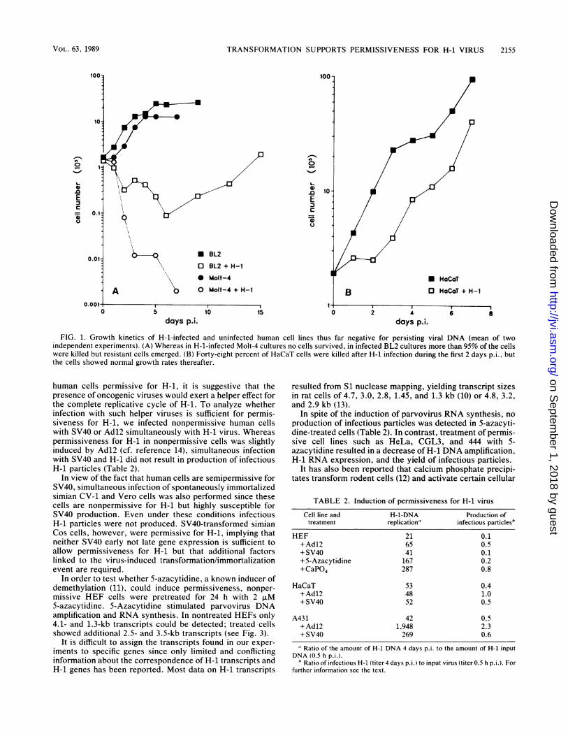
infectious H-1 particles (Table 1) and showed a reducedresponse to parvovirus-induced cell killing. Forty-eight per-cent of infected HaCaT cells were killed during the first 2days p.i., but then infected cultures grew normally (Fig. 1B).Two weeks p.i. parvovirus DNA was no longer detectable inthese cultures. Also, H-1-infected cultures of a subclone ofthe vulvar carcinoma cell line A431 failed to produce infec-tious H-1 progeny (Table 1), and over 95% of these cellswere killed in the course of parvovirus infection, but someresistant cells could be detected. These cells failed to growand died within 4 weeks. Since HaCaT and A431 cells werenot able to produce H-1, they were classified as nonpermis-sive, although they revealed cytopathic changes.The T-cell leukemia cell line Jurkat and the Burkitt's
lymphoma cell line BL2 also showed a reduced susceptibilityto H-1-induced cell killing, although these cells producedinfectious particles (Table 1). More than 95% of the cellswere killed within 6 days p.i., but 1 to 4 weeks p.i. resistantcells growing within infected cultures could be detected (Fig.1A) containing parvovirus DNA. A second infection of thesecells 4 weeks p.i. did not result in H-1-induced cell killing.Therefore, BL2 and Jurkat cells are classified as semiper-missive for H-1.
(iii) Results similar to those obtained with human cells wereobtained with simian cells. SV40-immortalized Cos cells werefully permissive for H-1, but spontaneously immortalizedparental CV-1 cells and Vero cells, which do not containdetectable viral DNA, were not permissive for H-1.
In all cells tested, parvovirus replicative form I DNA(double-stranded monomeric H-1 DNA) and replicative formII DNA (double-stranded dimeric H-1 DNA) could be de-tected after infection, indicating that DNA amplification/
replication is not the step at which the parvovirus replicativecycle is blocked in primary or other nonpermissive cells(data not shown).To elucidate whether permissiveness for H-1 is regulated
by positive or negative factors, we tested hybrids betweenthe permissive HPV-18-containing cervical carcinoma cellline HeLa and the nonpermissive normal human fibroblastsinitially developed by Srivatsan et al. (26). The hybrid cells(line 444) contain HPV-18 DNA but have lost the malignantphenotype. Also CGL3 cells, which are tumorigenic rever-tants of 444, were tested. HeLa, 444, and CGL3 cellsproduced almost the same amounts of infectious particlesand were killed by parvovirus infection during the first 5 to 6days p.i. These findings stress the possible role of thepresence ofHPV DNA within the cell, indicating that at leastunder conditions of in vitro cultivation the tumorigenicphenotype is not a prerequisite for H-1 permissiveness.
Induction of permissiveness for H-1. Nonpermissive humanfibroblasts became permissive after transformation by SV40or susceptible to H-1-induced cell killing after modificationby the chemical carcinogen 4-nitroquinoline 1-oxide or yirradiation (5, 6). In order to elucidate whether initiationevents are sufficient to induce permissiveness for H-1,primary human fibroblasts were treated with chemical orphysical carcinogens. Neither the chemical carcinogenMNNG (0.1 to 100 ,uM), DMBA (0.1 to 1.0 ,ug/ml), ormAMSA (0.01 to 10.0 ,uM) nor irradiation (UV light; 0.5 to60 J m-2) could stimulate replication of H-1 DNA or theproduction of infectious H-1 particles in a transient assay.This is in agreement with results of others (J. Rommelaereand J. Cornelis, personal communication).
Since virus-induced immortalization is sufficient to render
J. VIROL.
on Septem
ber 1, 2018 by guesthttp://jvi.asm
.org/D
ownloaded from
TRANSFORMATION SUPPORTS PERMISSIVENESS FOR H-1 VIRUS
100'
0
L-o 10-
0
.u 10
* BL2
O BL2 + H-1
* Molt-4
0 Molt-4 + H-1* HaCaT
O HaCaT + H-1
4
days p.i.FIG. 1. Growth kinetics of H-1-infected and uninfected human cell lines thus far negative for persisting viral DNA (mean of two
independent experiments). (A) Whereas in H-1-infected Molt-4 cultures no cells survived, in infected BL2 cultures more than 95% of the cellswere killed but resistant cells emerged. (B) Forty-eight percent of HaCaT cells were killed after H-1 infection during the first 2 days p.i., butthe cells showed normal growth rates thereafter.
human cells permissive for H-1, it is suggestive that thepresence of oncogenic viruses would exert a helper effect forthe complete replicative cycle of H-1. To analyze whetherinfection with such helper viruses is sufficient for permis-siveness for H-1, we infected nonpermissive human cellswith SV40 or Adl2 simultaneously with H-1 virus. Whereaspermissiveness for H-1 in nonpermissive cells was slightlyinduced by Adl2 (cf. reference 14), simultaneous infectionwith SV40 and H-1 did not result in production of infectiousH-1 particles (Table 2).
In view of the fact that human cells are semipermissive forSV40, simultaneous infection of spontaneously immortalizedsimian CV-1 and Vero cells was also performed since thesecells are nonpermissive for H-1 but highly susceptible forSV40 production. Even under these conditions infectiousH-1 particles were not produced. SV40-transformed simianCos cells, however, were permissive for H-1, implying thatneither SV40 early not late gene expression is sufficient toallow permissiveness for H-1 but that additional factorslinked to the virus-induced transformation/immortalizationevent are required.
In order to test whether 5-azacytidine, a known inducer ofdemethylation (11), could induce permissiveness, nonper-missive HEF cells were pretreated for 24 h with 2 ,uM5-azacytidine. 5-Azacytidine stimulated parvovirus DNAamplification and RNA synthesis. In nontreated HEFs only4.1- and 1.3-kb transcripts could be detected; treated cellsshowed additional 2.5- and 3.5-kb transcripts (see Fig. 3).
It is difficult to assign the transcripts found in our exper-iments to specific genes since only limited and conflictinginformation about the correspondence of H-1 transcripts andH-1 genes has been reported. Most data on H-1 transcripts
resulted from Si nuclease mapping, yielding transcript sizesin rat cells of 4.7, 3.0, 2.8, 1.45, and 1.3 kb (10) or 4.8, 3.2,and 2.9 kb (13).
In spite of the induction of parvovirus RNA synthesis, noproduction of infectious particles was detected in 5-azacyti-dine-treated cells (Table 2). In contrast, treatment of permis-sive cell lines such as HeLa, CGL3, and 444 with 5-azacytidine resulted in a decrease of H-1 DNA amplification,H-1 RNA expression, and the yield of infectious particles.
It has also been reported that calcium phosphate precipi-tates transform rodent cells (12) and activate certain cellular
TABLE 2. Induction of permissiveness for H-1 virus
Cell line and H-1-DNA Production oftreatment replication" infectious particles"
HEF 21 0.1+Ad12 65 0.5+SV40 41 0.1+5-Azacytidine 167 0.2+CaPO4 287 0.8
HaCaT 53 0.4+Adl2 48 1.0+ SV40 52 0.5
A431 42 0.5+Adl2 1,948 2.3+ SV40 269 0.6
" Ratio of the amount of H-1 DNA 4 days p.i. to the amount of H-1 inputDNA (0.5 h p.i.).
" Ratio of infectious H-1 (titer 4 days p.i.) to input virus (titer 0.5 h p.i.). Forfurther information see the text.
01-1
L.0.0EC
0
A
days p.i.6 8
VOL. 63, 1989 2155
on Septem
ber 1, 2018 by guesthttp://jvi.asm
.org/D
ownloaded from

2156 FAISST ET AL.
-1 Infection + 1 i- 2
days
AB
y.:08P04
CaPO4 +HPVIS-E2/E5
CaPO4 +HPVr1-E6SE7
.)
c
04
Lb
-28S-AD
I .:
4s4 YV 2.5-g |
.i ..|
I
,; 1.3-- 18S
Control
FIG. 2. In situ hybridization of calcium phosphate-treated, H-1-infected HEF cells with 32P-labeled H-1 DNA (21) 4 days p.i.Calcium phosphate treatment was performed during the 24 h beforeinfection (A), during the first 24 h p.i. (B), or during 24 h 2 days p.i.(C). Calcium phosphate precipitates containing HPV-16 DNA (openreading frames E2 to E5 and E6 and E7) did not induce theparvovirus replication cycle as effectively as precipitates free ofDNA. Control, Untreated H-1-infected HEF cells 4 days p.i.
genes (16). Treatment of the nonpermissive HEF cells withCaPO4 precipitates without DNA and subsequent infectionwith H-1 virus resulted in activation of parvovirus DNAamplification (Fig. 2) and RNA synthesis (Fig. 3). Similar tothe treatment with 5-azacytidine, additional 2.5- and 3.5-kbtranscripts were induced by CaPO4 treatment. The H-1 titerof these infected cells measured 4 days p.i. was still belowthe level of input virus, but six to eight times higher than inuntreated cells (Table 2). It could be shown that the inputtiter measured at 0.5 h p.i. was not modified by treatmentwith CaPO4. We subsequently analyzed by [3H-methyl]thy-midine labeling of newly synthesized virions whether therelatively increased H-1 titer was attributable to virus pro-duction. If any virus production was induced by calciumphosphate precipitates, this effect clearly was very marginaland not comparable to the effect of calcium phosphatemeasured for H-1 DNA amplification and RNA expression(data not shown).
In order to analyze whether increased concentration offree intracellular calcium was responsible for the inductionof parvovirus RNA expression, the calcium ionophoreA23187 was applied to HEF cells 24 h prior to infection.A23187 increases the concentrations of intracellular calciumand was applied in concentrations reported to have biologi-cal effects in fibroblasts (8, 17). A23187 had no effect atconcentrations between 0.5 and 50 ,uM on parvovirus RNAexpression, and there was no increase in the production ofinfectious particles.
DISCUSSIONParvovirus H-1-infected hamsters exhibited a decrease in
the incidence of spontaneous or chemically or virally in-
FIG. 3. Cytoplasmic RNA of H-1-infected HEF cells hybridizedwith 32P-labeled H-1 DNA (21). Untreated HEF cells reveal only4.1- and 1.2-kb bands. After treatment of the cells with 5-azacytidineor calcium phosphate precipitates, 3.5- and 2.5-kb transcripts wereinduced. The amounts of RNA in the three lanes were the same.
duced tumors compared with uninfected animals (27, 29, 30).Since previous experiments demonstrated that parvovirusH-1 can be propagated in human cells only after cell trans-formation accompanied by death of the infected cultures (4,28), it was of interest to investigate whether factors allowingpermissive growth of H-1 in human cells are linked tospecific functions related to tumorigenesis.
This report confirms that nontransformed diploid humanfibroblasts and human keratinocytes are nonpermissive forH-1, i.e., virus particles were not produced nor did CPEsbecome visible in spite of replication of H-1 DNA. Also, aline of spontaneously immortalized human keratinocytes didnot support a full replicative cycle of H-1 virus.
Interestingly, transformation of human fibroblasts bySV40 renders the cells permissive for H-1 (4). Our resultswith a variety of additional virus-transformed cells indicatethat virus-induced transformation provides functions permit-ting a complete replicative cycle of H-1 in human cells(cytopathic changes and production of infectious particles),irrespective of the type of transforming virus. Similar toprimary cells, most transformed cell lines devoid of detect-able viral DNA were nonpermissive or only semipermissivefor H-1 infection, although they exhibited cytopathicchanges following H-1 infection. A recent study demon-strated that cells of breast carcinoma lines not known tocontain viral DNA do not produce infectious H-1 particlesalthough they are killed after parvovirus infection (T. Du-pressoir, J. M. Vanackes, J. J. Cornelis, N. Duponchel, andJ. Rommelaere, Cancer Res., in press). Furthermore, it hasbeen shown that only a small fraction of cells transformed byirradiation or chemicals became permissive for H-1 (5, 6).Thus induction of cytopathogenic changes by parvovirusH-1 appears to be correlated with the transformed pheno-type of the infected cells: immortalization (as in HaCaT
J. VIROL.
on Septem
ber 1, 2018 by guesthttp://jvi.asm
.org/D
ownloaded from

TRANSFORMATION SUPPORTS PERMISSIVENESS FOR H-1 VIRUS 2157
cells) seems to be sufficient to induce some cytopathicchanges. Whether expression of the CPE-inducing protein ofH-1 (NS1) is facilitated or altered by functions present inimmortalized or transformed cells remains to be investi-gated. The data available thus far indicate that in thereplicative cycle of H-1 virus, DNA amplification, expres-sion of cytopathic functions, and synthesis of infectiousprogeny depend on different cellular functions. Our datashow that full permissiveness of human cells for H-1 (i.e.,production of infectious progeny) is greatly facilitated by orpossibly even dependent on the presence of the viral DNA ofoncogenic viruses in previously transformed or immortalizedcells. However, neither early nor late gene expression ofthese oncogenic viruses is sufficient to permit propagation ofH-1, as shown in this study with SV40 infection.However, the T-cell leukemia cell line Molt-4 and the
Burkitt's lymphoma cell line BJA-B, which until now havenot been found to contain viral DNA, were permissive forH-1. Possibly these lines may contain unidentified viralDNA. Since BJA-B was infected with EBV during the earlycourse of cell line establishment and has been passaged innude mice, it cannot be excluded that this cell line may havebeen contaminated by other viruses (15). It is possible thatthese cell lines could contain viruses as yet unidentified.
In nonpermissive primary human fibroblasts, which ex-press only a parvovirus 4.1-kb transcript, additional 2.5- and3.5-kb transcripts were induced by treatment with 5-azacy-tidine or calcium phosphate. Both substances have beenshown to induce cellular genes (11, 16). This suggests, thatpermissiveness is regulated by cellular genes not expressedin normal human cells. Since only parvovirus DNA replica-tion and gene expression, but not production of infectiousparticles, were induced by these substances, additionalcellular factors appear to be involved in the complete repli-cative cycle of H-1.
Hybrids between the permissive HPV-18 DNA-containingcervical carcinoma cell line HeLa and nonpermissive normalhuman fibroblasts (444 cells) (26) also contained HPV DNAbut lost the malignant phenotype. Tumorigenic HeLa, non-tumorigenic 444, and tumorigenic CGL3 cells (26), which aremalignant revertants of 444, were all permissive for H-1.This seems to emphasize that full permissiveness is linked tovirus-induced transformation rather than to the tumorigenicphenotype.The HPV-18 E6 and E7 open reading frames seem to be
under the control of negative regulatory factor(s) acting atthe viral 5' regulatory region (22). One of the enhancersequences of this region, the ACC(N6)GGT motif, is alsopresent in the 3' hairpin of the H-1 genome as a head-to-headdimer. Nearby, four additional ACC(N4)GGT motifs arefound. Another ACC(N6)GGT motif is found in the regula-tory region of the open reading frame coding for the H-1capsid proteins. Whether these structural similarities areinvolved in the permissiveness ofHeLa cells for H-1 remainsto be determined. Indeed there seems to be an interaction atthe level of transcriptional control, since in H-1-infectedHeLa cells HPV-18 expression is readily inhibited (J. R.Schlehofer, S. Faisst, and H. zur Hausen, manuscript inpreparation). This might result from competitive interactionsinvolving the regulatory regions of the genomes.The data presented here suggest that the tumor-suppres-
sive properties of H-1 infection might result from H-1-induced CPEs in transformed cells. On the basis of theresults presented here it is anticipated that the antitumori-genic effect of H-1 should be more pronounced in virus-transformed cells than in other tumor cells since in addition
to the CPEs, progeny virus could be provided to infectadjacent cells.The characterization of the interaction of H-1 with the
expression of tumor virus functions, apparently required tomaintain the tumorigenic phenotype, may further contributeto the understanding of tumor suppression by parvoviruses.
ACKNOWLEDGMENTS
We are indebted to E. Stanbridge for providing cell lines 444 andCGL3, to W. W. Franke for A431 C13 cells, to N. E. Fusenig forHaCaT cells, to M. Pawlita for BJA-B and Jurkat cells, to G. Lenoirfor BL-2 and BL-72 cells, to M. Durst for HPK I and HPK Il cells,to W. Waldeck for SV40 virus, and to S. L. Rhode for cloned H-1DNA (pSR1). Also we thank D. Gallahan, M. Durst, and F. Rosl forhelpful discussions and critical reading of the manuscript and R.Webler for preparing the photographs.
This work was supported by the Deutsches Forschungsgemein-schaft and in part by a research grant to J.R.S. within the cooper-ative program in cancer research by the Deutsche Krebsforschung-szentrum and the National Council for Research and Development(Israel).
LITERATURE CITED1. Boukamp, P., R. T. Petrusevska, D. Breitkreutz, J. Hornung, A.
Markham, and N. E. Fusenig. 1988. Normal keratinization in aspontaneously immortalized aneuploid human keratinocyte cellline. J. Cell. Biol. 106:761-771.
2. Burkle, A., T. Meyer, H. Hilz, and H. zur Hausen. 1987.Enhancement of N-methyl-N'-nitro-N-nitrosoguanidine-inducedDNA amplification in a simian virus 40-transformed chinesehamster cell line by 3-aminobenzamide. Cancer Res. 47:3632-3636.
3. Chen, C., and H. Okayama. 1987. High-efficiency transforma-tion of mammalian cells by plasmid DNA. Mol. Cell. Biol.7:2745-2752.
4. Chen, Y. Q., F. de Foresta, J. Hertoghs, B. L. Avalosse, J. J.Cornelis, and J. Rommelaere. 1986. Selective killing of simianvirus 40-transformed human fibroblasts by parvovirus H-1.Cancer Res. 46:3574-3579.
5. Cornelis, J. J., B. Avalosse, S. Mousset, M. Namba, and J.Rommelaere. 1986. Selective destruction by parvoviruses ofhuman and mouse fibroblasts transformed with -y-irradiation.Int. J. Radiat. Biol. 49:529.
6. Cornelis, J. J., P. Becquart, N. Duponchel, N. Salome, B. L.Avalosse, M. Namba, and J. Rommelaere. 1988. Transformationof human fibroblasts by ionizing radiation, a chemical carcino-gen, or simian virus 40 correlates with an increase in suscepti-bility to the autonomous parvoviruses H-1 virus and minutevirus of mice. J. Virol. 62:1679-1686.
7. de Villiers, J., and W. Schaffner. 1983. Transcriptional enhanc-ers from papovaviruses as components of eucaryotic expressionvectors, p. B507/1-B507/20. In R. A. Flavell (ed.), Techniquesin the life sciences, B5. Nucleic acid biochemistry. Elsevier,Ireland.
8. Drummond, I. A. S., A. S. Lee, E. Resendez, and R. A.Steinhardt. 1987. Depletion of intracellular calcium stores bycalcium ionophore A23187 induces the genes for glucose-regu-lated proteins in hamster fibroblasts. J. Biol. Chem. 262:12801-12805.
9. Durst, M., R. T. Dzarlieva-Petrusevska, P. Boukamp, N. E.Fusenig, and L. Gissmann. 1987. Molecular and cytogeneticanalysis of immortalized human primary keratinocytes obtainedafter transfection with human papillomavirus type 16 DNA.Oncogene 1:251-256.
10. Green, M. R., L. Lebowitz, and R. G. Roeder. 1979. Expressionof the autonomous parvovirus Hi genome: evidence for a singletranscriptional unit and multiple spliced polyadenylated tran-scripts. Cell 17:967-977.
11. Jones, P. A. 1985. Altering gene expression with 5-azacytidine.Cell 40:485-486.
12. Kerbel, R. S., C. Waghorne, M. S. Man, B. Elliot, and M. L.
VOL. 63, 1989
on Septem
ber 1, 2018 by guesthttp://jvi.asm
.org/D
ownloaded from

2158 FAISST ET AL.
Breitman. 1987. Alteration of the tumorigenic and metastaticproperties of neoplastic cells is associated with the process ofcalcium phosphate-mediated DNA transfection. Proc. Natl.Acad. Sci. USA 84:1263-1267.
13. Lebowitz, R. M., and R. G. Roeder. 1986. Parvovirus H-1expression: mapping of the abundant cytoplasmic transcriptsand identification of promoter sites and overlapping transcrip-tion units. J. Virol. 58:271-280.
14. Ledinko, N., S. Hopkins, and H. Toolan. 1969. Relationshipbetween potentiation of H-1 growth by human adenovirus 12and inhibition of the 'helper' adenovirus by H-1. J. Gen. Virol.5:19-31.
15. Menezes, J., W. Leibold, G. Klein, and G. Clements. 1975.Establishment and characterization of an Epstein-Barr virus(EBV)-negative lymphoblastoid B cell line (BJA-B) from anexceptional, EBV-genome-negative African Burkitt's lym-phoma. Biomedicine 22:276-284.
16. Pine, R., D. E. Levy, N. Reich, and J. E. Darnell. 1988.Transcriptional stimulation by CaPO4-precipitates. Nucleic Ac-ids Res. 16:1371-1378.
17. Reed, P. W., and H. A. Lardy. 1972. A23187: a divalent cationionophore. J. Biol. Chem. 247:6970-6977.
18. Rhode, S. L., III. 1973. Replication process of the parvovirusH-1 I. Kinetics in a parasynchronous cell system. J. Virol.11:856-861.
19. Rhode, S. L., III. 1987. Construction of a genetic switch forinducible trans-activation of gene expression in eucaryotic cells.J. Virol. 61:1448-1456.
20. Rhode, S. L., III, and S. M. Richard. 1987. Characterization ofthe trans-activation-responsive element of the parvovirus H-1P38 promoter. J. Virol. 61:2807-2815.
21. Rigby, P. W., C. Rhodes, and P. Berg. 1977. Labeling deoxyri-bonucleic acid to high specific activity in vitro by nick transla-tion with DNA polymerase I. J. Mol. Biol. 113:237-252.
22. Rosl, F., M. Durst, and H. zur Hausen. 1988. Selective suppres-
sion of human papillomavirus transcription in non-tumorigeniccells by 5-azacytidine. EMBO J. 7:1321-1328.
23. Schlehofer, J. R., M. Ehrbar, and H. zur Hausen. 1986. Vacciniavirus, herpes simplex virus and carcinogens induce DNA am-plification in a human cell line and support replication of ahelper virus dependent parvovirus. Virology 152:110-117.
24. Schlehofer, J. R., R. Heilbronn, B. Georg-Fries, and H. zurHausen. 1983. Inhibition of initiator-induced SV40 gene ampli-fication in SV40-transformed chinese hamster cells by infectionwith a defective parvovirus. lnt. J. Cancer 32:591-595.
25. Shein, J. M., and J. F. Enders. 1962. Multiplication and cyto-pathogenicity of simian vacuolating virus 40 in cultures ofhuman tissues. Proc. Soc. Exp. Biol. Med. 109:495-500.
26. Srivatsan, E. S., W. F. Benedict, and E. J. Stanbridge. 1986.Implication of chromosome 11 in the suppression of neoplasticexpression in human cell hybrids. Cancer Res. 46:6174-6179.
27. Toolan, H. 1967. Lack of oncogenic effect of the H-viruses forhamsters. Nature (London) 214:1036-1039.
28. Toolan, H., and N. Ledinko. 1965. Growth and cytopathogenic-ity of H-viruses in human and simian cell cultures. Nature(London) 208:812-813.
29. Toolan, H., and N. Ledinko. 1968. Inhibition by H-1 virus of theincidence of tumors produced by adenovirus 12 in hamsters.Virology 35:475-478.
30. Toolan, H., S. L. Rhode, and J. F. Gierthy. 1982. Inhibition of7,12-dimethylbenz(a)anthracene-induced tumors in Syrian ham-sters by prior infection with H-1 parvovirus. Cancer Res.42:2552-2555.
31. Yakobson, B., T. Koch, and E. Winocour. 1987. Replication ofadeno-associated virus in synchronized cells without the addi-tion of a helper virus. J. Virol. 61:972-981.
32. Yalkinoglu, 0. A., R. Heilbronn, A. Burkle, J. R. Schlehofer,and H. zur Hausen. 1988. DNA amplification of adeno-associ-ated virus as a response to cellular genotoxic stress. CancerRes. 48:3123-3129.
J. VIROL.
on Septem
ber 1, 2018 by guesthttp://jvi.asm
.org/D
ownloaded from