
Transfer of genetic epilepsy by embryonic brain grafts in the chicken
5
Proc. Nati. Acad. Sci. USA Vol. 88, pp. 6966-6970, August 1991 Neurobiology Transfer of genetic epilepsy by embryonic brain grafts in the chicken (avian embryonic chimeras/brain tissue transplantatlon/photlc epilepsy) MARIE-AIMEE TEILLET*t, ROBERT NAQUETt, GILDAS LE GAL LA SALLEO, PHILIPPE MERAT§, BERNADETTE SCHULER*, AND NICOLE M. LE DOUARIN* *Institut d'Embryologie Cellulaire et Moldculaire du Centre National de la Recherche Scientifique et du College de France, 49bis, Avenue de la Belle Gabrielle, 94736 Nogent-sur-Marne C6dex, France; tLaboratoire de Physiologie Nerveuse du Centre National de Recherche Scientifique, Avenue de la Terrasse, 91198 Gif-sur-Yvette, France; and §Laboratoire de G6n6tique Factorielle Centre National de Recherches Zootechniques, Institut National de la Recherche Agronomique, 78350 Jouy-en-Josas, France Contributed by Nicole M. Le Douarin, May 17, 1991 ABSTRACT In the Fayoumi chicken, a spontaneous re- cessive autosomal mutation (F.Epi) is responsible for high susceptibility to seizures that are especially inducible by inter- mittent light stimulation. Substitution of defined areas of the encephalic neuroepithelium in normal chicken embryos at 2 days of incubation by their counterparts from homozygous F.Epi embryos generates the epileptic phenotype in the chime- ras. It was found that grafting primordia of both prosenceph- alon and mesencephalon of homozygous F.Epi birds is neces- sary and sufficient for transfer of the full disease. When grafted alone, the homozygous F.Epi prosencephalon, although show- ing the typical epileptic interictal electroencephalogram, does not allow the complete epileptic seizures to occur in the hosts. Grafts of mesencephalon and/or rhombencephalon modify neither the behavior nor the electroencephalographic pattern of the recipient chickens. Cooperation of forebrain and mid- brain activities is therefore required to yield epileptic seizures in this model. Epilepsy, a well-characterized disease of the nervous system, is highly heterogeneous in its symptomatic manifestations and its etiology. In humans, certain epilepsies clearly result from localized or diffuse alterations of the brain, while the origin of others is difficult to assess. Some forms of human cryptogenic epilepsies, however, have a genetic origin (1-9). Various mammalian models of genetic forms of epilepsy exist and have led to a more precise knowledge of the brain structures involved in the disease (10-13). A line of chickens in which typical and reproducible seizures can be easily induced by intermittent light stimula- tion (ILS) has been established in the Fayoumi strain (14-18). The Fayoumi epileptic (F.Epi) mutation is controlled by a single recessive autosomal gene with complete penetrance. This avian model provides specific interesting features re- lated to the possibility of undertaking embryonic manipula- tions not feasible in mammals, such as the production of brain chimeras (19, 20). Microsurgical procedures allowing neural chimeras to be constructed were developed some years ago by one of us (21). So far, this method has been applied to interspecific combi- nations in which neuroepithelial grafts are performed be- tween quail and chicken embryos at day 2 of incubation (E2), prior to the onset of vascularization of the neural tube. This experimental design relies on the ability to distinguish quail and chicken cells by the structure of their nuclei (22), thus providing a cell marking technique to follow migrations of neural crest cells during ontogeny. Recently, such neural chimeras, in which either pieces of spinal cord (23, 24) or of encephalic vesicles (19, 20) were exchanged between quail and chicken embryos, were examined after hatching. Both types of chimeras turned out to be viable and exhibited a sensorimotor behavior compatible with their survival. Spe- cies-typical crowing behavior could be transferred from the quail donor to the chicken recipient by means of brain transplants (19). The quail -- chicken transplantation system thus leads to functional, albeit chimeric, brains. The limita- tion of the quail -- chicken combinations is that the grafted quail neural tissue is subjected to acute immunological re- jection at a variable time after birth. However, we know from the construction of embryonic chimeras involving non-neural tissues that in chicken -- chicken combinations virtually no immune rejection takes place, even if donor and host differ at the major histocompatibility complex (25). We could there- fore envisage replacing defined territories of the brain vesi- cles in normal E2 chicken embryos by their counterparts from the F.Epi strain. The question raised in the experiments related here was to determine whether transplantation of defined regions of the epileptic brain anlage into normal chicken embryos would result in the transfer of the epileptic phenotype. Previous experiments involving embryonic grafts between quail and chicken showed that neural transplantation per se never induces epileptic manifestations (19, 20). MATERIALS AND METHODS We used chicken embryos of commercial source (JA 57, Institut de Selection Animale, Lyon, France), known to be nonepileptic, as recipients and homozygous F.Epi or JA 57 (control) embryos as donors of neural epithelium. F.Epi embryonated eggs were obtained from our own breeding by artificial insemination of homozygous F.Epi chickens. Surgical Procedure. Microsurgery was performed in ovo at the 12- to 15-somite stage, when the encephalic vesicles are well defined by constrictions, which were used as limits for the operations. The graft included the neural crest and the superficial ectoderm, as in the case of the brain transplanta- tions previously reported (19, 20). Seven types of experiment were performed (Fig. 1). Six involved the replacement in normal JA 57 chicken embryos of the following regions of the brain by their counterparts from homozygous F.Epi embryos taken at the same developmental stage: experiment I, virtu- ally the whole brain including prosencephalon (with optic vesicles), mesencephalon, metencephalon, and anterior my- elencephalon; experiment II, the same brain territories minus Abbreviations: EEG, electroencephalogram; ILS, intermittent light stimulation; F.Epi, Fayoumi epileptic; Ch.Epi, chimeras con- structed for F.Epi analysis; E2, embryonic day 2. tTo whom reprint requests should be addressed. 6966 The publication costs of this article were defrayed in part by page charge payment. This article must therefore be hereby marked "advertisement" in accordance with 18 U.S.C. §1734 solely to indicate this fact.
Transcript of Transfer of genetic epilepsy by embryonic brain grafts in the chicken

Proc. Nati. Acad. Sci. USAVol. 88, pp. 6966-6970, August 1991Neurobiology
Transfer of genetic epilepsy by embryonic brain graftsin the chicken
(avian embryonic chimeras/brain tissue transplantatlon/photlc epilepsy)
MARIE-AIMEE TEILLET*t, ROBERT NAQUETt, GILDAS LE GAL LA SALLEO, PHILIPPE MERAT§,BERNADETTE SCHULER*, AND NICOLE M. LE DOUARIN**Institut d'Embryologie Cellulaire et Moldculaire du Centre National de la Recherche Scientifique et du College de France, 49bis, Avenue de la BelleGabrielle, 94736 Nogent-sur-Marne C6dex, France; tLaboratoire de Physiologie Nerveuse du Centre National de Recherche Scientifique, Avenue de laTerrasse, 91198 Gif-sur-Yvette, France; and §Laboratoire de G6n6tique Factorielle Centre National de Recherches Zootechniques, Institut National de laRecherche Agronomique, 78350 Jouy-en-Josas, France
Contributed by Nicole M. Le Douarin, May 17, 1991
ABSTRACT In the Fayoumi chicken, a spontaneous re-cessive autosomal mutation (F.Epi) is responsible for highsusceptibility to seizures that are especially inducible by inter-mittent light stimulation. Substitution of defined areas of theencephalic neuroepithelium in normal chicken embryos at 2days of incubation by their counterparts from homozygousF.Epi embryos generates the epileptic phenotype in the chime-ras. It was found that grafting primordia of both prosenceph-alon and mesencephalon of homozygous F.Epi birds is neces-sary and sufficient for transfer ofthe full disease. When graftedalone, the homozygous F.Epi prosencephalon, although show-ing the typical epileptic interictal electroencephalogram, doesnot allow the complete epileptic seizures to occur in the hosts.Grafts of mesencephalon and/or rhombencephalon modifyneither the behavior nor the electroencephalographic patternof the recipient chickens. Cooperation of forebrain and mid-brain activities is therefore required to yield epileptic seizuresin this model.
Epilepsy, a well-characterized disease ofthe nervous system,is highly heterogeneous in its symptomatic manifestationsand its etiology. In humans, certain epilepsies clearly resultfrom localized or diffuse alterations of the brain, while theorigin of others is difficult to assess. Some forms of humancryptogenic epilepsies, however, have a genetic origin (1-9).Various mammalian models ofgenetic forms of epilepsy existand have led to a more precise knowledge of the brainstructures involved in the disease (10-13).A line of chickens in which typical and reproducible
seizures can be easily induced by intermittent light stimula-tion (ILS) has been established in the Fayoumi strain (14-18).The Fayoumi epileptic (F.Epi) mutation is controlled by asingle recessive autosomal gene with complete penetrance.This avian model provides specific interesting features re-lated to the possibility of undertaking embryonic manipula-tions not feasible in mammals, such as the production ofbrainchimeras (19, 20).
Microsurgical procedures allowing neural chimeras to beconstructed were developed some years ago by one ofus (21).So far, this method has been applied to interspecific combi-nations in which neuroepithelial grafts are performed be-tween quail and chicken embryos at day 2 of incubation (E2),prior to the onset of vascularization of the neural tube. Thisexperimental design relies on the ability to distinguish quailand chicken cells by the structure of their nuclei (22), thusproviding a cell marking technique to follow migrations ofneural crest cells during ontogeny. Recently, such neuralchimeras, in which either pieces of spinal cord (23, 24) or of
encephalic vesicles (19, 20) were exchanged between quailand chicken embryos, were examined after hatching. Bothtypes of chimeras turned out to be viable and exhibited asensorimotor behavior compatible with their survival. Spe-cies-typical crowing behavior could be transferred from thequail donor to the chicken recipient by means of braintransplants (19). The quail -- chicken transplantation systemthus leads to functional, albeit chimeric, brains. The limita-tion of the quail -- chicken combinations is that the graftedquail neural tissue is subjected to acute immunological re-jection at a variable time after birth. However, we know fromthe construction ofembryonic chimeras involving non-neuraltissues that in chicken -- chicken combinations virtually noimmune rejection takes place, even ifdonor and host differ atthe major histocompatibility complex (25). We could there-fore envisage replacing defined territories of the brain vesi-cles in normal E2 chicken embryos by their counterpartsfrom the F.Epi strain.The question raised in the experiments related here was to
determine whether transplantation of defined regions of theepileptic brain anlage into normal chicken embryos wouldresult in the transfer of the epileptic phenotype. Previousexperiments involving embryonic grafts between quail andchicken showed that neural transplantation per se neverinduces epileptic manifestations (19, 20).
MATERIALS AND METHODSWe used chicken embryos of commercial source (JA 57,Institut de Selection Animale, Lyon, France), known to benonepileptic, as recipients and homozygous F.Epi or JA 57(control) embryos as donors of neural epithelium. F.Epiembryonated eggs were obtained from our own breeding byartificial insemination of homozygous F.Epi chickens.
Surgical Procedure. Microsurgery was performed in ovo atthe 12- to 15-somite stage, when the encephalic vesicles arewell defined by constrictions, which were used as limits forthe operations. The graft included the neural crest and thesuperficial ectoderm, as in the case of the brain transplanta-tions previously reported (19, 20). Seven types of experimentwere performed (Fig. 1). Six involved the replacement innormal JA 57 chicken embryos of the following regions of thebrain by their counterparts from homozygous F.Epi embryostaken at the same developmental stage: experiment I, virtu-ally the whole brain including prosencephalon (with opticvesicles), mesencephalon, metencephalon, and anterior my-elencephalon; experiment II, the same brain territories minus
Abbreviations: EEG, electroencephalogram; ILS, intermittent lightstimulation; F.Epi, Fayoumi epileptic; Ch.Epi, chimeras con-structed for F.Epi analysis; E2, embryonic day 2.tTo whom reprint requests should be addressed.
6966
The publication costs of this article were defrayed in part by page chargepayment. This article must therefore be hereby marked "advertisement"in accordance with 18 U.S.C. §1734 solely to indicate this fact.
Proc. Natl. Acad. Sci. USA 88 (1991) 6967
EXPERIMENTS Ch.Epi ILS EEG Born Sacrificed
Pros_Mes m 1 + NR 22-5-89 13-9-89MetMyel
lPro l 2 + NR 20-6-89 30-10-89
II MesMet l3 + Epileptic type 20-6-89
4 + Epileptic type 11-1-90 24-7-90Pro ______Mes
5 + NR 22-10-90
I|1ibs Pro 6 + NR 13-11-90 19-11-901/2Mes
|ITV Myel A7 ii T 7 - NR 1-5-90 10-8-90
8 + / - Epileptic type 30-5-90
9 + / - Epileptic type 30-5-90
V Pro l 10 + - Epileptic type 5-6-90 15-11-90
11 + I- NR 24-7-90
12 + / NR 11-9-90
VI Met 13 - normal 27-6-90 25-8-90Myel
VII Pro 14 - NR 28-8-90 5-11-90Mes
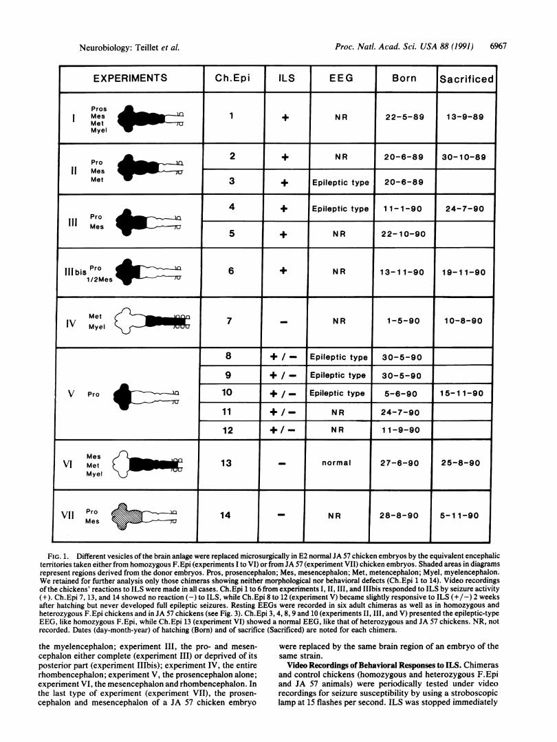
FIG. 1. Different vesicles of the brain anlage were replaced microsurgically in E2 normal JA 57 chicken embryos by the equivalent encephalicterritories taken either from homozygous F.Epi (experiments I to VI) or from JA 57 (experiment VII) chicken embryos. Shaded areas in diagramsrepresent regions derived from the donor embryos. Pros, prosencephalon; Mes, mesencephalon; Met, metencephalon; Myel, myelencephalon.We retained for further analysis only those chimeras showing neither morphological nor behavioral defects (Ch.Epi 1 to 14). Video recordingsof the chickens' reactions to ILS were made in all cases. Ch.Epi 1 to 6 from experiments I, II, III, and IIbis responded to ILS by seizure activity(+). Ch.Epi 7, 13, and 14 showed no reaction (-) to ILS, while Ch.Epi 8 to 12 (experiment V) became slightly responsive to ILS (+/-) 2 weeksafter hatching but never developed full epileptic seizures. Resting EEGs were recorded in six adult chimeras as well as in homozygous andheterozygous F.Epi chickens and in JA 57 chickens (see Fig. 3). Ch.Epi 3, 4, 8, 9 and 10 (experiments II, III, and V) presented the epileptic-typeEEG, like homozygous F.Epi, while Ch.Epi 13 (experiment VI) showed a normal EEG, like that of heterozygous and JA 57 chickens. NR, notrecorded. Dates (day-month-year) of hatching (Born) and of sacrifice (Sacrificed) are noted for each chimera.
the myelencephalon; experiment III, the pro- and mesen-cephalon either complete (experiment III) or deprived of itsposterior part (experiment IIIbis); experiment IV, the entirerhombencephalon; experiment V, the prosencephalon alone;experiment VI, the mesencephalon and rhombencephalon. Inthe last type of experiment (experiment VII), the -prosen-cephalon and mesencephalon of a JA 57 chicken embryo
were replaced by the same brain region of an embryo of thesame strain.Video Recordings of Behavioral Responses to ILS. Chimeras
and control chickens (homozygous and heterozygous F.Epiand JA 57 animals) were periodically tested under videorecordings for seizure susceptibility by using a stroboscopiclamp at 15 flashes per second. ILS was stopped immediately
Neurobiology: Teillet et al.

Proc. Natl. Acad. Sci. USA 88 (1991)
after initiation of convulsions. When convulsions did notoccur, ILS was lengthened up to 3 min in darkness.
Electroencephalogram (EEG) Recordings. For EEG record-ings, adult chickens were anesthetized with equithesin (2.5ml/kg, i.m.) and local analgesia was induced by applicationof small doses of 1% Xylocaine (lidocaine) at the variouspressure points provoked by the stereotaxic frame. Animalswere implanted with electrodes for EEG recordings. Fivestainless steel jeweler's screws (see Fig. 3) were inserted intothe skull on both sides at the anterior and posterior thirds ofthe cerebrum. Screw 4 served as reference electrode. Screwswere then connected to a five-female-pins socket and securedto the skull with dental acrylic cement. EEG recordings weremade under resting conditions following at least a 3-dayrecovery period. Each animal was previously tested forseizure susceptibility with ILS at 15 flashes per second.
RESULTSThree hundred transplants were performed and 14 viablechimeras (Ch.Epi 1 to 14) were retained for further analysis.We kept only those chimeras presenting neither obviousmalformations nor abnormal comportment at hatching (Fig.1). Their growth rates were similar to those ofJA 57 chickens.The chimeras always had feathers with the pigmentation ofthe donor Fayoumi strain at the level of the graft-i.e., eitherwhite or pigmented, but in both cases contrasting with theyellow color of the recipient JA 57 chickens (Fig. 2). Five ofthese birds (Ch.Epi 1 to 5) presented a typical epilepticphenotype. These belonged to experimental series I, II, andIII, involving the graft of at least the forebrain plus themidbrain. As in the case of homozygous F.Epi chickens
(14-18), these chimeras underwent seizures from hatching toadulthood, either spontaneously or under ILS. The ILS-provoked seizures were characterized by the stereotypedbehavior described by Crichlow and Crawford (15). In phase1, usually starting within 20 sec after initiation of ILS, thehead and neck are first slowly rotated and arched back andupward and the animals make pecking motions with excitedvocalizations. Phase 2 is characterized by extension of thewings with some loss of balance. In phase 3, the birds run inall directions, stagger, and then fall on the floor and flap theirwings violently with clonic movements ofthe legs resulting inthrashing and trumbling motions. Phase 3 may continue forseveral minutes. After the seizure, the chimeras, like thehomozygous F.Epi controls, were prostrate and showedtransitory wing paresis. Thereafter they progressively recov-ered a normal behavior. One bird in which the prosenceph-alon was grafted with only the anterior part of the mesen-cephalon (Ch.Epi 6, experiment I11bis) had convulsionsunder ILS a short time after hatching. Experiment VII, inwhich both pro- and mesencephalon were exchanged be-tween two embryos of the JA 57 strain, gave rise to a bird(Ch.Epi 14) showing no clinical sign of epilepsy.The birds in which the rhombencephalon alone (experi-
ment IV) or even both the rhombencephalon and the mes-encephalon (experiment VI) of a homozygous F.Epi embryowere implanted (Ch.Epi 7 and 13) never developed epilepticseizures.
Five chickens (Ch.Epi 8 to 12) carried grafts of the ho-mozygous F.Epi prosencephalon alone (experiment V). Incontrast to the birds of experiments I, II, and III, theyshowed no reaction to ILS during the first 2 weeks afterhatching. Thereafter, however, they all responded to ILS
FIG. 2. Ch.Epi 2 (left) and Ch.Epi 3, three days after hatching' These chimeras were constructed by transplanting jointly into E2 normalchicken embryos the pro-, mes-, and metencephalon extirpated from homozygous F.Epi chicken embryos ofsame stage (experiment II, see Fig.1). Host chickens (JA 57 strain) are yellow at hatching, whereas chickens of the Fayoumi epileptic (F.Epi) strain are either white or variegateddark and light brown. The chimeras show the F.Epi pigmentation in the graft area because neural crest cells were implanted together with thebrain vesicles.
6968 Neurobiology: Teillet et al.

Proc. Natl. Acad. Sci. USA 88 (1991) 6969
with a pattern of symptoms analogous to the first and secoiphases ofepileptic seizure (neck arched back and upward ailoss of balance) but did not progress to phase 3 as in titypical epileptic seizure described above. At the cessationILS, the behavior of these birds returned immediately to iprevious state.EEG recordings were taken in freely moving, awake no
epileptic and epileptic adult chickens during resting perio4and ILS stimulations but never during the full seizure, Iavoid interfering artifacts (Fig. 3). In both JA 57 and Fayourheterozygous carrier chickens, the EEG records were normand made up oflow-amplitude rhythms. A similar pattern wEfound in Ch.Epi 13, which received a graft of mesencephalcplus rhombencephalon (experiment VI). In Ch.Epi 3 and(experiments II and III), which exhibited clinical signs 4epilepsy as in homozygous F.Epi chickens, the interictrecords were characterized by high-amplitude continuouasynchronous slow waves, slow spikes, or spikes and wave,These abnormalities were prominent when the animals wetrelaxed. After any stimulus inducing an arousal reaction, threcord was transiently constituted by low-amplitude farrhythms. ILS produced a similar effect during phase 1 of thseizure, but rapidly the symptoms of the seizure itself did ncallow EEG activity to be distinguished from muscle spikeand movement artifacts. Immediately after the seizure, thlEEG activity was depressed with bursts of slow waveinterrupted by silences. Progressively, the amplitude, frequency, and shape of the waves returned as before theseizure.
2
r'
1 3
2 5
1 3
4
245
1 I f-3
2-.
1-3 F. htz
2-5
1-3 JA 57
2-5
1-3 Ch. Epi 3
*wM4Anff4i2-5
1-2 Ch.Epi 8
3-5
1-3 I F.Epi
2-5 50 /xVL1 sec
FIG. 3. Resting EEG ofa Fayoumi heterozygous chicken (F.htz);a JA 57 chicken, of the recipient strain; two chimeras [Ch.Epi 3(experiment II) and Ch.Epi 8 (experiment V)]; and a homozygousF.Epi chicken. Note the normal resting EEG in the F.htz and JA 57birds as compared with the continuous high-amplitude spikes,polyspikes, and slow wave discharges recorded in the homozygousF.Epi bird and the two chimeras. Although the characteristic spikesand spikes and waves have been recorded in Ch.Epi 8 and in otherchickens of experiment V (see Fig. 1), chimeras of this seriespresented symptoms of only phases 1 and 2 of the F.Epi seizure andnever the full seizure. Electrode positions are indicated in thediagram shown to the left of each pair of traces.
endnd.heofits
)n-dstomialasrn4of:alasS.re
Interestingly, recorded animals carrying the graft of aprosencephalon alone (Ch.Epi 8, 9, and 10, experiment V)had a resting EEG similar to that of homozygous F.Epichickens and of Ch.Epi 3 and 4. Moreover, during thesymptoms analogous to phases 1 and 2 of epileptic seizureinduced by ILS, the EEG showed only a blockade of theparoxysmal abnormalities constituting the EEG backgroundof the homozygous F.Epi chickens. The EEG returned to theprevious state immediately after cessation of ILS.
Eight chimeras were sacrificed (Fig. 1). Their brains weredissected and found similar to those of controls, without anydetectable malformation, thus showing the perfect integra-tion of the graft into the host's nervous system. The others(Ch.Epi 3, 5, 8, 9, 11, and 12) were still alive (see Fig. 1).Ch.Epi 3 (11/2 years old) had seizures well controlled by anadapted treatment with phenobarbital (16).
DISCUSSION AND CONCLUSIONSe These experiments demonstrate that the transfer of a patho-St logical genetic trait affecting the nervous system is possiblee through in situ transplantation of neuroepithelium during)t embryonic life.
After showing that transplantation of the whole brain [i.e.,e the four primitive encephalic vesicles (experiment I)] from anepileptic embryo to a normal embryo leads to the transfer ofthe full disease, implantation of selected regions of thee encephalic vesicles provided the opportunity of investigatingthe role of various neuroepithelial territories in the establish-ment of the seizure phenotype. We have found so far thatonly Ch.Epi birds into which at least both pro- and mesen-cephalon have been implanted presented the full spectrum ofthe epileptic manifestations. In contrast, the bird implantedat the level of the rhombencephalon (i.e., metencephalic andmyelencephalic vesicles, Ch.Epi 7) did not show the epilepticphenotype, nor did that in which rhombencephalon togetherwith mesencephalon were grafted (Ch.Epi 13). Up to theirsacrifice (at ages of about 21/2 and 2 months, respectively),they showed no sign of seizure activity, neither spontane-ously nor under repeated ILS. In contrast, they behaved inthese circumstances like normal JA 57 chickens. Theseresults, together with others previously reported (19, 20) andthe control experiment [grafting of a JA 57 pro- plus mesen-cephalon (Ch.Epi 14)], confirm that grafting by itself does notgenerate epileptic seizures, at least as long as it results innormal brain development.The birds with prosencephalic grafts, although exhibiting
typical interictal paroxysmal EEG, showed only very mildmanifestations under ILS; if they presented epileptic fits,they exhibited only the first and second phases of the seizure,which ceased with the withdrawal of the stimulation. On theother hand, the presence of the mutant mesencephalon(transplanted together with the rhombencephalon in Ch.Epi13) was not sufficient to induce epileptic manifestations. Itwas only when at least prosencephalon and anterior mesen-cephalon were grafted together that the complete neuraldisease was transmitted from a mutant to a normal chicken.The chimera in which both prosencephalon and anterior
mesencephalon were grafted (Ch.Epi 6) is particularly inter-esting. We know from the analysis of quail chicken brainchimeras that the caudal half of mesencephalon participatesin the formation of cerebellum (20). It is striking to see herethat transplantation of the rostral half of the mesencephalontogether with the prosencephalon appears to be necessaryand sufficient to induce the full epileptic phenotype. There-fore, the neuroepithelial territory yielding the cerebellum andcharacterized by expression of the Engrailed gene (26, 27)seems to be excluded from the epileptogenic area of the brainin this system.
Neurobiology: Teillet et al.

Proc. Natl. Acad. Sci. USA 88 (1991)
In conclusion, besides the fact that the F.Epi strain con-stitutes an interesting model to study genetic and pharmaco-logical aspects of "generalized epilepsy" (see ref. 28 for areview), we think that the experimental model developedhere may be a valuable means for investigating the impor-tance of certain discrete zones of the brain in eliciting thedisease in the genetically normal nervous system and body ofthe recipient. It is demonstrated here that neither the pres-ence of the epileptic forebrain characterized by typical in-terictal epileptic EEG nor the presence of a geneticallyepileptic midbrain is a sufficient condition for a completeepilepsy pattern to occur in this model. A cooperation ofthese two brain areas is necessary to generate the fullepileptic phenotype of the homozygous F.Epi chickens.
We are grateful to Dr. R. Crawford for sending us eggs of themutant epileptic strain that he discovered and to G. Coquerelle forestablishing in the Institut National de la Recherche Agronomique(Jouy-en-Josas, France), a colony of the Fayoumi epileptic chickensand providing us with homozygous breeders. We thank E. Bourson,S. Gournet, B. Henri, Y. Rantier, and S. Roy for their help in thepreparation of the manuscript and Profs. Jerome Engel, Jr. (Univer-sity of California, Los Angeles) and Charles Ordahl (University ofCalifornia, San Francisco) for critical reading of the article. Thiswork was supported by Centre National de la Recherche Scienti-fique, Institut National de la Sante et de la Recherche Mddicale,Association pour la Recherche coutre le Cancer, and Fondationpour la Recherche Mddicale.
1. Andermann, E. (1982) in Genetic Basis of the Epilepsies, eds.Anderson, V. E., Hauser, W. A., Penry, J. K. & Sing, C. F.(Raven, New York), pp. 355-374.
2. Andermann, E. (1985) in Genetic Aspects ofHuman Behavior,eds. Sakai, T. & Tsuboi, T. (Igakuhoin, Tokyo), pp. 129-145.
3. Nekhorocheff, M. I. (1950) Rev. Neurol. (Paris) 83, 601-602.4. Metrakos, J. D. & Metrakos, K. (1969) in Basic Mechanisms of
the Epilepsies, eds. Jasper, H. H., Ward, A. A. & Pope, A.(Little, Brown, Boston), pp. 700-708.
5. Doose, H., Gerken, H., Hien-Volpet, K. & Voelzke, E. (1969)Neuropadiatrie 1, 56-73.
6. Doose, H. & Gerken, H. (1973) Neuropadiatrie 4, 162-171.7. Naquet, R. (1975) in Growth and Development ofthe Brain, ed.
Brazier, M. A. B. (Raven, New York), pp. 219-230.
8. Delgado-Escueta, A. V., Greenberg, D. A., Treiman, L., Liu,A., Sparkes, R. S., Barbetti, A., Park, M. S. & Terasaki, P. I.(1989) Epilepsia 30, Suppl. 4, S8-S18.
9. Leppert, M., Anderson, V. E., Quattlebaum, T., Stauffer, D.,O'Connell, P., Nakamura, Y., Lalouel, J.-M. & White, R.(1989) Nature (London) 337, 647-648.
10. Killam, K. F., Killam, E. K. & Naquet, R. (1967) Electroen-ceph. Clin. Neurophysiol. 22, 497-513.
11. Seyfried, T. N. & Glaser, G. H. (1985) Epilepsia 26, 143-150.12. Le Gal La Salle, G. & Naquet, R. (1990) Brain Res. 518,
308-312.13. Silva-Barrat, C. & Menini, C. (1990) in Generalized Epilepsy:
Neurobiological Approaches, eds. Avoli, M., Gloor, P., Kost-opoulos, G. & Naquet, R. (Birkhaser, Boston), pp. 286-297.
14. Crawford, R. D. (1970) J. Hered. 61, 185-188.15. Crichlow, E. & Crawford, R. D. (1974) Can. J. Physiol. Phar-
macol. 52, 424-429.16. Johnson, D. D., Davis, H. L., Bailey, D. G. & Crawford,
R. D. (1977) Can. J. Physiol. Pharmacol. 55, 848-854.17. Johnson, D. D., Davis, H. L. & Crawford, R. D. (1979) Fed.
Proc. Am. Soc. Exp. Biol. 38, 2417-2423.18. Johnson, D. D., Jaju, A. T., Ness, L., Richardson, J. S. &
Crawford, R. D. (1981) Can. J. Physiol. Pharmacol. 59, 144-149.
19. Balaban, E., Teillet, M.-A. & Le Douarin, N. M. (1988) Science241, 1339-1342.
20. Hallonet, M., Teillet, M.-A. & Le Douarin, N. M. (1990)Development 108, 19-31.
21. Le Douarin, N. (1982) The Neural Crest (Cambridge Univ.Press, Cambridge, U.K.).
22. Le Douarin, N. (1969) Bull. Biol. Fr. Belg. 103, 435-452.23. Kinutani, M. & Le Douarin, N. (1985) Dev. Biol. 111, 243-255.24. Kinutani, M., Coltey, M. & Le Douarin, N. M. (1986) Cell 45,
307-314.25. Ohki, H., Martin, C., Corbel, C., Coltey, M. & Le Douarin,
N. M. (1987) Science 238, 1032-1035.26. Gardner, C. A., Darnell, D. K., Poole, S. J., Ordahl, C. P. &
Barald, K. F. (1988) J. Neurosci. Res. 21, 426-437.27. Patel, N. H., Martin-Blanco, E., Coleman, K. G., Poole, S. J.,
Ellis, M. C., Kornberg, T. B. & Goodman, C. S. (1989) Cell 58,955-968.
28. Johnson, D. D. & Tuchek, J. M. (1987) in Neurotransmittersand Epilepsy, eds. Jobe, P. C. & Laird, H. E., II (Humana,Clifton, NJ), pp. 95-114.
6970 Neurobiology: Teillet et al.