Transfection - GENEMOLgenemol.org/genemol/secondesemaine/pictures/Trans.pdf · development of...
60
Transfection G U I D E
Transcript of Transfection - GENEMOLgenemol.org/genemol/secondesemaine/pictures/Trans.pdf · development of...

TransfectionG U I D E

Recommended Promega Transfection Reagent for Commonly-Used Cell Lines.
Cell Cell TransFast™ Tfx™-10 Tfx™-20 Tfx™-50Origin Line Type Reagent Reagent Reagent Reagent
Human HeLa Epithelial XHuman Hep G2 Hepatocyte XHuman 293 Kidney
transformed XHuman K562 Lymphoblast XHuman Jurkat T-cell
leukemia XMonkey COS-7 Fibroblast XMonkey CV-1 Fibroblast XMouse NIH/3T3 Fibroblast XHamster BHK Fibroblast XHamster CHO Epithelial-
like XRat PC12 Pheochromo-
cytoma X XInsect Sf9 Ovary X X*Data were obtained from cells transiently transfected with plasmid DNA and using the Luciferase Assay System from Promega.Higher transfection efficiencies were generally obtained with reagent:DNA complexes incubated with the cells in serum-freemedium. Some cells exhibit similar transfection efficiencies with several different reagents, thus more than one suggested reagentis indicated for these cell lines.

Promega...
...the Source for DiscoveryPromega Corporation is a worldwide leader inapplying biochemistry and molecular biology to thedevelopment of innovative, high-value products forthe life sciences. Products such as the LuciferaseAssay Systema and the Dual-Luciferase™ ReporterAssay Systema,b have helped make Promega theleader in supplying genetic reporter systems for thestudy of eukaryotic gene expression and cellularphysiology.
Upstream from genetic reporting, Promega’s familyof transfection reagents are highly efficient, fast,easy to use and a tested complement to our reportersystems. Our commitment to your success intransfection and eukaryotic expression studies isreflected in the detail found in this guide. The guideis complete with protocols, references andtroubleshooting help, and features a cell line tableon the inside cover to direct you to the Promegatransfection reagent that will perform best withcommonly-used cell lines.
Please visit Promega’s website at
for the most current on-line references, applications,and other product information about our transfectionreagents and reporter systems. Please also look forthe Transfection Assistant to find transfectionconditions used successfully with Promegatransfection reagents. Over 120 cell lines arerepresented. Promega truly is your source fortransfection to detection reagents.
Promega’s Transfection, Reporter Assay Family and Eukaryotic Expression Vectors
Transfection ReagentsTransFast™ Transfection Reagentb
Tfx™-10 Reagentc
Tfx™-20 Reagentc
Tfx™-50 Reagentc
Transfectam® Reagentd for the Transfection of Eukaryotic Cells
ProFection® Mammalian Transfection Systems
• Calcium Phosphate
• DEAE-Dextran
Reporter Vectors and Assay SystemsDual-Luciferase™ Reporter Assay Systema,b
Luciferase Assay Systema
CAT Enzyme Assay System
β-Galactosidase Enzyme Assay System
Luciferase Reporter Vectors
• pGL3 Vectorse,f
Renilla Luciferase Control Vectors
• pRL Vectorsg
CAT Reporter Vectors
pSV-β-Galactosidase Control Vector
Eukaryotic Expression VectorspCI-neo Mammalian Expression Vectorh,i
pCI Mammalian Expression Vectorh
pSI Mammalian Expression Vector
pTARGET™ Mammalian Expression Vector Systemh,j
Transfection Guide 1
aU.S. Pat. Nos. 5,283,179, 5,641,641 and 5,650,289 have been issued to Promega Corporation for a firefly luciferase assay method, which affords greater light output withimproved kinetics as compared to the conventional assay.bPatent Pending.cThe cationic lipid component of the Tfx™ Reagents is covered by U.S. Patent No. 5,527,928 assigned to The Reagents of the University of California and pending foreignpatents.dTransfectam® Reagent is covered by U.S. Patent No. 5,171,678. The Transfectam® product was developed by J.P. Behr and J.P. Loeffler (under license from CNRS-ULPStrasbourg).eU.S. Pat. No. 5,670,356 has been issued to Promega Corporation for a modified luciferase technology.fThe method of recombinant expression of Coleoptera luciferase is covered by U.S. Pat. No. 5,583,024 assigned to The Regents of the University of California.gThe cDNA encoding luciferase from Renilla reniformis is covered by U.S. Pat. No. 5,292,658 assigned to the University of Georgia Research Foundation, Inc., and sublicensed from SeaLite Sciences, Inc., Norcross, GA. The pRL family of Renilla luciferase cDNA vectors is for research use only.hThe CMV vector technology is the subject of U.S. Pat. No. 5,168,062 assigned to the University of Iowa Research Foundation.iU.S. Pat. No. 4,766,072 has been issued to Promega Corporation for transcription vectors having two different bacteriophage RNA polymerase promoter sequences separated by a series of unique restriction sites into which foreign DNA can be inserted.jLicensed under one or both of U.S. Pat. Nos. 5,487,993 and European Pat. No. 0 550 693.
PrefaceT R A N S F E C T I O N T O D E T E C T I O N . . .

Transfection Guide2

Transfection Guide 3
Historical Background. . . . . . . . . . . . . . . . . . . . . . . . . . . . . . . . . . . . . . . . . . . 5Transfection Technologies . . . . . . . . . . . . . . . . . . . . . . . . . . . . . . . . . . . . . . . 6
Preparation of DNA for Transfection . . . . . . . . . . . . . . . . . . . . . . . . . . . . . . . 9Preparation of Cells for Transfection . . . . . . . . . . . . . . . . . . . . . . . . . . . . . . 11Transient Expression vs. Stable Transfection. . . . . . . . . . . . . . . . . . . . . . . 11
Chapter 2
Cationic Lipid Transfection ReagentsTransfectam®, TransFast™ and Tfx™
Reagents for the Transfection of Eukaryotic Cells. . . . . . . . . . . . . . . . . . . 15Liposome-Based Transfection ProtocolsTransFast™ and Tfx™ Reagents . . . . . . . . . . . . . . . . . . . . . . . . . . . . . . . . . . 17Transfection Protocols - Transfectam® Reagentfor the Transfection of Eukaryotic Cells. . . . . . . . . . . . . . . . . . . . . . . . . . . . 25Optimization of Transfection Efficiency. . . . . . . . . . . . . . . . . . . . . . . . . . . . 26
Chapter 3
Introduction. . . . . . . . . . . . . . . . . . . . . . . . . . . . . . . . . . . . . . . . . . . . . . . . . . . 29Calcium Phosphate-Mediated Transfection. . . . . . . . . . . . . . . . . . . . . . . . 29Glycerol or DMSO Shock . . . . . . . . . . . . . . . . . . . . . . . . . . . . . . . . . . . . . . . 30DEAE-Dextran-Mediated Transfection . . . . . . . . . . . . . . . . . . . . . . . . . . . . 31
Chapter 4
General Troubleshooting Tips . . . . . . . . . . . . . . . . . . . . . . . . . . . . . . . . . . . 35Cationic Lipid Reagent Troubleshooting . . . . . . . . . . . . . . . . . . . . . . . . . . 35Calcium Phosphate Transfection Troubleshooting . . . . . . . . . . . . . . . . . . 36DEAE-Dextran Transfection Troubleshooting . . . . . . . . . . . . . . . . . . . . . . 36
Chapter 5
Introduction. . . . . . . . . . . . . . . . . . . . . . . . . . . . . . . . . . . . . . . . . . . . . . . . . . . 39Firefly Luciferase Reporter Gene Systems. . . . . . . . . . . . . . . . . . . . . . . . . 40Dual-Luciferase™ Reporter Assay System. . . . . . . . . . . . . . . . . . . . . . . . . 42CAT Reporter Gene Systems . . . . . . . . . . . . . . . . . . . . . . . . . . . . . . . . . . . . 43β-Galactosidase Reporter Gene System . . . . . . . . . . . . . . . . . . . . . . . . . . 44Mammalian Expression Vectors . . . . . . . . . . . . . . . . . . . . . . . . . . . . . . . . . 45
Chapter 6
Cited References. . . . . . . . . . . . . . . . . . . . . . . . . . . . . . . . . . . . . . . . . . . . . . 47
Cited References
References Using Promega Transfection Reagents. . . . . . . . . . . . . . . . . 49
Appendix A
Ordering Information. . . . . . . . . . . . . . . . . . . . . . . . . . . . . . . . . . . . . . . . . . . 53
Appendix B
Contents
Chapter 1
T A B L E O F C O N T E N T S
A N I N T R O D U C T I O N T O T R A N S F E C T I O N M E T H O D S
P R E P A R A T I O N F O R T R A N S F E C T I O N
C A T I O N I C L I P I D T R A N S F E C T I O N R E A G E N T S
PROFECTION ® MAMMALIAN TRANSFECTION SYSTEMS
T R O U B L E S H O O T I N G T R A N S F E C T I O N R E A C T I O N S
G E N E T I C R E P O R T E R S Y S T E M S
R E F E R E N C E S
A D D I T I O N A L R E F E R E N C E S
O R D E R I N G I N F O R M A T I O N
pCAT, pGEM, PolyATtract, ProFection, RiboClone and Stop & Gloare trademarks of Promega Corporation and are registered withthe U.S. Patent and Trademark Office.
Dual-Luciferase, TransFast, pTARGET and Tfx are trademarks ofPromega Corporation.
Calbiochem is a registered trademark of Calbiochem-Novabiochem Corporation.
Corning is a trademark of Corning, Inc.
Fisherbrand is a registered trademark of Fisher Scientific.
Geneticin is a registered trademark of Life Technologies, Inc.
Kimwipes is a registered trademark of Kimberly Clark Corporation.
Transfectam is a registered trademark of BioSepra, Inc.Transfectam product developed by J.P. Behr and J.P. Loeffler(License of CNRS-ULP Strasbourg).
Product claims are subject to change. Please contact PromegaTechnical Services or access the Promega on-line catalog for themost up-to-date information on Promega products.
Applications mentioned in Promega literature are provided for informational purposes only. Promega does not warrant that referenced applications have been tested in Promega laboratories.
1998 Promega Corporation. All Rights Reserved.Prices and specifications subject to change without prior notice.

Transfection Guide4

Historical BackgroundThe ability to introduce nucleic acids into cells hasenabled the advancement of our knowledge ofgenetic regulation and protein function withineukaryotic cells, tissues and organisms. Thesuccessful pioneering studies of Vaheri and Pagano(1), and Graham and van der Eb (2) with DEAE-dextran and calcium phosphate-mediatedtransfection techniques, paved the way for futureexperiments necessitating DNA transfer intocultured eukaryotic cells. The process of introducingnucleic acids into cells by non-viral methods, suchas the DEAE-dextran and calcium phosphatetechniques, is defined as “transfection”. Thisprocess is distinct from “infection”, which is a viralmethod of nucleic acid introduction into cells.
Progress in transfection technology was relativelyslow until the advent of molecular biologytechniques for cloning plasmid DNA. Thesetechniques provided the means to prepare andmanipulate DNA sequences and the ability toprepare virtually unlimited amounts of relatively pureDNA for transfection experiments. Clonedsequences could also be used to generate RNA invitro with phage RNA polymerase using DNAtemplates with the corresponding polymerasepromoter (3). As the ability to prepare DNA and RNAfor transfection became easier, additional methods,such as electroporation and liposome-mediatedtransfer, were developed to enable more efficienttransfer of the nucleic acids to a broad range ofcultured mammalian cells (4,5).
The development of reporter gene systems andselection methods for stable gene expression oftransferred DNA greatly expanded the applicationsfor gene transfer technology (Figure 1.1). In 1982,Gorman et al. initiated the reporter gene concept
with the bacterial chloramphenicol acetyltransferase(CAT) gene and associated CAT assay system (6).Using a reporter gene that is not endogenous to thecell, coupled with a sensitive assay system for thatgene product, allows investigators to cloneregulatory sequences of interest upstream of thereporter gene to study expression of the reportergene under various conditions. This technology,together with the availability of transfection reagents,provides the foundation for studying promoter andenhancer sequences, trans-acting proteins such astranscription factors, mRNA processing, protein/protein interactions, translation, and recombinationevents (7). Since the introduction of the CAT geneand assay system several other reporter systemshave been developed for various in vitro and in vivoapplications including luciferase, β-galactosidase,alkaline phosphatase and green fluorescent protein(7). See Chapter 6 for detailed descriptions ofPromega’s luciferase, CAT and β-galactosidasereporter vectors and assay systems.
Integration of DNA into the chromosome, or stableepisomal maintenance, of reporter genes and othergenes occurs with a relatively low frequency. Theability to select for these cells is made possibleusing genes that encode resistance to a lethal drug.An example of such a combination is the markergene for neomycin phosphotransferase with thedrug Geneticin® (8). Individual cells that survive thedrug treatment expand into clonal groups that canbe individually selected, propagated and analyzed.
Today the study of gene regulation, the analysis ofthe expression and function of proteins withinmammalian cells, the generation of transgenicorganisms and in vivo/ex vivo gene therapystrategies are all made possible by the availability ofof gene transfer technologies, nucleic acidmolecular biology and reporter gene systems.
Transfection Guide 5
Figure 1.1. Reporter Gene Systems.
RNA in situ β-Galactosidase
Reporter Gene Plasmid DNA
ReporterProteinNucleus
Ribosomes
Cell Lysates
Enzyme ActivityLuciferase
CAT
β-Galactosidase
Chapter 1A N I N T R O D U C T I O N T O T R A N S F E C T I O N M E T H O D S

Transfection TechnologiesMany transfection techniques have been developed.Desirable features include high efficiency transfer ofnucleic acid to the appropriate cellular organelle (forexample, DNA into the nucleus), minimal intrusion orinterference with normal cell physiology, low toxicity,ease of use, reproducibility, successful generation ofstable transfectants, and in vivo efficacy. Thetechniques developed for gene transfer can bebroadly classified as either chemical reagents orphysical methods.
Chemical ReagentsDEAE-dextran was one of the first chemical reagentsused for transfer of nucleic acids into culturedmammalian cells (1,9). The ProFection® MammalianTransfection System-DEAE-Dextran providesreagents for this transfection technique (see Chapter4 for further information). DEAE-dextran is a cationicpolymer that associates with negatively chargednucleic acids. An excess of positive charge,contributed by the polymer in the DNA/polymercomplex allows the complex to come into closerassociation with the negatively charged cellmembrane. Uptake of the complex is presumably byendocytosis. This method is successful for delivery
of nucleic acids into cells for transient expression;that is, for short-term expression studies of a fewdays in duration. However, this technique is notgenerally useful for stable transfection studies thatrely upon integration of the transferred DNA into thechromosome (10). Other synthetic cationic polymershave been used for the transfer of DNA into cells,including polybrene (11), polyethyleneimine (12)and dendrimers (13,14).
Calcium phosphate co-precipitation became apopular transfection technique following thesystematic examination of this method by Grahamand van der Eb in the now-classic paper publishedin 1972 (2). Their study examined the effect ofdifferent cations, cationic and phosphateconcentrations, and pH on the parameters oftransfection. Calcium phosphate co-precipitation iswidely used because the components are easilyavailable and reasonable in price, the protocol iseasy to use and many different types of culturedcells can be transfected. This method is routinelyused for both transient and stable transfection of avariety of cell types. The protocol involves mixingDNA with calcium chloride, adding this in acontrolled manner to a buffered saline/phosphatesolution and allowing the mixture to incubate at roomtemperature. This step generates a precipitate that
Transfection Guide6
Figure 1.2. Schematic representation of various transfection technologies based on chemical reagents.
DEAE-Dextran
Artificial Liposomes
+
+
+++
+++
+
++
+
+ ++
+
++
++
+
+++
CalciumPhosphate
Ca++
Ca++
Ca++
Ca++
Ca++
––
––
–
––
––
–
DNA
Ca++
––
––
–
––
––
–
DNA
––
––
–
––
––
–
DNA
+
+
+
++
+++
+
++
++
+
+
+ + + ++
+
++
++
+
+++
Chapter 1A N I N T R O D U C T I O N T O T R A N S F E C T I O N M E T H O D S

is dispersed onto the cultured cells. The precipitateis taken-up by the cells via endocytosis orphagocytosis. The calcium phosphate also appearsto provide protection against intracellular and serumnucleases (15). Promega’s ProFection® MammalianTransfection System-Calcium Phosphate providesreagents for this transfection technique (see Chapter4 for further information).
By 1980, artificial liposomes were being used todeliver DNA into cells (5). The next advancement inliposomal vehicles was the development of syntheticcationic lipids by Felgner and colleagues (16).Liposome-mediated delivery offers advantagessuch as relatively high efficiency of gene transfer,ability to transfect certain cell types that areintransigent to calcium phosphate or DEAE-dextran,successful delivery of DNA of all sizes fromoligonucleotides to yeast artificial chromosomes (16-20), delivery of RNA (21), and delivery of protein(22). Cells transfected by liposome techniques canbe used for transient and for longer termexperiments that rely upon integration of the DNAinto the chromosome or episomal maintenance.Unlike the DEAE-dextran or calcium phosphatechemical methods, liposome-mediated nucleic aciddelivery can be used for in vivo transfer of DNA andRNA to animals and humans (23).
A lipid with overall net positive charge atphysiological pH is the most common synthetic lipidcomponent of liposomes developed for genedelivery (Figure 1.3). Often the cationic lipid is mixedwith a neutral lipid such as L-dioleoyl phosphatidyl-ethanolamine (DOPE) (Figure 1.4). The cationicportion of the lipid molecule associates with thenegatively charged nucleic acids, resulting incompaction of the nucleic acid in a liposome/nucleicacid complex. For cultured cells, an overall netpositive charge of the liposome/nucleic acid complexgenerally results in higher transfer efficiencies,presumably because this allows closer association of the complex with the negatively charged cellmembrane. Following endocytosis, the complexesappear in the endosomes, and later in the nucleus. It is unclear how the nucleic acids are released fromthe endosomes and traverse the nuclear membrane.DOPE is considered a “fusogenic” lipid (24) and it isthought that its role may be to release thesecomplexes from the endosomes, as well as tofacilitate fusion of the outer cell membrane with the liposome/nucleic acid complexes.
Promega provides a variety of transfection reagentsthat use cationic lipids for the delivery of nucleicacids to eukaryotic cells. These include TransFast™Transfection Reagent, the Tfx™ Reagents andTransfectam® Reagent. See Chapter 3 for moreinformation on the use of these reagents.
Physical MethodsDirect microinjection into cultured cells or nuclei is an effective, although laborious technique to delivernucleic acids into cells. This method has been usedto transfer DNA into embryonic stem cells that areused to produce transgenic organisms (25).However, this technique is not appropriate for studiesthat require a large number of transfected cells.
Electroporation was first reported for gene transferstudies in 1982 (4). This technique is often used for cell types such as plant protoplasts that areparticularly recalcitrant to milder methods of genetransfer. The mechanism for entry into the cell isbased upon perturbation of the cell membrane byan electrical pulse, which forms pores that allow thepassage of nucleic acids into the cell (26). Thetechnique requires fine-tuning and optimization forduration and strength of the pulse for each type ofcell used. A critical balance must be achievedbetween conditions that allow efficient delivery andconditions that kill cells.
Another physical method of gene delivery is biolistic particle delivery. This method relies upon highvelocity delivery of nucleic acids on microprojectiles to recipient cells (27). This method has beensuccessfully employed to deliver nucleic acid tocultured cells, as well as to cells in vivo (28).
Transfection Guide 7
Figure 1.3. General structure of a synthetic cationic lipid.
O
O
O
C
CN
O
+
CationicHeadGroup
LipidLink
Figure 1.4. Structure of DOPE (L-dioleoyl phosphatidylethanolamine).
H3N
O
OO
P
O
O
O-
O
O
C
C+ H
O
Chapter 1A N I N T R O D U C T I O N T O T R A N S F E C T I O N M E T H O D S

Notes
Transfection Guide8
Chapter 1A N I N T R O D U C T I O N T O T R A N S F E C T I O N M E T H O D S

Preparation of DNA for TransfectionThe quality of the DNA used for transfection iscritical. Purified plasmid DNA should be free fromprotein, RNA and chemical contamination. DNA maybe purified using a plasmid preparation protocol, aCsCl gradient, or column chromatography. Onemeasure of DNA purity is the ratio of absorbance at260 to 280nm; for transfection the A260:A280 ratioshould be at or above 1.8. The purified DNA shouldbe ethanol precipitated and resuspended in sterileTE buffer to a final concentration of approximately1mg/ml. The optimal amount of DNA to use fortransfection depends on both the cell type and thereagent used.
Plasmid Preparation ProtocolWe have successfully purified transfection qualityplasmid DNA using a modified alkaline lysis protocol(29). In the following procedure, membrane lipidsare solubilized using SDS. Sodium hydroxide is usedto denature and break up a large amount of thechromosomal DNA, which is then precipitated byaddition of potassium acetate. Treatment with RNase Aand ammonium acetate removes ribonucleic acids(30). Polyethylene glycol (PEG) is used to furtherpurify the plasmid DNA by precipitating it away fromother contaminating material (30). Any remainingproteins and oligosaccharides are removed by a highsalt phenol extraction; the acid phenol extractionserves to remove residual chromosomal and nickedplasmid DNA.
All reagents used should be molecular biologygrade and solutions should be freshly prepared fromreliable, nuclease-free stocks.
Materials to Be Supplied by the User(Solution compositions are provided at the end ofthis chapter.)
• 25mM Tris-HCl, 50mM EDTA• 0.1M NaOH, 1% SDS • 5M potassium acetate• TE (pH 8.0)• 5M ammonium acetate• 5M NaCl• PEG precipitation solution• DNase-free RNase• chloroform:isoamyl alcohol (24:1)• sterile water• 2M sodium acetate• high salt phenol• acid phenol• Miracloth (Calbiochem®)• 2-propanol• 100% ethanol• 70% ethanol• sterile nuclease-free water
1. Harvest the bacterial cells from a 1 literovernight culture by centrifugation at 6,000 x gfor 10 minutes. If necessary, the cell pellet maybe stored at –20°C or –70°C.
2. Resuspend the pellet in 50ml of 25mM Tris-HCl(pH 8.0), 50mM EDTA.
3. Add 100ml of freshly prepared 0.1M NaOH, 1%SDS; mix gently by swirling the container for ~15seconds. Do not vortex. Incubate for 10 minuteson ice.
4. Add 75ml of ice-cold 5M potassium acetate. Mix gently and incubate on ice for 5 minutes. A precipitate will form.
5. Centrifuge at 6,000 x g for 15 minutes. Filter the supernatant through Miracloth or through 4 layers of cheesecloth.
6. Add 135ml of 2-propanol, mix and incubate atroom temperature for 30 minutes.
7. Centrifuge at 6,000 x g for 15 minutes. Decantand discard the supernatant.
8. Resuspend the pellet in 20ml TE (pH 8.0). Add20ml 5M ammonium acetate. Incubate on ice for20 minutes.
9. Centrifuge at 12,000 x g for 10 minutes. Decantsupernatant into a fresh tube.
10. Add 80ml of 100% ethanol to the supernatant.Incubate on ice for 15 minutes. Centrifuge at12,000 x g for 10 minutes.
11. Dissolve pellet in 2ml TE (pH 8.0). Add 20µl of10mg/ml DNase-free RNase. Incubate for 15minutes at 37°C.
12. Add 600µl 5M NaCl and 650µl PEG precipitationsolution. Mix and incubate on ice for 30 minutes.Centrifuge at 12,000 x g for 15 minutes at 4°C.Discard the supernatant. Drain the pellets byinverting the tubes onto paper towels and blotthe rim of the tube with a Kimwipes® tissue or apaper towel.
13. Dissolve the pellet in 1ml TE (pH 8.0). Extract the remaining PEG by adding an equal volumeof chloroform:isoamyl alcohol (24:1). Mix well by inversion and spin in a microcentrifuge for 5 minutes (or 1,600 x g for 10 minutes if usinganother rotor).
14. Remove the upper aqueous phase to freshtubes. Add NaCl to a final concentration of 0.5M(a total of 100µl of 5M NaCl). Extract with anequal volume of high salt phenol. Spin for 5minutes in a microcentrifuge tube. Remove theupper, aqueous phase to a fresh tube.
Transfection Guide 9
Chapter 2P R E P A R A T I O N F O R T R A N S F E C T I O N

15. Add two volumes of 100% ethanol. Incubate onice for 15 minutes. Spin for 10 minutes in amicrocentrifuge (20 minutes at 12,000 x g).Discard the supernatant and drain the pelletsbriefly by inverting the tube onto paper towels.
16. Dissolve pellets in a total of 960µl water. Add15µl of 5M NaCl and 25µl of 2M sodium acetate(pH 4.0). Extract with an equal volume of acidphenol (31). Centrifuge for 5 minutes at roomtemperature in a microcentrifuge (phenol maycrystallize at colder temperatures).
17. Extract any remaining phenol by adding anequal amount of chloroform:isoamyl alcohol(24:1). Invert to mix and centrifuge for 5 minutesin a microcentrifuge. Remove the upperaqueous phase to a fresh tube.
18. Add two volumes of 100% ethanol. Incubate for20 minutes on ice or store overnight at –20°C.Spin for 10 minutes in a microcentrifuge.Discard the supernatant.
19. Wash the pellet by adding 70% ethanol.Centrifuge for 10 minutes in a microcentrifuge.Carefully remove the supernatant withoutdisturbing the pellet. Dry the pellet briefly undervacuum.
20. Resuspend the DNA in 600µl of sterile,nuclease-free TE. Determine the exact DNAconcentration by measuring the absorbance at260nm. Run an aliquot on a 0.7% agarose gelstained with ethidium bromide to check for thesize, purity and integrity of the purified plasmidDNA.
The above protocol is time consuming, butgenerates high quality DNA that works well intransfections. Alternatively, DNA purified by thealkaline lysis method may be further purified using acesium chloride (CsCl) gradient.
Cesium Chloride Equilibrium GradientThe CsCl equilibrium centrifugation methodproduces transfection quality DNA. Standardprotocols for this procedure can be found inreferences 29 and 30. In this procedure, a high-speed centrifugation step follows a crude DNAisolation protocol such as the alkaline lysisprocedure (29). Ethidium bromide (EtBr; a mutagen)is then added to the DNA along with CsCl and themixture is centrifuged to equilibrium. The DNA“band” is removed, leaving many contaminantsbehind. A second EtBr and CsCl centrifugationremoves additional protein and RNA contaminants.Once the DNA has been isolated, both EtBr andCsCl must be removed. EtBr may be removed using a Dowex AG50 column or by extraction with 1-butanol. The DNA must then be dialyzed orethanol precipitated and washed thoroughly with70% ethanol to remove excess CsCl. It is importantto remove residual CsCl ions as they can react withcharged liposomes or other transfection reagents,making transfection less effective. The purified DNAis resuspended in TE buffer.
Anion Exchange ChromatographyColumn chromatography is by far the quickestmethod of preparing high quality plasmid DNAsuitable for transfection. However, care must betaken in choosing the type of column used, as somecommercially available columns leave contaminantsin the DNA preparation that adversely affecttransfection efficiency, sometimes to a dramaticdegree.
Transfection Guide1 0
Chapter 2P R E P A R A T I O N F O R T R A N S F E C T I O N

Preparation of Cells for Transfection
Trypsinization Procedure forRemoving Adherent CellsTrypsinizing cells for purposes of subculturing or cellcounting is an important technique that is critical tosuccessful cell culture. The following techniqueworks consistently well when passaging cells.
Materials to Be Supplied by the User(Solution compositions are provided at the end ofthis chapter.)
• 1X trypsin-EDTA solution (0.05% trypsin, 0.5mMEDTA)
1. Prepare a sterile trypsin-EDTA solution in acalcium- and magnesium-free salt solution suchas 1X PBS or 1X HBSS. The 1X solution can befrozen and thawed for future use, but the activityof the trypsin will decline with each freeze-thawcycle. The trypsin-EDTA solution may be storedfor up to 1 month at 4°C.
2. Remove the media from the tissue culture dish.Add enough PBS or HBS solution to cover thecell monolayer: 2ml for a 150mm flask, 1ml for a 100mm plate. Rock the plates to distribute thesolution evenly. Remove and repeat the wash.Remove the final wash. Add enough trypsinsolution to cover the cell monolayer.
3. Place the plates in a 37°C incubator until thecells just begin to detach (usually 1-2 minutes).
4. Remove the flask from the incubator. Strike thebottom and sides of the culture vessel sharplywith the palm of your hand to help dislodge theremaining adherent cells. View the cells under a microscope to check whether all cells havedetached from the growth surface. If necessary,the cells may be returned to the incubator for anadditional 1-2 minutes.
5. When all cells have detached, add mediacontaining serum to the cells to inactivate thetrypsin. Gently pipet the cells up and down tobreak up cell clumps. The cells may then becounted using a hemacytometer and/ordistributed to fresh plates for subculturing.
Transient Expression vs. Stable Transfection
Transient ExpressionCells are typically harvested 48-72 hours post-transfection for studies designed to analyzetransient expression of the transfected genes. The optimal time interval depends upon the celltype, the doubling time of the cells and the specificcharacteristics of expression for the transferredgene. Analysis of gene products may requireisolation of RNA or protein for enzymatic activityassays or immunoassays. The method used for cellharvest will depend upon the end-product beingassayed.
Extracts may be prepared using Promega’s ReporterLysis Buffer. This allows extracts to be assayed forluciferase, CAT and β-galactosidase activity. If onlyluciferase activity is to be assayed, Promega’s CellCulture Lysis Reagent may be used. Passive LysisBuffer is best for the Dual-Luciferase™ ReporterAssay System. For further information on the pre-paration and assay of cell extracts, see Chapter 6.
Stable TransfectionThe goal of stable, long-term transfection is to isolate and propagate individual clones containingtransfected DNA. Therefore it is necessary todistinguish nontransfected cells from those that have taken up the exogenous DNA. This screeningcan be accomplished by drug selection when anappropriate drug resistance marker is included inthe transfected DNA. Alternatively, morphologicaltransformation can be used as a selectable trait incertain cases. For example, bovine papilloma virusvectors produce a morphological change intransfected mouse CI127 cells (32).
Typically, cells are maintained in nonselective mediumfor 1-2 days post-transfection, then trypsinized andreplated in selective medium containing the drug.The use of the selective medium is continued for 2-3 weeks, with frequent changes of medium toeliminate dead cells and debris, until distinctcolonies can be visualized. Individual colonies arethen trypsinized and subcloned to multiwell platesfor further propagation in the presence of selectivemedium.
Transfection Guide 1 1
Chapter 2P R E P A R A T I O N F O R T R A N S F E C T I O N

Several different drug selection markers arecommonly used for long-term transfection studies.For example, cells transfected with recombinantvectors containing the bacterial gene for neomycinphosphotransferase can be selected for stabletransformation in the presence of the neomycinanalog G418 (8). Similarly, expression of the genefor hygromycin B phosphotransferase from thetransfected vector will confer resistance to the drughygromycin B (33).
An alternative strategy is to use a vector carrying anessential gene that is defective in a given cell line.For example, CHO cells deficient in expression ofthe dihydrofolate reductase (DHFR) gene surviveonly in the presence of added nucleosides.
However, these cells, when stably transfected withDNA expressing the DHFR gene, will synthesize therequired nucleosides (34). An additional advantageof using DHFR as a marker is that gene amplificationof DHFR and associated transfected DNA occurswhen cells are exposed to increasing doses ofmethotrexate (35).
Before using a particular drug for selectionpurposes, it is important to determine the amount of drug necessary to kill the cells you will be using. This may vary greatly between cell types. Designexperiments using various concentrations of thedrug to determine the amount to use for selection of resistant clones (29).
Transfection Guide1 2
Chapter 2
Figure 2.1. Schematic representations of stable and transient transfections.
DNA sample
Assay forGene Expression
Transient Transfection
Day 1 Day 3 or 4
Apply SelectivePressure
Select clonal cells that stably replicate and express transfected DNA.
Stable Transfection
Day 2 or 3 2-3 Weeks
DNA sample
Day 1
P R E P A R A T I O N F O R T R A N S F E C T I O N

Composition of Buffers and Solutions5M ammonium acetateDissolve 385g of ammonium acetate in 1 literdistilled water. Filter through a 0.2µm filter. Store at 4°C.
DNase-free RNase APrepare a 10mg/ml stock solution of PancreaticRNase A in 10mM Tris-HCl (pH 7.5), 15mM NaCl.Aliquot to tubes and heat in a boiling water bath for15 minutes. Cool slowly to room temperature. Storeat –20°C.
1X HBSS (Hanks Balanced Salt Solution)5mM KCl
0.3mM KH2PO4138mM NaCl
4mM NaHCO30.3mM Na2HPO45.6mM D-glucose
The final pH should be 7.1
1X PBS137mM NaCl2.7mM KCl4.3mM Na2HPO4
1.47mM KH2PO4
The final pH should be 7.1
PEG Precipitation Solution (30% PEG-8000, 1.5M NaCl)300g PEG 8000
(molecular biology grade)300ml 5M NaCl
Add deionized water to a final volume of 1L. This solution may have to be heated slightly tocompletely dissolve the PEG. Store at 4°C.
Acid Phenol (phenol saturated with TE + 50mM sodium acetate)Phenol is caustic; work in a chemical safety hoodand wear protective safety equipment. Melt phenolby placing in a 50°C or warmer water bath. Add anequal volume of 50mM sodium acetate (pH 4.0). ThepH of the sodium acetate solution is important. Stirwith a Teflon-coated magnetic stir bar until the twophases become completely mixed. Stop stirring andallow the phases to separate. Remove and discardthe top aqueous phase. Add 50mM sodium acetate(pH 4.0), mix and allow the phases to separate.Remove the aqueous phase. Repeat two more timesor until the pH of the aqueous phase after extractionis between 4.0 and 4.2. Add back 1/10 volume of50mM sodium acetate (pH 4.0) to the phenol. Storeprotected from light at 4°C.
High Salt Phenol (phenol saturated with TE + 0.5M NaCl)Phenol is caustic; work in a chemical safety hoodand wear protective safety equipment. Melt phenolby placing in a 50°C or warmer water bath. Add anequal volume of TES (TE + 1/10 volume of 5M NaCl).Stir with a Teflon-coated magnetic stir bar until thetwo phases become completely mixed. Stop stirringand allow the phases to separate. Remove anddiscard the top aqueous phase. Add TES, mix, andallow the phases to separate; remove the aqueousphase. Repeat two more times. Add back 1/10volume of TES to the phenol. Store protected fromlight at 4°C.
5M Potassium Acetate Solution60ml 5M potassium acetate
11.5ml glacial acetic acid28.5ml deionized water
Store at 4°C. This solution is 3M with respect topotassium and 5M with respect to acetate.
2M Sodium Acetate, pH 4.015g NaOH
115ml deionized water115ml glacial acetic acid
Dissolve the NaOH slowly in 115ml of water. Slowlyadd the glacial acetic acid. Adjust the final volume to1L with deionized water. The final pH of the solutionshould be 4.0. This solution provides a 40X stock forthe 50mM sodium acetate solution used to prepareacid phenol.
TE10mM Tris-HCl (pH 8.0)1mM EDTA
1X Trypsin-EDTA solution0.05% (w/v) trypsin
0.53mM EDTA
Dissolve in a calcium- and magnesium-free saltsolution such as 1X PBS or 1X HBSS.
Transfection Guide 1 3
Chapter 2P R E P A R A T I O N F O R T R A N S F E C T I O N

Transfection Guide1 4
Notes
Chapter 2P R E P A R A T I O N F O R T R A N S F E C T I O N

Cationic Lipid Transfection Reagents -Transfectam®, TransFast™ and Tfx™ Reagents for the Transfection of Eukaryotic Cells
Introduction to Promega’s Cationic Lipid ReagentsPromega provides three types of cationic lipid-based transfection reagents, Transfectam® Reagent,TransFast™ Reagent and the Tfx™-10, Tfx™-20 andTfx™-50 Reagents. The cationic lipid component ofthese reagents associates with negatively chargednucleic acids, resulting in a lipid/nucleic acidcomplex that has a net neutral or positive chargeand therefore allows closer association of the DNAwith the negatively charged cell membrane.
Transfectam® Reagent for the Transfection ofEukaryotic Cells is a cationic lipid reagent consistingof dioctadecylamidoglycyl spermine (DOGS), asynthetic, cationic lipopolyamine. The sperminegroup is covalently attached through a peptide bondto the lipid moiety (Figure 3.1). The strong positivecharge contributed by the spermine headgroupgives the molecule a high affinity for DNA (105-106M –1). The positively charged headgroupeffectively coats the negatively charged DNA with acationic lipid layer, allowing it to fuse with the plasmamembrane of eukaryotic cells, resulting ininternalization of the DNA.
Liposome Based Transfection ReagentsThe term “liposome” refers to lipid bilayers that formcolloidal particles in an aqueous medium (36).Liposome reagents specifically designed fortransfection applications incorporate syntheticcationic lipids (16), often formulated together withthe neutral lipid DOPE (Figure 1.4), which has beendemonstrated to enhance the gene transfer ability ofcertain synthetic cationic lipids (37,38).
Incubation of cationic lipid-containing liposomesand nucleic acids results in quick association and acompaction of the nucleic acid (39,40), presumablyfrom electrostatic interactions between thenegatively charged nucleic acid and the positivelycharged head group of the synthetic lipid. Entry ofthe liposome complex into the cell may occur by theprocesses of endocytosis, or fusion with the plasmamembrane via the lipid moieties of the liposome(41). Once inside the cell, the complexes oftenbecome trapped in endosomes and lysosomes.Endosomal disruption is facilitated by DOPE (24),which allows the complexes to escape into thecytoplasm. The cytoplasm is the site of action forRNA or anti-sense oligonucleotides delivered via theliposomes. The nucleus is the target for most DNAdelivery and it is not known precisely how thetransfected DNA or liposome/DNA complex gainsentry to the nucleus.
Promega’s TransFast™ and Tfx™ Reagents facilitateliposome-mediated transfer of nucleic acids intoeukaryotic cells. The TransFast™ Reagent iscomposed of the synthetic cationic lipid, N,N [bis (2-hydroxyethyl)]-N-methyl-N-[2,3 di(tetradecanoyloxy)propyl] ammonium iodide (Figure 3.2a) and theneutral lipid, (DOPE) (Figure 1.4).
The Tfx™ Reagents contain a mixture of a synthetic,cationic lipid molecule [N,N,N’,N’-tetramethyl-N,N’-bis)2-hydroxy-ethyl)-2,3,-dioleoyloxy-1,4-butanediammonium iodide] (Figure 3.2b) and DOPE(Figure 1.4). All of the Tfx™ Reagents (Tfx™-10, Tfx™-20, and Tfx™-50) contain the same concentration ofthe cationic lipid component, but contain differentmolar ratios of the fusogenic lipid, DOPE.
The best transfection reagent and conditions for aparticular cell type must be empirically andsystematically tested because inherent properties ofthe cell influence the success of any specifictransfection method.
Transfection Guide 1 5
Figure 3.1. Structure of Transfectam® Reagent.
+NH3
ON C
NH
OC
+NH2
+NH2
+NH3
Figure 3.2b. Structure of the synthetic cationic lipid component of the Tfx™ Reagents.
HO
HO
N
NI
I
O
OO
O C
C–
–+
+
Figure 3.2a. Structure of the synthetic cationic lipid component of the TransFast™ Reagent.
HO
HO
CH3
N
I
O
O
OO–
+
Chapter 3C A T I O N I C L I P I D T R A N S F E C T I O N R E A G E N T S

Advantages of Using Cationic Lipid Reagents for TransfectionCationic lipid reagents designed for transfectionapplications are more versatile than many othertraditional methods. The advantages includeversatility in the macromolecules delivered, in vitroand in vivo applications, ability to reproduciblytransfect cells that are recalcitrant to other methods,and suitability for transient and stable transfectionparadigms. For example, several different types ofmacromolecules can be delivered to cells usingthese methods, including RNA and DNA of all sizesranging from oligonucleotides to plasmids and yeastartificial chromosomes (17-21,42).
TransFast™ Reagent, Transfectam® Reagent and theTfx™ Reagents offer the advantages that they are easyto optimize and work well for a variety of cell types(43-46). In addition, these reagents are excellent foruse with primary cells as they can be used in thepresence of serum, can be used for both transientand stable transfections and are of low toxicity.
The Tfx™ and Transfectam® Reagents can also beused for in vivo transfection (47-49). It has beenshown that Tfx™-50 Reagent is highly active in thepresence of amniotic fluid (50), which has implicationsfor its use in intra-amniotic injection and transfection.
Factors Influencing Transfection EfficiencyWith any transfection reagent or method, cell health, degree of confluency, passage number,contamination, and DNA quality and quantity areimportant parameters that can greatly influencetransfection efficiency. Plasmid DNA fortransfections should be free of protein, RNA andchemical contamination (See Chapter 2). Suspendethanol-precipitated DNA in sterile water or TE bufferto a final concentration of 0.2-1mg/ml. The optimalamount of DNA to use in the transfection will varywidely depending upon the type of DNA and thetarget cell line.
It is essential to optimize specific transfectionconditions to gain optimal transfection efficiencies.
Transfection Guide1 6
Figure 3.3. Relative levels of gene expression as a function of Tfx™ Reagent, DNA amount and reagent:DNA charge ratio. CHO Cells (Panel A), HeLacells (Panel B), BHK cells (Panel C) and 293 cells (Panel D) were plated at a density of 50,000 cells/well in 24 well plates. Transfections wereperformed in the absence of serum using the indicated Tfx™ Reagent and pGL3-Control Vector at reagent:DNA ratios of 2:1 and 4:1. Alltransfections were overlaid with serum-containing media after one hour, and cells were harvested for luciferase assays after 48 hours. Theresults represent the mean of 6 replicates and are expressed as relative light units per well of cells. The single Tfx™-50 Reagent conditionsreflect the optimal DNA amount and reagent:DNA ratio determined from previous optimization experiments.
25
4:1 Ratio 2:1 Ratio
4:1 Ratio 2:1 Ratio 4:1 Ratio 2:1 Ratio
4:1 Ratio 2:1 Ratio
125 250 500 1,000 125 250 500 1,000 1,000 125 250 500 1,000 125 250 500 1,000 250
50
45
45
40
35
30
25
20
15
10
5
0
40
35
30
25
20
15
10
5
0
180
160
140
120
100
80
60
40
20
0
20
15
10
5
0
Rela
tive
Ligh
t Uni
ts (x
10-4
)
Rela
tive
Ligh
t Uni
ts (x
10-2
)
Rela
tive
Ligh
t Uni
ts (x
10-4
)
Rela
tive
Ligh
t Uni
ts (x
10-2
)
ng DNA
Tfx™-10 Tfx™-20 Tfx™-50
125 250 500 1,000 125 250 500 1,000 500 125 250 500 1,000 125 250 500 1,000 1,000
ng DNA
Tfx™-10 Tfx™-20 Tfx™-50
ng DNA
Tfx™-10 Tfx™-20 Tfx™-50
ng DNA
Tfx™-10 Tfx™-20 Tfx™-50
A. CHO Cells
C. BHK Cells D. 293 Cells
B. HeLa Cells
Chapter 3C A T I O N I C L I P I D T R A N S F E C T I O N R E A G E N T S

The important parameters to optimize in order tomaximize transfection efficiencies are the chargeratio of transfection reagent to DNA, the amount oftransfected DNA, the length of time the cells areexposed to the transfection reagent and thepresence or absence of serum. Figure 3.3 shows anexample of optimization experiments for 4 differentcell lines using different amounts of pGL3 ControlDNA, different reagent:DNA charge ratios and thethree different Tfx™ Reagents. Expression ofluciferase activity from the transfected DNA isindicated in relative light units on the Y-axis. Thegraphs show the results of comparisons among thethree Tfx™ Reagents: for CHO cells, Tfx™-10Reagent and 500ng DNA at a 2:1 reagent:DNAcharge ratio was most effective; for HeLa cells, Tfx™-20 Reagent and 250ng DNA at a 2:1 chargeratio was most effective; for BHK cells Tfx™-10Reagent and 1,000ng DNA at a 2:1 ratio was mosteffective; and for 293 cells Tfx™-20 Reagent and500ng of DNA at a 2:1 Reagent:DNA ratio was mosteffective. It should be noted that Figure 3.3 is acomparison of the performance of the three Tfx™
Reagents in these cell lines. For CHO and 293 cells,TransFast™ Reagent has been found to performbetter than Tfx™ Reagents.
The transfection efficiency achieved using all ofPromega’s cationic lipid-based transfectionreagents varies depending on the cell type beingtransfected and the transfection conditions used.
Liposome Based Transfection Protocols -TransFast™ and Tfx™ Reagents
General ConsiderationsCharge Ratio of Transfection Reagent to DNAThe amount of positive charge contributed by thecationic lipid component of the transfection reagentshould equal or exceed the amount of negativecharge contributed by the phosphates on the DNAbackbone, resulting in a net neutral or positive chargeon the multilamellar vesicles associating with theDNA. Charge ratios of 2:1 to 4:1 Tfx™ Reagent:DNAand 1:1 to 2:1 TransFast™ Reagent:DNA have workedwell with various cultured cells but ratios outside ofthis range may be optimal for other cell types orapplications. Each of the Tfx™ Reagents contains thesame amount of cationic lipid (1mM when thecontents of each vial are resuspended in therecommended 400µl volume), but contains varyingamounts of the neutral lipid component, DOPE.
DNAThe optimal amount of DNA to use in the transfectionwill vary depending upon the type of DNA and thetarget cell line used. For example, HeLa cells areoptimally transfected with 0.25µg of pGL3-ControlDNA using Tfx™-20 Reagent while NIH/3T3 cells areoptimally transfected with TransFast™ Reagent. Foradherent cells, we recommend initially testing 0.25,0.50, 0.75 and 1µg of DNA in a 24 well plate formatat a transfection reagent:DNA ratio of 3:1 for each ofthe Tfx™ Reagents and at transfection reagent:DNAratios of 2:1 and 1:1 for TransFast™ Reagent.Increasing the amount of DNA does not necessarilyresult in higher transfection efficiencies.
TimeThe optimal transfection time is dependent upon thecell line and DNA used. For the first tests, use a onehour transfection interval. However, in optimizationexperiments, test transfection times from 30 minutesto 4 hours. Monitor cell morphology during thetransfection interval, particularly when the cells aremaintained in serum-free medium, as some cell lineslose viability under these conditions. Thetransfection time with the TransFast™ and Tfx™
Reagents is usually significantly shorter than thatrequired with other cationic lipid compounds, andcan be decreased to as little as 30 minutes withcertain cell lines (Figure 3.4). In addition to savingtime, this shortened transfection time maysignificantly reduce the risk of cell death during thetransfection procedure.
Transfection Guide 1 7
Figure 3.4. Effect of transfection interval on transfection of CHO cellsusing TransFast™ Reagent. CHO cells were transfected with 250ng ofpGL3-Control DNA using TransFast™ Reagent at a 2:1 reagent:DNA charge ratio for various times in the absence of serum. Alltransfections were performed in 24 well plates and cell lysates wereharvested 2 days post-transfection. The results represent the meanof 6 replicates and are expressed as relative light units per well.
300,000
250,000
200,000
150,000
100,000
50,000
Rela
tive
Ligh
t Uni
ts
Transfection Interval
30 Minutes 1 Hour 2 Hours 4 Hours
Chapter 3C A T I O N I C L I P I D T R A N S F E C T I O N R E A G E N T S

SerumTransfection protocols often require serum-freeconditions for optimal performance, as serum caninterfere with many commercially availabletransfection reagents. The TransFast™ and Tfx™
Reagents can be used in transfection protocols inthe presence of serum, allowing transfection of celltypes or applications that require continuousexposure to serum. Figure 3.5 shows the effect of thepresence or absence or serum on transfection ofCOS-7 cells using TransFast™ Reagent.
Stable TransfectionThe TransFast™ and Tfx™ Reagents can be used forthe production of stable transfectants. However, werecommend first optimizing the transfectionconditions using transient transfection studies.
Lipid CarrierThe TransFast™ and each of the Tfx™ Reagents workoptimally for different cell lines. For example, wehave determined that BHK cells are optimallytransfected with Tfx-10™ Reagent, HeLa cells withTfx™-20 Reagent, and 293 cells with TransFast™Reagent (see the table on the inside front cover ofthis guide). The optimal transfection reagent foreach cell line needs to be determined empirically.Table 3.1 gives specific transfection conditions thathave worked well for the various Tfx™ Reagents andTransFast™ Reagent in some commonly-used celllines.
Transfection Guide1 8
Figure 3.5. Effect of serum on transfection of COS-7 cells usingTransFast™ Reagent. Cells were transfected with 500ng of a CMV-promoter driven luciferase reporter plasmid DNA per well, at 1:1 reagent:DNA charge ratios in 10% serum-supplemented orserum-free medium. The transfection interval was one hour. Alltransfections were performed in 24 well plates and cell lysateswere harvested 2 days post-transfection. The results represent the mean of 6 replicates and are expressed as relative light unitsper well.
50,000
100,000
150,000
200,000
250,000
Rela
tive
Ligh
t Uni
ts/W
ell
+ Serum – Serum
Chapter 3C A T I O N I C L I P I D T R A N S F E C T I O N R E A G E N T S

Transfection Guide 1 9
Transfection Solution per ChargeCell Line Cell Type Reagent Well in a 24 Well Plate Ratio
293 Attached; TransFast™ 250ng plasmid DNA 1:1Human Epithelial; Reagent 0.75µl TransFast™ ReagentAd5-transformed S.F. Media to 200µlembryonic kidney
BHK Attached; Tfx™-10 1.0µg plasmid DNA 2:1Hamster Fibroblasts; 3.0µl Tfx™-10Kidney S.F. Media to 200µl
CHO Attached; TransFast™ 500ng plasmid DNA 1:1Hamster Epithelial-like; Reagent 1.5µl TransFast™ Reagent (2:1)Ovary S.F. Media to 200µl
TransFast™ 1.0µg plasmid DNA 1:1Reagent 3µl TransFast™º Reagent
Serum + Media to 200µl
COS-7 Attached; TransFast™ 500ng plasmid DNA 1:1Monkey Fibroblasts; Reagent 1.5µl TransFast™ ReagentAfrican Green Serum + Media to 200µlMonkey Kidney; (or S.F. Media to 200µl)SV40 Transformed
CV-1 Attached; TransFast™ 1.0µg plasmid DNA 1:1Monkey Fibroblasts; Reagent 3µl TransFast™ ReagentAfrican Green S.F. Media to 200µlMonkey Kidney
HeLa Attached; Tfx™-20 250ng plasmid DNA 2:1Human Epithelial; 0.75µl Tfx™-20cervical carcinoma S.F. Media to 200µl
HepG21 Attached; Tfx™-20 250ng plasmid DNA 4:1Human Epithelial; 1.5µl Tfx™-20Hepatoblastoma S.F. Media to 200µl
Tfx™-50 250ng plasmid DNA 3:11.1µl Tfx™-50 (or 1.5µl) (4:1)Serum + Media to 200µl
Jurkat2 Suspension; TransFast™ 3µg plasmid DNA 1:1Human T-lymphocytes; Reagent 9µl TransFast™ ReagentT cell leukemia S.F. Media to 1ml per 6 well plate
K5622 Suspension; TransFast™ 4µg plasmid DNA 1:1Human Lymphoblast; Reagent 12µl (or 24µl) TransFast™ Reagent (2:1)Myelogenous Leukemia S.F. Media to 1ml per 6 well plate
NIH/3T3 Attached; TransFast™ 1.0µg plasmid DNA 1:1Mouse Fibroblasts; Reagent 3.0µl TransFast™ ReagentNIH Swiss Mouse embryo S.F. Media to 200µl
PC12 Attached; Tfx™-20 1.0µg plasmid DNA 1.5:1Rat Adrenal; 2.25µl Tfx™-20Pheochromocytoma S.F. Media to 200µl
TransFast™ 1.0µg plasmid DNA 2:1Reagent 6µl TransFast™ Reagent
S.F. Media to 200µl
SF9 Insect Tfx™-20 500ng plasmid DNA 2:11.5µl Tfx™-20 ReagentS.F. Media to 200µl
TransFast™ 500ng plasmid DNA 2:1Reagent 3µl TransFast™ Reagent
S.F. Media to 200µl
N.D. = Not Determined
S.F. = Serum-Free1TransFast™ Reagent Not Tested.2Procedures are different for suspension cells. See page 23.
Table 3.1. A Comparison of Transfection Conditions Used for TransFast™, Tfx™-10, Tfx™-20 and Tfx™-50 Reagents with Various Cell Lines.
Note: Conditions for these cell lines were determined using cells obtainedfrom the American Type Culture Collection (ATCC). All attached cells weretested at low passage number and 80% confluency.
Chapter 3C A T I O N I C L I P I D T R A N S F E C T I O N R E A G E N T S

ProtocolA general protocol for use with Promega’s liposome-based transfection reagents (TransFast™, Tfx™-10,Tfx™-20 and Tfx™-50 Reagents) is provided below.Figure 3.6 gives a general overview of the stepsinvolved in the procedure. This protocol can be usedwith serum-supplemented or serum-free medium. Forfurther information on each system, please requestthe TransFast™ Transfection Reagent TechnicalBulletin #TB260, or the Tfx™ Reagents TechnicalBulletin #TB216. These Technical Bulletins are alsoavailable on the Internet at www.promega.com.
For a list of references using the Tfx™ Reagents in avariety of cell lines, see Appendix A.
Materials to Be Supplied by the User• cell culture medium with serum
(i.e., complete medium; appropriate for the celltype being transfected)
• serum-free cell culture medium• 24 well plates or 60mm or 100mm cell culture
plates
Transfection Guide2 0
Figure 3.6. Overview of cationic lipid - mediated transfection with adherant cells.
Day One Plate cells, resuspend
and freeze liposome reagent.
Day Two Dilute DNA in medium.
Add thawed liposome reagent to the DNA /medium mixture and vortex briefly.
Incubate the DNA/liposome reagent mixturefor 10-15 minutes at room temperature.
Add the transfection mixture to the cells and return them to the 37°C incubator.
After an incubation period (usually 1 hour),add complete growth medium to cells.
Return the cells to the incubator for the appropriate length of time before analysis.
Perform the desired assay.
Remove the growth medium from the cells.
Liposomes
2. Incubate 10-15 minutes.
3. Add DNA/ liposome complex to cells.
1. Add liposome reagent to the DNA/Medium mixture and vortex briefly.
DNA/LiposomeComplex
Media+
DNA
Chapter 3C A T I O N I C L I P I D T R A N S F E C T I O N R E A G E N T S

Plating CellsCells should be almost confluent by the time they areharvested 48 hours after transfection. The degree ofconfluency on the day of transfection is a parameterthat needs to be optimized for each individual cellline.
As a general guideline, plate cells one day beforethe transfection experiment so that they will beapproximately 50-80% confluent on the day of thetransfection. Some cell lines, such as HeLa cells,exhibit higher toxicity effects when transfected atlower cell densities. As a general guideline, plate 5 x 10⁴ cells per well (24 well plate) or 5.5 x 10⁵
cells (60mm culture dish). Change cell numbersproportionately for differently sized plates (see Table 3.2).
Table 3.2. Area of Culture Plates for Cell Growth.
Preparation of Liposome Reagent Stock Solution1. The day before transfection, warm the vial of
TransFast™ or Tfx™ Reagent to roomtemperature. Dissolve the contents of the vial in400µl of Nuclease-Free Water at roomtemperature (1mM final concentration of thecationic lipid component). After adding theNuclease-Free Water, vortex the samplevigorously for 10 seconds to dissolve the lipidfilm. (For Tfx™ Reagents, place the vial in a 65°Cwater bath for one minute after vortexing. Makesure the level of the water is above the level ofthe liquid in the vial. Vortex again.) Store thesuspended reagent at –20°C overnight. Beforeeach use, thaw and vortex the solution. Storeany remaining suspended reagent at –20°C,where it is stable for 8 weeks.
Note: It is necessary to freeze the reagent prior to use.
Note: It is normal for the lipid suspension toappear cloudy and contain particulate matter. A slight, residual “ring” of material may remain in the vial after suspension.
2. Before each use, thaw at room temperature andvortex the solution. If liquid has condensed atthe top of the vial or in the vial cap, collect theliquid by placing the reagent vial inside a 50mlcentrifuge tube and centrifuging briefly at 300 x g. After use, store the remaining stock inthe vial at –20°C.
Optimization of TransfectionPlasmids with reporter gene functions can be usedto monitor transfection efficiencies (see Chapter 6).An ideal reporter gene product is one that is uniqueto the cell, can be expressed from plasmid DNA and can be assayed conveniently. Generally,reporter gene assays are performed 2-3 days aftertransfection.
We recommend testing various amounts of trans-fected DNA (0.25, 0.5, 0.75 and 1.0µg per well),using a one-hour exposure time and charge ratios ofTransFast™ Reagent:DNA of 1:1 and 2:1, or a 3:1ratio of Tfx™ Reagent:DNA. This can be done underserum-free conditions with adherent cells in a 24 wellplate format. Figure 3.7 outlines a typicaloptimization matrix.
Transfection Guide 2 1
Figure 3.7. Typical optimization matrices for TransFast™ Reagent andTfx™ Reagent:DNA Ratios.
0.25
0.50
0.75
1.0
1:1 Charge Ratio of TransFast™ Reagent:DNA
µg DNA/well
0.25
0.50
0.75
1.0
3:1 Charge Ratio of Tfx™ Reagent:DNA
µg DNA/well
Growth AreaSize of Plate (cm2)a Relative Areab
24 well 1.88 1 X96 well 0.32 0.2 X12 well 3.83 2 X6 well 9.4 5 X35mm 8.0 4.2 X60mm 21 11 X100mm 55 29 X
aThis information was calculated for Corning™ culture dishes.
bRelative area is expressed as a factor of the total growth area of the 24 well plate recommended for optimization studies. To determine theproper plating density, multiply 5 x 10⁴ cells by this factor.
Chapter 3C A T I O N I C L I P I D T R A N S F E C T I O N R E A G E N T S

Table 3.3. Optimization Protocol Using a 1:1 Charge Ratio of TransFast™Reagent to DNA.
To test 2:1 ratios of TransFast™ Reagent:DNA, simplydouble the amount of reagent used for each DNAamount.
Table 3.4. Optimization Protocol Using a 3:1 Charge Ratio of Tfx™ Reagent to DNA.
1. For a 24 well plate, the total volume of themedium, DNA and liposome reagent should be200µl per well. The volumes in Tables 3.3 and3.4 were calculated for seven wells, adequatefor 6 replicates for each DNA concentration. In asterile tube, combine the indicated amount ofserum-free medium (prewarmed to 37°C) andplasmid DNA and vortex. Add the indicatedamount of liposome reagent and vorteximmediately.
2. Incubate for 10-15 minutes at room temperature.Incubations longer than 30 minutes result in alowered transfection efficiency.
3. Carefully remove the medium from the cells byaspiration.
4. Briefly vortex the liposome reagent/DNA mixture.Add the mixture to the cells (200µl per well) andreturn the plates to the incubator for 1 hour.During the incubation, warm complete medium(cell culture medium containing serum) to 37°C.
5. At the end of the 1 hour incubation period,gently overlay the cells with 1ml of completemedium (prewarmed to 37°C). Do not removethe transfection medium containing theliposome reagent/DNA mixture. Return the cellsto the incubator and continue the incubation forthe appropriate length of time before analysis.For many reporter systems (luciferase, CAT and β-galactosidase) a 48 hour incubation issufficient.
6. Check the transfection efficiency using an assayappropriate for the reporter system.
Transfection Protocol for Adherent CellsAfter the transfection parameters have beenoptimized, use the empirically determinedconditions for experimental transfections. If youchoose not to optimize the transfection parameters,use the general conditions recommended below.Volumes and amounts are given for transfectionsperformed in 60mm plates (values for 100mm platesare given in parentheses).
1. The total volume of medium, DNA and liposomereagent per 60mm dish is 2ml (6ml). To a steriletube add the appropriate amount of medium,prewarmed to 37°C. Add 2.5-10µg of plasmidDNA to the medium (7.5-30µg) and vortex. Werecommend 5µg of DNA per 60mm dish (15µg),a 1:1 reagent:DNA ratio for TransFast™ Reagentand a 3:1 reagent:DNA ratio for Tfx™ Reagentsfor initial transfection experiments. Add theamount of liposome reagent indicated in Table3.5 and vortex immediately.
Note: The TransFast™ Reagent and the Tfx™
Reagents are at a final concentration of 1mMcationic lipid per suspension. However, thecationic lipid in the Tfx™ Reagents has twopositive charges per molecule while theTransFast™ Reagent has one positive chargeper molecule. Therefore, twice the volume of theTransFast™ Reagent is required to provide thesame charge ratio to DNA.
Table 3.5. Relationship Between Volume of TransFast™ Reagent andTransFast™ Reagent:DNA Charge Ratio.
2. Incubate the liposome reagent/DNA mixture for10-15 minutes at room temperature.
3. Remove the medium from the cells.
4. Add 2ml (or 6ml) of the liposome reagent/DNAmixture to each plate and return the cells to theincubator. Incubate the plates for 1 hour. Duringthe incubation, warm an appropriate volume ofserum-containing medium to 37°C.
Transfection Guide2 2
Amount of DNA Per Well
0.25µg 0.5µg 0.75µg 1µg
Medium (to final volume) 1,400µl 1,400µl 1,400µl 1,400µlDNA 1.8µg 3.5µg 5.3µg 7.0µgTransFast™ Reagent* 5.3µl 10.5µl 15.8µl 21µl
*Volumes given are for use with TransFast™ Reagent suspended in400µl/vial and for use with 24 well plates at 200µl/well.
Amount of DNA Per Well
0.25µg 0.5µg 0.75µg 1µg
Medium (to final volume) 1,400µl 1,400µl 1,400µl 1,400µlDNA 1.8µg 3.5µg 5.3µg 7.0µgTfx™ Reagent* 7.9µl 15.8µl 23.6µl 31.5µl
*Volumes given are for use with Tfx™ Reagent suspended in 400µl/vialand for use with 24 well plates at 200µl/well.
Charge Ratio of Volume of Volume ofLiposome Reagent TransFast™ Reagent Tfx™ Reagent
to DNA Per µg of DNA* Per µg of DNA
1:1 3.0µl 1.5µl2:1 6.0µl 3.0µl3:1 9.0µl 4.5µl4:1 12.0µl 6.0µl
*Volumes given are for use with reagents suspended in 400µl/vial.
Chapter 3C A T I O N I C L I P I D T R A N S F E C T I O N R E A G E N T S
5. At the end of the incubation period, gentlyoverlay the cells with 4ml (12ml) of theprewarmed medium. Again, do not remove thetransfection medium containing the liposomereagent. Return the cells to the incubator andcontinue incubation for the appropriate length of time before analysis.
6. Check the transfection efficiency using anappropriate reporter assay (Figure 3.8). For transient transfection, cells are typicallyharvested 48 hours after transfection.
Transfection Protocol for Suspension CellsOptimization of transfection parameters can beperformed with suspension cells using the followinggeneral guidelines: For 1x10⁶ cells, test 1, 2, 3 and4µg DNA at an initial charge ratio of liposomereagents to DNA of 1:1 for TransFast™ Reagent and3:1 for the Tfx™ Reagents. Incubate for 1 hour in theabsence of serum. Additional optimization studies,to test the effect of serum and other liposomereagent:DNA charge ratios can be performed oncethe optimal amount of DNA has been determined.We recommend also testing a 2:1 charge ratio forboth TransFast™ Reagent and Tfx™ Reagents foroptimization.
1. Suspend the liposome reagent the day beforethe transfection and store at –20°C.
2. On the day of the transfection, determine the celldensity using a hemacytometer and spin downenough cells to complete the transfection
experiments; 1 x 10⁶ cells per transfection isusually sufficient. Spin the cells for 5 minutes at300 x g in a swinging bucket rotor. Resuspendthe cell pellet such that the cells are at aconcentration of 2 x 10⁶ cells/ml in serum-freemedia. Re-count the cells and adjust the volumeif necessary.
3. Prepare the liposome reagent/DNA mixture. To asterile tube add the indicated amount of medium(prewarmed to 37°C) and DNA to a total volumeof 0.5ml and vortex. Add the indicated amountof liposome reagent and vortex immediately(see Table 3.6).
4. Allow the liposome reagent and DNA mixture(s)to incubate for 10-15 minutes at roomtemperature.
Table 3.6. Optimization Protocols for Suspension Cells.
TransFast™ Reagents (1:1 Charge Ratio)
Tfx™ Reagents (3:1 Charge Ratio)
5. While the liposome reagent/DNA mixtures areincubating, aliquot 0.5ml of cells (1 x 10⁶ cells) to each well of a 6 well plate.
7. Briefly vortex the liposome reagent/DNA mixtureand add to the cells (0.5ml/well). Return the cells to the incubator for 1 hour. During theincubation, warm complete medium (containingserum) to 37°C.
8. At the end of the incubation period, add 5ml of the prewarmed medium per well. Return the cells to the incubator and continue theincubation for the appropriate length of timebefore analysis. For many reporter systems(e.g., luciferase, CAT and β-galactosidase), a 48 hour incubation is sufficient.
9. Check the transfection efficiency using an assayappropriate for the reporter system.
Transfection Guide 2 3
Amount of DNA Per Tube
1µg 2µg 3µg 4µg
Medium (to final volume) 0.5ml 0.5ml 0.5ml 0.5mlDNA 1µg 2µg 3µg 4µgTransFast™ Reagent* 3µl 6µl 9µl 12µl
*Volumes given are for use with TransFast™ Reagent suspended in400µl/vial.
Amount of DNA Per Tube
1µg 2µg 3µg 4µg
Medium (to final volume) 0.5ml 0.5ml 0.5ml 0.5mlDNA 1µg 2µg 3µg 4µgTfx™ Reagent* 4.5µl 9µl 13.5µl 18µl
*Volumes given are for use with Tfx™ Reagent suspended in 400µl/vial.
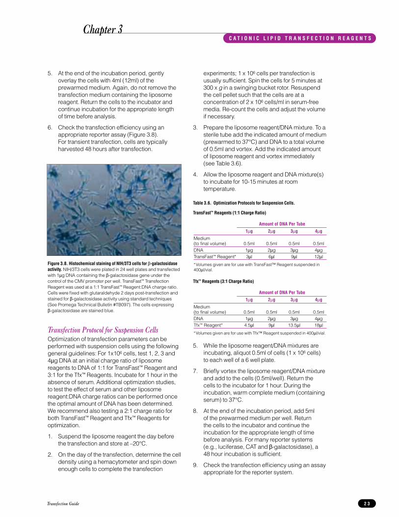
Figure 3.8. Histochemical staining of NIH/3T3 cells for β-galactosidaseactivity. NIH/3T3 cells were plated in 24 well plates and transfectedwith 1µg DNA containing the β-galactosidase gene under thecontrol of the CMV promoter per well. TransFast™ TransfectionReagent was used at a 1:1 TransFast™ Reagent:DNA charge ratio.Cells were fixed with glutaraldehyde 2 days post-transfection andstained for β-galactosidase activity using standard techniques(See Promega Technical Bulletin #TB097). The cells expressing β-galactosidase are stained blue.
Chapter 3C A T I O N I C L I P I D T R A N S F E C T I O N R E A G E N T S

Stable TransfectionsFor stable transfections, cells should be transfectedwith a plasmid containing a gene for drugresistance, such as neomycin phosphotransferase(see Chapter 6 for details of vectors available fromPromega). As a negative control, transfect the cellsusing DNA that does not contain the drug resistancemarker.
1. Prior to transfection, determine the killingconcentration of the selective drug being used.
2. Forty-eight hours post transfection, trypsinizeadherent cells and re-plate at several differentdilutions (for example, 1:100, 1:500) in mediacontaining the appropriate selection drug.
3. For the next 14 days, replace the drug-containing media every 3 to 4 days.
4. During the second week monitor the cells fordistinct “islands” of surviving cells. Cell deathshould occur in cultures transfected with thenegative control plasmid.
5. Transfer individual clones by standardtechniques (e.g., using cloning cylinders) to 96 well plates and continue to maintain culturesin medium containing the appropriate drug.
Neomycin (G418) SelectionG418 blocks protein synthesis in mammalian cellsby interfering with ribosomal function. It is anaminoglycoside, similar in structure to neomycin,gentamycin, and kanamycin (8).
Varying concentrations of G418 should be tested as cells differ in their susceptibility to G418. Use 100 to 800 µg/ml of G418 in complete medium.G418 should be prepared in a highly bufferedsolution (eg. 100 mM HEPES, pH 7.3) so that theaddition of drug does not alter the pH of the medium.
Different lots of G418 can have different potencies,causing many investigators to buy a large amount of one lot to standardize selection conditions. G418 concentration should be calculated using the amount of active drug (usually indicated on eachlot label) so that variance is controlled.
Cells will divide once or twice in the presence oflethal doses of G418, so the effects of the drug takeseveral days to become apparent. Completeselection can take up to 3 weeks of growth inselective media.
Calculating Stable Transfection EfficiencyThe following procedure may be used to determinethe percentage of stable transfectants obtained.
Note: The stained cells will not be viable after thisprocedure.
1. After approximately 14 days of selection in theappropriate drug, monitor the culturesmicroscopically for the presence of viable cellclones. When distinct “islands” of surviving cellsare visible and non-transfected cells have diedout, proceed with step 2.
2. Prepare stain containing 2% methylene blue in50-70% methanol
3. Remove the growth media from the cells byaspiration.
4. Add stain to the cells, sufficient to cover thebottom of the dish.
5. Incubate for 5 minutes.
6. Remove the stain and rinse gently underdeionized cold water. Shake off excessmoisture.
7. Allow the plates to air dry. The plates can bestored at room temperature.
8. Count the number of colonies and calculate thepercent of transfectants based on the celldilution and original cell number.
For further information on stable transfections seereference 29.
Transfection Guide2 4
Chapter 3C A T I O N I C L I P I D T R A N S F E C T I O N R E A G E N T S

Transfection Protocols - Transfectam® Reagent for theTransfection of Eukaryotic CellsTable 3.7. List of Cell Lines Transfected Using Transfectam® Reagent.
For further references using Transfectam® Reagentin a variety of cell lines, see Appendix A.
Plating CellsPlate cells the day before the transfectionexperiment according to the guidelines given in theprotocol for Tfx™ and TransFast™ Reagents.
Suspension cells can be transfected by the followingprotocol using the equivalent of 106 suspended cellsper assay. The volume of reagents can be scaled upor down proportionately depending on the numberof cells used per assay.
Preparation of Transfectam® Reagent Stock Solution1. Resuspend Transfectam® Reagent in 100%
ethanol (dehydrated) with vortexing (finalconcentration is 2mM) and incubate at roomtemperature for at least 5 minutes. Overnightstorage at 4°C ensures complete solubilizationand may give improved transfection efficiencies.Store the resuspended Transfectam® Reagent at4°C, where it is stable for 6 months.
Note: For some cells, it may be desirable tominimize the ethanol concentration applied. If so, Transfectam® Reagent may be dissolvedwith vortexing in as little as 1/10 volume ofethanol (dehydrated), incubated at roomtemperature for 5 minutes, and then furtherdiluted to working concentration in water.
2. Mix the solution before each use. Store theremaining stock at 4°C.
Transfection Protocol for Media Without SerumWe recommend using medium with no added serumfor transfection. Some components in serum maydegrade the Transfectam® Reagent. The presenceof albumin, heparin, trypsin or EDTA in the mediumalso will decrease the efficiency of transfection.However, if cell viability is low in medium withoutserum, use the alternative protocol, provided below.
Materials to Be Supplied by the User• cell culture medium appropriate for the cell type
usedThe reagent volumes in this protocol are based onuse of 60mm culture dishes. Optimizationexperiments can be performed using adherent cellsin 24 well plates. Scale the reagent volumes up ordown proportionally if using different sized plates(see Table 3.8).
Table 3.8. Area of Culture Plates for Cell Growth.
Transfection Guide 2 5
Growth AreaSize of Plate (cm2)a Relative Areab
96 well 0.32 0.02 X24 well 1.88 0.09 X12 well 3.83 0.18 X6 well 9.4 0.45 X35mm 8.0 0.38 X60mm 21 1.00 X
100mm 55 2.62 XaThis information is for Corning™ culture dishes.
bRelative area is expressed as a factor of the growth area of the 60mmdish. To determine the approximate plating density, multiply 5 x 10⁵
cells by this factor. To determine the reagent volumes needed for platesother than 60mm plates, multiply the volumes by the appropriate“Relative Area” factor.
Cell Cell Origin Line Type References
Established Cell LinesHamster CHO Fibroblast 43,52-55Human 293 Embryonic kidney 56-58Human HeLa Epthelial 43Human Hep G2 Hepatocyte 59Monkey COS Fibroblast 53,60-63Mouse C2C12 Myoblast 64Mouse F9 Teratocarcinoma 43,65Mouse LM (tk-) Fibroblast 43,65,66Mouse NIH/3T3 Fibroblast 46Mouse AtT20 Hypophysis 43,67Mouse S49 Lymphocyte 43Mouse Balb/3T3 Fibroblast 68Mouse MEL Erythroleukemia 69Rat PC12 Pheochromocytoma 70Mink Mv1LU Lung epithelial 71Human A549 Lung carcinoma 72Human LoVo/Dx Colon adenocarinoma 68Human Lymphoblast 73Human CCRF- Leukemic T-
CEM/VLB lymphoblast 68Primary CellsRat Cerebellum neurons 67Rat Striatium neurons 67Rat Cortical neurons 67Rat Astrocytes 67Rat Adipocytes 67Rat Anterior pituitary 43Chicken embryo (in vivo) 74Chicken Heart 75Pig Melantrope cells 43,67Bovine Chromaffin cells 43,67
Chapter 3C A T I O N I C L I P I D T R A N S F E C T I O N R E A G E N T S

1. Add 1-5µg of plasmid DNA to 500µl of serum-free medium in a sterile tube and vortex(Solution A). We recommend 5µg per 60mmdish for the initial tests.
2. For each microgram of plasmid DNA used in Solution A, add between 1.5 and 5µl ofTransfectam® Reagent to 500µl of serum-freemedium in a sterile tube and mix (Solution B).For the initial tests, use 10µl of Transfectam®
Reagent per 60mm dish per 5µg plasmid DNA.
3. Immediately mix Solutions A and B and adddirectly to the cells. The final volume will be1.0ml for a 60mm plate or per 106 suspendedcells.
4. Leave in contact with the cells 30 minutes toovernight. Use 2 hours for the initial tests.
5. At the end of the incubation period, gentlyoverlay the cells with 4ml of complete mediumwith serum (37°C). It is not necessary to removethe transfection medium containing theTransfectam® Reagent/DNA mixture. Return the cells to the incubator and continue theincubation for the appropriate length of timebefore analysis. For many reporter assays 48 hours after addition of DNA is sufficient.
6. Check the transfection efficiency using theappropriate reporter assay.
Transfection Protocol for Medium With SerumMaterials to Be Supplied By the User• 0.15M NaCl (sterile)• complete medium with serum
The reagent volumes in this protocol are based on use of 60mm culture dishes. Scale the reagentvolumes up or down proportionately if using differentsize plates (see Table 3.8).
1. Add 1-5µg of plasmid DNA to 50µl of 150mMNaCl solution in a sterile tube and vortex(Solution A). We recommend 5µg per 60mmdish for the initial tests.
2. For each microgram of plasmid DNA used in Solution A, add between 1.5 and 5µl ofTransfectam® Reagent to 50µl of 150mM NaClsolution in a sterile tube and mix (Solution B). For the initial tests, use 10µl of Transfectam®
Reagent per 60mm dish, for 5µg plasmid DNA.
3. Immediately mix solutions A and B, wait 10 minutes, then add to the cells.
4. Leave in contact with the cells 30 minutes toovernight. Use 2 hours for the initial tests.
5. At the end of the incubation period, gentlyoverlay the cells with 4ml of the completemedium (37°C). It is not necessary to removethe transfection medium containing theTransfectam® Reagent/DNA mixture. Return the cells to the incubator and continue theincubation for the appropriate length of timebefore analysis. For many reporter assays 48 hours after addition of the DNA is sufficient.
6. Check the transfection efficiency using theappropriate reporter assay.
Optimization of Transfection EfficiencyFollow these recommendations to obtain the bestresults possible:
• Optimize the volume/weight ratio of Transfectam®
Reagent/DNA in the range of 1.5-5µl/µg DNA.Ten microliters of Transfectam® Reagent stock to5µg DNA is a good initial test for a 60mm plate.
• Optimize the amount of DNA used in the rangeof 1-10µg DNA. It may not be necessary toincrease the quantity of DNA significantly toobtain optimal results. In fact, if the firsttransfection results are satisfactory, a reducedDNA quantity can be tested (while keeping theoptimal Transfectam® Reagent/DNA ratioconstant).
• The transfection time depends on the specificDNA and cell system used and should beoptimized between 30 minutes and overnight. It may be necessary to monitor cell viability ifusing serum-free medium for a prolongedperiod because some cells do not thrive underthis condition. Usually, the transfection timeusing the Transfectam® Reagent is significantlyshorter than that with standard techniques,reducing the risk of cell death significantlyduring transfection.
• Calibrate the system using a test plasmid withreporter gene function (see Chapter 6).
Transfection Guide2 6
TipStudies by Boukhnikachvili et al. suggest that the efficiency of transfection using DOGS (Transfectam® Reagent) is related to the structure of the lipid/DNA complex formed, and increasing both pH and ionic strength (76) can increaseformation of such complexes.
Chapter 3C A T I O N I C L I P I D T R A N S F E C T I O N R E A G E N T S

Transfection Guide 2 7
Notes
Chapter 3C A T I O N I C L I P I D T R A N S F E C T I O N R E A G E N T S

Transfection Guide2 8
Chapter 3
Notes
C A T I O N I C L I P I D T R A N S F E C T I O N R E A G E N T S

IntroductionThe ProFection® Mammalian Transfection Systemsoffer the choice of calcium phosphate or DEAE-Dextran mediated transfection procedures. Both ofthese methods appear to facilitate DNA binding tocell membranes and entry of the DNA into the cellvia endocytosis. Calcium phosphate also appears to provide protection against intracellular and serumnucleases (77).
Calcium phosphate transfection may be used for theproduction of long-term stable transfectants, workswell for transient expression of transfected genesand can be used with most adherent cell lines.DEAE-Dextran transfection is also an efficientmethod for introducing DNA into many cell types,including some cell suspensions. However, itssuitability is limited to transient expression studiesand it is not recommended for the production ofstable transfectants (10). For transient expressionstudies using a particular cell type, both protocolsshould be tried in order to determine the mostefficient method.
For a list of references using the ProFection®
Mammalian Transfection Systems in a variety of celllines, see Appendix A.
Factors That Affect Efficiency of Gene TransferTransfection efficiencies can be increased in manycell types by additional treatments after the primaryexposure of the cells to calcium phosphate-DNA orDEAE-Dextran and DNA. The most effective androutinely used agents are glycerol (78,79), dimethylsulfoxide (DMSO) (79-81), chloroquine (82) andsodium butyrate (83). Since each of these chemicalsis toxic to cells, the conditions for transfection ofindividual cell types must be carefully optimized forreagent concentration and exposure time.
Calcium Phosphate-MediatedTransfectionA precipitate containing calcium phosphate andDNA is formed by slowly mixing a HEPES-bufferedphosphate solution with a solution containingcalcium chloride and DNA. These DNA precipitatesare then taken into eukaryotic cells by an endocytic-type mechanism.
Plating Cells for TransfectionPlate cells the day before the transfectionexperiment according to the guidelines given inChapter 3.
Transfection ProtocolThe protocol given below is for a 60mm plate. Fordifferent size plates scale the volumes and amountsproportionally according to the information given inChapter 3, Table 3.8.
1. Plate the cells the day before transfection asdescribed in Chapter 3.
2. Three hours prior to transfection, remove themedium from the cells and replace it with freshgrowth medium.
3. Thaw all system components and warm them toroom temperature. Mix each componentthoroughly by swirling the container or vortexing.
Transfection Guide 2 9
Figure 4.2. NIH/3T3 cells were transfected with the ProFection®
Mammalian Transfection System-Calcium Phosphate and DNA containingthe green fluorescent protein reporter gene.
Figure 4.1. CHO cells were transfected with the ProFection® MammalianTransfection System-Calcium Phosphate and DNA containing the greenfluorescent protein reporter gene. Cells were counter-stained withpropidium iodide.
Chapter 4PROFECT ION ® MAMMALIAN TRANSFECT ION SYSTEMS

4. For each transfection, prepare the DNA and 2XHBS solutions in separate sterile tubes. Add theDNA and water to the first tube, mix well, thenadd the CaCl2 and mix again. Add the specifiedamount of 2X HBS to the second tube.
per60mm dish
DNA 6-12µg2M CaCl2 37µl
sterile, deionized water to final volume 0.3ml
2X HBS 0.3ml
5. Working in a tissue culture hood, gently vortexthe tube containing the 2X HBS solution. Thespeed should be adjusted such that the tubecan be vortexed safely with the cap off and canaccommodate the addition of the prepared DNAsolution. Continue to vortex while slowly addingthe prepared DNA solution dropwise to the 2X HBS. (Alternatively, bubble air with a pipetthrough the 2X HBS while slowly adding theCaCl2/DNA solution). When the DNA addition is complete, the solution should appear slightlyopaque due to the formation of a fine calciumphosphate-DNA coprecipitate. Incubate thesolution at room temperature for 30 minutes.
6. Vortex the transfection solution again just prior toadding it to the cells. Add the solution dropwiseto the plates. Swirl the plates to distribute theprecipitate evenly over the cells. Return theplates to a 37°C CO2 incubator.
7. When working with sensitive cells, the culturemedium should be changed 4-16 hours aftertransfection. The length of the incubation shouldbe optimized for individual cell lines. Primarycells are particularly sensitive and should not be exposed to calcium phosphate for more than 4 hours.
8. In general cells may be harvested or selectivemedia applied 48-72 hours after transfection.
Glycerol or DMSO Shock
Glycerol ShockHigh transfection efficiencies can be obtained byleaving the DNA/calcium phosphate solution on thecells until the cells are harvested or selectivepressure is applied. HeLa cells, for example,respond well to this treatment. However, transfectionof some cell lines, such as CHO cells, is enhancedby a glycerol shock step.
The glycerol shock step may be performed 4-16hours after transfection. In general, if cells cantolerate the calcium phosphate solution, it is best toleave it on for as long as possible and perform theglycerol shock 16 hours after transfection. Cell linesthat are more sensitive to the calcium phosphatesolution may respond better to a glycerol shock stepperformed earlier, such as 4 hours after exposure tothe DNA. Do not expose the cells to the glycerolsolution for more than 2 minutes. The optimum timeinterval before performing the glycerol shock shouldbe determined empirically for each cell line.
Materials to Be Supplied by the User(Solution compositions are provided at the end ofthis chapter.)
• glycerol shock solution (15% glycerol)• wash solution: 1X PBS or 1X HBSS
1. Prepare a fresh glycerol shock solution in 1XHBS and warm it to 37°C, along with growthmedium and wash solution.
2. Wash the cells once with 5ml of wash solutionper 60mm plate.
3. Add 2ml of the glycerol shock solution per60mm plate.
4. Incubate for up to 2 minutes at room temperature.
5. Remove the glycerol shock solution and washthe cells twice with 5ml of wash solution per60mm plate.
6. Add regular growth medium and return the cellsto a 37°C incubator.
DMSO ShockCertain cell types exhibit enhanced transfectionefficiencies after exposure to dimethyl sulfoxide(DMSO). The DMSO step can be added to either thecalcium phosphate or DEAE-Dextran transfectionprotocols. DMSO, like glycerol, is toxic to cells andthe concentration and exposure times requirecareful optimization for each cell type. Most cellsshould not be exposed to DMSO for more than2.5 minutes. One representative protocol for aDMSO shock is provided.
Transfection Guide3 0
Chapter 4
TipStrontium chloride can be used in place of calcium chloride if the cells being transfected are sensitive to the high calciumconcentration present in the calcium phosphate/DNAprecipitate (84).
TipTo increase efficiency of transfection of some cell types, glycerolmay be added during incubation with the calcium phosphate/DNA precipitate (85).
PROFECT ION ® MAMMALIAN TRANSFECT ION SYSTEMS

Materials to Be Supplied by the User(Solution compositions are provided at the end ofthis chapter.)
• DMSO shock solution (10% DMSO)
1. Remove the medium from the cells.
2. Immediately before use, prepare the DMSOshock solution and warm it to 37°C. Prepare 2ml per 60mm plate.
3. Add the DMSO shock solution to the cells andincubate for 2.5 minutes. Do not return the platesto an incubator during this time.
4. Remove the DMSO shock solution, wash thecells twice and add 5ml of regular growthmedium per 60mm plate. Return the cells to the37°C incubator.
DEAE-Dextran-Mediated TransfectionDEAE-Dextran, a polymeric cation, associates tightlywith the negatively charged DNA and carries it intothe cell. As DEAE-Dextran is toxic to cells,transfection conditions for individual cell lines mayrequire careful optimization of both DEAE-Dextranconcentration and exposure times. At higher DEAE-Dextran concentrations, the exposure time to cellscan be shortened in order to minimize cell death.This protocol may be inappropriate for certain celllines for which DEAE-Dextran is highly toxic.
Two different protocols for DEAE-Dextrantransfections are given. The standard protocolinvolves concurrent exposure of cells to DEAE-Dextran and DNA. The second protocol involvespretreatment of the cells with DEAE-Dextran and is a modification of the procedure described by Al-Molish, et al. (86). It offers the advantages oflimited DEAE-Dextran exposure and longer DNAincubation, allowing maximal DNA uptake. The bestprotocol for a particular cell line should bedetermined experimentally. The addition of 80µMchloroquine along with the DNA is an option for bothprotocols. For some cell lines, chloroquinedramatically increases transfection efficiencies; forothers, it has a minimal effect and may be quitecytotoxic. The optimal amount of DNA to use fortransfection will vary with the cell line and type ofreporter construct being used. Generally, 2-6µg ofDNA will be sufficient for a 60mm plate and 4-10µgDNA will be sufficient for a 100mm plate.
Both protocols require a sterile calcium- andmagnesium-free salt solution for the wash steps.This wash solution is not provided with the system.1X PBS or another salt solution such as 1X HBSSworks well for this purpose.
Materials to Be Supplied by the User(Solution Compositions are provided at the end ofthis chapter.)
• wash solution: 1X PBS or 1X HBSS• optional: 8mM chloroquine (in sterile water)
Standard DEAE-Dextran ProtocolThe protocol given below is for a 60mm plate. Fordifferent size plates scale the volumes and amountsproportionally according to the information given inChapter 3, Table 3.8.
1. Plate cells the day before the transfectionexperiment according to the guidelines given inChapter 3.
2. Prepare the wash solution (1X PBS or 1X HBSS)and warm it to 37°C. Ten milliliters of washsolution are required for each 60mm plate.Warm the DEAE-Dextran solution to 37°C.
3. Dilute the 10X PBS stock 10-fold with sterilewater. You will need approximately 0.4ml of 1X PBS per 60mm plate. Prepare the trans-fection solutions as outlined below:
Per 60mm dish: Using a sterile tube, dilute 2-6µgDNA to a final volume of 326µl in 1X PBS. Add17µl of the 10mg/ml DEAE-Dextran and mix bygently tapping the tube.
4. Remove the medium from the cells. Wash thecells twice with wash solution using 2 x 5ml per60mm plate.
5. Add the DNA/DEAE-Dextran mixture anddisperse it evenly over the cells. The finalconcentration of DEAE-Dextran in the saltsolution is approximately 0.5mg/ml.
6. Incubate the plates at 37°C for 30 minutes. Rockthe plates occasionally to keep the cells moist.
7. Gently add 3.5ml of growth medium per 60mmplate. Incubate up to 2.5 hours at 37°C or untilcytotoxicity is apparent. Gently change themedium or follow with a DMSO shock.
Optional: Add 35µl of 8mM chloroquine per60mm plate along with the medium during the2.5 hour incubation step. If chloroquine isadded, the culture medium must be replacedafter 4 hours (or earlier, if signs of cytotoxicity areapparent). The time that the cells are exposed tochloroquine must be empirically determined foreach cell line.
8. Harvest the cells 48 hours after transfection.
Transfection Guide 3 1
Chapter 4PROFECT ION ® MAMMALIAN TRANSFECT ION SYSTEMS

DEAE-Dextran Pretreatment Protocol1. Plate the cells the day before the transfection
experiment as described in Chapter 3.
2. Prepare the wash solution and warm it to 37°C.Fifteen milliliters of wash solution are required foreach 60mm plate.
3. Dilute the 10X PBS stock 10-fold with sterilewater. You will need approximately 3ml of 1XPBS per 60mm plate. Prepare the transfectionsolutions as outlined below:
Dilute the DEAE-Dextran stock solution 1:10 inthe 1X PBS solution prepared above. You willneed 2ml of diluted DEAE-Dextran per 60mmplate.
Dilute the DNA in 1X PBS to a final volume of325µl for a 60mm plate.
4. Remove the medium from the cells. Add 5ml ofsterile wash solution for 60mm plates. Incubatefor 15 minutes at room temperature.
5. Remove the wash solution from the cells. Add2ml of the diluted DEAE-Dextran solution per60mm plate. Incubate for 9 minutes at roomtemperature.
6. Remove the DEAE-Dextran solution. Very gentlywash the cells twice with 2 x 5ml of washsolution per 60mm plate. Be careful not todislodge the cells, which may begin to detachafter exposure to DEAE-Dextran.
7. Remove the final wash. Add the diluted DNAand disperse it evenly over the cells. Incubatefor 30 minutes in a 37°C CO2 incubator. Rock the plates occasionally to keep the cells moist.
8. Add 3.5µl of regular growth medium per 60mmplate.
Optional: Add 35ml of 8mM chloroquine per 60mmplate together with the medium. If chloroquine isadded, the culture medium must be replacedafter 4 hours (or earlier, if signs of cytotoxicity areapparent). The time that the cells are exposed tochloroquine must be empirically determined foreach cell line.
9. Return the plates to a 37°C CO2 incubator.
10. Harvest the cells 48 hours after transfection.
Composition of Buffers and Solutions2X HBS (HEPES-Buffered Saline)
50mM HEPES (pH 7.1)280mM NaCl1.5mM Na2HPO4
The final pH should be 7.1
1X PBS (Phosphate Buffered Saline)137mM NaCl2.7mM KCl4.3mM Na2HPO4
1.47mM KH2PO4
The final pH should be 7.1
1X HBSS (Hanks Balanced Salt Solution)5mM KCl
0.3mM KH2PO4138mM NaCl
4mM NaHCO30.3mM Na2HPO45.6mM D-glucose
The final pH should be 7.1.
1X Trypsin-EDTA solution0.05% (w/v) trypsin
0.53mM EDTA
Dissolve these components in a calcium- andmagnesium-free salt solution such as 1X PBS or 1X HBSS.
TE buffer10mM Tris-HCl (pH 8.0)1mM EDTA
Glycerol shock solution1X HBS
15% Glycerol
DMSO shock solution1X PBS
10% DMSO (tissue culture grade)
Transfection Guide3 2
Chapter 4PROFECT ION ® MAMMALIAN TRANSFECT ION SYSTEMS

Notes
Transfection Guide 3 3
Chapter 4PROFECT ION ® MAMMALIAN TRANSFECT ION SYSTEMS

Notes
Transfection Guide3 4
Chapter 4PROFECT ION ® MAMMALIAN TRANSFECT ION SYSTEMS

General Troubleshooting Tips
Cationic Lipid Reagent Troubleshooting
Transfection Guide 3 5
Symptoms Possible Causes Comments
No transfection or low Charge ratio of Optimize the reagent to DNA charge ratio. Charge transfection efficiency reagent to DNA ratios of 1:1, 2:1, 3:1 and 4:1 work well for many cell
is sub-optimal lines, but ratios outside this range may be optimal for a particular cell type or application.
Excessive cell death Decrease the time of exposure of the cells to the reagent.Lower the amount of input DNA and cationic lipid reagent, while holding the charge ratio constant.Increase cell density for the transfection step.Remove cationic lipid reagent/DNA mixture from the cells after the transfection period and prior to adding complete medium.
Symptoms Possible Causes Comments
No transfection or Poor quality DNA The DNA should be purified on CsCl gradients orlow transfection equivalent methods. The A260:A280 ratio of the DNA efficiency should be 1.8 or greater.Variable transfection Cells are Test cultures for Mycoplasma contamination. Destroyin replicate cultures contaminated contaminated cultures and start a new culture from a experiments with Mycoplasma fresh stock.
Suboptimal growth Transfection efficiency may decrease if cells have been of cells passaged for many generations. Start a fresh culture
from cell stocks that were frozen at an early passage. Some cells, particularly lymphocytes, will exhibit variability in transfection efficiency if they are left in culture beyond 1-2 weeks.
Variable cell Maintain a consistent cell density at the time of density transfection for each experiment.
Chapter 5T R O U B L E S H O O T I N G T R A N S F E C T I O N R E A C T I O N S

Calcium Phosphate Transfection Troubleshooting
DEAE-Dextran Transfection Troubleshooting
Transfection Guide3 6
Symptoms Possible Causes Comments
No transfection or Excessive Decrease the concentration of DEAE-Dextran or low transfection cell death shorten the time during which the cells are exposed toefficiency DEAE-Dextran.
Decrease the time of exposure to chloroquine.Certain types of cells that are very sensitive to DEAE-Dextran toxicity, such as primary cell cultures, may require a higher cell concentration at the time of transfection.For some cell lines, lower concentrations of DNA can be used for standard DEAE-Dextran transfections compared to calcium phosphate transfection. Establish a dose-response curve to determine the optimal DNAconcentration to use.
Symptoms Possible Causes Comments
No transfection or Poor precipitate The CaCl2/DNA and 2X HBS solutions should be at roomlow transfection formation temperature (22-25°C) when they are mixed. Higher or efficiency lower temperatures for precipitate formation can lead to
decreased transfection efficiency.The addition of CaCl2/DNA to the 2X HBS solution should be performed dropwise and with continuous mixing.The concentration of DNA can affect the size of the precipitate. Low amounts of DNA (less than 1µg) can be supplemented with sheared carrier DNA such as salmon or herring sperm DNA. However, there are conflicting reports in the literature as to the efficacy of adding carrier DNA (80,87).The precipitate should be added dropwise around the dish to the medium bathing the cells, and the medium should be mixed thoroughly at the end of the addition. This helps to evenly distribute the precipitate and avoid the localized acidification of cells.After the addition of the calcium phosphate precipitate to the cells, the pH of the medium should be between 7.2 and 7.4. The CO2 concentration in the incubator should be maintained at an appropriate level (generally 5-10%, depending on the composition of the culture medium).
pH not optimal The pH of the HBS solution should be 7.1. A large volume of added DNA in Tris buffer could change the pH. The DNA should be resuspended in water, 1mM Tris or, if present in 10mM Tris, should be fairly concentrated so that a relatively small volume of the DNA solution is added to the HBS.pH of the 2X HBS may have changed on storage. Check the pH and adjust it to 7.1 if necessary.
Chapter 5T R O U B L E S H O O T I N G T R A N S F E C T I O N R E A C T I O N S

Notes
Transfection Guide 3 7
Chapter 5T R O U B L E S H O O T I N G T R A N S F E C T I O N R E A C T I O N S

Transfection Guide3 8
Notes
Chapter 5T R O U B L E S H O O T I N G T R A N S F E C T I O N R E A C T I O N S

IntroductionGenetic reporter systems have contributed greatly to the study of eukaryotic gene expression andregulation. Although reporter genes have played asignificant role in numerous applications (88), theyare most frequently used as indicators oftranscriptional activity in cells (89). Typically, areporter gene is joined to a promoter sequence in an expression vector that is transfected. Followingtransfer, the cells are assayed for the presence of the reporter by directly measuring the amount ofreporter mRNA, the reporter protein itself or theenzymatic activity of the reporter protein. An idealreporter gene is not endogenously expressed in thecell type of interest, and is amenable to assays thatare sensitive, quantitative, rapid, easy, reproducibleand safe.
The most popular systems for monitoring geneticactivity in eukaryotic cells include chloramphenicolacetyltransferase (CAT), β-galactosidase, fireflyluciferase, growth hormone (GH), β-glucuronidase(GUS), alkaline phosphatase (AP) and, mostrecently, green fluorescent protein (GFP) and Renillaluciferase (90,91)
A control vector can be used to normalize fortransfection efficiency or cell lysate recoverybetween treatments or transfection experiments(92). Typically, the control reporter gene is driven by a strong, constitutive promoter and is co-transfected with experimental vectors. Theexperimental regulatory sequences are linked to adifferent reporter gene so that the relative activitiesof the two reporter gene products can be assayedindividually. Control vectors can also be used tooptimize transfection methods. Gene transferefficiency is typically monitored by assaying reporteractivity in cell lysates, or by staining the cells in situto estimate the percentage of cells expressing thetransferred gene (80).
Promega’s Reporter SystemsPromega currently offers reporter vectors and assaysystems for chloramphenicol acetyltransferase(CAT), β-galactosidase, firefly luciferase, and an integrated dual-reporter assay system for thesequential quantitation of firefly and Renilla (seapansy) luciferases.
Luciferase Reporter Assay SystemThe luciferase enzyme used most frequently forreporter gene technology is derived from the codingsequence of the luc gene cloned from the fireflyPhotinus pyralis (91,93,94). Compared to CAT, thefirefly luciferase protein has a shorter half-life intransfected mammalian cells (95,96), making the
luciferase reporter especially suited for transientassays designed to assess inducible and short-livedeffects.
The firefly luciferase enzyme catalyzes a reactionusing D-luciferin and ATP in the presence of oxygenand Mg2+, resulting in light emission. The totalamount of light measured during a given timeinterval is proportional to the amount of luciferasereporter activity in the sample. The assay has beenimproved by including coenzyme A in the reaction,which provides a longer, sustained light reaction withgreater sensitivity (97). Light emission is typicallyquantified over a defined assay period. Theextended “glow” reaction of the enhanced luciferaseassay allows for accurate measurement of theluminescence reaction when using a luminometer orscintillation counter. The sensitivity of the luciferaseassay is in the subattomole range, approximately 30-1,000 times greater compared to the sensitivity of CAT assays (96). An added advantage is thatluciferase assay results can be obtained in minutescompared to hours, or even days, for the radioactiveCAT assay. The linear range of the firefly luciferaseassay extends over an impressive 8 orders ofmagnitude of firefly luciferase concentration.
Dual-Luciferase™ Reporter Assay SystemPromega’s Dual-Luciferase™ Reporter (DLR™) Assay System combines the speed, sensitivity andconvenience of two luciferase reporter enzymes into an integrated, single-tube, dual-reporter assayformat. The DLR™ Assay is designed to providerapid, sequential quantitation of firefly luciferase and sea pansy (Renilla reniformis) luciferase in celllysates or cell-free translation systems. Because the firefly and Renilla luciferases are of distinctevolutionary origins, they have dissimilar enzymestructures and substrate requirements. Thesedifferences make it possible to selectivelydiscriminate between their respective biolumi-nescent reactions. Thus, the luminescence from thefirefly luciferase reaction may be quenched whilesimultaneously activating the luminescent reactionof Renilla luciferase (the control reporter).
Renilla luciferase is a 36kDa monomeric protein that utilizes oxygen and coelenterate luciferin(coelenterazine) to generate light emission (98). In the integrated Dual-Luciferase™ Reporter Assaychemistry, the kinetics of the Renilla luciferasereaction provide a glow-type luminescent signal thatdecays slowly over the course of the measurement.Similar to firefly luciferase, the luminescent reactioncatalyzed by Renilla luciferase provides highsensitivity and a linear range extending over 7 ordersof magnitude of Renilla luciferase concentration.
Transfection Guide 3 9
Chapter 6G E N E T I C R E P O R T E R S Y S T E M S

CAT Reporter Assay SystemThe CAT gene is derived from transposon 9 of E. coli(99). CAT is a trimeric protein comprising threeidentical subunits of 25kDa (100). The CAT protein isrelatively stable in mammalian cells, although themRNA has a comparatively short half-life, makingthe CAT reporter especially suited for transientassays designed to assess accumulation ofexpressed protein (95).
CAT catalyzes the transfer of the acetyl group fromacetyl-CoA to the substrate, chloramphenicol. Theenzyme reaction can be quantitated by incubatingcell lysates with [14C]chloramphenicol and followingproduct formation by physical separation with thinlayer chromatography (TLC) or organic extraction(101,102).
β-Galactosidase Reporter Assay SystemThe E. coli lacZ gene encodes β-galactosidase, a tetrameric enzyme that catalyzes the hydrolysis of β-galactoside sugars such as lactose. Theenzymatic activity in cell extracts can be assayedwith various specialized substrates that allowquantitation of enzyme activity using a spectro-photometer, a fluorometer or a luminometer. A majorstrength of this reporter gene is the ability to easilyassay in situ expression with histochemical staining(see Figure 6.1).
The β-galactosidase reporter gene is frequentlyused as a control vector for normalizing transfectionefficiency when co-transfected with chimeric DNAslinked to other reporter genes (92). One potentiallimitation of this reporter gene is that certainmammalian cells have endogenous lysosomal β-galactosidase activity. Enzyme assays performedat a higher pH of 7.3-8.0, or with cell extracts pre-heated to 50°C, preferentially favor the E. colienzyme (88,103). However, because of endogenous
cellular β-galactosidase activity, it is important toinclude negative control extracts or cells that havenot been transfected as comparisons for the cell-free and in situ analyses.
Firefly Luciferase Reporter Gene Systems
Firefly Luciferase Reporter VectorsPromega’s Luciferase pGL2 and pGL3 ReporterVectors and Luciferase Assay Reagents provide abasis for rapid, quantitative analysis of factors thatpotentially regulate gene expression. The pGL2 andpGL3 Luciferase Reporter Vectors contain the cDNAencoding luciferase (luc) cloned from the NorthAmerican firefly (Photinus pyralis), as well asnumerous features that aid in the characterizationand manipulation of cloned regulatory sequences.Changes in luciferase reporter activity directlycorrelate to the transcriptional activity of the clonedregulatory element when expressed in transfectedcells.
All pGL3 Vectors contain a modified firefly luciferasecDNA, designated luc+, and a vector backbone thathas been designed to provide enhanced reportergene expression (Figure 6.2). These modificationshelp to ensure that the luciferase reporter gene,itself, does not contribute spurious transcriptionalsignals. Further details on these modifications areprovided in Technical Manual #TM033.
The combination of modifications embodied in thepGL3 Vector family provides greater flexibility inperforming genetic manipulations, minimalbackground activity and luciferase expression levelsthat are dramatically higher than previously obtainedwith the pGL2 Reporter Vectors (104). Using thepGL3 Vectors, it is now possible to obtain
Transfection Guide4 0
Figure 6.1. Histochemical staining of HeLa and BHK cells for β-galactosidase activity. HeLa (Panel A) and BHK (Panel B) cells wereplated in 24 well plates and transfected with pCI-lacZ vector DNA. BHK cells were transfected with Tfx™-10 Reagent at a 2:1Reagent:DNA ratio, with 1,000ng of DNA. HeLa cells were transfected with Tfx™-20 Reagent at a 2:1 Reagent:DNA ratio, with250ng of DNA. The transfections were performed in the absence of serum for one hour. Cells were fixed with glutaraldehyde 48 hours post-transfection and stained for β-galactosidase using standard techniques. The cells expressing β-galactosidaseare stained blue.
A. HeLa Cells B. BHK Cells
Chapter 6G E N E T I C R E P O R T E R S Y S T E M S

measurable luciferase expression in cell types thatare difficult to transfect, when studying weakpromoter elements, or when performing in vivoluminescence measurements. It is important torecognize that absolute light unit values and relativeexpression profiles of reporter vectors will varybetween different cell types (104). The appropriatecontrol vector should always be included inexperiments utilizing genetic reporter systems.
General Considerations for FireflyLuciferase Reporter AssaysPromega’s firefly Luciferase Assay System offersseveral advantages over conventional assays forluciferase (97). The reaction catalyzed by fireflyluciferase is oxidation of beetle luciferin withconcomitant production of a photon. Underconventional reaction conditions, the oxidationoccurs from an enzyme intermediate, luciferyl-AMP.However, recent investigation has revealed thatcoenzyme A is a substrate in the luminescentreaction. In the presence of CoA, oxidation occurspresumably through luciferyl-CoA. The result is lightproduction without the characteristic self-inhibition ofluciferase observed in other assays (97).
The conventional assay for luciferase generates a“flash” of light that rapidly decays after enzyme andsubstrate are mixed, thus requiring automatedinjection luminometers for measurements of photonproduction. Promega’s Luciferase Assay System
allows for greater enzymatic turnover of luciferase(97), which results in greater light intensity that isnearly constant for measurements of up to severalminutes (Figure 6.3).
The constant light intensity generated in Promega’sassay eliminates the need for rapid mixing protocolsand automated injection devices. The simplifiedassay procedure is adaptable to different measure-ment methods for light production, such as scintil-lation counting or exposure to photographic film.
Please request Promega Technical Bulletins #TB101or #TB161 for a detailed Luciferase Reporter AssayProtocol.
Transfection Guide 4 1
Figure 6.2. Circle maps of the pGL3 Vectors.
SV40 Enhancer
Xba I 1742
Kpn ISac IMlu INhe ISma IXho IBgI IIHind III
511152128323653
pGL3-EnhancerVector
(5064bp)
f1 ori
ori
Sal IBamH I
22562250
Nar I 121
Nco I 86luc+
Synthetic poly(A) signal / transcriptional pause site(for background reduction)
SV40 late poly(A) signal (for luc+ reporter)
Hpa I 1902
Ampr
Xba I 1742
Ampr
Kpn ISac IMlu INhe ISma IXho IBgI IIHind III
511152128323653
pGL3-BasicVector
(4818bp)
f1 ori
ori
20102004
Sal IBamH I Nar I 121
Nco I 86luc+
SV40 late poly(A) signal (for luc+ reporter)
Hpa I 1902
Synthetic poly(A) signal / transcriptional pause site(for background reduction)
SV40 Enhancer
Xba I 1934
Ampr
Kpn ISac IMlu INhe ISma IXho IBgI II
5111521283236
pGL3-ControlVector
(5256bp)
f1 ori
ori
Sal IBamH I
24482442
Nar I 313
Nco I 278
Hind III 245
luc+
SV40 Promoter
SV40 late poly(A) signal (for luc+ reporter)
Hpa I 2094
Synthetic poly(A) signal / transcriptional pause site(for background reduction)
Xba I 1934
Ampr
Kpn ISac IMlu INhe ISma IXho IBgI II
5111521283236
f1 ori
ori
Sal IBamH I
22022196
Nco I 278
Hind III 245luc+
SV40 Promoter
pGL3-PromoterVector
(5010bp)
SV40 late poly(A) signal (for luc+ reporter)
Hpa I 2094
Synthetic poly(A) signal / transcriptional pause site(for background reduction)
Figure 6.3. Comparison of Promega’s Luciferase Assay System to theconventional luciferase assay method.
160
120
80
40
0
Ligh
t Int
ensi
ty
Time (seconds)
0 10 20 30 40 50 60
Luciferase Assay System
Conventional Method
Chapter 6G E N E T I C R E P O R T E R S Y S T E M S

Dual-Luciferase™ Reporter Assay System
pRL Renilla Luciferase VectorsThe pRL family of Renilla luciferase vectors, usedwith the Dual-Luciferase™ Reporter Assay System, provides options for Renilla luciferase expression intransfected mammalian cells. The pRL Vectors maybe used in combination with any experimental fireflyluciferase vectors to co-transfect mammalian cells.Thus, the expression of Renilla luciferase canprovide an internal control value to which expressionof the experimental firefly luciferase reporter genemay be normalized.
The pRL family of control reporter vectors contain thecDNA encoding Renilla luciferase (Rluc) cloned fromthe anthozoan coelenterate Renilla reniformis, thesea pansy (105), with some minor modifications forconvenience as a genetic reporter. The constitutiveexpression of Rluc is provided by one of severalavailable promoter elements. The pRL Vectors arecurrently available in three promoter configurationsand one promoter-less configuration (Figure 6.4).
General Considerations for Co-Transfection ExperimentsThe pRL Vector of choice may be used in combinationwith any experimental reporter vector to co-transfectmammalian cells. However, it is important to realizethe potential for trans effects between co-transfection
promoters that may affect reporter gene expression(106). Primarily, this is of concern when working withvery strong promoter/enhancer elements resident on one or the other, or both, of the control andexperimental reporter vectors. The occurrence andmagnitude of such effects will depend on 1) thecombination and activities of the genetic regulatoryelements present on the co-transfected vectors; 2) the relative ratio of experimental vector to controlvector introduced into the cells; and 3) on the celltype itself.
To help ensure independent genetic expressionbetween experimental and control reporter genes,perform preliminary co-transfection experiments tooptimize both the amount of vector DNA and theratio of co-reporter vectors added to the transfectionmix. The extreme sensitivity of both the firefly andRenilla luciferase assays, and the very large linearrange of luminometers (typically 5-6 logs) allowsaccurate measurement of substantially differentexperimental and control luminescence values.Therefore, relatively small quantities of a pRL co-reporter vector are needed to provide low-level,constitutive expression of Renilla luciferase controlactivity. Ratios of luciferase co-transfection vectorsof 50:1 or greater are feasible, and in some instanceswill be preferable to aid in suppressing trans effectsbetween promoter elements.
Transfection Guide4 2
Figure 6.4. Circle maps of the pRL-SV40, pRL-CMV, pRL-TK and pRL-null Vectors.
T7 Promoter
Rluc HSV TKPromoter
SV40 latepoly(A)
Ampr
ori
(1040) Csp45 I
1971 Xba IBgl II 1
Hind III 760EcoR I 649
Nhe I 1024
2223 BamH I
pRL-TKVector
(4045bp)
T7 PromoterRluc
SV40 EarlyEnhancer/Promoter
SV40 latepoly(A)
Ampr
ori
Csp45 I 700
1631 Xba I
Kpn I 58
Pst I 462
Bgl II 1
Hind III 420
Nhe I 684
1883 BamH I pRL-SV40Vector
(3705bp)
T7 Promoter
Rluc
SV40 late poly(A)
Ampr
ori
Csp45 I 3151246 Xba INhe I 299
1498 BamH I
pRL-nullVector
(3320bp)
151210152430323441475158656777
Bgl IIXho ISac IEcoICR IHind IIINde INsi ISph ISpe INar ISal IMlu IEcoR IXma ISma IPst I
T7 Promoter
RlucCMV Immediate EarlyEnhancer/Promoter
SV40 latepoly(A)
Ampr
ori
1074 Csp45 I
2005 Xba I
Pst I 836
Bgl II 1
Hind III 754EcoICR I 725
Nhe I 1058
2257 BamH I
pRL-CMVVector
(4079bp)
Chapter 6G E N E T I C R E P O R T E R S Y S T E M S

General Considerations for the Dual-Luciferase™
Reporter Assay SystemThe luminescent signal from each of the twoluciferase reporter enzymes may be quantitatedimmediately following lysate preparation without theneed for dividing samples or performing additionaltreatments. The firefly luciferase reporter assay isinitiated by adding an aliquot of lysate to LuciferaseAssay Reagent II. Quenching of firefly lumines-cence, and concomitant activation of the Renillaluciferase, is accomplished by adding Stop & Glo®
Reagent to the sample tube immediately uponcompletion of the firefly reaction. Luminescencesignal from the firefly reaction is quenched by atleast a factor of 100,000 (to ≤0.001% residual lightoutput) within one second following the addition ofStop & Glo® Reagent (see Figure 6.5).
Complete activation of Renilla luciferase is alsoachieved within this one-second period. When using a manual luminometer, the time required toquantitate both luciferase reporter activities will beapproximately 30 seconds.
Instrument ConsiderationsThe Turner Designs Model TD-20/20 Luminometer(Promega Cat.# E2061) is ideally suited for low-throughput processing of Dual-Luciferase™ ReporterAssays. The instrument is pre-programmed tocomplete sequential readings of both firefly andRenilla luciferase reporter activities with a single“Start” command. Further, the instrument is pro-grammed to provide the individual and normalizedluciferase values, as well as statistical analyses ofvalues measured within replicate groups.
Please request Promega Technical Manuals #TM040 or #TM046 for more information on the Dual-Luciferase™ Reporter Assay System protocol.
CAT Reporter Gene Systems
pCAT ® Reporter VectorsPromega provides two families of CAT gene reportervectors: pCAT® and pCAT® 3 Reporter Vectors. Eachfamily of vectors contains four plasmids which arereferred to as pCAT®- or pCAT® 3-Basic (lackingeukaryotic promoter and enhancer sequences),Enhancer (with the SV40 early enhancer element 3′ of the CAT gene), Promoter (with the SV40 earlypromoter driving expression of the CAT gene) andControl (with the SV40 early promoter drivingexpression of the CAT gene and the SV40 earlyenhancer 3′ of the gene). Figure 6.6 provides vectormaps for the pCAT® 3 Vectors; maps for the pCAT®
Vectors are provided in Technical Bulletins #TB080-083.
The increase in CAT expression observed with thepCAT® 3 Vectors provides greater sensitivity. It may now be possible to obtain measurable CATexpression in cell types that are difficult to transfector when studying weak promoter elements. Users ofthe pCAT® and pCAT®3 Vectors should be aware,however, that relative expression profiles varybetween different cell types (107). Therefore, it isimportant to include the appropriate control vectorsin all experiments.
Details on pCAT®3 Reporter Vector cloningstrategies and analyses are provided in TechnicalManual #TM036.
CAT Enzyme AssaysChloramphenicol acetyltransferase (CAT), encodedby a bacterial drug-resistance gene, inactivateschloramphenicol by acetylating the drug at one orboth of its two hydroxyl groups (108). This gene isnot found in eukaryotes, and therefore eukaryoticcells contain no background CAT activity. Thischaracteristic, along with the sensitivity of the assayfor CAT activity, has made the CAT gene the mostwidely used reporter gene for studies of mammaliangene expression (90,109). CAT activity may bemonitored by two alternative methods using Promega’sCAT Enzyme Assay System.
Please request Promega Technical Bulletin #TB084for a detailed CAT assay protocol.
Transfection Guide 4 3
Figure 6.5. Measurement of luciferase activities before and after theaddition of Stop & Glo® Reagent.
1,000,000
100,000
10,000
1,000
100
1
0.10
10
FireflyLuciferase
Activity
RenillaLuciferase
Activity
FireflyLuciferase
Activity afterQuench
80,600
0.28
116,800
0.0004%Residual Activity
Rela
tive
Ligh
t Uni
ts
Chapter 6G E N E T I C R E P O R T E R S Y S T E M S

β-Galactosidase Reporter Gene SystemThe enzyme β-galactosidase is widely used as areporter molecule for both in vitro and transgenicapplications. Promega’s pSV-β-GalactosidaseControl Vector (Cat.# E1081; Figure 6.7) is designedas a positive control vector for monitoring transfectionefficiencies of mammalian cells. The SV40 earlypromoter and enhancer drive transcription of thebacterial lacZ gene, which encodes β-galactosidase.β-Galactosidase is an excellent reporter enzyme(89,110) that can be assayed quickly and directly incell extracts using a spectrophotometric assay(111), or in fixed cells by in situ staining (29).
A protocol for histochemical staining for β-Galactosidasecan be found in Promega Technical Bulletin #TB097.
Transfection Guide4 4
Figure 6.6. Circle maps of the pCAT®3 Vectors.
SV40 Enhancer
Xba I 971
Kpn ISac IMlu INhe ISma IXho IBgI II(Hind III)
511152128323653
pCAT3-EnhancerVector
(4293bp)
f1 ori
ori
Sal IBamH I
14851479
Nco I 309(Hind III) 276
CAT
Synthetic poly(A) signal and transcriptional pause site(for background reduction)
SV40late poly(A)
region
Ampr
Hpa I 1131
Xba I 971
Kpn ISac IMlu INhe ISma IXho IBgI II(Hind III)
511152128323653
pCAT3-BasicVector
(4047bp)
f1 ori
ori
Sal IBamH I
12391233
Nco I 309(Hind III) 276
CAT
Synthetic poly(A) signal and transcriptional pause site(for background reduction)
SV40late poly(A)
region
Ampr
Hpa I 1131
SV40 Enhancer
SV40 Promoter
Xba I 1163
Kpn ISac IMlu INhe IXma IXho IBgI II
5111521283236
pCAT3-ControlVector
(4485bp)
f1 ori
ori
Sal IBamH I
16771671 Nco I 501
(Hind III) 486
(Hind III) 245
CAT
Synthetic poly(A) signal and transcriptional pause site(for background reduction)
SV40late poly(A)
region
Ampr
Hpa I 1323
SV40 Promoter
Xba I 1163
Kpn ISac IMlu INhe ISma IXho IBgI II
5111521283236
pCAT3-PromoterVector
(4239bp)
f1 ori
ori
Sal IBamH I
14311425
Nco I 501
(Hind III) 245
(Hind III) 468CAT
Synthetic poly(A) signal and transcriptional pause site(for background reduction)
SV40late poly(A)
region
Ampr
Hpa I 1323
Figure 6.7. Circle map of the pSV-β-Galactosidase Control Vector.
pSV-β-GalactosidaseVector
(6821bp)
Ampr
ori
lac Z
EcoR I 3701
Hind III 414
BamH I 4151Sal I 4163
Pst I 4173
EcoR I 6815
SV40 Promoterand Enhancer
Chapter 6G E N E T I C R E P O R T E R S Y S T E M S
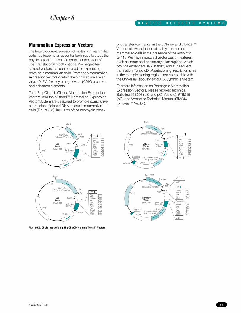
Mammalian Expression VectorsThe heterologous expression of proteins in mammaliancells has become an essential technique to study thephysiological function of a protein or the effect ofpost-translational modifications. Promega offersseveral vectors that can be used for expressingproteins in mammalian cells. Promega’s mammalianexpression vectors contain the highly active simianvirus 40 (SV40) or cytomegalovirus (CMV) promoterand enhancer elements.
The pSI, pCI and pCI-neo Mammalian ExpressionVectors, and the pTARGET™ Mammalian ExpressionVector System are designed to promote constitutiveexpression of cloned DNA inserts in mammaliancells (Figure 6.8). Inclusion of the neomycin phos-
photransferase marker in the pCI-neo and pTARGET™
Vectors allows selection of stably transfectedmammalian cells in the presence of the antibiotic G-418. We have improved vector design features,such as intron and polyadenylation regions, whichprovide enhanced RNA stability and subsequenttranslation. To aid cDNA subcloning, restriction sitesin the multiple cloning regions are compatible withthe Universal RiboClone® cDNA Synthesis System.
For more information on Promega’s MammalianExpression Vectors, please request TechnicalBulletins #TB206 (pSI and pCI Vectors), #TB215(pCI-neo Vector) or Technical Manual #TM044(pTARGET™ Vector).
Transfection Guide 4 5
Figure 6.8. Circle maps of the pSI, pCI, pCI-neo and pTARGET™ Vectors.
➞
ori
Intron
f1 ori
SV40 Latepoly(A)
CMV I.E.Enhancer/Promoter
pCIVector
(4008 bp)
BamH I
Pst I
Afl II
Afl II
Ampr
T7Nhe IXho IEcoR IMlu IKpn IXba ISal I Acc ISma IBstZ INot I
Bgl II
10521058106310691079108110871088109410981098
Nhe IXho IEcoR IMlu IXba ISal IAcc I Sma IBstZ INot I
➞
ori
Intron
f1 ori
SV40 Latepoly(A)
SV40 EarlyEnhancer/Promoter
pSIVector
(3634 bp)
Bgl II
BamH I
Pst IAfl II
Afl II
Ampr
T7678684689695707713714720724724
Sgf I 664
I-Ppo I 851
Bgl II 5665
SV40 Enhancer/EarlyPromoter
SV40 Latepoly (A)
f1 oriSynthetic poly(A)
Ampr
ori
CMVEnhancer/Promoter
Intron
Neo
pTARGET ™Vector
(5670 bp)
TT
EcoR IBamH INhe IXho IMlu I
lacZ
lacZ
Sma IKpn ISal IAcc INot IEcoR I
T7
➞
12501256126412701276
129313011303130413111318
T overhang
I-Ppo I
Sgf I
BamH I
Ampr
Neo
T7
T3
Bgl II
108510911096110211141120112111271131
Intron
CMV I.E.Enhancer/Promoter
f1 ori
SV40 Enhancer/Early Promoter
Syntheticpoly(A)
SV40 Latepoly(A)
pCI-neoVector
(5474bp)
➞
➞
Nhe IXho IEcoR IMlu IXba ISal IAcc ISma INot I
ori
Chapter 6G E N E T I C R E P O R T E R S Y S T E M S
Transfection Guide4 6
Notes
Chapter 6G E N E T I C R E P O R T E R S Y S T E M S

1. Vaheri, A. and Pagano, J.S. (1965) Virology 27, 434.
2. Graham, F.L. and van der Eb, A.J. (1973) Virology 52,456.
3. Melton, D. et al. (1984) Nucl. Acids. Res. 12, 7035.
4. Wong, T.K. and Neumann, E. (1982) Biochem.Biophys. Res. Commun. 107, 584.
5. Fraley, R. et al. (1980) J. Biol. Chem. 255, 10431.
6. Gorman, C.M., Moffat, L.F. and Howard, B.H. (1982)Mol. Cell. Biol. 2, 1044.
7. Groskreutz, D. and Schenborn, E.T. (1997) In:Methods in Molecular Biology 63, 11 ed. R. Tuan,Humana Press, NJ.
8. Southern, P.J. and Berg, P. (1982) J. Mol. Appl. Gen.1, 327.
9. McCutchan, J.H. and Pagano, J.S. (1968) J. Natl.Cancer Inst. 41, 351.
10. Gluzman, Y. (1981) Cell 23, 175.
11. Kawai, S. and Nishizawa, M. (1984) Mol. Cell. Biol. 4,1172.
12. Boussif, O. et al. (1995) Proc. Natl. Acad. Sci. USA 92,7297.
13. Haensler, J. and Szoka, F.C. (1993) Bioconj. Chem. 4,372.
14. Kukowska-Latallo, J.F. et al. (1996) Proc. Natl. Acad.Sci. USA 93, 4897.
15. Loytner, S., Scangos, G.A. and Ruddle, F.H. (1982)Proc. Natl. Acad. Sci. USA 79, 422.
16. Felgner, P.L. et al. (1987) Proc. Natl. Acad. Sci. USA84, 7413.
17. Capaccioi, S. et al. (1993) Bioch. Biophys. ResearchCommunications 197, 818.
18. Felgner, J., Bennett, F. and Felgner, P.L. (1993)Methods 5, 67.
19. Lee, J.T. and Jaenisch, R. (1996) Nucl. Acids Res. 24,5054.
20. Lamb, B.T. and Gearhart, J.D. (1995) Current Opinionin Genetics and Development 5, 342.
21. Malone, R.W., Felgner, P.L. and Verma, I.M. (1989)Proc. Natl. Acad. Sci. USA 86, 6077.
22. Debs, R.J. et al. (1990) J. Biol. Chem. 265, 10189.
23. Felgner, P.L. et al. (1995) Ann. NY Acad. Sci. 772, 126.
24. Farhood, H., Servina, N.S. and Huang, L. (1995)Biochim. Biophys. Acta 1235, 289.
25. Cappechi, M.R. (1980) Cell 22, 479.
26. Shigekawa, K. and Dower, W.J. (1988) BioTechniques6, 742.
27. Ye, G.N., Danielle, H. and Sanford, J.C. (1990) PlantMolec. Biol. 15, 809.
28. Klein, T.M. et al. (1987) Nature 327, 70.
29. Ausubel, F. M. et al. (1995) Current Protocols inMolecular Biology. Wiley Interscience and GreenePublishing Associates.
30. Sambrook, J., Fritsch, E.F. and Maniatis, T. (1989)Molecular Cloning: A Laboratory Manual, SecondEdition. Cold Spring Harbor Laboratory, Cold SpringHarbor, NY.
31. Zasloff, M., Ginder, G.D. and Felsenfeld, G. (1978)Nucl. Acids Res. 5, 1139.
32. Sarver, N.P. et al. (1981) Mol. Cell. Biol. 1, 486.
33. Blochlinger, K. and Diggelmann, H. (1984) Mol. Cell.Biol. 4, 2929.
34. Stark, G.R., and Wahl, G.M. (1984) Ann. Rev.Biochem. 53, 337.
35. Schimke, R.T. (1988) J. Biol. Chem. 263, 5989.
36. Sessa, G. and Weissmann, G. (1968) J. Lipid Res. 9,310.
37. Felgner, P.L. et al. (1994) J. Biol. Chem. 269, 2550.
38. Wheeler, C.J. et al. (1996) Biochim. Biophys. Acta1280, 1.
39. Kabanov, A.V. and Kabanov, V.A. (1995) BioconjugateChem. 6, 7.
40. Moleur, F.L. et al. (1996) Gene Therapy 3, 1010.
41. Gao, X. and Huang, L. (1995) Gene Therapy 2, 710.
42. Wilson, T., Papahadjopoulos, D. and Taber, R. (1979)Cell 17, 77.
43. Behr, J.-P. et al. (1989) Proc. Natl. Acad. Sci. USA 86,6982.
44. Loeffler, J.-P. et al. (1990) J. Neurochem. 54, 1812.
45. Barthel, F. et al. (1993) DNA and Cell Biology 12, 553.
46. Remy, J.S. et al. (1994) Bioconj. Chem. 5, 647.
47. Keogh, M-C. et al. (1997) Gene Therapy 4, 162.
48. Demeneix, B.A. et al. (1994) BioTechniques 16, 496.
49. Tsukamoto, M. et al. (1995) Nature Gen. 9, 243.
50. Douar, A-M. et al. (1996) Gene Therapy 3, 789.
51. Gao, X., and Huang, L. (1996) Biochemistry 35 (3),1027.
52. Foley, B.T., Moehring, J.M. and Moehring, T.J. (1995)J. Biol. Chem. 270, 23218.
53. Harada, N. et al. (1997) J. Biol. Chem. 272, 15232.
54. Smit, M.J. et al. (1996) J. Biol. Chem. 271, 7574.
55. Zhang, L., David, G. and Esko, J.D. (1995) J. Biol.Chem. 270, 27127.
56. Hinz, M., Moore, M.J. and Bindereif, A. (1996) J. Biol.Chem. 271, 19001.
57. Nibbs, R.J.B. et al. (1997) J. Biol. Chem. 272, 12495.
58. Venkatakrishnan, G. and Exton, J.H. (1996) J. Biol.Chem. 271, 5066.
59. Fisher, E.A. et al. (1997) J. Biol. Chem. 272, 20427.
60. Jeannin, P. et al. (1997) J. Biol. Chem. 272, 15613.
61. Klafki, H.-W. et al. (1996) J. Biol. Chem. 271, 28655.
62. Olofsson, A. et al. (1995) J. Biol. Chem. 270, 31294.
63. Von Krempelhuber, A., Muller, F, and Fuhrmann, U.(1994) J. Steroid Biochem. Mol. Biol. 48 (5-6), 511.
64. Epstein, J.A. et al. (1995) J. Biol Chem. 20, 11719.
65. Choi, Y.-C. and Chae, C.-B. (1993) Mol. Cell. Biol. 13(9), 5538.
66. Kizer, N., Guo, X.-L. and Hruska, K. (1997) Proc. Natl.Acad. Sci. USA 94, 1013.
Transfection Guide 4 7
Cited ReferencesR E F E R E N C E S

67. Loeffler, J.-P. and Behr, J.-P. (1993) Meth. Enzymol.217, 599.
68. Capaccioli, S. et al. (1993) Biochem. Biophys. Res.Comm. 197 (2), 818.
69. Nemoto, Y. et al. (1996) J. Biol. Chem. 271, 13542.
70. Gaiddon, C. et al. (1994) J. Biol. Chem. 269 (36),22663.
71. Ichijo, H. et al. (1997) Science 275, 90.
72. Döhr, O. et al. (1997) Mol. Pharmacol. 51, 703.
73. Lesch, K.-P. et al. (1996) Science 274, 1527.
74. Demeneix, B.A. et al. (1994) BioTech. 16 (3), 496.
75. Rosoff, M.L., Wei, J. and Nathanson, N.M. (1996)Proc. Natl. Acad. Sci. USA 93, 14889.
76. Boukhnikachvili, T. et al. (1997) FEBS Lett. 409 (2),188.
77. Loyter, A., Scangos, G.A. and Ruddle, F.H. (1982)Proc. Natl. Acad. Sci. USA 79, 422.
78. Frost, E. and Williams, J. (1978) Virology 91, 39.
79. Lopata, M.A., Cleveland, D.W. and Sollner-Webb, B.(1984) Nucl. Acids Res. 12, 5707.
80. Lowy, D.R., Rands, E. and Scolnick, E.M. (1978) J.Virology 26, 291.
81. Lewis, W.H. et al. (1980) Somat. Cell Genet. 6, 333.
82. Luthman, H. and Magnusson, G. (1983) Nucl. AcidsRes. 11, 1295.
83. Gorman, C.M., Howard, B.H. and Reeves, R. (1983)Nucl. Acids Res. 11, 7631.
84. Brash, D.E. et al. (1987) Mol. Cell Biol. 7, 2031.
85. Wilson, S.P., and Smith, A.L. (1996) Anal. Biochem.246, 148.
86. Al Molish, M.I. and Dubes, G.R. (1973) J. Gen. Virol.18, 189.
87. Freshney, R.I., (1987) Culture of Animal Cells A.R.Liss, Inc., NY.
88. Alam, J. and Cook, J.L. (1990) Anal. Biochem. 188,245.
89. Rosenthal, N. (1987) Meth. Enzymol. 152, 704.
90. Groskreutz, D. and Schenborn, E. (1996) ReporterSystems, In: Recombinant Proteins: Detection andIsolation. Tuan, R., ed., Humana Press, Clifton, NJ.
91. Wood, K.V. (1995) Curr. Opin. Biotech. 6, 50.
92. Hollon, T. and Yoshimura, F. K. (1989) Anal. Biochem.182, 411.
93. DeWet, J.R. et al. (1985) Proc. Natl. Acad. Sci. USA82, 7870.
94. DeWet, J.R. et al. (1987) Cell. Biol. 7, 725.
95. Thompson, J.F. et al. (1991) Gene 103, 171.
96. Pazzagli, M. et al. (1992) Anal. Biochem. 204, 315.
97. Wood, K.V. (1991) In: Bioluminescence andChemiluminescence: Current Status. Stanley, P.E. andKricka, L.J., eds., John Wiley and Sons, NY.
98. Matthews, J.C.et al. (1977) Biochemistry 16, 85.
99. Alton, N.K. and Vapnek, D. (1979) Nature 282, 864.
100. Leslie, A.G.W. et al. (1988) Proc. Natl. Acad. Sci. USA85, 4133.
101. Seed, B. and Sheen, J-Y. (1988) Gene 67, 271.
102. Neumann, J.R. et al. (1987) BioTechniques 5, 444.
103. Young, D.C. (1993) Anal. Biochem. 215, 24.
104. Groskreutz, D.J. et al. (1995) Promega Notes 50, 2.
105. Lorenz, W.W. et al. (1991) Proc. Natl. Acad. Sci. USA88, 4438.
106. Farr, A. and Roman, A. (1991) Nucl. Acids Res. 20,920.
107. Groskreutz, D.J. et al. (1996) Promega Notes 55, 2.
108. Shaw, W.V. (1975) Meth. Enzymol. 43, 737.
109. Cullen, B. et al. (1987) Meth. Enzymol. 152, 687.
110. Hall, C.V. et al. (1983) J. Molec. Applied Gen. 2, 101.
111. Silhavy, T. et al. (1972) In: Experiments in MolecularGenetics, Miller, J.H., ed., Cold Spring HarborLaboratory, Cold Spring Harbor, NY.
Transfection Guide4 8
Cited ReferencesR E F E R E N C E S

References Using Promega Transfection Reagents
Tfx™ Reagents for the Transfection of Eukaryotic Cells1. Fearon, I.M., et al. (1997) Hypoxia inhibits the
recombinant α1C subunit of the human cardiac L-typeCa2+ channel. J. Physiol. 500.3, 551-556.
Systems Used: Tfx™-50 Reagent.
Summary: The reagent was used to transfect 293 Cells.
2. Gobin, S.J.P. et al. (1997) Site α is crucial for two routesof IFNγ-induced MHC Class I transactivation: TheISRE-mediated route and a novel pathway involvingCIITA. Immunity 6, 601.
Systems Used: Tfx™-50 Reagent; pGL3 Basic Vector;pGL3 Enhancer Vector; Universal RiboClone® cDNASynthesis System
Summary: The Tfx™-50 Reagent was used to transfectK562 cells. Cells were assayed for expression of asurface protein by FACS analysis.
3. Gründemann, D. et al. (1997) Primary structure andfunctional expression of the apical organic cationtransporter from kidney epithelial LLC-PK1 cells. J.Biol. Chem. 272, 10408.
System Used: Tfx™-50 Reagent.
Summary: The reagent was used to produce transientlytransfected LLC-PK1 and 293 cells as well as stablytransfected 293 cells.
4. Ichijo, H. et al. (1997) Induction of apoptosis by ASK1,a mammalian MAPKKK that activates SAPK/JNK andp38 signaling pathways. Science 275, 90.
Systems Used: Tfx™-50 Reagent; Transfectam®
Reagent.
Summary: Tfx™-50 Reagent was used to producetransient transfectants of human 293 cells. Eight hourspost-transfection, the cells were treated with apoptoticagents for 16 hours and assayed.
5. Inui, T. et al. (1997) Cathepsin K antisense oligo-deoxynucleotide inhibits osteoclastic bone resorption. J. Biol. Chem. 272, 8109.
System Used: Tfx™-50 Reagent.
Summary: The reagent was used to transiently transfectosteoclasts with antisense oligonucleotides.
6. Keogh, M.-C. et al. (1997) High efficiency reportergene transfection of vascular tissue in vitro and in vivousing a cationic lipid-DNA complex. Gene Therapy, 4,162.
System Used: Tfx™-50 Reagent.
Summary: The reagent was used to transfect pGL3Vector into HepG2, HUVEC, and human, rat and rabbitprimary cells. The optimized conditions were used totransfect rabbit arteries in vivo and human arteries in vitro.
7. Ko, B.C.B. et al. (1997) Identification andcharacterization of multiple osmotic responsesequences in the human aldose reductase gene. J. Biol. Chem. 272, 16431.
Systems Used: Tfx™-50; pGL3 Basic Vector; pGL3Promoter Vector.
Summary: Studies were performed in Chang liver cells using 2µg of luciferase reporter and 1µg of β-galactosidase control in serum-free medium. Cellswere transfected 1hr at 37°C, the medium removedand fresh medium applied. Twenty-four hours later the cells were assayed.
8. Nishitoh, H. et al. (1996) Identification of type I andtype II serine/threonine kinase receptors forgrowth/differentiation factor-5. J. Biol. Chem. 271,21345.
System Used: Tfx™-50 Reagent.
Summary: The reagent was used to transiently transfectCOS-1 cells and R mutant Mv1Lu cells.
9. Nork, T.M. et al. (1997) p53 regulates apoptosis inhuman retinoblastoma. Arch. Opthamol. 115, 213.
System Used: Tfx™-50 Reagent.
Summary: The reagent was used to transfect WERI-Rb1 human retinoblastoma tissue culture cells.
10. Rodriquez-Viciana, P. et al. (1997) Role of phospho-inositide 3-OH kinase in cell transformation and controlof the actin cytoskeleton by Ras. Cell 89, 457.
System Used: Tfx™-50 Reagent
Summary: NIH/3T3 cells were stably transfected witheither puromycin or G418 selection. Cells wereassayed 2 weeks after transfection and selected forfoci formation in soft agar.
11. Schenborn, E. and Goiffon, V. (1997) Transfection ofinsect cells with Tfx™-20 Reagent. Promega Notes 63,13.
12. Schenborn, E., Oler, J. and Goiffon, V. (1996) A Trio ofTfx™ Transfection Reagents for Eukaryotic Cells.Promega Notes 59, 24.
13. Urban, R.J. and Bodenburg, Y. (1996) Transcriptionalactivation of the porcine P450 11A insulin-like growthfactor response element in MCF-7 breast cancer cells.J. Biol. Chem. 271, 31695.
System Used: Tfx™-50 Reagent.
Summary: The reagent was used to transiently transfectMCF-7 cells.
14. Urban, R.J., Nagamani, M. and Bodenburg, Y. (1996)Tumor necrosis factor α inhibits transcriptional activityof the porcine P45011A insulin-like growth factorresponse element. J. Biol. Chem. 271, 31699.
System Used: Tfx™-50 Reagent
Summary: The reagent was used to transiently transfecttwo variants of the NIH/3T3 cells, NWTb3 and KR1.
Transfection Guide 4 9
Appendix AA D D I T I O N A L R E F E R E N C E S

15. Velcich, A. et al. (1997) Organization and regulatoryaspects of the human intestinal mucin gene (MUC2)locus. J. Biol. Chem. 272, 7968.
Systems Used: Tfx™-50 Reagent; pGL2 Basic Vector;pGL2 Enhancer Vector; pGL2 Promoter Vector.
Summary: Studies were performed in the intestinal celllines, HT29 and LS174T, and the HeLa cell line. TheTfx™-50 reagent was used to transfect the intestinal cell lines.
TransFast™ Reagent1. Schenborn, E., Goiffon, V. and Oler, J. (1998) An
efficient new transfection reagent for eukaryotic cells:TransFast™ Reagent. Promega Notes 65, 2.
Transfectam® Reagent for the Transfection ofEukaryotic Cells1. Carey, D.J., Bendt, K.M. and Stahl, R.C. (1996) The
cytoplasmic domain of syndecan-1 is required forcytoskeleton association but not detergent insolubility.Identification of essential cytoplasmic domainresidues. J. Biol. Chem. 271, 15253.
Summary: The reagent was used to produce stablytransfected Schwann cells.
2. Fisher, E.A. et al. (1997) The degradation ofapolipoprotein B100 is mediated by the ubiquitin-proteasome pathway and involves heat shock protein70. J. Biol. Chem. 272, 20427.
Summary: Transfectam® Reagent was used to transfectHepG2 cells.
3. Foley, B.T., Moehring, J.M and Moehring, T.J. (1995)Mutations in the elongation factor 2 gene which conferresistance to diphtheria toxin and Pseudomonasexotoxin A. J. Biol. Chem. 270, 23218.
Summary: The reagent was used to produce stablytransfected CHO cells.
4. Harada, N. et al. (1997) Human IgGFc binding protein(FcBP) in colonic epithelial cells exhibits mucin-likestructure. J. Biol. Chem. 272, 15232.
Summary: COS-7 cells were transfected with 10µg ofDNA and 5µl of Transfectam® Reagent per 500µl ofRPMI 1640 media. After 6 hours the medium waschanged the cells were cultured for an additional 48hours prior to assay. CHO cells were plated at 2 x 10⁵
cells per plate 24 hrs prior to transfection as detailedfor COS-7 cells. Stably transfected CHO cells werechosen 14 days later after growth in G418.
5. Hinz, M., Moore, M.J. and Bindereif, A. (1996) Domainanalysis of human U5 RNA: Cap trimethylation, proteinbinding and spliceosome assembly. J. Biol. Chem.271, 19001.
Summary: Transfectam® Reagent was used totransiently transfect 293 cells.
6. Ichijo, H. et al. (1997) Induction of apoptosis by ASK1,a mammalian MAPKKK that activates SAPK/JNK andp38 signaling pathways. Science 275, 90.
Systems Used: Transfectam® Reagent; Tfx™-50Reagent.
Summary: Transfectam® Reagent was used to producestable transfectants of Mv1LU mink lung epithelial cellsusing hygromycin selection.
7. Ito, M., Jameson, J.L. and Ito, M. (1997) Molecularbasis of autosomal dominant neurohypophysealdiabetes insipidus: Cellular toxicity caused by theaccumulation of mutant vasopressin presursors withinthe endoplasmic reticulum. J. Clin. Invest. 99, 1897.
Systems Used: Transfectam® Reagent; CAT AssaySystem.
Summary: Stable transfectants of Neuro2A cells weregenerated in G418-containing media. The stabletransfectants were assayed for the production ofarginine vasopressin and CAT enzyme.
8. Jeannin, P. et al. (1997) CD86 (B7-2) on human B cells:A functional role in proliferation and selectivedifferentiation into IgE- and IgG4-producing cells. J.Biol. Chem. 272, 15613.
Summary: Studies were performed in COS cells andthe cells were assayed 4 days post-transfection.
9. Kennedy, E.D. et al. (1996) Glucose-stimulated insulinsecretion correlates with changes in mitochondrial andcytosolic Ca2+ in aequorin-expressing INS-1 cells. J.Clin. Invest. 98, 2524.
Summary: INS-1 cells were stably transfected with a neomycin-resistant plasmid and an aequorinexpression vector. The cells were assayed byimmunocytochemistry, various assays of insulinsecretion and Ca2+ channel activity.
10. Kizer, N., Guo, X.-L. and Hruska, K. (1997)Reconstitution of stretch-activated cation channels byexpression of the α-subunit of the epithelial sodiumchannel cloned from osteoblasts. Proc. Natl. Acad.Sci. USA 94, 1013.
Summary: LM(TK–) mouse cells were stably transfectedand selected in media containing hygromycin. The resulting cells were analyzed by Northern blot,Western blot and patch-clamp protocols.
11. Klafki, H.-W. et al. (1996) The carboxyl termini of β-amyloid peptides 1-40 and 1-42 are generated bydistinct γ-secretase activities. J. Biol. Chem. 271,28655.
Summary: The reagent was used to transiently transfectCOS-1 cells.
Transfection Guide5 0
Appendix AA D D I T I O N A L R E F E R E N C E S

12. Lesch,K.-P. et al. (1996) Association of anxiety-relatedtraits with a polymorphism in the serotonin transportergene regulatory region. Science 274, 1527.
Systems Used: Transfectam® Reagent; pGL3 BasicVector; pGL3 Control Vector; pSV-β-GalactosidaseControl Vector.
Summary: Studies were performed in humanlymphoblasts. For transient expression, lymphoblasts(2 x 10⁵ cells) were exposed for 24 hours to 5µg ofconstruct DNA complexed with 5µl of Transfectam®
Reagent in 5ml of RPMI1640. Cells were grown anadditional 24 hours, then assayed for luciferase and β-galactosidase activity.
13. Nemoto, Y. et al. (1996) Regulatory function ofdelta/YY-1 on the locus control region-like sequence of mouse glycophorin gene in erythroleukemia cells. J. Biol. Chem. 271, 13542.
Summary: Transfectam® Reagent was used to transfectMEL Murine Erythroleukemia cells.
14. Nibbs, R.J.B. et al. (1997) Cloning and characterizationof a novel murine β chemokine receptor, D6:Comparisonto three other related macrophage inflammatoryprotein-1α receptors, CCR-1, CCR-3 and CCR-5. J. Biol. Chem. 272, 12495.
Summary: The Transfectam® Reagent was used togenerate G418-resistant, stably-transfected HEK293cells.
15. Olofsson, A. et al. (1995) Efficient association of anamino-terminally extended form of human latenttransforming growth factor-β binding protein with theextracellular matrix. J. Biol. Chem. 270, 31294.
Summary: Transfectam® Reagent was used totransiently transfect COS-1 cells.
16. Rosoff, M.L., Wei, J. and Nathanson, N.M. (1996)Isolation and characterization of the chicken m2acetylcholine receptor promoter region: Induction ofgene transcription by leukemia inhibitory factor andciliary neurotrophic factor. Proc. Natl. Acad. Sci. USA93, 14889.
Systems Used: Transfectam® Reagent; pGL3 BasicVector; pSV-β-Galactosidase Control Vector.
Summary: Transfectam® Reagent was used at 3.5µl/µgof DNA to transfect primary chicken heart cultures. TheDNA/ Transfectam® solution was left in contact with the cells for 6-7 hours. Thirty hours post-transfection, the cells were lysed and assayed for luciferase and β-galactosidase activity.
17. Smit, M.J. et al. (1996) Two distinct pathways forhistamine H2 receptor down-regulation: H2 Leu124-αAla receptor mutant provides evidence for a cAMP-independent action of H2 agonists. J. Biol. Chem. 271,7574.
Summary: The reagent was used to stably transfectCHO cells.
18. Venkatakrishnan, G. and Exton, J.H. (1996)Identification of determinants in the α -subunit of Gqrequired for phospholipase C activation. J. Biol. Chem.271, 5066.
Summary: Transfectam® Reagent was used totransiently transfect 293 cells.
19. Zhang, L., David, G. and Esko, J.D. (1995) Repetitive Ser-Gly sequences enhance heparan sulfate assemblyin proteoglycans. J. Biol. Chem. 270, 27127.
Summary: Transfectam® Reagent was used to producestably transfected CHO cells.
Profection® Mammalian Transfection System-CaPO41. Behrooz, A. and Ismail-Beigi, F. (1997) Dual control of
glut1 glucose transporter gene expression by hypoxiaand by inhibition of oxidative phosphorylation. J. Biol.Chem. 272, 5555.
Systems Used: ProFection® Mammalian TransfectionSystem -CaPO4; Dual-Luciferase™ Reporter AssaySystem; pGL2 Basic Vector; pRL-TK Vector; pCAT®
Control Vector.
Summary: The Renilla luciferase activity was used tonormalize the firefly luciferase activity of transfectedClone 9 cells.
2. Chu, B. et al. (1996) Sequential phosphorylation bymitogen-activated protein kinase and glycogensynthase kinase 3 represses transcriptional activationby heat shock factor-1. J. Biol. Chem. 271, 30847.
Summary: NIH/3T3 cells were seeded at 2.5 x 10⁵
cells/100mm dish 24 hours prior to transfection. Cellswere assayed 48 hours post-transfection.
3. Hoock, T.C., Peters, L.L. and Lux, S.E. (1997) Isoformsof ankyrin-3 that lack the NH2-terminal repeatsassociate with mouse macrophage lysosomes. J. CellBiol. 136, 1059.
Summary: The ProFection® Mammalian TransfectionSystem was used to transiently transfect COS cells.The DNA precipitates were left in contact with the cellsfor 16 hours and 48 hours later the cells were analyzedby immunofluorescence.
4. Obiri, N.I. et al. (1997) Modulation of interleukin (IL)-13binding and signaling by the γc chain of the IL-2receptor. J. Biol. Chem. 272, 20251.
System Used: ProFection® Mammalian TransfectionSystem -CaPO4
Summary: The system was used to produce stabletransfectants in the ML-RCC renal carcinoma cell line.
Transfection Guide 5 1
Appendix AA D D I T I O N A L R E F E R E N C E S

5. Walsh, A.A. et al. (1996) Identification of a novel cis-acting negative regulatory element affectingexpression of the CYP1A1 gene in rat epidermal cells.J. Biol. Chem. 271, 22746.
Systems Used: ProFection® Mammalian TransfectionSystem -CaPO4; pGL2 Basic Vector; Luciferase AssaySystem.
Summary: Studies were performed in rat epidermalkeratinocytes. Cells were grown to 50% confluency in60mm dishes. Cells were transfected with 15µg ofplasmid DNA for 24 hours then the media wasreplaced and the cells assayed 24 hours later.
Profection® Mammalian Transfection System-DEAE-Dextran1. Pierce, R.A. et al. (1996) Monocytic cell type-specific
transcriptional induction of collagenase. J. Clin. Invest.97, 1890.
Summary: U-937 cells (1 x 10⁷cells) were transfected insuspension with 10µg of DNA and 75µg of DEAE-Dextran for 20 min at room temperature. The cells weredivided and allowed to recover overnight. The cellswere treated with a reagent and CAT activity assayed24 hours later.
2. Rohlff, C. et al. (1997) Modulation of transcriptionfactor Sp1 by cAMP-dependent protein kinase. J. Biol.Chem. 272, 21137.
Systems Used: ProFection® Mammalian TransfectionSystem-DEAE-Dextran; pCAT® Basic Vector; pCAT®
Promoter Vectors, Recombinant Human Sp1.
Summary: Reporter studies were performed in HL-60and HL-60/AR (doxorubicin-resistant isolate) leukemiacells. DNA was transfected by electroporation in thepresence of DEAE-Dextran and many details of thetransfection are reported. Recombinant Sp1 was invitro phosphorylated with the cAMP dependent proteinkinase catalytic subunit and used in gel shift assays.
3. Zhang, M., Magit, D and Sager, R. (1997) Expressionof maspin in prostate cells is regulated by a positiveEts element and a negative hormonal responsiveelement site recognized by androgen receptor. Proc.Natl. Acad. Sci. USA 94, 5673.
Summary: LNCaP human prostate tumor cells, CF3normal human epithelial cells and 70N mammaryepithelial cells were transfected at 75% confluency in a100mm dish. Cells were assayed 48 hours post-transfection for β-gal and CAT activity.
References Using Promega’s Reporter AssaySystems and Expression VectorsReferences using Promega’s reporter assay systems andexpression vectors are available on the Internet atwww.promega.com.
Transfection Guide5 2
Appendix AA D D I T I O N A L R E F E R E N C E S

Transfection SystemsProduct Size Cat.#TransFast™Transfection Reagent 1.2mg E2431
Contains sufficient reagent to transfect 400µg of DNA (at a 1:1TransFast™ Reagent:DNA ratio).
Product Size Cat.#Tfx™-10 Reagent 9.3mg E2381Tfx™-20 Reagent 4.8mg E2391Tfx™-50 Reagent 2.1mg E1811
Each system contains sufficient reagent to transfect 200µg ofDNA (at a 4:1 Tfx™ Reagent:DNA ratio).
Product Size Cat.#Tfx™ Reagents Transfection Trio 5.4mg E2400
Contains one tube each of Tfx™-10, Tfx™-20 and Tfx™-50Reagents. Each system contains sufficient reagent to transfect200µg of DNA (at a 4:1 Tfx™ Reagent:DNA ratio).
Product Size Cat.#Transfectam® Reagent for the Transfection of Eukaryotic Cells 1mg E1231
0.5mg E1232ProFection® Mammalian Transfection System - Calcium Phosphate 40 reactions E1200ProFection® Mammalian Transfection System - DEAE-Dextran 40 reactions E1210
Eukaryotic Expression VectorsProduct Size Cat.#pCI-neo Mammalian Expression Vector 20µg E1841pCI Mammalian Expression Vector 20µg E1731pSI Mammalian Expression Vector 20µg E1721pTARGET ™ Mammalian Expression Vector 20µg A1410
Reporter Assays and Vectors
Luciferase Reporter Vectors and Assay Systems
Product Size Cat.#pRL-TK Vector 20µg E2241pRL-CMV Vector 20µg E2261pRL-null Vector 20µg E2271pRL-SV40 Vector 20µg E2231pGL3-Control DNA 20µg E1741pGL3-Basic DNA 20µg E1751pGL3-Enhancer DNA 20µg E1771pGL3-Promoter DNA 20µg E1761pGL2-Control DNA 20µg E1611pGL2-Basic DNA 20µg E1641pGL2-Enhancer DNA 20µg E1621pGL2-Promoter DNA 20µg E1631Dual-Luciferase™
Reporter Assay System 100 assays E1910Luciferase Assay System With Reporter Lysis Buffer 100 assays E4030Luciferase Assay System 100 assays E1500
CAT Reporter Vectors and Assay Systems
Product Size Cat.#pCAT®3-Basic Vector 20µg E1871pCAT®3-Enhancer Vector 20µg E1881pCAT®3-Promoter Vector 20µg E1861pCAT®3-Control Vector 20µg E1851pCAT®-Basic DNA 20µg E1041pCAT®-Enhancer Vector 20µg E1021pCAT®-Control DNA 20µg E1011pCAT®-Promoter DNA 20µg E1031CAT Enzyme Assay System With Reporter Lysis Buffer E1000 Chloramphenicol Acetyltransferase (CAT) 100u E1051
β-Galactosidase Reporter Vectors and Assay Systems
Product Size Cat.#pSV-β-Galactosidase Control Vector 20µg E1081β-Galactosidase Enzyme Assay System With Reporter Lysis Buffer 65 assays E2000Reporter Lysis Buffer 5X 30ml E3971
continued
Transfection Guide 5 3
Appendix BO R D E R I N G I N F O R M A T I O N

Luminometers
Product Cat.#Turner Designs Luminometer Model TD-20/20 Genetic Reporter Instrumentation Package for Stabilized Assays E2041Turner Designs Luminometer Model TD-20/20 Genetic Reporter Instrumentation Package for Stabilized Assays, with Printer E2051Turner Designs Luminometer Model TD-20/20 Genetic Reporter Instrumentation Package for Stabilized Assays, with Printer and Dual Auto Injector System E2061Turner Designs Luminometer Model TD-20/20 Genetic Reporter System with Single Auto Injector E2351Turner Designs Luminometer Model TD-20/20 Genetic Reporter System with Dual Auto Injector E2361
Transfection Guide5 4
Appendix BO R D E R I N G I N F O R M A T I O N

Notes
Transfection Guide 5 5
Appendix BO R D E R I N G I N F O R M A T I O N

Notes
Transfection Guide5 6
Appendix BO R D E R I N G I N F O R M A T I O N

ARGENTINA AND URUGUAYBiodynamics, Srl.Buenos AiresTel: 1 383 3000FAX: 1 384 7316E-mail: [email protected]
BRAZILbioBRASBelo HorizonteTel: 31 291 9877FAX: 31 291 5369E-mail:[email protected]
PromicroSao PaoloTel/FAX: 11 869 0699E-mail: [email protected]
CHILEFERMELO, S.A.SantiagoTel: 2 247 2976FAX: 2 247 2977
COLOMBIABiologia Molecular, Ltda. (BioMol)BogotaTel/FAX: 1 271 5319E-mail: [email protected]
ECUADORElectromedicsQuitoTel: 3 258 2483FAX: 3 251 9363E-mail: [email protected]
MEXICOUniparts, S.A.Mexico CityTel: 5 281 4718FAX: 5 281 4722E-mail: [email protected]
VENEZUELAVargas Scientific Supplies, C.A.CaracasTel: 2 265 0891FAX: 2 263 0924E-mail: [email protected]
EGYPTLab TechnologyCairoTel: 2 245 1785FAX: 2 242 8366E-mail: [email protected]
ISRAELORNAT Biochemicals & LaboratoryEquipment
RehovotTel: 89477077FAX: 89363034E-mail: [email protected]
LEBANONAtom Medical Company, sarlBeirutTel: 1 249836FAX: 1 249838E-mail: [email protected]
SOUTH AFRICAWhitehead ScientificCape TownTel: 21 981 1560FAX: 21 981 5789E-mail: [email protected]
TURKEYERMANAK MISKCIYANIstanbulTel: 216 385 8321FAX: 216 385 4649E-mail: [email protected]
▲ AUSTRALIAPromega CorporationSydneyTel: 02 9565 1100Freecall: 1 800 225 123FAX: 02 9550 4454Free FAX: 1 800 626 017E-mail: [email protected]
MelbourneTel: 03 9818 1466Freecall: 1 800 335 435FAX: 03 9818 1244
CHINA, PEOPLE'S REPUBLIC▲ Representative OfficePromega Beijing Beijing Tel: 10 6849 8287FAX: 10 6849 8390E-mail: [email protected]
CHINA (Joint Venture)Shanghai Promega Biological Products, Ltd.ShanghaiTel: 21 6483 5136FAX: 21 6470 0176E-mail: [email protected]
SABC, Sino-American Biotechnology Co.Luo YangTel: 379 432 2510FAX: 379 432 2505E-mail: [email protected]
HONG KONGBio-Gene TechnologyTel: 2646 6101FAX: 2686 8806E-mail: [email protected]
INDIAHYSEL INDIANew DelhiTel: 11 684 6565Telex: 3175354 HYSLINFAX: 11 691 5917E-mail: [email protected]
INDONESIAPT Diastika BiotekindoJakarta SelatanTel: 21 489 1718FAX: 21 475 4707E-mail: [email protected]
▲ JAPANPromega KKTokyo, OsakaTel: 03 3669 7981FAX: 03 3669 7982E-mail: [email protected]
KOREASeoulin Scientific Co., Ltd.SeoulTel: (02) 478 5911FAX: (02) 478 5572
MALAYSIAResearch Instruments SDN. BHD.Petaling Jaya SelangorTel: 3 704 8600FAX: 3 704 8599E-mail: [email protected]
NEW ZEALANDDADE Behring DiagnosticsNew ZealandAucklandTel: 9 366 4784FAX: 9 379 8308
PAKISTANSS DiagnosticsRawalpindiTel: 92 51 518 469FAX: 92 51 522 243E-mail: [email protected]
SINGAPORE▲ Representative OfficeTel: 65 452 4230FAX: 65 452 2518E-mail: [email protected]
Research InstrumentsTel: 775 7284FAX: 775 9228
E-mail: [email protected]
TAIWANGenelabs Life ScienceTaipeiTel: 02 238 25378FAX: 02 231 18524E-mail: [email protected]
THAILAND, VIETNAM ANDMYANMARDiagnostic Biotechnology Co., Ltd.BangkokTel: 66 2 295 2532FAX: 66 2 295 3632
Promega Corporation Worldwide Distribution
Rev.010299
LATIN AMERICA
▲ AUSTRIAPromega GmbHMannheimTel: (+49)(0)621-8501-0Free Phone: +800-77663422FAX: (+49)(0)621-8501-222Free FAX: +800-77663423E-mail: [email protected]
CZECH REPUBLICEast Port ScientificPragueTel: 02 206 10151FAX: 02 206 11664E-mail: [email protected]
DENMARKBie & Berntsen A-SRoedovreTel: 44 94 88 22FAX: 44 94 27 09
FINLANDBioFellowsHelsinkiTel: 358 9 755 2550FAX: 358 9 755 25555E-mail: [email protected]
▲ FRANCEPromega FranceLyonTel: 04 37 22 50 00Numero Vert Tel: 0800 48 79 99FAX: 04 37 22 50 10E-mail: [email protected]
▲ GERMANYPromega GmbHMannheimTel: (+49)(0)621-8501-0Free Phone: +800-77663422FAX: (+49)(0)621-8501-222
Free FAX: +800-77663423E-mail: [email protected]
GREECEBioAnalytica, S.A.AthensTel: 1 6436138FAX: 1 6462748E-mail: [email protected]
HUNGARYEast Port ScientificBudapestTel: 1 251 0344FAX: 1 183 3787E-mail: [email protected]
IRELANDMedical Supply Co., Ltd.DublinTel: 01 8224222FAX: 01 8224100E-mail: [email protected]
▲ ITALYPromega Italia S.R.L.MilanTel: 02 290 6651Numero Verde: 800 69 1818FAX: 02 2901 7365E-mail:[email protected]
▲ THE NETHERLANDSBelgium and Luxembourg
Promega Benelux B.V.LeidenTel: (+31) (0) 71-5324244FAX: (+31) (0) 71-5324907Free phone BE: 0800-18098
Free FAX BE: 0800-16971Free Tel NL: 0800-0221910Free FAX NL: 0800-0226545E-mail: [email protected]
NORWAYAH Diagnostics ASOsloTel: 22 71 50 90FAX: 22 71 92 90E-mail: [email protected]
POLANDSymbios Sp. zo.o.GdanskTel/FAX: (58) 341 4726Tel/FAX: (58) 344 1980E-mail: [email protected]
PORTUGALBiocontec Biotecnologia e Ambiente, Ltda.CarcavelosTel: 1 361 3620FAX: 1 362 5615E-mail: [email protected]
RUSSIABionMoscow Tel/FAX: 095 135 4206E-mail: [email protected]
SLOVAK REPUBLICEast Port ScientificTel: 95 632 4729FAX: 95 632 4728E-mail: [email protected]
SLOVENIAKemomed d.o.o.Tel: (0) 64 351 510 FAX: (0) 64 351 511E-mail: [email protected]
SPAINInnogenetics Diagnostica y TecnologiaBarcelonaTel: 93 404 52 14FAX: 93 404 54 85E-mail:[email protected]
SWEDEN AND ICELANDSDS Scandinavian DiagnosticServicesFalkenbergTel: 0346 83050FAX: 0346 84840E-mail: [email protected]
▲ SWITZERLANDCatalys AGWallisellenTel: 01 830 70 37FAX: 01 830 55 78E-mail: [email protected]
▲ UNITED KINGDOMPromega UKSouthamptonFree Phone: 0800 378994Free FAX: 0800 181037Technical Direct Free Phone:
0800 559900Tel: 01703 760225FAX: 01703 767014E-mail: [email protected]
EUROPE
MIDDLE EAST/AFRICA
Corporate HeadquartersMadison, WisconsinTel: (800) 356-9526
(608) 274-4330FAX: (800) 356-1970
(608) 277-2516
Fisher Scientific Pittsburgh, PennsylvaniaTo order: (800) 766-7000FAX: (800) 926-1166
VWR Scientific Products To order: (800) 932-5000
On the World Wide Webwww.promega.com
In Europewww.euro.promega.com
CANADAFisher Scientific, Ltd.Nepean, OntarioToll Free Tel: 800 267 7424Tel: 613 226 8874FAX: 613 226 8639
NORTH AMERICA
PACIFIC ASIA
▲ Indicates Promega Branch Office or Representative Office

1999 Promega Corporation. All RightsReserved. Prices and specificationssubject to change without prior notice.
Printed in USA Rev. 01/99 Part #BR041
Promega Corporation2800 Woods Hollow RoadMadison, WI 53711-5399 USATelephone 608-274-4330Fax 608-277-2516Internet www.promega.comEurope www.euro.promega.com