Tragacanth Gum: Structural Composition, Natural - Orbit
96
General rights Copyright and moral rights for the publications made accessible in the public portal are retained by the authors and/or other copyright owners and it is a condition of accessing publications that users recognise and abide by the legal requirements associated with these rights. Users may download and print one copy of any publication from the public portal for the purpose of private study or research. You may not further distribute the material or use it for any profit-making activity or commercial gain You may freely distribute the URL identifying the publication in the public portal If you believe that this document breaches copyright please contact us providing details, and we will remove access to the work immediately and investigate your claim. Downloaded from orbit.dtu.dk on: Jan 04, 2019 Tragacanth Gum: Structural Composition, Natural Functionality and Enxymatic Conversion as Source of Potential Prebiotic Activity Ahmadi Gavlighi, Hassan; Mikkelsen, Jørn Dalgaard; Meyer, Anne S. Publication date: 2013 Document Version Publisher's PDF, also known as Version of record Link back to DTU Orbit Citation (APA): Ahmadi Gavlighi, H., Mikkelsen, J. D., & Meyer, A. S. (2013). Tragacanth Gum: Structural Composition, Natural Functionality and Enxymatic Conversion as Source of Potential Prebiotic Activity. Kgs. Lyngby: Technical University of Denmark (DTU).
Transcript of Tragacanth Gum: Structural Composition, Natural - Orbit

General rights Copyright and moral rights for the publications made accessible in the public portal are retained by the authors and/or other copyright owners and it is a condition of accessing publications that users recognise and abide by the legal requirements associated with these rights.
Users may download and print one copy of any publication from the public portal for the purpose of private study or research.
You may not further distribute the material or use it for any profit-making activity or commercial gain
You may freely distribute the URL identifying the publication in the public portal If you believe that this document breaches copyright please contact us providing details, and we will remove access to the work immediately and investigate your claim.
Downloaded from orbit.dtu.dk on: Jan 04, 2019
Tragacanth Gum: Structural Composition, Natural Functionality and EnxymaticConversion as Source of Potential Prebiotic Activity
Ahmadi Gavlighi, Hassan; Mikkelsen, Jørn Dalgaard; Meyer, Anne S.
Publication date:2013
Document VersionPublisher's PDF, also known as Version of record
Link back to DTU Orbit
Citation (APA):Ahmadi Gavlighi, H., Mikkelsen, J. D., & Meyer, A. S. (2013). Tragacanth Gum: Structural Composition, NaturalFunctionality and Enxymatic Conversion as Source of Potential Prebiotic Activity. Kgs. Lyngby: TechnicalUniversity of Denmark (DTU).

Tragacanth Gum: Structural Composition, Natural
Functionality and Enzymatic Conversion as Source of
Potential Prebiotic Activity
Hassan Ahmadi Gavlighi
PhD Thesis
2012

i
Preface
In the name of Allah, the Most Gracious and the Most Merciful
This thesis is submitted in candidacy for the PhD degree from the Technical University of Denmark (DTU).This
PhD thesis contains the result of research undertaken at the Department of Chemical and Biochemical Engineering,
DTU from 1th of June 2009 until 30th of November 2012 under supervisions of Prof. Jørn D. Mikkelsen and Prof.
Anne S. Meyer. The PhD study was financed by scholarships from the Ministry of Science, Research and
Technology of Iran and Tarbiat Modares University.
The most experimental work was done at the Center for Bioprocess Engineering. I thank the department for
providing the facilities for conducting my experimental work.
I am immensely pleased to place on record my profound gratitude and heartfelt thanks to my supervisors, Prof. Jørn
D. Mikkelsen and Prof. Anne S. Meyer for their guidance during my research at DTU and provided inspiring
guidance for the successful completion of my research work. I am forever grateful to my friend Dr. Mohammad
Amin Mohammadifar for their constant support and encouragement throughout my research work. Further, I would
like to thanks my colleagues, Dayang N.A. Zaidel who helped me in emulsion part and Malwina Michalak who
assisted for prebiotic test.
At this Juncture I think of my parents whose selfless sacrificial life and their great efforts with pain and tears and
unceasing prayers has enabled me to reach the present position in life. Especially, I would like to give my special
thanks to my wife Farzaneh whose patient love enabled me to complete this work. Finally, I thank all those who
have helped me directly or indirectly in the successful completion of my thesis in BioEng.
Hassan Ahmadi Gavlighi
November, 2012

ii
Abstract
Gum tragacanth derived from the plant (Astragalus sp.) has a long history of use as a stabilizing, viscosity-
enhancing agent in food emulsions. The gum is mainly produced in the Middle East, and permitted for food use in
the US as well as in Europe (E-number E413). Gum tragacanth is known to confer very high viscosities when in
aqueous solution, and is described as a complex, highly branched, heterogeneous hydrophilic polysaccharide. The
gum contains pectinaceous arabinogalactans and fucose-substituted xylogalacturonans. The objective of this PhD
study were to evaluate tragacanth samples from six species of Iranian Astragalus for their emulsion stabilizing
effects and their detailed chemical composition in order to examine any possible correlation between the make-up
and the emulsion stabilizing properties of gum tragacanth. Also, enzymatic modification of highly fucose content of
tragacanth gum and separation via membrane technique to get different molecular size. Furthermore, examination of
compositional structure and effect of different molecular size on potential prebiotic was evaluated.
The first part of the present study was selected of six different species of Astragalus and exudates of gum and
fractionated by centrifugation to soluble and insoluble. To examine correlation between composition structure, sugar
composition and methoxyl and acetyl content was determined. The six gum samples varied with respect to their
levels and ratios of water-soluble and water-swellable fractions, their monosaccharide composition, methoxylation,
and acetylation degrees. Emulsion and rheological properties of different gum solution was assessed with WPI as an
emulsifier in protein base emulsion and correlation of each composition on emulsion stability was established.
Tragacanth gum solution added in emulsion and without emulsion showed shear thinning properties in all gums. The
emulsion stabilization effect correlated linearly and positively to the methoxylation degree, and galacturonic acid
content of the gums, but not to acetyl or fucose content. A particularly high correlation was found between methoxyl
level in the soluble gum part and emulsion stabilization.
The results of this work provide some important clues to the emulsion stabilization mechanisms in relation to the
structure composition of tragacanth gums.
From our knowledge and many research for application of this gum in food industry and unique properties of this
gum with arabinogalactan and fucoxylogalacturonans in the structure of we decided to evaluate bioactivity of this
gum. To date, different commercial of prebiotic compound available but still new compound is needed and
interested. The main process for the production of prebiotic is enzymatic process. Thus, the next study of work was
using commercial pectinolytic enzyme to get different molecular size and purified with membrane technique and
get three different fraction : HAG1 < 2 kDa; 2 kDa < HAG2 < 10 kDa; HAG3 > 10 kDa. HPAEC results shown that
these three fractions varied with respect to composition and HAG1 and HAG2 were enriched in arabinose,
galactose, and galacturonic acid, but low in fucose and xylose; whereas HAG3 was high in xylose, fucose and
galacturonic acid, but low in arabinose and galactose. The structural composition of different fractions with linkage
analysis shown that the structure of gum tragacanth fractions was different and included 1,4-bonded galacturonic
acid backbone with terminally linked fucose and (1,),2-linked xylose, as well as terminally linked xylose called

iii
fucoxylogalacturonan. In addition, the presence of (1,),4-galactose linkages and 1,5 Ara linkage presumably
correspond to arabinogalactan-derived galactan.
Determination of prebiotic effect of different fraction in vitro were assessed on seven different probiotic strains in
single culture fermentations on: Bifidobacterium longum subsp. longum (2 strains), B. longum subsp. infantis (3
strains), Lactobacillus acidophilus, B. lactis, and on one pathogenic strain of Clostridium perfringens. The fractions
HAG1 and HAG2 consistently promoted higher growth of the probiotic strains than HAG3, especially of the three
B. longum subsp. infantis strains, and the growth promotion on HAG1 and HAG2 was better than that on galactan
(control). HAG3 completely inhibited the growth of the Cl. perfringens strain.
In summary of this study:
• Emulsion stabilization of the gum is related to the gum composition and structure, and mainly galacturonic
acid content and degree of esterification are important
• low molecular size oligosaccharides produced enzymatically has higher potential prebiotic activity than
longer chain gum saccharides
• Tragacanth gum can be a new source for development of innovative functional foods with health claims
iv
Dansk Sammenfatning
Tragantgummi afledt af planten (Astragalus sp.) Har en lang historie for anvendelse som en stabiliserende,
viskositetsforøgende middel i fødevarer emulsioner. Gummiet er hovedsageligt produceres i Mellemøsten, og tilladt
til anvendelse i fødevarer i USA såvel som i Europa (E-nummer E413). Tragantgummi er kendt for at give meget
høje viskositeter, når de er i vandig opløsning, og er beskrevet som en kompleks, særdeles forgrenet, heterogene
hydrofil polysaccharid. Gummiet indeholder pectinaceous Arabinogalactaner og fucose-substituerede
xylogalacturonans. Formålet med denne ph.d.-studiet var at evaluere tragant prøver fra seks arter af iranske
Astragalus for deres emulsionsstabiliserende effekter og deres detaljerede kemiske sammensætning med henblik på
at undersøge eventuelle sammenhæng mellem make-up og emulsionsstabiliserende egenskaber tragacanthgummi.
Også, enzymatisk modifikation af meget fucose indhold af tragacanthgummi og adskillelse via membranen teknik få
forskellige molekylstørrelse. Endvidere blev undersøgelsen af kompositorisk struktur og virkningen af forskellige
molekylære størrelse på potentiel præbiotisk evalueret.Den første del af denne undersøgelse blev valgt af seks
forskellige arter af Astragalus og ekssudater af gummi og fraktioneres ved centrifugering til opløselige og
uopløselige. At undersøge sammenhængen mellem sammensætning struktur, sukker sammensætning og methoxyl
og acetylindhold blev bestemt. De seks tyggegummiprøverne varierede med hensyn til deres niveauer og forhold af
vandopløselige og vandkvældbare fraktioner, deres monosaccharidkomposition, methoxyleringsgrad og acetylering
grader. Emulsion og rheologiske egenskaber af forskellige gummi opløsning blev vurderet med WPI som emulgator
i proteinbasis emulsion og korrelation af hver sammensætning på emulsionsstabiliteten blev etableret.
Tragantgummi tilsat i emulsionen og uden emulsion viste forskydningsfortyndende egenskaber i alle gummier.
Emulsionen stabiliseringsvirkning korrelerede lineært og positivt på methoxyleringsgrad graden og galacturonsyre
indhold af tandkødet, men ikke til acetyl eller fucose indhold. En særlig høj korrelation mellem methoxyl niveau i
den opløselige gummi del og emulsion stabilisering.Resultaterne af dette arbejde giver nogle vigtige fingerpeg til
emulsionen stabiliseringsmekanismer i forhold til strukturen ifølge tragant gummier.Fra vores viden og mange
forsknings for anvendelsen af denne tyggegummi i fødevareindustrien og unikke egenskaber for denne tyggegummi
med arabinogalactan og fucoxylogalacturonans i strukturen af vi besluttet at evaluere bioaktivitet af denne gummi.
Til dato er forskellig forretningsmæssig af præbiotisk forbindelse tilgængelig, men stadig ny forbindelse, der
behøves og interesseret. Den vigtigste fremgangsmåde til fremstilling af præbiotikum er enzymatisk proces. Således
blev den næste undersøgelse af arbejde ved hjælp af kommercielt pectinolytisk enzym for at få forskellige
molekylstørrelse og oprenset med membran teknik og få tre forskellige fraktion: HAG1 <2 kDa, 2 kDa <HAG2 <10
kDa; HAG3> 10 kDa. HPAEC resultater vist, at disse tre fraktioner varieres med hensyn til sammensætning og
HAG1 og HAG2 blev beriget i arabinose, galactose og galacturonsyre, men lav i fucose og xylose, og at HAG3 var
høj i xylose, fucose og galacturonsyre, men lav i arabinose og galactose. Den strukturelle sammensætning af
forskellige fraktioner med bindingsanalyse viste, at strukturen af tragantgummi fraktioner var forskellige og
omfattede 1,4-bundet galacturonsyre-backbone med terminalt bundet fucose og (1,) ,2-bundet xylose, samt terminalt
bundet xylose kaldet fucoxylogalacturonan. Endvidere. Tilstedeværelse af (1,) ,4-galactose-bindinger og 1,5 Ara
sammenkædning formentlig svarer til arabinogalactan-afledte galactan Bestemmelse af præbiotisk effekt af

v
forskellig fraktion in vitro blev vurderet på syv forskellige probiotiske stammer i enkelt kultur fermenteringer om:
Bifidobacterium longum subsp. longum (2 stammer), B. longum subsp. infantis (3 stammer), Lactobacillus
acidophilus, B. lactis, og på en patogen stamme af Clostridium perfringens. De fraktioner HAG1 og HAG2
konsekvent fremmes højere vækst i de probiotiske stammer end HAG3, især af de tre B. longum subsp. infantis
stammer, og væksten fremme på HAG1 og HAG2 var bedre end på galactan (kontrol). HAG3 fuldstændigt
inhiberede væksten af Cl. perfringens stamme.
I sammenfatning af denne undersøgelse:
• Emulsion stabilisering af gummiet er relateret til gummi sammensætning og struktur, og fortrinsvis galacturonsyre
indhold og grad af esterificering er vigtige
• lavmolekylære størrelse produceres oligosaccharider enzymatisk har større potentiale præbiotisk aktivitet end
længere kæder gum saccharider
• traganth kan være en ny kilde til udvikling af innovative funktionelle fødevarer med sundhedsanprisninger

vi
Table of Content
Preface ............................................................................................................................................................................i
Abstract......................................................................................................................................................................... ii
Dansk Sammenfatning ..................................................................................................................................................iv
Table of Content ...........................................................................................................................................................vi
List of Figures ............................................................................................................................................................ viii
List of Tables ................................................................................................................................................................ix
List of Publications ........................................................................................................................................................ x
Hypothesis and objectives ............................................................................................................................................xi
1. Introduction ...................................................................................................................................................... 1
1.1 Emulsion ......................................................................................................................................................... 1
1.2 Particle size distribution .................................................................................................................................. 2
1.3 Creaming characterization of emulsion .......................................................................................................... 3
1.4 Emulsion Preparation methods ....................................................................................................................... 3
1.5 Whey protein as emulsifier ............................................................................................................................. 4
1.6 Rheology ......................................................................................................................................................... 5
1.7 Classification of rheological behavior ............................................................................................................ 5
2. Gum Tragacanth ............................................................................................................................................... 8
2.1 History ............................................................................................................................................................ 8
2.2 Structure .......................................................................................................................................................... 8
2.3 Composition, structure and molecular size ................................................................................................... 11
2.4 Functional properties, stability and application ............................................................................................ 11
2.5 Mechanism of tragacanth gum stabilization.................................................................................................. 12
2.6 Rheological characterization and particle size of tragacanth gum ................................................................ 14
3. Effect of the galacturonic acid content and methoxylation degree of gum tragacanth (Astragalus spp.) on
emulsion stability .................................................................................................................................................... 17
3.1 Significance of study ..................................................................................................................................... 17
3.2 Experimental considerations ......................................................................................................................... 17
3.3 Highlights...................................................................................................................................................... 18
4. Prebiotic.......................................................................................................................................................... 40
4.1 Source and production of prebiotics ............................................................................................................. 40
4.2. Enzymatic depolymerization of gum Tragacanth: Prebiotic, bifidogenic potential of low molecular weight
oligosaccharides .................................................................................................................................................. 41
4.3 Significance of study ..................................................................................................................................... 41
4.4 Experimental considerations ......................................................................................................................... 42

vii
4.5 Highlights...................................................................................................................................................... 42
Enzymatic depolymerization of gum Tragacanth: Prebiotic, bifidogenic potential of low molecular weight
oligosaccharides .................................................................................................................................................. 44
5. Tragacanth gum: Functionality and new Prebiotics Potential ....................................................................... 64
6. Conclusion ...................................................................................................................................................... 76
6.1 Future perspectives ....................................................................................................................................... 77
7. References ...................................................................................................................................................... 79

viii
List of Figures
Figure 1.1: Physical mechanisms of destabilization food emulsions: creaming, sedimentation, flocculation,
coalescence, and phase inversion................................................................................................................................... 2
Figure 1.2: Schematic representation of monodisperse and polydisperse emulsions.. ................................................. 3
Figure 1.3: Illustration of two alternative procedures for stabilization of oil droplets by protein–polysaccharide ...... 4
Figure 1.4: Flow curves (shear stress vs shear rate) for different types of flow behavior. ........................................... 6
Figure 1.5: Rheogram of idealized shear-thinning (pseudoplastic) behavior. .............................................................. 7
Figure 2.1: Gum tragacanth fractions ........................................................................................................................... 9
Figure 2.2: Structure of partial part of gum tragacanth structure ......................................................................... 9
Figure2.3: The scheme of stabilization mechanism of protein-emulsified emulsions by Gum Tragacanth ............... 14
Figure 2.4: Flow curves of apparent viscosity versus shear rate of gum tragacanth suspensions in different
concentrations (w/w %) at 25 °C ................................................................................................................................. 16
Figure 3.1: Correlation between methoxyl content in the soluble fraction of different gum tragacanth samples from
different Astragalus species and Creaming Index. ...................................................................................................... 19
Figure 4.1: Mechanisms of prebiotic action against pathogens. adapted from. .......................................................... 41
Figure 5.1: Differential growth of bacterial strains on enzyme catalyzed (Pectinex BE Colour) degradation products
from gum tragacanth against potato galactan used as control...................................................................................... 43

ix
List of Tables
Table 2.1: Linkage analysis of gum tragacanth (Astragalus gossypinus)..............................................................10
Table 2.2: Maximum usage levels (%) of gum tragacanth permitted in accordance with the FDA Code of federal
regulations………………………………………………………………………………………………….............13
Table 2.3: Particle size characteristics of six species of gum tragacanth dispersions………….…………….15
x
List of Publications
The present thesis is based on the work contained in the following papers:
I. Stabilization of emulsions by gum tragacanth (Astragalus spp.) correlates to the galacturonic acid content
and methoxylation degree of the gum
Hassan Ahmadi Gavlighi, Anne S. Meyer, Dayang N.A. Zaidel, Mohammad Amin Mohammadifar and J.
Dalgaard Mikkelsen, Food Hydrocolloids, Volume 31, Issue 1, May 2013, Pages 5-14
II. Enzymatic depolymerization of gum Tragacanth: Prebiotic, bifidogenic potential of low molecular weight
oligosaccharides
Hassan Ahmadi Gavlighi, Malwina Michalak, Anne S. Meyer and J. Dalgaard Mikkelsen, Submitted to
Journal of Agricultural and Food Chemistry
III. Compositional analysis and rheological characterization of gum tragacanth exudates from six species of
Iranian Astragalus
Sima Balaghi, Mohammad Amin Mohammadifar, Azizollaah Zargaraan, Hassan Ahmadi Gavlighi and
Mehrdad Mohammadi, Food Hydrocolloids, Volume 25, Issue 7, October 2011, Pages 1775-1784
IV. Tragacanth gum: Functionality and new prebiotics potential
Hassan Ahmadi Gavlighi, Anne S. Meyer and J. Dalgaard Mikkelsen, Submitted to Agro FOOD industry
hi-tech
Paper not included in the PhD thesis
V. Enhanced enzymatic cellulose degradation by cellobiohydrolases via product removal
Hassan Ahmadi Gavlighi, Anne S. Meyer and J. Dalgaard Mikkelsen, Biotechnology Letters, In Press

xi
Hypothesis and objectives
These PhD study work was built on analysis structural and composition one of exudates gum namely, tragacanth
gum, from six different species and explain mechanisms of emulsions stabilization based on structure. Also, because
of unique composition on it, healthy beneficial effect of enzymatic product is examined.
The hypothesis of these works is:
• It is different species of gum tragacanth has different chemical composition, physicochemical and
functional properties
• It is relationship between the make-up of gum tragacanth and its stabilization in emulsions
• It is possible to produce of different molecular size of gum tragacanth
• Tragacanth gum products has potential prebiotic activity effect
• There is relationship between molecular size and bioactivity of enzymatic products
Aim of these PhD work:
• To analysis of the chemical composition of tragacanth gum obtained from six Astragalus species and also
soluble and insoluble fraction
• To make correlations between compositional structure of gums and emulsion stability
• To elucidate of mechanism of the stabilization of six gums in protein based emulsion
• To establish process to produce of different molecular size with different composition via enzymatic
method
• To examine enzymatic products for bioactivity effect with pure culture of healthy and pathogen bacteria

1
1. Introduction
1.1 Emulsion
An emulsion is an immiscible dispersion of one liquid in another. Many food products such as soft drinks, milk,
cream, salad dressings, mayonnaise, soups, sauces, dips, butter and margarine exists in the form of an emulsion.
Emulsions are classified in two groups: A: A system that consists of oil droplets dispersed in an aqueous phase is
called an oil-in-water or O/W emulsion, for example, milk, cream, dressings, mayonnaise, beverages, soups, and
sauces. B: A system that consists of water droplets dispersed in an oil phase is called a water in- oil or W/O
emulsion, for example, margarine and butter. An emulsions are produced by homogenizing oil and aqueous phases
together in the presence of one or more emulsifiers (Guzey & McClements, 2006). Emulsions are metastable
systems that tend to destabilize through a number of mechanisms (creaming, sedimentation, coalescence and
flocculation) (Figure1.1) (McClements, 2004). Therefore, increasing emulsion stability is a key factor for its
commercial applications. The use of emulsifiers such as proteins or surfactants is essential for stabilization of
emulsions. Many proteins are surface-active molecules that can be used as emulsifiers because of their ability to
facilitate the formation, improve the stability and produce desirable physicochemical properties in oil-in-water
emulsions. Proteins adsorb to the surfaces of freshly formed oil droplets created by homogenization of oil–water–
protein mixtures, where they facilitate further droplet disruption by lowering the interfacial tension and retard
droplet coalescence by forming protective membranes around the droplets. The ability of proteins to generate
repulsive interactions (e.g., steric and electrostatic) between oil droplets and to form an interfacial membrane that is
resistant to rupture also plays an important role in stabilizing the droplets against flocculation and coalescence
during long-term storage (Home, 1996).
Furthermore, most of hydrocolloids have been widely used in the food industry for stabilize emulsions through
viscosity effects, steric hindrance and electrostatic interactions but only a few can act as emulsifiers (emulsifying
agents). Among the most common stabilizers are such hydrocolloids as xanthan, gum arabic, modified starches,
pectin, carrageenan and tragacanth gum (Sima Balaghi, etal., 2011; Eric Dickinson, 2009). It has been shown that
presence of some polysaccharides, such as arabic gum, at certain concentration enhances the rate of creaming of
dispersed droplets due to depletion flocculation (Chanamai & McClements, 2001). As stabilizer in food emulsions,
some gums were found to migrate slowly to air–water and oil–water interfaces and exhibit some surface and
interfacial activities (Garti, 1999). These researchers have suggested that gums participate or adsorb onto oil droplets
and stericaly stabilise emulsions against flocculation and coalescence. However, it was shown that both adsorbing
and non-adsorbing hydrocolloids are capable of stabilising the system. The term ‘adsorbing’ is related to charged
polysaccharides, which can interact with proteins via electrostatic forces and the interaction is highly dependent on
pH and ionic strength of solution. In case of using this type of hydrocolloids, stability of system is due to
electrostatic repulsion, steric repulsion or both of them. On the other hand, ‘non-adsorbing’ hydrocolloids can
prevent serum separation by increasing the viscosity of continuous phase, entrapping water in a network and
immobilising the particles (Azarikia & Abbasi, 2010).

2
In general terms, the stability properties of emulsions prepared with mixtures of proteins and polysaccharides can
be related to the thermodynamics of the mixed biopolymer solutions at the corresponding pH, ionic strength and
protein: polysaccharide ratio (Eric Dickinson, 2008) .
Figure1.1 physical mechanisms of destabilization food emulsions: creaming, sedimentation, flocculation,
coalescence, and phase inversion (McClements, 2004).
1.2 Particle size distribution
Particle size of fat globules (oil phase) and their size distribution play a predominant role in deciding the stability of
emulsion and emulsion based products with precisely controlled particle size exhibit better emulsion stability .
Creaming, and more specifically the creaming rate, is directly affected by the size of droplets. The stability of an
emulsion to gravitational separation can be enhanced by reducing the droplet size. As a rule, large globules tend to
coalesce faster than the small ones (McClements, 2004). Many of the most important properties of emulsion-based
food products are determined by the size of the droplets that they contain, for example, shelf life, appearance,
texture, and flavor. Also, stability of the emulsion in the finished products can be predicted by determination particle
size (E. Dickinson & McClements, 1996). Consequently, it is important for food scientists to be able to reliably
control, predict, measure, and report the size of the droplets in emulsions. Many techniques have been developed to
measure droplet size distribution; the most used techniques are microscopy, light scattering, ultrasonic methods and
more recently low-resolution NMR (Denkova, et al., 2004).
If all the droplets in an emulsion are of the same size it is referred to as a monodisperse emulsion, but if there is a
range of droplet sizes present it is referred to as a polydisperse emulsion (Fig 1.2).

3
Figure1.2 Schematic representation of monodisperse and polydisperse emulsions. In a monodisperse emulsion all
the droplets have the same size, but in a polydisperse emulsion they have a range of different sizes.
A widely used method for determination of particle size is light scattering method that expressing the mean particle
size is the area–volume mean diameter (d32), which is related to the average surface area of droplets exposed to the
continuous phase per unit volume of emulsion. Another commonly used method of expressing the mean particle size
of a polydisperse emulsion is the volume–length diameter (d43), which is the sum of the volume ratio of droplets in
each size-class multiplied by the mid-point diameter of the size-class. It should be noted that d43 is more sensitive to
the presence of large particles in an emulsion than d32, hence it is often more sensitive to phenomenon such as
flocculation (McClements, 2004).
1.3 Creaming characterization of emulsion
Creaming or sedimentation process occurring in emulsion can be easily assessed by optical observations. Indeed, in
most cases, creaming is characterized by a whitish/yellowish layer at the top of emulsion, while a layer appears at
the bottom of an emulsion if sedimentation occurs. Creaming/sedimentation rate can be determined by measuring
the volume of cream/sediment in the emulsion with time. This can be done by placing the emulsion in a tube and
measuring the height of the cream/sediment. In some cases, visual observations are not accurate enough to measure
the creaming then used to measure the creaming rate, using light scattering.
1.4 Emulsion Preparation methods
There are two alternative ways in which emulsion droplets can be stabilized by protein– polysaccharide complexes.
These are illustrated schematically in (Figure1.3). Method A involves first preparing a primary emulsion with the
protein as the sole emulsifier, and then adding the charged polysaccharide to the aqueous phase of the emulsion to
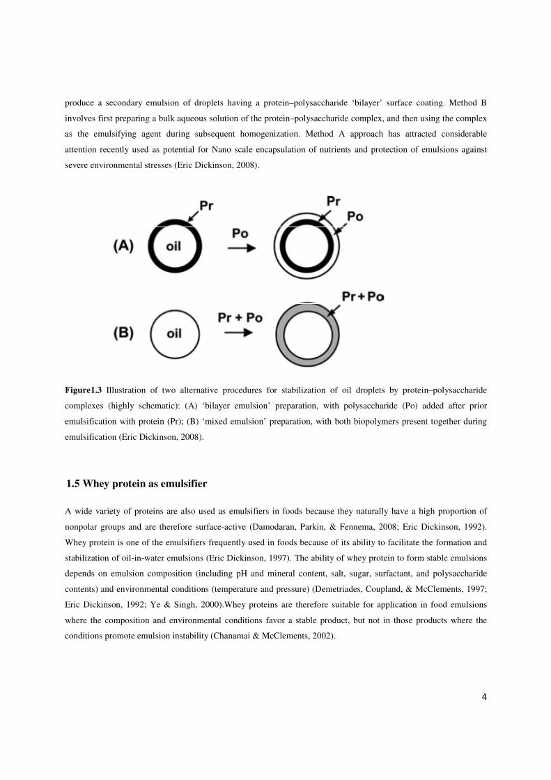
4
produce a secondary emulsion of droplets having a protein–polysaccharide ‘bilayer’ surface coating. Method B
involves first preparing a bulk aqueous solution of the protein–polysaccharide complex, and then using the complex
as the emulsifying agent during subsequent homogenization. Method A approach has attracted considerable
attention recently used as potential for Nano scale encapsulation of nutrients and protection of emulsions against
severe environmental stresses (Eric Dickinson, 2008).
Figure1.3 Illustration of two alternative procedures for stabilization of oil droplets by protein–polysaccharide
complexes (highly schematic): (A) ‘bilayer emulsion’ preparation, with polysaccharide (Po) added after prior
emulsification with protein (Pr); (B) ‘mixed emulsion’ preparation, with both biopolymers present together during
emulsification (Eric Dickinson, 2008).
1.5 Whey protein as emulsifier
A wide variety of proteins are also used as emulsifiers in foods because they naturally have a high proportion of
nonpolar groups and are therefore surface-active (Damodaran, Parkin, & Fennema, 2008; Eric Dickinson, 1992).
Whey protein is one of the emulsifiers frequently used in foods because of its ability to facilitate the formation and
stabilization of oil-in-water emulsions (Eric Dickinson, 1997). The ability of whey protein to form stable emulsions
depends on emulsion composition (including pH and mineral content, salt, sugar, surfactant, and polysaccharide
contents) and environmental conditions (temperature and pressure) (Demetriades, Coupland, & McClements, 1997;
Eric Dickinson, 1992; Ye & Singh, 2000).Whey proteins are therefore suitable for application in food emulsions
where the composition and environmental conditions favor a stable product, but not in those products where the
conditions promote emulsion instability (Chanamai & McClements, 2002).

5
1.6 Rheology
Rheology is defined as “the science of deformation and flow of matter.” (Macosko & Larson, 1994). Knowledge of
the rheological behavior of food products is essential for process design and evaluation, quality control, and
consumer acceptability. In addition, used by food scientists as an analytical tool to provide fundamental insights
about the structural organization and interactions of the components within emulsions, for example, measurements
of viscosity versus shear rate can be used to provide information about the strength of the colloidal interactions
between droplets (Quemada & Berli, 2002).
1.7 Classification of rheological behavior
The major types of fluid flow behavior can be described by means of basic shear diagram of shear rate versus shear
stress (Figure 1.4).
Newtonian Behavior: With Newtonian fluids, the shear rate is directly proportional to the shear stress and the plot
begins at the origin. Typical Newtonian foods are those containing compounds of low molecular weight (e.g.,
sugars) and that do not contain large concentrations of either dissolved polymers (e.g., pectins, proteins, starches) or
insoluble solids. Examples of Newtonian foods include water, sugar syrups, most honeys, most carbonated
beverages, edible oils, filtered juices and milk.
Shear-Thinning Behavior: With shear-thinning fluids, the curve begins at the origin of the shear stress-shear rate
plot but is concave upwards, that is, an increasing shear rate gives a less than proportional increase in shear stress.
Most non-Newtonian foods exhibit shear thinning behavior, including many salad dressings and some concentrated
fruit juices.
Shear-Thickening Behavior :In shear-thickening behavior also, the curve begins at the origin of the shear stress
shear rate plot and is concave downwards, that is, an increasing shear stress gives a
less than proportional increase in shear rate. This type of flow has been encountered in partially gelatinized starch
dispersions (Rao, 2007).

6
Figure1.4 Flow curves (shear stress vs shear rate) for different types of flow behavior (Norton, Spyropoulos, & Cox,
2011).
On the other hand, rheological models used to describe the behavior of fluids. There are different models that can be
used based on flow behavior such as power law that is widely used as a model for materials of shear thinning fluids
behaviour:
η� ��� ����
Where η is shear viscosity, γ� is shear rate m is the consistency index and n is the flow behaviour index (Tischer,
Iacomini, & Gorin, 2002). The model can describe a Newtonian, shear-thinning and shear-thickening behaviour,
depending on the value of the flow behaviour index, n. For a Newtonian material, n is equal to 1, and the equation
reduces to the Newtonian model. If n is less than 1, the fluid is shear thinning, whereas if it is greater than 1, then the
fluid is shear thickening (dilatant) (Miri, 2011). Shear-thinning behavior is very common in fruit and vegetable
products, polymer melts, as well as cosmetic and toiletry products. During flow, these materials may exhibit three
distinct regions (Figure1.5): a lower Newtonian region where the apparent viscosity (η0 ), called the limiting
viscosity at zero shear rate, is constant with changing shear rates; a middle region where the apparent viscosity (η) is
changing (decreasing for shear-thinning fluids) with shear rate and the power law equation is a suitable model for
the phenomenon; and an upper Newtonian region where the slope of the curve (η∞ ), called the limiting viscosity at
infinite shear rate, is constant with changing shear rates. The middle region is most often examined when
considering the performance of food processing equipment.
n<1
n=1
n>1

7
Figure1.5 Rheogram of idealized shear-thinning (pseudoplastic) behavior (Steffe, 1996).
8
2. Gum Tragacanth
2.1 History
Gum tragacanth was first described by Theophrastus several centuries before Christ. The name "tragacanth" comes
from the appearance of the exuded gum, which tends to form ribbons similar in appearance to a goat horn (from the
Greek "tragos" meaning goat and "akantha" meaning horn). The gum is obtained from small shrubs of the
Astragalus genus, are small, low bushy perennial shrubs having a large tap root along with branches, and grow
wildly in the dry deserts and mountainous regions of South West Asia, from Pakistan to Greece, and in particular, in
Iran and Turkey (Whistler, 1993). The main areas of commercial production are the arid and mountainous regions of
Iran (accounting for ~70% of the supplies). Fifty years ago, Iran exported annually more than 4,000 t, but in the
1970s and 1980s this amount greatly decreased, due to a number of reasons (Al-Tamimi, Palframan, Cooper,
Gibson, & Rastall, 2006). At present, the world market for gum tragacanth is estimated to be no more than 500
t/year (about 300 t) (FAO 1995). Plants develop a mass of gum in the centre of the root, which swells in the summer
heat. If the stem is slit, soft gum is exuded. The gum exudes readily from these cuts in the form of ‘ribbons’ or
‘flakes’ which become brittle on drying.
2.2 Structure
Gum tragacanth is a highly branched, heterogeneous hydrophilic carbohydrate with polymer. The molecular weight
is about 840 kDa. It is a complex, slightly acidic polysaccharide bounded with small proportions of protein (below
than 4%) (S. Balaghi, Mohammadifar, & Zargaraan, 2010), and with trace amounts of starch and cellulosic material
present. Calcium, magnesium and potassium are the associated cations (Anderson & Grant, 1988). After acid
hydrolysis, gum tragacanth commonly produces sugars of D-galacturonic acid, D-galactose, L-fucose (6-deoxy-L-
galactose), D-xylose, L-arabinose, L-rhamnose. The exact proportion of each sugar varies between different species
and in gums from different locations (Sima Balaghi, et al., 2011). The easy separation of tragacanthin and bassorin
suggests that the two polysaccharides are in a physical mixture and not chemically bonded (Lapasin & Pricl, 1995).
It has been reported that gum tragacanth consists of two fractions (Figure 2.1) (Phillips & Williams,). One fraction,
termed ‘Tragacanthic acid’ or Bassorin which represents 60–70% of the total gum with a molar mass of
approximately 105 Da, though insoluble in water, has the capacity to swell and form a gel. Another small fraction,
termed Tragacanthin is soluble in water with a molar mass of approximately 104 Da (Elias, 1992) to give a colloidal,
hydrosol solution. Bassorin, a pectic component, has a chain of (1-4)-linked α-D-galacturonic acid units some of
which are substituted at O-3 with β-D-xylopyranosyl units and some of these being terminated with D-Gal or L-Fuc
( Table 1) (Phillips & Williams, 2009).
The water soluble tragacanthin is reported as a neutral, highly branched arabinogalactan (of type II) comprising (1-
6)- and (1-3)- linked core chain containing galactose and arabinose (both in furanose and pyranose forms) and side
groups of (1-2)-, (1-3)- and (1-5)-linked arabinose units occurring as monosccharide or oligosaccharides (Table2.1)
(Tischer, et al., 2002). Depending on the species, the ratio of the water-swellable to the water-soluble fraction varies
(Sima Balaghi, et al., 2011).

9
Figure2.1 Gum tragacanth fractions (Sima Balaghi, et al., 2011)
Linkage analysis of the gum tragacanth showed that this material was composed of mostly terminal substituted
Fucp, 2-Xylp and 4-GalAp that made up fucoxylogalactronan structure in gum tragacanth and the results agree with
those structure previously presented by Aspinall & Baillie (1963) (Figure 2.2). Also, the presence of 1,5 Araf, 1,4-
Galp and 1,3,6 Galp linkages in the structure indicate of occurrence arabinogalactan in the tragacanth gum
(Tischer, et al., 2002) .
Figure2.2 Structure of partial part of gum tragacanth structure (Aspinall & Baillie, 1963)
Ethanol Insoluble (Tragacanthic Acid)
Water Soluble Fraction
(Tragacanthin)
Gum Tragacanth
Water Insoluble SwellingFraction
(Bassorin)
Ethanol Soluble minor Fraction
(Arabinogalactan)

10
Monosaccharide Linkage type % peak area
L-Fucose t-Fucp 23.9(69.3)
1,3-Fucp 2.8(8.1)
1,4-Fucp 3.2(9.3)
1,2-Fucp 3.8(11)
1,3,4-Fucp 0.8(2.3)
L-Rhamnose t-Rhap 0.1(3.3)
1,2,4 Rhap 2.7(90)
1,4 Rhap 0.2(6.7)
L-Arabinose t-Araf 5.2(53.6)
1,5 Araf 3.4(35)
1,2,5 Araf 1.1(11.4)
D-Galactose
t-Galp 1.7(29.8)
1,4-Galp 2.6(45.6)
1,6 Galp 0.2(3.5)
1,4,6 Galp 0.1(1.8)
1,3,6 Galp 1.1 (19.3)
D-Glucose t-Glup 0.2(11.8)
1,4-Glup 1.4(82.3)
1,2,4-Glup 0.1(5.9)
D-Xylose t-Xylp 7.2(21.2)
1,2,4-Xylp 0.8(2.4)
1,2-Xylp 24.9(73.5)
1,2,3-Xylp 1(2.9)
D-Galacturonic acid t-GalAp 0.1(3.3)
1,4-GalAp 2.8(93.3)
1,4,6-GalAp 0.1(3.3)
Table 2.1 Linkage analysis of gum tragacanth (Astragalus gossypinus)

11
2.3 Composition, structure and molecular size
Fully understand of functional and biological properties of polysaccharides needs to use special methods to find
relation between structure and functional properties. Hence, the approach to assessment of quantitative of the
monosaccharaides composition of gum tragacanth was done by means of acid hydrolysis (TFA 2M, 121 °C for 2 h)
where the efficiency of this method has been established previously byArnous and Meyer (2008).
The monosaccharide content of the hydrolysates was determined with the chromatography method HPAEC-PAD.
The separation and the quantification method using HPAEC-PAD were accomplished in one single run without the
need for pre-derivative treatment.
To determine effect molecular size of oligomers produce via enzymatic reaction on bioactivity properties of gum
tragacnth, High-performance size exclusion chromatography (HPSEC) has been done. The separation principle is
achieved by the differential exclusion from the pores of the packing material, of the sample molecules as they pass
through a bed of porous particles (Rasmussen & Meyer, 2009). The elution of polysaccharide is then achieved by
the time that larger molecules takes less time in the pores elute first and the smaller molecules elute later (Cutié &
Martin, 1995).
The complete structural elucidation of polysaccharide and enzymatic hydrolysis of oligomers has been done by
methylation analysis to reveal essential structural information on structure and linkage. This method (an ingenious
approach for determining linkage positions) was developed in the late 1960s and early 1970s. For glycosyl linkage
analysis, the sample was permethylated (Ciucanu & Kerek, 1984), depolymerized, reduced, and acetylated; and the
resulting partially methylated alditol acetates (PMAAs) analyzed by gas chromatography-mass spectrometry (GC-
MS) as described by York, etal., (1986). The PMAAs were analyzed on a Hewlett Packard 5975C GC interfaced to a
7890A MSD (mass selective detector, electron impact ionization mode); separation was performed on a 30m
Supelco 2330 bonded phase fused silica capillary column. However, the analyses of these linkage data are
sophisticated, usually incomplete and difficult, at least because of the following reasons: (1) most polysaccharides
are heterogeneous with high molecular weights, (2) there are many combinations of linkage types between any two
monosaccharides, and (3) there is a lack of a complete set of standard materials of all linkage types that could be
used as reference standards(Lo, Kang, Wang, & Chang, 2007).
2.4 Functional properties, stability and application
Gum tragacanth solutions are acidic, usually in the pH range of 5-6. Its maximum initial viscosity is at ph 8, but
usually exhibited maximum stability near pH 5 (Schwarz, Levy, & Kawagoe, 1958). So compared to other gums,
tragacanth gum is fairly stable over a wide pH range down to extremely acidic conditions at about pH 2 (Levy &
Schwarz, 1958).
In spite of the availability of alternative materials, the continued use of the gum is the result of its unique functional
properties (viscosity and emulsification) combined with a high degree of stability in a range of conditions. The flow
behavior of six species of Iranian gum tragacanth dispersions was investigated and results shown that all of the gum
dispersions had shear-thinning natures. Depending on the species of the gum, the viscosity of 1.5% solutions range
from 1.66 up to 34.6 Pa s (S. Balaghi, et al., 2010) .
12
Gum tragacanth, regarded as a bifunctional emulsifier, is a most efficient natural emulsifier for acidic oil-in-water
emulsions. Gum tragacanth has well-defined surface activity properties and produces a rapid lowering of the surface
tension of water at low concentration, less than 0.25% and facilitate emulsification (S. Balaghi, et al., 2010; karaya,
Phillips, & Williams, 2000). It thickens the aqueous phase and also lowers the interfacial tension between oil and
water. Dickinson et al., (1988) working with gum arabic has shown that polypeptide present is involved with the
surface activity and emulsification properties.
The use of gum tragacanth in foods has to be in accordance with the FDA Code of federal regulations (title 21,
section 184.1351; Table2.2).Gum tragacanth, a highly acid-resistant hydrocolloid, has been accepted since 1961 as
GRAS at the level of 0.2–1.3% (Anderson & Bridgeman, 1985) and in Europe, gum tragacanth has E-number E413
on the list of additives approved by the Scientific Committee for Food of the European Community. It has been used
for many years as a stabiliser, thickener, emulsifier and suspending agent in the food, pharmaceutical, cosmetic,
textile and leather industries as well as in technical applications based on its high viscosity at low concentration,
good suspending action, unusually high stability to heat and acidity and effective emulsifying properties. It also is
pourable and has a creamy mouth feel and good flavour-release properties (Phillips & Williams, 2009) and very
long shelf life (Levy, et al., 1958). Gum tragacanth is used in the food industry in citrus oil emulsions (Taherian,
Fustier, & Ramaswamy, 2008), salad dressings, condiments, sauces, bakery emulsions, oil and flavour emulsions,
bakery fruit-based fillings and toppings (give a shiny, clear appearance and a creamy texture) (Whistler, 1993) ,
confectionery, soft drinks, jellies, desserts, ice creams (provide texture to the product) , flavours , spices (Phillips &
Williams, 20). In chewy sweets, such as lozenges, it acts as a thickener and provides texture. The gum is used in
fruit tablets, gum drops, and pastilles as a binding agent during compression. It also provides body and mouthfeel
and ensures good flavor-release during consumption. In icings, gum tragacanth is used as a water binder,
maintaining pliability and preventing cracks and breaks. It also provides consistency, smooth texture, and creamy
taste to the product (Verbeken, Dierckx, & Dewettinck, 2003). Taherian et al. (2008) reported that the emulsions
produced based on arabic gum and tragacanth have a greater stability than the emulsions containing arabic gum and
xanthan which was related to higher surface activity and greater acid and heat resistances by tragacanth gum.
Recently, the use of gum Tragacanth to maintain the quality of bell peppers (one of the most important commercial
vegetables) during long-term storage has been recommended (Mohebbi, Amiryousefi, Hasanpour, & Ansarifar,
2012). Gum tragacanth addition to nonfat fermented milk drink (doogh) was found beneficial for improving physical
properties and prevents serum separation (Gorji, Mohammadifar, & Ezzatpanah, 2011). Use of gum tragacanth in
dairy cream fat showed that fat reduction from 30 wt% to 14 wt% without significant changes to sensory properties,
shelf life or packaging requirements (Nasirian, Vaziri, Safekordi, & Ardjmand, 2010).
2.5 Mechanism of tragacanth gum stabilization
There have been different elucidations to how gum tragacanth can stabilize of different emulsion system.
Yorkoyama, Srinivasan and Fogeler (1988) shown that the stabilization effect of tragacanth is a result of the steric
repulsion force and the stability can be controlled by changing pH. (Yokoyama, Srinivasan, & Fogler, 1988) . On the
other hand, the ability of tragacanth gum in stabilizing the beverage emulsions could be due to its residual surface

13
activity and enhancement of emulsion viscosity (Rezvani, Schleining, & Taherian, 2012). Azarikia, et al. (2010)
stated that tragacanth gum has zeta potential around -21 mV and could stabilize doogh (Iranian yoghurt drink) via
electrostatic interactions. This phenomenon has been related to the negatively charged carboxylic groups of
galacturonic acid as the main backbone of tragacanthin as the soluble part of the tragacanth gum. In addition, they
reported that bassorin is probably unable to interact with caseins, and its main effect on the stabilization of doogh is
to increase the viscosity of the continuous phase.
Table2.2 Maximum usage levels (%) of gum tragacanth permitted in accordance with the FDA Code of federal
regulations
Food (as served) Percentage Function
Baked goods and baking mixes 0.2 Emulsifier, emulsifier salt,
formulation aid, stabilizer/thickener
Condiments and relishes 0.7 Emulsifier, emulsifier salt,
formulation aid, stabilizer/thickener
Fats and oils 1.3 Emulsifier, emulsifier salt,
formulation aid, stabilizer/thickener
Gravies and sauces 0.8 Emulsifier, emulsifier salt,
formulation aid, stabilizer/thickener
Meat products 0.2 Formulation aid, stabilizer/thickener
Processed fruits and fruit juices 0.2 Emulsifier, emulsifier salt,
formulation aid, stabilizer/thickener
All other food categories 0.1 Emulsifier, emulsifier salt,
formulation aid, stabilizer/thickener
Anionic hydrocolloids (λ-carrageenan, carboxymethyl cellulose, pectin and gum tragacanth) interact with the
positive charges on the surface of casein micelles and reduce the syneresis via the formation of protein–
polysaccharide complexes and strengthen the casein network through a bridging mechanism. The electrostatic
interactions between gum tragacanth and milk proteins have been studied in both real and model systems. The effect
of pH and ionic strength on the formation of complexes between β-lactoglobulin and soluble part of gum tragacanth
(exudates form A. gossypinus) suggests an electrostatic nature of their interactions (Mohammadifar, Musavi, &
Williams, 2007). It is well-recognized that emulsion stabilization by whey proteins may be improved by
polysaccharides (and particularly so at pH near the isoelectric point). For pectin-β-lactoglobulin complexes it has
been shown that this effect occurs when the protein associates to the polysaccharide, and soluble, charged

complexes are formed, which have an overall charge as the polysaccharide (
complex is thus stabilized by electrostatic repulsion. From model
appears that the polysaccharide effect is mainly due to enhanced steric stabilization provided by the bulkiness of the
hydrophilic polysaccharides that provides a stabilizing charged layer around the protein stabilized oil droplets which
protects the protein stabilized oil droplets against flocculation under conditions where electrostatic stabilization is
less favourable (Akhtar & Dickinson, 2003).
In the summary, the tragacanth gum stabilization of protein
mechanisms: Firstly, formation of non
interaction, and secondly viscosity increase
2.6 Rheological characterization and particle size of tragacanth gum
Rheological characterization of polysaccharides can be importance as it provides fundamental information required
for assessment of some of the final properties of a product, such as quality, storage stability, effect of formulation
variables on product characteristics
of Iranian gum tragacanth dispersions was in
concentration range (0.05–1.5% w/w) using a controlled shear rate rheometer. The steady shear measurements
showed that all of the gum dispersions had shear
In order to have a better understanding about functional properties of different species of gum tragacanth, also the
particle size distribution of all gum dispersions was determined. Size measurements were reported as the volume
weighted mean diameter (S. Balaghi, et al., 2010)
d32 = ∑nidi3/∑nidi
2
d43 = ∑nidi4/∑nidi
3
where ni is the number of particles with diameter
complexes are formed, which have an overall charge as the polysaccharide (Sperber,et
is thus stabilized by electrostatic repulsion. From model-studies with dextran-
appears that the polysaccharide effect is mainly due to enhanced steric stabilization provided by the bulkiness of the
at provides a stabilizing charged layer around the protein stabilized oil droplets which
protects the protein stabilized oil droplets against flocculation under conditions where electrostatic stabilization is
less favourable (Akhtar & Dickinson, 2003).
the summary, the tragacanth gum stabilization of protein-emulsified emulsions is probably a result of two
mechanisms: Firstly, formation of non-covalent protein–(gum) polysaccharide complexes via electrostatic
interaction, and secondly viscosity increase by insoluble fraction (Bassorin) (Figure2.3).
Rheological characterization and particle size of tragacanth gum
Rheological characterization of polysaccharides can be importance as it provides fundamental information required
of the final properties of a product, such as quality, storage stability, effect of formulation
variables on product characteristics (Ramachandran, Chen, & Etzler, 1999).Hence, the flow behavior of six species
of Iranian gum tragacanth dispersions was investigated at different temperatures and ionic strengths, within a
1.5% w/w) using a controlled shear rate rheometer. The steady shear measurements
showed that all of the gum dispersions had shear-thinning natures (Figure2.4) (S. Balaghi, et al., 2010)
In order to have a better understanding about functional properties of different species of gum tragacanth, also the
particle size distribution of all gum dispersions was determined. Size measurements were reported as the volume
(S. Balaghi, et al., 2010):
is the number of particles with diameter di.
Figure2.3 The scheme of stabilization mechanism of
emulsions by Gum Tragacanth
14
,et al., 2009). The soluble
-whey protein conjugates it
appears that the polysaccharide effect is mainly due to enhanced steric stabilization provided by the bulkiness of the
at provides a stabilizing charged layer around the protein stabilized oil droplets which
protects the protein stabilized oil droplets against flocculation under conditions where electrostatic stabilization is
emulsified emulsions is probably a result of two
(gum) polysaccharide complexes via electrostatic
Rheological characterization of polysaccharides can be importance as it provides fundamental information required
of the final properties of a product, such as quality, storage stability, effect of formulation
.Hence, the flow behavior of six species
vestigated at different temperatures and ionic strengths, within a
1.5% w/w) using a controlled shear rate rheometer. The steady shear measurements
Balaghi, et al., 2010).
In order to have a better understanding about functional properties of different species of gum tragacanth, also the
particle size distribution of all gum dispersions was determined. Size measurements were reported as the volume
The scheme of stabilization mechanism of protein-emulsified

15
The results showed that different value on particle size parameters (Table2.3). The variation in the particle size
distribution of different species of gum dispersions may be attributed to differences in the swelling power of
different gum particles which seems to be related to ratio of soluble to insoluble part. It was shown that multi-factors
such as different amount of neutral sugars, uronic acid, and also methoxyl group content as well as molecular weight
and conformational and configurational properties play their own crucial role in rheological properties.
In general, the results indicated that the six varieties of gum tragacanth studied exhibited significantly different
rheological properties; therefore, these different gums may find use in a variety of applications as stabilisers,
thickeners, emulsifiers and suspending agents depending on their rheological behaviour. Consequently, any research
carried out on gum tragacanth or any industrial application of this gum without respect to the plant species will lead
to misleading results.
Table 2.3 Particle size characteristics of six species of gum tragacanth dispersions (0.05% w/w) (S. Balaghi, et al., 2010)
A. parrowianus A. fluccosus A. rahensis A. gossypinus A. microcephalus A.compactus
D [3,2]* 98.72d 98.63d 97.70d 143.63a 112.43c 127.60b
D [4,3]* 142.50d 136.34e 155.49d 357.63a 263.69b 196.27c
*Units: micrometer a–e Means with different letters within the same row differed significantly (p < 0.05)

16
A.microcephalus
A.gossypinus
A.compactus
A.fluccosus
A.rahensis
A.parrowianus
Figure2.4 Flow curves of apparent viscosity versus shear rate of gum tragacanth
suspensions in different concentrations (w/w %) at 25 °C (S. Balaghi, et al., 2010)

17
3. Effect of the galacturonic acid content and methoxylation degree of gum
tragacanth (Astragalus spp.) on emulsion stability
This chapter examines correlation of compositional make-up of six different gum tragacanth exudates from
Astragalus spp on protein based emulsions (Paper I,III).
Paper I: Stabilization of emulsions by gum tragacanth (Astragalus spp.) correlates to the
galacturonic acid content and methoxylation degree of the gum
Hassan Ahmadi Gavlighi, Anne S. Meyer, Dayang N. A. Zaidel, Mohammad Amin Mohammadifar,
J. Dalgaard Mikkelsen
Food Hydrocolloids, Volume 31, Issue 1, May 2013, Pages 5–14
http://dx.doi.org/10.1016/j.foodhyd.2012.09.004
3.1 Significance of study
Gum tragacanth has been reported to have both emulsifying and stabilizing properties in emulsions, i.e. being “bi-
functional” by having both the capacity to facilitate emulsification as well as providing stabilization of the emulsion
after its formation (Weiping, 2000). There are indications in the literature that compositional differences of tragacanth
gums obtained from different species of Astragalus affect the rheological properties, including the viscosity of
tragacanth solutions (Balaghi et al., 2011). However, despite the long and extensive use of gum tragacanth as a
stabilizer in emulsion systems, surprisingly little is known about structure function relationships, and the traits of the
gum that confer emulsifying properties are presently unknown. It is of course tempting to hypothesize that the
composition of tragacanth gum affects its stabilization effects in emulsions, and it has been reported that the terminal
deoxyhexoxyl groups (i.e. fucose) or the methoxyl groups of the homogalacturonan in the tragacanth gum structure may
play a role in the emulsion stabilization (Stephen, 1990). However, to our knowledge, no unequivocal evidence has up
until now been provided for this hypothesis. Hence, the exact significance of specific structural components of
tragacanth gum in relation to viscosity and emulsion stabilization properties is unclear. The purpose of this study was to
evaluate the stabilization of emulsions by tragacanth gum, and to aim at obtaining an understanding of any relationships
between the make-up of gum tragacanth and its stabilization properties in emulsions.
3.2 Experimental considerations
In the beginning of work, six different of tragacanth gum from Astragalus species (A. parrowianus, A. fluccosus, A.
rahensis, A. gossypinus, A. microcephalus, and A. compactus) growing in different regions of Iran were collected .The
dried raw gums were ground with a coffee mill, sieved, solubilized in deionized water, then freeze dried, and finally
used in this study. In order to determine the ratio between the water-soluble and the water-insoluble fractions of the
gums, each gum sample was re-suspended overnight in deionized water (1 % dry matter gum weight/volume) and the
separation of the soluble and insoluble fraction was done by centrifugation. The monosaccharide composition and

18
methyl and acetyl group of each of the gum tragacanth samples was determined. To confirm our hypoesthesia about
effect of methoxyl content , saponifiaction of tragacanth gum solution has been done (Leroux, et al., 2003).
Emulsion was prepared and stabilization and particle size determined via method explained previously by Zaidel et al
(2012) to investigate correlation between composition structure of gums and creaming index and particle size. Particle
size of fat globules (oil phase) and their size distribution play a predominant role in deciding the stability of emulsion
and emulsion based products with precisely controlled particle size exhibit better emulsion stability (McClements,
1999).The stability of an emulsion to gravitational separation can be enhanced by reducing the droplet size. As a rule,
large globules tend to coalesce faster than the small ones (Chiewchan, Phungamngoen, & Siriwattanayothin, 2006).
WPI was used as an emulsifier because whey proteins, having pI near 5, are amphiphilic molecules near the isoelectric
point 4<pH<6. Whey proteins, essentialy β-lactoglobulin, are widely used as emulsifiers in food applications because
they are inexpensive, natural, readily available, and have the ability to facilitate formation and stabilization of oil-in-
water emulsions in systems having pH 4-6 (Dickinson, 2001). In this particular system a pH of 4.5, slightly below the
pI of the whey protein, was used to enhance the electrostatic attraction between the (potentially pectic) negatively
charged gum tragacanth polysaccharides and the protein.
Also, tragacanth gum rheological properties of different gum solution and emulsions were evaluated to compare any
difference in interaction between gums in the emulsion system.
3.3 Highlights
It has been demonstrated that different tragacanth gum samples obtained from different species of Astragalus have
different composition, and produce different levels of soluble and insoluble gum fractions. The results shown that the
gums from A. parrowianus and A. fluccosus had relatively high tragacanthin:bassorin ratios of ~66:34 and ~75:25,
respectively, whereas in the other gums this ratio approached 50:50 (A. rahensis, A. microcephalus, A. compactus) or
tipped towards higher bassorin than tragacanthin (A. gossypinus). The monosaccharide make-up of the six gums also
varied, but all the gums contained relatively high levels of galacturonic acid (∼100-330 mg/g), arabinose (50-360 mg/g),
xylose (∼150-270 mg/g), and galactose (∼40-140 mg/g), and also contained fucose, rhamnose, and glucose.
Galacturonic acid was high in the soluble part of all species whereas L-fucose and partially xylose was major in
insoluble fraction. A positive correlation between the methoxyl content of the soluble part of the gums and creaming
index indicate that methoxyl groups may have properties that play a role in the emulsification properties of tragacanth
gums Fig 1. The results obtained after removing the methyl groups from the A. fluccosus gum also confirmed that gum
tragacanth with methoxyl was acting better as an emulsion stabilizer than the corresponding gum with no methyl
groups.
The viscosities of the emulsions were considerably higher than the gum tragacanth solutions, but the overall flow
behavior in the emulsions with gum tragacanth added were quite similar to the behavior observed in the gum tragacanth
solutions (See Fig. 4 in paper). Increased viscosity of emulsions may be a result of hydrophobic bonding between
tragacanth gum and WPI emulsified emulsion particles. Clearly, the viscosity is a factor, but apparently also the
composition, notably the total galacturonic acid content in the gum, the amount of methoxyl groups and probably also
fucose in the solubilized part are factors determining how tragacanth gums work to stabilize emulsions.

19
The n (flow behavior index) was found to be lower than 1 for all tragacanth gum solutions as well as for all the
emulsions (See Table 2 in paper); this value of less than 1 confirmed the shear thinning behavior of the samples.
The tragacanth gum stabilization of protein-emulsified emulsions is probably a result of two mechanisms: Firstly,
formation of non-covalent protein–(gum) polysaccharide complexes, and secondly viscosity increase. It is well-
recognized that emulsion stabilization by whey proteins may be improved by polysaccharides (and particularly so at pH
near the isoelectric point).
To conclude, the work provides some important clues to the emulsion stabilization mechanisms in relation to the
composition of tragacanth gums mainly soluble methoxyl content and galacturonic acid content.
Figure3.1 Correlation between methoxyl content in the soluble fraction of different gum tragacanth samples from
different Astragalus species and Creaming Index.
y = -0.0283x + 0.6927R² = 0.9294
0
0.1
0.2
0.3
0.4
0.5
0.6
0.7
0.8
0 5 10 15 20 25 30
Cre
am
ing
In
dex
ra
tio
Soluble Methoxyl Content (mg/g)

lable at ScienceDirect
Food Hydrocolloids 25 (2011) 1775e1784
Contents lists avai
Food Hydrocolloids
journal homepage: www.elsevier .com/locate/ foodhyd
Compositional analysis and rheological characterization of gum tragacanthexudates from six species of Iranian Astragalus
Sima Balaghi a, Mohammad Amin Mohammadifar a,*, Azizollaah Zargaraan a,Hassan Ahmadi Gavlighi b, Mehrdad Mohammadi a
aDepartment of Food Science and Technology, National Nutrition and Food Technology Research Institute, Faculty of Nutrition Sciences and Food Technology,Shahid Beheshti University of Medical Sciences, P.O. Box 19395-4741, Tehran, IranbBioProcess Engineering Center, Department of Chemical Engineering, Technical University of Denmark, DK-2800 Kgs. Lyngby, Denmark
a r t i c l e i n f o
Article history:Received 3 July 2010Accepted 11 April 2011
Keywords:Gum tragacanthSugar compositionStrain sweepFrequency sweepIonic strength
* Corresponding author. Tel.: þ98 2122648120; faxE-mail address: [email protected] (M.A. Mohamm
0268-005X/$ e see front matter � 2011 Elsevier Ltd.doi:10.1016/j.foodhyd.2011.04.003
a b s t r a c t
The sugar composition and viscoelastic behaviour of Iranian gum tragacanth exuded by six species ofAstragaluswas investigated at a concentration of 1.3% and varying ionic strength using a controlled shear-rate rheometer. Compositional analysis of the six species of gum tragacanth by high-performance anion-exchange chromatography with pulsed amperometric detection suggested the occurrence of arabinose,xylose, glucose, galactose, fucose, rhamnose and galacturonic acid residues in the gum structure;however, the proportions of each sugar varied significantly among the gums from the different species ofAstragalus, and this variation led to interesting differences in functional properties. Rheologicalmeasurements performed on dispersions of the six species of gum tragacanth demonstrated viscoelasticproperties. The mechanical spectra derived from strain sweep and frequency sweep measurementsindicated that the different gum tragacanth dispersions had distinctive viscoelastic behaviours. Inves-tigation of the viscoelastic properties of the different gum dispersions in the presence of NaCl revealedthat the addition of NaCl could lead to slight to drastic decreases in the G0 , G00 or h* values of the variousgums. In general, the results indicated that the six varieties of gum tragacanth studied exhibitedsignificantly different rheological properties; therefore, these different gums may find use in a variety ofapplications as stabilisers, thickeners, emulsifiers and suspending agents depending on their rheologicalbehaviour.
� 2011 Elsevier Ltd. All rights reserved.
1. Introduction
Gum tragacanth, a dried exudate obtained from the stems andbranches of Asiatic species of Astragalus, is a very complex hetero-geneous anionic polysaccharide of highmolecularweight (Weiping,2000) and consists of two main fractions: a water-insolublecomponent called bassorin, which has the capacity to swell andform a gel, and a water-soluble component called tragacanthin(Balaghi, Mohammadifar, & Zargaraan, 2010). The easy separation oftragacanthin and bassorin suggests that the twopolysaccharides arein a physical mixture and not chemically bonded (Lapasin & Pricl,1999). It has been reported that gums tragacanth from differentspecies of Astragalus have different ratios of the two fractions,different chemical compositions and also varying physicochemicalproperties; therefore, different functionalities and applications for
: þ98 2122376470.adifar).
All rights reserved.
each species are expected (Balaghi et al., 2010). Gum tragacanth,a highly acid-resistant hydrocolloid, has been accepted since 1961 asGRAS at the level of 0.2e1.3% (Anderson&Bridgeman,1985) and hasbeen used for many years as a stabiliser, thickener, emulsifier andsuspending agent in the food, pharmaceutical, cosmetic, textile andleather industries as well as in technical applications based on itshigh viscosity at low concentration, good suspending action,unusually high stability to heat and acidity and effective emulsifyingproperties. It also is pourable and has a creamymouth feel and goodflavour-release properties (Weiping, 2000) and very long shelf life(Levy & Schwarz,1958). Gum tragacanth is used in the food industryin salad dressings, condiments, sauces, bakery emulsions, oil andflavour emulsions, fillings and toppings, confectionery, soft drinks,jellies, desserts, ice creams, flavours and spices (Weiping, 2000).
To date, a wide variety of gums has been extensively investi-gated for their structural characteristics, functional properties andapplication properties. A number of studies have been devotedto elucidate the effects of various processes (salting, heating,acidification, ultrasonication, irradiation, high pressure, etc.) and

S. Balaghi et al. / Food Hydrocolloids 25 (2011) 1775e17841776
processing parameters (time, temperature and rate of a process) aswell as the ratio, type and concentration of ingredients on thefunctional properties of a broad range of hydrocolloids (Ahmed &Ramaswamy, 2004; Dogan, Kayacier, & Ic, 2007; Farhoosh & Riazi,2007; Mu et al., 2010). Dynamic rheology is one of the methodsmost extensively used to assess the viscoelastic behaviour ofpolysaccharide solutions/dispersions or gels. The viscoelasticproperties of various gums such as xanthan, guar (Mills & Kokini,1984), pectin (Gigli, Garnier, & Piazza, 2009), gelatin (Gómez-Guillén et al., 2002) and mucilage (Medina-Torres, Brito-De LaFuente, Torrestiana-Sanchez, & Katthain, 2000) have been reportedby numerous researchers. Although the physicochemical propertiesand steady-state rheological evaluation of gum tragacanth havebeen recently established (Balaghi et al., 2010), only a few studieshave dealt with the viscoelastic behaviour of gum tragacanth(Mohammadifar, Musavi, Kiumarsi, & Williams, 2006). However,the viscoelastic properties of gum solutions/dispersions areparticularly important for food processors in adjusting processingparameters, monitoring consistency as well as predicting thestability of fluid food systems and the final textural attributes offormulated foods.
One of the factors that seem to have a direct bearing on therheological behaviour of the gum is the sugar composition. Thesugar compositions of many hydrocolloids such as xanthan, locust-bean galactomannans (Lazaridou, Biliaderis, & Izydorczyk, 2001)and mucilage gum (Medina-Torres et al., 2000) have been reportedin the literature. The determination of sugar composition might beuseful in developing microstructureefunction relationships forsystems of gum dispersions and intermolecular interactions. In1988, Anderson and Grant determined the sugar composition ofseveral gums tragacanth by formic acid hydrolysis followed bypaper chromatography (Anderson & Grant, 1988).
The ionic strength of the medium is a crucial variable influ-encing the networking mechanism of charged biopolymers. Theeffects of NaCl addition on the linear viscoelastic properties ofvarious gum dispersions and polysaccharide gels have been previ-ously investigated. Since the addition of salt may alter the chainflexibility and conformation features of a polymer, differentphenomena were observed in these investigations. The formationof junction zones in the gel network can be controlled by manyfactors such as the content and type of the counter ion. However,when gelation does not involve the formation of an orderedstructure, the behaviour is strongly influenced by the presence ofa counter ion (Silva, Brito, de Paula, Feitosa, & Paula, 2003). To thebest of our knowledge, the effect of NaCl on the linear viscoelasticproperties of gum tragacanth has not been examined.
Owing to the fact that various varieties of gum tragacanthpossess different chemical compositions and physicochemicalproperties (Balaghi et al., 2010), understanding the rheologicalproperties of gum tragacanth is essential for evaluating the gumsfrom different species and their potential applications and use asfood thickeners, stabilisers or emulsifiers. Unfortunately, most ofthe previous studies have been done on gums tragacanth fromunknown botanical sources; moreover, the experimental condi-tions such as the applied shear rate, temperature and solvent werenot clearly explained (Anderson & Grant, 1988; Azarikia & Abbasi,2010; Chenlo, Moreira, & Silva, 2010; Stauffer & Andon, 1975;Zahedi, Vedadi, & Dollimore, 1979). Therefore, the aim of thepresent study was to determine the sugar compositions and tocharacterise the dynamic rheological properties of six varieties ofgum tragacanth at different ionic strengths, as this information isnecessary to gain insight into the functional properties of thisbiopolymer and should be useful in the processing of tragacanthgum for scientific research as well as for potential industrialapplications.
2. Materials and methods
2.1. Plant materials and standards
Iranian gum tragacanth exuded by various species of Astragalus(Astragalus parrowianus, Astragalus fluccosus, Astragalus rahensis,Astragalus gossypinus, Astragalus microcephalus and Astragalus com-pactus) was collected from plants growing in different provincesof Iranincluding Isfahan, Khorasan, Semnan and Shiraz. The plantswere tappedwith a knife by making careful longitudinal incisions inthe taproot and the bark of the branches. The gum readily exudedfrom these cuts in the formof ‘ribbons’ that becamebrittle on drying.Taxonomic identification of the specimens was done by Dr. AliMasoumi, an academicmember of the Forest, Range, andWatershedManagement Organisation of Iran. Deionised (Milli-Q) waterwas used for all experimental work. Neutral monosaccharide stan-dards, L-fucose, L-rhamnose, D-arabinose, D-galactose, D-glucose,D-mannose, and D-galacturonic acid were obtained from SigmaAldrich Chemical Co. (St. Louis, MO, USA). Trifluoroacetic acid (99%)was from Merck (Darmstadt, Germany). NaOH standard solutionHPLC grade was from Fluka/SigmaeAldrich Chemical Co. (St. Louis,MO, USA).
2.2. Native gum tragacanth sample preparation
The raw gum tragacanth was powdered in a high-speedmechanical blender and later sieved to obtain uniform samples.Powdered gum with a mesh size between 200 and 500 mm wasused in this study.
2.3. Sample preparation for high-performance anion-exchangechromatography with pulsed amperometric detection (HPAEC-PAD)
Approximately 15e20 mg of powdered gum tragacanth sample(200e500 mmparticle size) was transferred to a screw-cap vial plusdeionised water for 2e4 h (depends on variety of Tragacanthbecause some of them is dissolved faster than others) at roomtemperature. After dissolving samples Trifluoroacetic acid (TFA;2 M, 600 mL) was added by micropipette. The apparatus was thenheated in a drying oven maintained at 121 �C for 2 h without stir-ring and then allowed to cool at room temperature. Hydrolysateswere lyophilised and kept at�20 �C under N2 until analysis. Prior toanalysis by HPAEC-PAD, the sample hydrolysates were redissolvedin 5 mL of double deionised water and then filtered through0.20mmnylon syringe filters into the sample vials used for the AS50auto sampling chromatography system (Arnous & Meyer, 2008).
2.4. Determination of carbohydrate composition
The monosaccharide contents of the hydrolysates and standardswere determined with a BioLC system consisting of a GS50 gradientpump, an ED50 electrochemical detector and an AS50 chromato-graph coupled to an AS50 autosampler (Dionex Corp., Sunnyvale,CA). Separations were performed using a CarboPacTM PA20(3 mm� 150 mm) analytical column (Dionex Corp., Sunnyvale, CA)according to the method of Arnous and Meyer (2008). The eluentflow rate was 0.5 mL/min. The monosaccharides were separatedwith adequate resolution using a two-eluent system consisting ofdeionised water (18.2 mU at 25 �C) and 500 mM aqueous NaOH.Neutral monosaccharides were eluted isocratically with 2.5 mMNaOH for 20 min followed by a second isocratic elution at highNaOH (500 mM) for 10 min to elute any acidic monosaccharidespresent (galacturonic). This high concentration of NaOH simulta-neouslywashed the column. Before each injection (10 mL), a columnre-equilibration program was run for 5 min with 100 mM NaOH

S. Balaghi et al. / Food Hydrocolloids 25 (2011) 1775e1784 1777
followed by 5 min with 2.5 mM NaOH. The different carbohydratestandards were mixed proportionally to resemble the matrix of thestudied samples. The following pulse potentials and durations wereused for detections: E1¼0.1 V, t1¼400 ms; E2¼�2 V, t2¼ 20 ms;E3¼ 0.6 V, t3¼10 ms; E4¼�0.1 V, t4¼70 ms, data-collection rate:0.2 Hz (Arnous & Meyer, 2008).
2.5. Preparation of dispersions for rheological analysis
Gum tragacanth powder (1.3 g) was accurately weighed anddispensed into 100 mL pure deionised water and also in 100 mL of0.2 M NaCl. The whole gum dispersions were kept on a magneticstirrer at room temperature and gently stirred for 2 h. Later, thegum dispersions were allowed to stand at 3�1 �C for 24 h toenable biopolymer hydration.
2.6. Rheological measurements
Oscillatory shear measurements were performed with a PhysicaMCR 301 rheometer (Anton-Paar, GmbH, Graz, Austria) usinga serrated plate-and-plate system (25 mm in diameter, 0.6 mmgap). Temperature control was carried out with a Peltier systemequipped with a fluid circulator. All samples were left standing forfive minutes to allow structure recovery and temperature equili-bration. The samples were covered with a solvent trap to preventevaporation. All experiments were carried out at 25 �C. Strainsweep tests were performed (0.01e1000%, 1 Hz) to determine: 1)the limiting value of the linear viscoelastic range (LVE or gL); 2) thestructural strength (G0 at LVE); 3) the resistance to mechanical forceor yield stress(sy), which is also a measure of structural strengthand can be determined from the limiting value of LVE range interms of shear stress (it determined visually or manually from thelinear viscoelastic plateau and from the data table at which the G0
values are just beginning to deviate noticeably from the previouslyconstant values.; 4) the flow point (sf), the stress at which theinternal structure breaks to the extent that it causes the material toflow (G0 ¼G00); and 5) the damping factor (tan d) or the ratio of lossmodulus to elastic modulus, to provide a direct view of whether thesamples behaved as liquids or solids (Mezger, 2006). Frequencysweep tests were performed using a frequency ramp from 0.01 to100 Hz. The experimental data were fitted to a power-law model.
2.7. Statistical analysis
Analytical values are based on the mean and standard deviationof three replicates. For all rheological measurements, the reportedvalues are based on the mean of three replicates. Analysis of vari-ance (ANOVA) was used for the data analysis (SPSS, 16). When theF-values were significant (p< 0.05) in ANOVA, Duncan’s multiple-range test was used to compare treatment means. Spearman
Table 1Sugar compositions of gums tragacanth from six species of Astragalus.
A. parrowianus A. fluccosus A.
Arabinose 39b 23d 51Xylose 10e 24b 11Glucose 10b e 13Fucose 7d 8d e
Galactose 8a 7a 7Rhamnose e e e
Galacturonic acid 21d 34b 9Galacturonicþ fucose 28e 42c 9Arabinose/(xyloseþ fucose) 2.29b 0.71d
Values with different letters in each row are significantly different (p< 0.05).In all cases the standard deviations were lower than 2%.
correlation-coefficient analyses were used to evaluate potentialrelations between the sugar compositions of the samples and theirrheological parameters and also between the different rheologicalparameters.
3. Results and discussion
3.1. Sugar composition
The sugar compositions of the six varieties of gum tragacanthdetermined by HPAEC-PAD are listed in Table 1. The results revealedthe presence of galacturonic acid, xylose, fucose, arabinose, galactose,glucose, and traces of rhamnose; however, the proportions of eachsugar varied among the gums from various species of Astragalus.
It has been reported that gums with higher viscosity containhigh proportions of fucose, xylose, galacturonic acid and methoxylgroups and low proportions of arabinose and nitrogenous fractions(Anderson & Grant, 1988). However, by considering our previousresearch data regarding the consistency coefficient of gum traga-canth (Table 2) from different Astragalus species (Balaghi et al.,2010), we didn’t find any relationship between consistency coeffi-cient and the amount of each individual sugar; but we found thatgums with higher amounts of galacturonic acid and fucose andusually lower amounts of xylose and arabinose had higher values ofthe consistency coefficients. The Spearman correlation coefficientsand p values between the sum of galacturonic acid and fucose andthe consistency coefficient (m) as well as between arabinose andthe consistency coefficient, fucose and the consistency coefficient,and between arabinose/(xyloseþ fucose) and the consistencycoefficient were þ1 (p¼ 0), �0.943 (p¼ 0.005), þ0.900 (p¼ 0.037)and �0.943 (p¼ 0.005), respectively. The relations between thesugar compositions and the consistency coefficients are illustratedin Fig. 1. According to Fig. 1, gums with higher amount of gal-acturonic acid and fucose have higher values of consistency coef-ficients. On the contrary, gums with higher amount of xylose andarabinose have higher lower values of consistency coefficients.Spearman correlation coefficient and p value between consistencycoefficient and sum of xylose and arabinose were �0.943 andp¼ 0.005.
The results showed that the content of galacturonic acid variedbetween 9 and 37% among the six varieties of gum tragacanth. Thecontent of arabinose, which has been reported to be the majorcomponent of the side chains of the 1,4-linked a-D-galacturonicacid backbone of gum tragacanth, had a wide percentage range(1e51%) among the six gums. In contrast with A. gossypinus, whichcontained 1% arabinose and 37% galacturonic acid, gum tragacanthfrom A. rahensis interestingly had arabinose and galacturonic acidcontents of 51% and 9%, respectively. The results also indicated thatthe fucose contents of the different gums ranged from 0% forA. rahensis to 35% for A. compactus. It was also observed that
rahensis A. gossypinus A. microcephalus A. compactusa 1f 35c 7ee 32a 19d 21ca 1c 10b 2c
23b 9c 35aa 1b 2b 2b
1a 1a 1ae 37a 21d 30cf 60b 30d 65a
4.6a 0.02f 1.25c 0.12e
Table 2Consistency coefficients for 1.5% dispersions of the six varieties of gum tragacanth in the absence of added NaCl at 25 �C (Balaghi et al., 2010).
A. parrowianus A. fluccosus A. rahensis A. gossypinus A. microcephalus A. compactus
Consistency coefficient(Pa sn) 11.29 18.46 1.66 23.70 13.06 36.04Ratio of soluble to insoluble fractions 1.74 3.15 1.30 0.51 1 0.84
S. Balaghi et al. / Food Hydrocolloids 25 (2011) 1775e17841778
A. fluccosus gum had no glucose residues. The contents of xylose,the other constituent of the side chains linked to the galacturonicacid main chain, varied from 10 to 32% among the six species(Table 1). This wide variation in the neutral sugar and uronic acidcontents of gum tragacanth from different species of Astragalusmay imply the occurrence of various numbers as well as differentlengths of the side chains, which are involved in the primarystructure of gum tragacanth.
The accurate names of the gum tragacanth fractions based onthe work of Aspinall and Baillie and Norman has been illustrated inFig. 2 (Aspinall & Baillie, 1963a, 1963b; Norman, 1931). There hasbeen a large number of publications such as books and articlesabout polysaccharides and hydrocolloids (Chenlo et al., 2010;Izdorczyk, Cui, & Wang, 2005; Ramsden, 2004; Vebeken, Dierckx,& Dewettinck, 2003; Weiping, 2000; William, Phillips, Stephen, &Churms, 2006), which is for the most part cited by researchpapers, incorrectly reporting that gum tragacanth consists oftragacanthic acid as an insoluble fraction and arabinogalactan asa soluble fraction, and in some cases it is stated that tragacanthin isa neutral arabinogalactan minor fraction, or it is stated that bas-sorin and tragacanthic acid are the same compound. These state-ments are completely incorrect and probably arose asa misunderstanding. It should be noted that the minor neutralarabinogalactan component of gum tragacanth was isolated byextraction with ethanolewater (7:3) (Aspinall & Baillie, 1963a,1963b) and should not be confused with the water-soluble frac-tion of the gum (tragacanthic acid), which usually (but not always)seems to be an arabinogalacturonan.
Contrary to the reports which states that soluble fraction of thegum tragacanth is an arabinogalactan, in the current study, theSpearman correlation between the percentages of the solublefraction (Table 2) and arabinose content (Table 1) was not statisti-cally significant because, for instance, gum exudates from speciessuch as A. fluccosus, with 75% soluble part, had just 23% arabinoseand gum exudates from A. gossypinus, with 50% the soluble part,had only a 1% arabinose content.
According to the extensive studies of Aspinall and Baillie, trag-acanthic acid contains residues of a-galacturonic acid, D-xylose,L-fucose, D-galactose and trace amounts of D-glucuronic acid. It
0
5
10
15
20
25
30
35
8 28 48 68
Con
sist
ency
Coe
ffic
ient
(Pa.
sn )
Sum(%)
Galacturonic acid + Fucose
Xylose + Arabinose
Fig. 1. Consistency coefficient of gum tragacanth as a function of the sum of the gal-acturonic acid and fucose or as the sum of the xylose and arabinose.
was suggested that tragacanthin is based on linear chains of 1,4-linked galacturonic acid residues. It has been reported in theliterature that the primary structures of gum tragacanth appear tobe arabinogalactans and arabinogalacturonan methyl esters,incorporating lesser quantities of xylose, rhamnose, and fucose(Walter,1998); however, this is not in complete agreementwith ourresults because for species such as A. fluccosus and A. gossypinus themajor sugar is xylose. Thus, A. fluccosus and A. gossypinus could beclassified as xylogalacturonans; moreover, the major neutral sugarof A. compactus is fucose, so it seems to be a fucogalacturonan.Therefore, assigning the classification arabinogalacturonan to gumtragacanth from all species would seem to be incorrect.
3.2. Strain sweep
From the preliminary step in the analysis of the oscillatory flowdata, namely, the strain sweep tests, the limits of the linear visco-elastic domains for the six gum tragacanth dispersions weredetermined. However, the strain sweep graphs of just two gums inthe presence and absence of added NaCl is depicted in Fig. 3. For thesix gums, it was possible to discriminate two different regions,namely, a linear viscoelastic region, in which G0 and G00 werepractically constant, and a nonlinear region, in which G0 and G00
started to decrease with increasing strain, with both moduli finallytending to crossover. The lengths of the LVE indicated that G0 and G00
values were independent of the oscillation strain (reversible elas-ticity). The strain sweep tests were performed at a constantfrequency of 1 Hz.
As illustrated in Fig. 3, for A. parrowianus both in the presenceand absence of NaCl, the G0 value was approximately equal to G00 inthe LVE range. In this case, the values of the two moduli werebalanced; this behaviour is termed “being at the gel point”. It showsthat the sample exists at the borderline between liquid and gel-likestates at f¼ 1 Hz (Mezger, 2006).
For A. rahensis, A. gossypinus, A. microcephalus and A. compactus,both in the presence and absence of added NaCl, G0 dominated G00. Ithas been reported that if G0 >G00 the test material exhibits a certainrigidity; this is typical for solids or stable pastes. However, there aremany dispersions such as coatings or foodstuffs that exhibit low-viscosity flow behaviour at medium and high shear rates butG0 >G00 in the LVE range. Indeed, A. rahensis, A. gossypinus,A. microcephalus and A. compactus, both in the presence andabsence of added NaCl indicate a gel-like structure at f¼ 1 Hz;although this is a weak-gel structure, they exhibits a certainstability of form. For A. fluccosus in the absence of NaCl, G0
Fig. 2. The right and accurate names of the gum tragacanth fractions based on Aspinalland Baillie (1963a, 1963b) and Norman (1931).

1
10
0.1 1 10 100
G',
G"(
Pa)
Strain(%)
A. goossypinus-G'-I=0
A. gossypinus-G'-I=0.2
A. gossypinus-G"-I=0
A. parrowianus-G'-I=0
A. parrowianus-G"-I=0
A. gossypinus-G"-I=0.2
A.parrowianus-G'-I=0.2
A. parrowianus-G"-I=0.2
Fig. 3. Strain sweep at 1 s�1 frequency modulus G0 and G00 for A. parrowianus andA. gossypinus dispersions at 1.3% w/w concentration in the presence (I¼ 0.2) andabsence (I¼ 0) of added NaCl (25 �C).
S. Balaghi et al. / Food Hydrocolloids 25 (2011) 1775e1784 1779
dominated G00; however, by adding NaCl, the reverse was observed.In this case, the viscous character dominates the elastic one, so thesample exhibits liquid or sol character in the LVE range. It has beenshown that A. fluccosus has a fairly high consistency coefficient(Balaghi et al., 2010); however, even high-viscosity samples withentangled molecular chains but without a chemical or physical-force network show this behaviour. These materials usually arenot form-stable at rest because they creep or flow; however, thisprocess can occur at a very low flow velocity (Mezger, 2006).
The limiting values of strain (gL), tan d and s obtained within theLVE range are presented in Table 3. gL was high for A. fluccosus,A. parrowianus and A. compactus (20, 12, and 10%, respectively),which indicates that these samples have longer LVE ranges, implyinga higher stability of the viscoelastic material under the g-amplitude.The addition of NaCl caused the gL to increase in dispersions ofA.fluccosus,A. gossypinusandA.microcephalus,whereas it caused thegL to decrease in A. parrowianus, A. rahensis and A. compactus. Itseems that the gL of the six gum varied in relation to the ratio of thesoluble to insoluble fractions(Table 2); that is, gums with higherratios of soluble to insoluble fractions had higher limiting values ofstrain (Spearman correlation coefficient 0.829, p< 0.05).
Table 3Structural strength G0
LVE, limiting value of strain gL, and loss-tangent value tan dLVE in thestress with corresponding modulus Gf: G0 ¼G00 for 1.3% dispersions of the six varieties odetermined by strain sweep tests at 25 �C and a frequency of 1 s�1.
G0LVE (Pa) gL (%) tan dLVE
I¼ 0 I¼ 0.2 I¼ 0 I¼ 0.2 I¼ 0
A. parrowianus 7.34d 5.18e 12.8b 9.04b 1.1a
A. fluccosus 13.50c 7.79d 20.4a 24.5a 0.99b
A. rahensis 2.69e 2.81f 1.95d 1.46e 0.87c
A. gossypinus 37.5b 19.7c 1.69d 4.94c 0.52e
A. microcephalus 45.3a 32a 1.78d 3.53d 0.40f
A. compactus 38.1b 21b 10.4c 5.04c 0.70d
Values with different letters in each column are significantly different (p< 0.05).In all cases the standard deviations were lower than 2%.
A. microcephalus and A. gossypinus had the lowest values oftan dLVE. The addition of NaCl caused tan dLVE of A. gossypinus,A. microcephalus and A. compactus to decrease which is due to themore severe fall of G00. Among the sugars, the Spearman correlationcoefficient showed that there is a statistically significant relationbetween the amount of galactose and the value of tan dLVE. Gumswith higher amounts of galactose residues had higher values oftan dLVE (Spearman correlation coefficient, 0.883, p¼ 0.02); thegalactose residues probably contribute to the degree of liquid or solcharacter of the samples. Table 3 shows that sy was highest forA. compactus and A. fluccosus both in the presence and absence ofNaCl, which indicates that these samples require highermechanicalforces to flow. Although A. compactus had the highest value of sf(12.8 Pa), the addition of NaCl caused sf to decrease drastically, to3.97 Pa. The structural strengths (G0 at LVE) of A. microcephalus,A. compactus and A. gossypinus gums were higher than those fromthe other species. A. gossypinus and A. microcephalus gums, bothwith high structural strengths, had low sf values. A. gossypinus andA. compactus had the most sensitivity to the addition of NaClbecause the structural strength of these samples was reduced bynearly half. Subsequent experiments were carried out in the linearviscoelastic domain with the deformation applied to the sixsystems of gum tragacanth dispersions varying between 0.4% and1.5%.
3.3. Frequency sweep
It has been previously argued that the dispersions of the sixgums are polydisperse systems due to the presence of tragacanthinand bassorin (Balaghi et al., 2010); additionally, these polydispersesystems differ in their percentages of bassorin and tragacanthin.These two fractions differ greatly in size (the hydrodynamicdiameter of tragacanthin is 0.12 mm vs. the Sauter diameter of302 mm for bassorin) (Mohammadifar et al., 2006) and this providesa sufficient reason to classify the system as polydisperse. Thesoluble part of the gum (tragacanthin) readily dissolves inwater butthe insoluble fraction (bassorin) can swell in water and form a gel.
As shown in Fig. 4, for A. parrowianus and A. fluccosus disper-sions there were transition from a predominantly viscous responseat longer time scales (G00 >G0) to a predominantly elastic responseat shorter time scales (G0 >G00). At frequencies lower than uc,the moduli were within the viscous limit and transitioned toflow region (Barnes, 2000); at u>uc, the moduli approachedthe rubbery region, G0 >G00. The crossover point occurred forA. parrowianus at uc¼ 8.11 rad/s and Gc¼ 9.5 Pa, whereas forA. fluccosus it occurred at uc¼ 3.4 rad/s and Gc¼ 10.25 Pa. A lowercrossover value indicates a larger elastic contribution (de Brito,Sierakowski, Reicher, Feitosa, & de Paula, 2005). For theA. fluccosus dispersion, the crossover point occurred at a lowerfrequency than with the A. parrowianus dispersion. For unlinked
linear viscoelastic range, yield stress at the limit of the LVE range, sy, and flow-pointf gum tragacanth in the absence (I¼ 0) and the presence (I¼ 0.2) of added NaCl, as
sy (Pa) sf (Pa) Gf (Pa)
I¼ 0.2 I¼ 0 I¼ 0.2 I¼ 0 I¼ 0.2 I¼ 0 I¼ 0.2
1.12a 1.4c 0.70d e e e e
1.17a 3.85b 2.94a 4.20b e 13.19d e
0.54b 0.07f 0.05e 0.12d 0.17d 2.35e 1.64d
0.36c 0.71e 1.03c 2.25c 2.24c 19.47c 7.97c
0.34c 0.87d 1.20b 3.15b 4.21a 21.75b 14.29a
0.57b 4.84a 1.22b 12.8a 3.97b 24.73a 12.47b

ω
ω
ηη η
ηη
η
ηη
ω
Fig. 4. Frequency sweeps of moduli G0 and G00 for A. parrowianus, A. fluccosus, A. rahensis, A. gossypinus, A. microcephalus, and A. compactus dispersions at LVE range and frequencysweeps of complex viscosity (h*) in the absence (I¼ 0) and presence (I¼ 0.2) of added NaCl at 1.3% w/w concentration (25 �C).
S. Balaghi et al. / Food Hydrocolloids 25 (2011) 1775e17841780
polymers, the values of both G00 and G0 fall constantly towards lowerfrequencies (Mezger, 2006). It seems that in NaCl-free dispersionsboth A. parrowianus and A. fluccosus frequency sweep pattern aresimilar to that of unlinked polymers(i.e. when the polymerconcentration is high enough, the polymer chains begin to formentanglements; however these entanglements seem to be themechanical interactions and they probably are neither chemicalbonds nor physicalechemical bonds, therefore, macromoleculesare able to move slowly even under the small shear force and glidealong each other showing partial or even complete disentangle-ment). It has been reported that an entanglement network systemshows G00 and G0 curves intersecting at the middle of the frequencyrange, indicating a clear tendency for more solid-like behaviour athigher frequency (Chamberlain & Rao, 2000). The A. parrowianusand A. fluccosus dispersions behaved as an entangled network
system. The behaviour of these dispersions can be compared todeacetylated Sterculia striata polysaccharide (de Brito et al., 2005)and sulphoacetate derivatives of cellulose (Chauvelon, Doublier,Buléon, Thibault, & Saulnier, 2003).
Although the tests were performed withinwhat is known as thelinear viscoelastic region, bonds between structural units generallybreak and reform (Everett & McLeod, 2005). As shown in Fig. 5Aand B, the trend of tan d curves for A. fluccosus and A. parrowianusimply that at low frequencies (lower than 0.06 rad/s) the bondsbetween structural flow units (tragacanthin and bassorin) stretchand relax with very little breakage taking place. Thus, despite thepresence of a 60e75% tragacanthin fraction as a dissipating agent inthese systems, there would be only slight dissipated energy.In these systems very large particles of bassorin act as an energy-storing agent. Therefore long molecules of bassorin govern the

A
B
ω
ω
δδ
Fig. 5. (A) tan d curves of A. parrowianus, A. microcephalus and A. rahensis 1.3%dispersions in the absence (I¼ 0) and presence (I¼ 0.2) of added NaCl as a function ofangular frequency at 25 �C. (B) tan d curves of A. fluccosus, A. compactus andA. gossypinus 1.3% dispersions in the absence (I¼ 0) and presence (I¼ 0.2) of addedNaCl as function of angular frequency at 25 �C.
S. Balaghi et al. / Food Hydrocolloids 25 (2011) 1775e1784 1781
behaviour of the system at low frequencies, where the entangle-ment between the tragacanthin and bassorin has not been broken,and this is reflected in the low values for tan d at low frequencies,where solid-like behaviour predominates (G0 >G00). It seems thatdue to long time scale of the experiments there was enough timeduring one period of oscillation for the bonds or entanglements tobreak and form new linkages. As the frequency increased, some ofthe bonds or entanglements between flow units broke and did nothave sufficient time to form new linkages before a second oscilla-tion occurred, and the material became increasingly more liquid-like(Everett & McLeod, 2005), as reflected by higher values for tan din Fig. 5A and B; It appears that as the frequency increased, trag-acanthin, as amajor component of the system, governed the systembehaviour and the dispersion became liquid-like. At the highestfrequencies, another mechanism in our samples likely occurred.Probably the most tenacious bonds did not have time to break andreform during a rapid period of oscillation. This period is shorterthan the average characteristic bond-relaxation time; therefore, thetan d decreased (Fig. 5A and B). The high-frequency behaviourcould be explained by the Deborah number (Yasar, Kahyaoglu, &Sahan, 2009), which states that all materials tend to show greatersolid-like characteristics over shorter experimental time scales(such as at higher frequencies).
The addition of NaCl changed the behaviour of theA. parrowianus dispersion. In the presence of NaCl, G0 predominatedover the investigated frequency window and no crossover pointoccurred; gel-like behaviour then prevailed. As shown in Fig. 4, itseems that NaCl was involved in the long range rather thanthe short-range cross-linking of the aqueous dispersion ofA. parrowianus. The behaviour of the A. fluccosus dispersion in thepresence of NaCl was similar to that of the NaCl-free dispersion;
however, the addition of NaCl salt to the A. fluccosus dispersionshifted the crossover point to a higher frequency (uc¼ 9.98 rad/s,Gc¼ 1.03 Pa).
As can be seen in Fig. 5A, the tan d values of the NaCl-freedispersion of A. parrowianus were significantly higher than that ofthe same sample with NaCl. Fig. 4 also shows that the added saltdecreased both G0 and G00 overmost of the frequency range studied;however, it seems that NaCl decreased the viscous character moresignificantly than the elastic character, as samples with NaCl dis-played more solid-like behaviour (tan d< 1). Addition of salt to thedispersions of the gum tragacanth exuded by six species of Astra-galus, caused G0 and G00 values to decrease over thewhole frequencyrange. It seems that external salt concentration plays a large role onthe viscosity and elasticity of water-soluble polysaccharide solutionhaving polyelectrolyte properties. This effect might be caused byscreening effect on long-range electrostatic repulsion among theuronic acid residues of gum tragacanth. Probably with increasingionic strength, chain conformations changes from extendedconformations to the compact ones with reduced hydrodynamicsize. It has been reported that the decrease of viscosity and elasticityis largerwhen the flexibility of the polymer is larger; this is based onthe contribution of electrostatic persistence length (Le) compared tothe intrinsic value of persistence length (Lp). Le depends only on theionic concentration, then the larger is Lp the lower is the effect ofsalt on the rheological properties. It seems that in NaCl-freedispersions, polysaccharide chains are stiffer than NaCl-containeddispersions which results in a higher viscosity and elasticity.
For the A. fluccosus gum, the addition of NaCl enhanced thevalues of tan d significantly (Fig. 5B). Both G0 and G00 significantlydecreased with addition of NaCl. The moduli of A. fluccosusdispersion was higher than those of A. parrowianus, demonstratinga greater degree of elasticity and structural integrity (Khondkar,Tester, Hudson, Karkalas, & Morrow, 2007).
Increasing the frequency increased both the G0 and G00 values ofaqueous dispersions of A. microcephalus and A. compactus gums,both in the presence and absence of NaCl (Fig. 4), with the elasticcomponent dominating over the viscous component throughoutthe entire frequency range examined (G0/G00 x 3 for theA. microcephalus aqueous dispersion and G0/G00 x 2 for A. compactusaqueous dispersion) and themoduliwere almost parallel both in thepresence and absence of added NaCl. A typical strong gel spectrumconsists of two nearly horizontal straight lines, where G0 is typicallyone to two orders of magnitude higher than G00. In weak gels, bothmoduli show a slight frequency dependence, with G0 exceeding G00
at all frequencies (Martínez-Ruvalcaba, Chornet, & Rodrigue, 2007).Therefore, dispersions of A. microcephalus and A. compactus gumsare considered weak gels because their G0 and G00 values showedslight frequency dependence. However, the rheology of theA.microcephalusdispersion can be compared to a 2%w/wof xanthangum (Medina-Torres et al., 2000). In the spectra of A. microcephalusand A. compactus, only rubbery regions were observed. Themechanical spectra shown in the figures for these samples aresimilar to those of sparsely cross-linked polymers (Mezger, 2006).Cross-linked polymers refers to a network of bridges between themacromolecules which are fixed by chemical (primary valency)bonds. This makes it possible for the molecule chains tomove alongeach other without destruction of the chemical network.
For the A. gossypinus aqueous dispersion (Fig. 4), G0 and G00
increased with frequency and G0 was greater than G00 throughoutthe frequency range covered. The G0 increased with u within therange 0.66<u< 195 rad/s, followed by an almost constant valuefor range 0.06<u< 0.41 rad/s indicating frequency independence;a similar response was also observed for G00.
The tan d values of A. gossypinus, A. compactus andA. microcephalus gums, which have bassorin fractions over 50%,

Table 4tan d value and the corresponding frequency of the peak of the tan d curves for 1.3%dispersions of the six varieties of gum tragacanth in the absence (I¼ 0) and presence(I¼ 0.2) of added NaCl at 25 �C.
Ionic strength I¼ 0 I¼ 0.2
Species tan d. Max u (rad/s) tan d. Max u (rad/s)
A. parrowianus 1.29 0.42 0.894 4.44A. fluccosus 1.29 0.26 1.78 0.26A. rahensis 1.67 7 e e
A. gossypinus 0.807 29 0.796 76A. microcephalus 0.393 76 0.431 76A. compactus 0.789 0.16 0.809 2.85
S. Balaghi et al. / Food Hydrocolloids 25 (2011) 1775e17841782
were less than unity; this is indicative of more solid-like behaviourof these samples. It seems that bassorin (the insoluble fraction ofthe gum), with its swelling power and its greater Sauter diameterthan tragacanthin (the soluble fraction), contributed to the gel-likecharacter of the samples. The tan d curve of these three gumssuggests that as the frequency was increased, the energy-dissipating agent of the system increased G00, while the magni-tude of G0 remained greater than G00 due to the dominance of thelarger bassorin particles. Indeed, the longer molecules of the bas-sorin particles are probably less able to glide along each other;therefore, their entanglements began to form a temporary network,resulting in G0 dominating G00.
The tan d values of the aqueous dispersion of A. microcephalusgum in the presence and absence of NaCl (Fig. 5A) were verysimilar; both were less than unity and had slight frequencydependence (tan d w0.2 to w0.4), indicating solid-like behaviourthroughout the entire frequency range examined. The similarity ofthe tan d values of the NaCl-added and NaCl-free aqueous disper-sions implies that addition of NaCl caused G0 to decrease to thesame extent as G00. However, the tan d values of the aqueousdispersion of A. compactus gum indicated frequency dependence,ranging from w0.8 to w0.3 with increasing frequency. Therefore,the NaCl-free aqueous dispersion of A. compactus gum had a moresolid-like behaviour at higher frequencies. In contrast, the tan dvalues of A. compactus gum in the presence of NaCl increased fromw0.5 to w0.8 up to the frequency of w3 rad/s, indicating a moreliquid-like behaviour, and then at frequencies higher than 3 rad/s itbegan to decrease tow0.45, indicating a more solid-like behaviour.
The ratio of G00 to G0 for A. gossypinus was greater than 0.1 andless than unity, meaning that this sample was not a true gel. Thus,the A. gossypinus dispersion had a structure between that ofa concentrated biopolymer and a true or strong gel. As this samplehad a G0 higher than G00, with themoduli almost parallel, it could becharacterised as a weak gel, like A. microcephalus and A. compactus.(Chamberlain & Rao, 2000).
In contrast to the NaCl-free dispersion, the A. gossypinus NaCl-added aqueous dispersion (Fig. 4) showed a crossover point ata high frequency (uc¼ 128 rad/s, Gc¼ 14.78 Pa). At 0.06<u<
122 rad/s, the G0 values were higher than G00. At frequencies higherthan uc, the G0 and G00 values were very noisy. The addition of NaCldecreased the G0 and G00 values (Fig. 4) and tan d significantly(Fig. 5B), but it is worth noting that in the presence of NaCl thedifference between G0 and G00 values was considerable (G0 [G00).This implies that NaCl caused the viscous component to decreasemore drastically than the elastic component; however, the tan dvalues were still greater than 0.1, so the salt-added aqueousdispersion of A. gossypinuswas not a true gel. The features of the G0-curve and the occurrence of a crossover point at high frequencyimplied that there might be a sparse cross-linking between themolecules which can be indicative of a flexible gel or dispersionsshowing low structural strength at rest. The larger effect of theaddition of NaCl to A. gossypinus compared to A. microcephalus andA. parrowianus dispersions was probably associated with higheruronic acid content of A. gossypinus (Table 2).
For the A. rahensis dispersion, increasing the frequencyincreased both the G0 and G00 moduli (Fig. 4), with the elasticbehaviour dominating up to crossover point (uc¼ 0.84 rad/s andGc¼ 0.49 Pa). At frequencies higher than uc, the reverse wasobserved. This supports the fact that a transition from a weak-gel-like structure to a fluid-like one occurred. The tan d values of theA. rahensis aqueous dispersion were larger than unity overthe entire frequency range and the tan d values increased with thefrequency (Fig. 5A). The G0 and G00 values of the A. rahensissalt-added aqueous dispersionwere very noisy and did not coincideafter running a series of increasing frequency ramps. For the
A. rahensis gum, with a tragacanthin fraction over 55%, a greatsensitivity to increasing frequency was observed. Similarly to theA. parrowianus and A. fluccosus gums, at low frequencies the bondsbetween structural flow units stretched and relaxed with very littlebreakage taking place and solid-like behaviour was observed. Asthe frequency was increased, most of the bonds between flow unitsbroke and did not have enough time to form new linkages beforea second oscillation occurred, and the dispersion became increas-ingly liquid-like as the tan d values increased from 0.5 to 7.Surprisingly, the liquid-like behaviour of the A. rahensis dispersionremained at the highest frequencies, and it did not change toa solid-like character at the shortest time scale of the experiment.
The tan d has previously been argued to be an indicator of bond-relaxation behaviour. The peak values for tan d correspond to anaverage bond-relaxation time between structural flow units in thesystem, which is equal to the reciprocal of the frequency. It must bepointed out that the bonds between structural flow units havea spectrum of relaxation times rather than a single characteristicrelaxation time (Everett & McLeod, 2005). The tan d values and thecorresponding frequencies of the peaks of the tan d curves for thesix gums in the presence and absence of added NaCl are presentedin Table 4. The peak values differed among the six species.For A. parrowianus, A. fluccosus and A. compactus, the peak valuesoccurred at low frequencies (0.1e0.4 rad/s); however, forA. rahensis, A. gossypinus and A. microcephalus, it occurred at highfrequencies. The addition of NaCl to A. parrowianus, A. gossypinusand A. compactus dispersions caused peak values to shift to higherfrequencies. The frequency of the peak value was not sensitive tothe addition of NaCl with A. fluccosus and A. microcephalus.
As shown in Fig. 4, the complex viscosity value h* of the sixspecies decreased with increased frequency, showing a non-Newtonian shear-thinning behaviour. With decreasing frequency,the h* of cross-linked polymers rise to an infinitely high value, andthe zero-shear viscosity cannot be calculated (Mezger, 2006).This observation in the case of the cross-linked molecules ofA. microcephalus, A. compactus and A. gossypinus indicated gel-likecharacter and therefore form stability at rest. For the non-cross-linked molecules of A. parrowianus and A. fluccosus aqueousdispersions, according to their h* functions (Fig. 4), the plateauvalues of zero-shear viscosity cannot be calculated. Indeed, forA. parrowianus and A. fluccosus, both with over 60% tragacanthinfractions, with decreasing frequency it seems that h*�u wouldapproach the plateau region, but, due to the polydispersity of thesystem and the presence and impact of the large particles of bas-sorin, it could not flatten out, and thus rose to a high value witha lower slope than the other gums.
3.4. Modelling of the mechanical spectra
The power-law parameters used to model the frequency depen-dence of G0 for the aqueous dispersions of the six gums are shown inTable 5. Here, the coefficient a represents the magnitude of G0 at
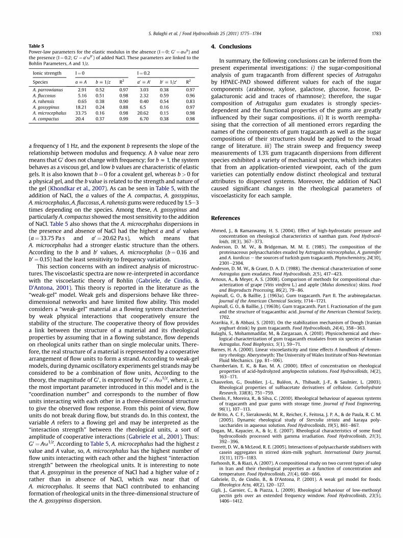
Table 5Power-law parameters for the elastic modulus in the absence (I¼ 0; G0 ¼ aub) andthe presence (I¼ 0.2; G0 ¼ a0ub0) of added NaCl. These parameters are linked to theBohlin Parameters, A and 1/z.
Ionic strength I¼ 0 I¼ 0.2
Species ah A bh 1/z R2 a0 h A0 b0 h 1/z0 R2
A. parrowianus 2.91 0.52 0.97 3.03 0.38 0.97A. fluccosus 5.16 0.51 0.98 2.32 0.59 0.96A. rahensis 0.65 0.38 0.90 0.40 0.54 0.83A. gossypinus 18.21 0.24 0.88 6.5 0.16 0.97A. microcephalus 33.75 0.16 0.98 20.62 0.15 0.98A. compactus 20.4 0.37 0.99 6.70 0.38 0.98
S. Balaghi et al. / Food Hydrocolloids 25 (2011) 1775e1784 1783
a frequency of 1 Hz, and the exponent b represents the slope of therelationship between modulus and frequency. A b value near zeromeans that G0 does not change with frequency; for bz 1, the systembehaves as a viscous gel, and low b values are characteristic of elasticgels. It is also known that b¼ 0 for a covalent gel, whereas b> 0 fora physical gel, and the b value is related to the strength and nature ofthe gel (Khondkar et al., 2007). As can be seen in Table 5, with theaddition of NaCl, the a values of the A. compactus, A. gossypinus,A.microcephalus,A.fluccosus,A. rahensisgumswere reducedby1.5e3times depending on the species. Among these, A. gossypinus andparticularly A. compactus showed themost sensitivity to the additionof NaCl. Table 5 also shows that the A. microcephalus dispersions inthe presence and absence of NaCl had the highest a and a0 values(a¼ 33.75 Pa s and a0 ¼ 20.62 Pa s), which means thatA. microcephalus had a stronger elastic structure than the others.According to the b and b0 values, A. microcephalus (b¼ 0.16 andb0 ¼ 0.15) had the least sensitivity to frequency variation.
This section concerns with an indirect analysis of microstruc-tures. The viscoelastic spectra are now re-interpreted in accordancewith the viscoelastic theory of Bohlin (Gabriele, de Cindio, &D’Antona, 2001). This theory is reported in the literature as the“weak-gel” model. Weak gels and dispersions behave like three-dimensional networks and have limited flow ability. This modelconsiders a “weak-gel” material as a flowing system characterisedby weak physical interactions that cooperatively ensure thestability of the structure. The cooperative theory of flow providesa link between the structure of a material and its rheologicalproperties by assuming that in a flowing substance, flow dependson rheological units rather than on single molecular units. There-fore, the real structure of a material is represented by a cooperativearrangement of flow units to form a strand. According to weak-gelmodels, during dynamic oscillatory experiments gel strandsmay beconsidered to be a combination of flow units, According to thetheory, the magnitude of G0, is expressed by G0 ¼ Au1/z, where, z, isthe most important parameter introduced in this model and is the“coordination number” and corresponds to the number of flowunits interacting with each other in a three-dimensional structureto give the observed flow response. From this point of view, flowunits do not break during flow, but strands do. In this context, thevariable A refers to a flowing gel and may be interpreted as the“interaction strength” between the rheological units, a sort ofamplitude of cooperative interactions (Gabriele et al., 2001). Thus:G0 ¼ Au1/z. According to Table 5, A. microcephalus had the highest zvalue and A value, so, A. microcephalus has the highest number offlow units interacting with each other and the highest “interactionstrength” between the rheological units. It is interesting to notethat A. gossypinus in the presence of NaCl had a higher value of zrather than in absence of NaCl, which was near that ofA. microcephalus. It seems that NaCl contributed to enhancingformation of rheological units in the three-dimensional structure ofthe A. gossypinus dispersion.
4. Conclusions
In summary, the following conclusions can be inferred from thepresent experimental investigations: i) the sugar-compositionalanalysis of gum tragacanth from different species of Astragalusby HPAEC-PAD showed different values for each of the sugarcomponents (arabinose, xylose, galactose, glucose, fucose, D-galacturonic acid and traces of rhamnose); therefore, the sugarcomposition of Astragalus gum exudates is strongly species-dependent and the functional properties of the gums are greatlyinfluenced by their sugar compositions. ii) It is worth reempha-sising that the correction of all mentioned errors regarding thenames of the components of gum tragacanth as well as the sugarcompositions of their structures should be applied to the broadrange of literature. iii) The strain sweep and frequency sweepmeasurements of 1.3% gum tragacanth dispersions from differentspecies exhibited a variety of mechanical spectra, which indicatesthat from an application-oriented viewpoint, each of the gumvarieties can potentially endow distinct rheological and texturalattributes to dispersed systems. Moreover, the addition of NaClcaused significant changes in the rheological parameters ofviscoelasticity for each sample.
References
Ahmed, J., & Ramaswamy, H. S. (2004). Effect of high-hydrostatic pressure andconcentration on rheological characteristics of xanthan gum. Food Hydrocol-loids, 18(3), 367e373.
Anderson, D. M. W., & Bridgeman, M. M. E. (1985). The composition of theproteinaceous polysaccharides exuded by Astragalus microcephalus, A. gummiferand A. kurdicus e the sources of turkish gum tragacanth. Phytochemistry, 24(10),2301e2304.
Andeson, D. M. W., & Grant, D. A. D. (1988). The chemical characterization of someAstragalus gum exudates. Food Hydrocolloids, 2(5), 417e423.
Arnous, A., & Meyer, A. S. (2008). Comparison of methods for compositional char-acterization of grape (Vitis vinifera L.) and apple (Malus domestica) skins. Foodand Bioproducts Processing, 86(2), 79e86.
Aspinall, G. O., & Baillie, J. (1963a). Gum tragacanth. Part II. The arabinogalactan.Journal of the American Chemical Society, 1714e1721.
Aspinall, G. O., & Baillie, J. (1963b). Gum tragacanth. Part I. Fractionation of the gumand the structure of tragacanthic acid. Journal of the American Chemical Society,1702.
Azarikia, F., & Abbasi, S. (2010). On the stabilization mechanism of Doogh (Iranianyoghurt drink) by gum tragacanth. Food Hydrocolloids, 24(4), 358e363.
Balaghi, S., Mohammadifar, M., & Zargaraan, A. (2010). Physicochemical and rheo-logical characterization of gum tragacanth exudates from six species of IranianAstragalus. Food Biophysics, 5(1), 59e71.
Barnes, H. A. (2000). Linear viscoelasticity and time effects A handbook of elemen-tary rheology. Aberystwyth: The University of Wales Institute of Non-NewtonianFluid Mechanics. (pp. 81e106).
Chamberlain, E. K., & Rao, M. A. (2000). Effect of concentration on rheologicalproperties of acid-hydrolyzed amylopectin solutions. Food Hydrocolloids, 14(2),163e171.
Chauvelon, G., Doublier, J.-L., Buléon, A., Thibault, J.-F., & Saulnier, L. (2003).Rheological properties of sulfoacetate derivatives of cellulose. CarbohydrateResearch, 338(8), 751e759.
Chenlo, F., Moreira, R., & Silva, C. (2010). Rheological behaviour of aqueous systemsof tragacanth and guar gums with storage time. Journal of Food Engineering,96(1), 107e113.
de Brito, A. C. F., Sierakowski, M. R., Reicher, F., Feitosa, J. P. A., & de Paula, R. C. M.(2005). Dynamic rheological study of Sterculia striata and karaya poly-saccharides in aqueous solution. Food Hydrocolloids, 19(5), 861e867.
Dogan, M., Kayacier, A., & Ic, E. (2007). Rheological characteristics of some foodhydrocolloids processed with gamma irradiation. Food Hydrocolloids, 21(3),392e396.
Everett, D. W., & McLeod, R. E. (2005). Interactions of polysaccharide stabilisers withcasein aggregates in stirred skim-milk yoghurt. International Dairy Journal,15(11), 1175e1183.
Farhoosh, R., & Riazi, A. (2007). A compositional study on two current types of salepin Iran and their rheological properties as a function of concentration andtemperature. Food Hydrocolloids, 21(4), 660e666.
Gabriele, D., de Cindio, B., & D’Antona, P. (2001). A weak gel model for foods.Rheologica Acta, 40(2), 120e127.
Gigli, J., Garnier, C., & Piazza, L. (2009). Rheological behaviour of low-methoxylpectin gels over an extended frequency window. Food Hydrocolloids, 23(5),1406e1412.

S. Balaghi et al. / Food Hydrocolloids 25 (2011) 1775e17841784
Gómez-Guillén, M. C., Turnay, J., Fernández-Díaz, M. D., Ulmo, N., Lizarbe, M. A., &Montero, P. (2002). Structural and physical properties of gelatin extracted fromdifferent marine species: a comparative study. Food Hydrocolloids, 16(1), 25e34.
Izdorczyk,M., Cui, S.W., &Wang,Q. (2005). Polysaccharide gums: structures, functionalproperties and applications Food carbohydrates: Chemistry, physical properties, andapplications. United States of America: Taylor and Francis Group, LLC. (pp. 282).
Khondkar, D., Tester, R. F., Hudson, N., Karkalas, J., & Morrow, J. (2007). Rheologicalbehaviour of uncross-linked and cross-linked gelatinised waxy maize starchwith pectin gels. Food Hydrocolloids, 21(8), 1296e1301.
Lapasin, R., & Pricl, S. (1999). Rheology of polysaccharide systems Rheology ofindustrial polysaccharides: Theory and applications. United States of America: AnAspen Publication. (pp. 250e309).
Lazaridou, A., Biliaderis, C. G., & Izydorczyk, M. S. (2001). Structural characteristicsand rheological properties of locust bean galactomannans: a comparison ofsamples from different carob tree populations. Journal of the Science of Food andAgriculture, 81, 68e75.
Levy, G., & Schwarz, T. W. (1958). Tragacanth solutions I. The relation of method ofpreparation to the viscosity and stability. Journal of the American PharmaceuticalAssociation, 47(6), 451e454.
Martínez-Ruvalcaba, A., Chornet, E., & Rodrigue, D. (2007). Viscoelastic properties ofdispersed chitosan/xanthan hydrogels. Carbohydrate Polymers, 67(4), 586e595.
Medina-Torres, L., Brito-De La Fuente, E., Torrestiana-Sanchez, B., & Katthain, R.(2000). Rheological properties of the mucilage gum (Opuntia ficus indica). FoodHydrocolloids, 14(5), 417e424.
Mezger, T. G. (2006). The rheology handbook: For users of rotational and oscillatoryrheometers (pp. 114e141) (2nd ed.). Hannover: Vincentz Network.
Mills, P. L., & Kokini, J. L. (1984). Comparison of steady shear and dynamic visco-elastic properties of guar and karaya gums. Journal of Food Science, 49(1), 1e4.
Mohammadifar, M. A., Musavi, S. M., Kiumarsi, A., & Williams, P. A. (2006). Solutionproperties of targacanthin (water-soluble part of gum tragacanth exudate fromAstragalus gossypinus). International Journal of Biological Macromolecules, 38(1),31e39.
Mu, L., Zhao, M., Yang, B., Zhao, H., Cui, C., & Zhao, Q. (2010). Effect of ultrasonictreatment on the graft reaction between soy protein isolate and gum acacia andon the physicochemical properties of conjugates. Journal of Agricultural andFood Chemistry, 58(7), 4494e4499.
Norman, A. G. (1931). Studies on the gums. II. Tragacanthin e the soluble constit-uent of gum tragacanth. The Biochemical Journal, 25(1), 200e204.
Ramsden, L. (2004). Plant and algal gums and mucilages. In P. Tomasik (Ed.),Chemical and functional properties of food saccharides (pp. 244). United States ofAmerica: CRC Press LLC.
Silva, D. A., Brito, A. C. F., de Paula, R. C. M., Feitosa, J. P. A., & Paula, H. C. B. (2003).Effect of mono and divalent salts on gelation of native, Na and deacetylatedSterculia striata and Sterculia urens polysaccharide gels. Carbohydrate Polymers,54(2), 229e236.
Stauffer, K. R., & Andon, S. A. (1975). comparison of the functional characteristics oftwo grades of tragacanth. Food Technology, 4, 46e51.
Vebeken, D., Dierckx, S., & Dewettinck, K. (2003). Exudate gums: occurrence,production, and applications. Applied Microbiology and Biotechnology, 63,10e21.
Walter, R. H. (1998). In S. L. Taylor (Ed.), Polysaccharide dispersion: Chemistry andtechnology in food.
Weiping, W. (2000). Tragacanth and karaya. In G. O. Philips, & P. A. Williams (Eds.),Handbook of hydrocolloids (pp. 231e246). Cambridge, England: CRC Press.
William, P. A., Phillips, G. O., Stephen, A. M., & Churms, C. C. (2006). Gums andmucilages. In G. O. Phillips, A. M. Stephen, & P. A. Williams (Eds.), Food poly-saccharides and their applications (2nd ed.). (pp. 474e475) United States ofAmerica: Taylor and Francis Group.
Yasar, K., Kahyaoglu, T., & Sahan, N. (2009). Dynamic rheological characterization ofsalep glucomannan/galactomannan-based milk beverages. Food Hydrocolloids,23(5), 1305e1311.
Zahedi, A. H., Vedadi, I., & Dollimore, D. (1979). Effect of gum tragacanth sizereduction on DTA and TG behaviour and its relationship to solution viscosity.Thermochimica Acta, 34(2), 281e286.

at SciVerse ScienceDirect
Food Hydrocolloids 31 (2013) 5e14
Contents lists available
Food Hydrocolloids
journal homepage: www.elsevier .com/locate/ foodhyd
Stabilization of emulsions by gum tragacanth (Astragalus spp.) correlates to thegalacturonic acid content and methoxylation degree of the gum
Hassan Ahmadi Gavlighi a,b, Anne S. Meyer a,*, Dayang N.A. Zaidel a,d, Mohammad Amin Mohammadifar c,J. Dalgaard Mikkelsen a
aCenter for Bioprocess Engineering, Department of Chemical and Biochemical Engineering, Technical University of Denmark (DTU), Building 229, DK-2800 Kgs. Lyngby, DenmarkbDepartment of Food Science and Technology, Faculty of Agriculture, Tarbiat Modares University, P. O. Box 14115-336, Tehran, IrancDepartment of Food Science and Technology, National Nutrition and Food Technology Research Institute, Faculty of Nutrition Sciences and Food Technology, Shahid BeheshtiUniversity of Medical Sciences, P.O. Box 19395-4741, Tehran, IrandDepartment of Bioprocess Engineering, Faculty of Chemical Engineering, Universiti Teknologi Malaysia, 81310 Skudai, Johor, Malaysia
a r t i c l e i n f o
Article history:Received 15 April 2012Accepted 11 September 2012
Keywords:Emulsion stabilizationGalacturonic acidBassorinTragacanthinMethoxylationAcetylation
* Corresponding author. Tel.: þ45 45252800.E-mail address: [email protected] (A.S. Meyer).
0268-005X/$ e see front matter � 2012 Elsevier Ltd.http://dx.doi.org/10.1016/j.foodhyd.2012.09.004
a b s t r a c t
Gum tragacanth samples from six species of Iranian Astragalus bush plants (“goat’s-horn”) wereevaluated for their emulsion stabilizing effects and their detailed chemical composition in order toexamine any possible correlation between the make-up and the emulsion stabilizing properties of gumtragacanth. The six gum tragacanth samples were exudates from the species Astragalus parrowianus,Astragalus fluccosus, Astragalus rahensis, Astragalus gossypinus, Astragalus microcephalus, and Astragaluscompactus. The six gum samples varied with respect to their levels and ratios of water-soluble(tragacanthin) and water-swellable (bassorin) fractions, their monosaccharide composition, methox-ylation, and acetylation degrees. The gums from A. parrowianus and A. fluccosus had relatively hightragacanthin:bassorin ratios of w66:34 and w75:25, respectively, whereas in the other gums this ratioapproached 50:50 (A. rahensis, A. microcephalus, A. compactus) or tipped toward higher bassorin thantragacanthin (A. gossypinus). The monosaccharide make-up of the six gums also varied, but all the gumscontained relatively high levels of galacturonic acid (w100e330 mg/g), arabinose (50e360 mg/g), xylose(w150e270 mg/g), and galactose (w40e140 mg/g), and also contained fucose, rhamnose, and glucose.The ability of the gums to act as stabilizers in whey protein isolate based emulsions varied. The bestemulsion stabilization effect, measured as lowest creaming index ratio after 20 days, was obtained withthe A. fluccosus gum. The emulsion stabilization effect correlated linearly and positively to themethoxylation degree, and galacturonic acid content of the gums, but not to acetyl or fucose content. Aparticularly high correlation was found between methoxyl level in the soluble gum part and emulsionstabilization. The work provides some important clues to the emulsion stabilization mechanisms inrelation to the monosaccharide composition of tragacanth gums.
� 2012 Elsevier Ltd. All rights reserved.
1. Introduction
The natural plant exudate gum tragacanth is obtained from thestem of the bush like plant “goat’s-horn”, Astragalus species. Gumtragacanth has beenused commercially forwell over 2000 years andis currently widely used as an emulsifier and thickener in emulsionsystems in different food, pharmaceutical and cosmetic applications(Whistler, 1993). Gum tragacanth has a bland flavor and goodstability to heat and acid and is allowed for food uses in accordancewith the FDA Code of Federal Regulations as a food additive at the
All rights reserved.
level of 0.2e1.3% by weight of the product (FDA, 2006). In Europe,gum tragacanth has E-number E413 on the list of additives approvedby the Scientific Committee for Food of the European Community.The main area of commercial production is the Middle East withIran, supplying 70% of the commercially used gum tragacanth, fol-lowed by Turkey and Syria as main producers (Anderson, 1989).
Gum tragacanth is known to confer very high viscosities when inaqueous solution, and is described as a complex, highly branched,heterogeneous hydrophilic polysaccharide thatmay form complexeswith salts, notably salts with divalent cations, such as Ca2þ salts(Balaghi, Mohammadifar, & Zargaraan, 2010). The molecular weightof a typical gumhas been reported to be about 840kDa, calculated bySvedberg’s method and formula, and the shape of the flake gumwas
H.A. Gavlighi et al. / Food Hydrocolloids 31 (2013) 5e146
originally described as being elongated with dimensions of 450 nmby 1.9 nm (Gralén & Kärrholm, 1950). The polysaccharides of gumtragacanthwere studied in detail by James and Smith (1945a,1945b)already almost 70 years ago and intensively by Aspinall and Baillie(1963a, 1963b) in the sixties . Tragacanthin is the water-solublefraction, and bassorin is the insoluble part which swells in water toform a gel (Aspinall & Baillie, 1963a). It is well known that the ratiobetween the water-soluble and water-insoluble fraction of gumtragacanth varies significantly among gums obtained from differentAstragalus species (Balaghi et al., 2010), but it is unclear to whatextent the composition of the polysaccharides affects the ratio oftragacanthin to bassorin.
With respect to the compositional make-up of the gum traga-canth polysaccharides, it has been amply reported that gum trag-acanth contains L-arabinose, D-galactose, D-glucose, D-xylose, L-fucose, L-rhamnose, and D-galacturonic acid upon acid hydrolysis(Balaghi, Mohammadifar, Zargaraan, Gavlighi, & Mohammadi,2011; Tischer, Iacomini, & Gorin, 2002). Recently, it has becomeevident, that the understanding up until now that the solublefraction (tragacanthin) consists of arabinogalactan and that theinsoluble fraction (bassorin) is made up of “tragacanthic acid” isprobably not correct (Balaghi et al., 2011). Rather, at least thewater-soluble fraction (tragacanthin) appears to resemble pectin, andseems to contain linear chains of galacturonic acid (probably 1,4-a-linked); hence, gum tragacanth species rich in xylose with minorlevels of fucose may contain xylogalacturonans and some fuco-xylogalacturonans as the main components of the soluble frac-tion, whereas those having high fucose levels may mainly containfuco-xylogalacturonan in the tragacanthin part (Balaghi et al.,2011). In most Astragalus species, the bassorin and tragacanthinhave also been found to contain methyl groups, probably repre-sentingmethoxylated galacturonic acid. The insoluble bassorin partgenerally appears to have less methoxyl substitutions than thesoluble tragacanthin part (Anderson & Grant, 1988).
Gum tragacanth has been reported to have both emulsifying andstabilizing properties in emulsions, i.e. being “bi-functional”by having both the capacity to facilitate emulsification as wellas providing stabilization of the emulsion after its formation(Weiping, 2000). There are indications in the literature thatcompositional differences of tragacanth gums obtained fromdifferent species of Astragalus affect the rheological properties,including the viscosity of tragacanth solutions (Balaghi et al., 2011).However, despite the long and extensive use of gum tragacanth asa stabilizer in emulsion systems, surprisingly little is known aboutstructure function relationships, and the traits of the gum thatconfer emulsifying properties are presently unknown. It is of coursetempting to hypothesize that the composition of tragacanthgum affects its stabilization effects in emulsions, and it has beenreported that the terminal deoxyhexoxyl groups (i.e. fucose) or themethoxyl groups of the homogalacturonan in the tragacanthgum structure may play a role in the emulsion stabilization(Stephen, 1990). However, to our knowledge, no unequivocalevidence has up until now been provided for this hypothesis.Hence, the exact significance of specific structural components oftragacanth gum in relation to viscosity and emulsion stabilizationproperties is unclear.
The purpose of this study was to evaluate the stabilization ofemulsions by tragacanth gum, and to aim at obtaining anunderstanding of any relationships between the make-up of gumtragacanth and its stabilization properties in emulsions. This wasdone by comparing the chemical composition of tragacanth gumobtained from six Astragalus species growing in different regions ofIran, and by systematically assessing whether there were anycorrelations between emulsion stabilization properties and theprevalence of specific chemical components in the gums.
2. Materials and methods
2.1. Materials and gum preparation
Gum tragacanth samples exuded from various species of Astra-galus (Astragalus parrowianus, Astragalus fluccosus, Astragalusrahensis, Astragalus gossypinus, Astragalus microcephalus, andAstragalus compactus) were collected from plants growing indifferent provinces of Iran. The plants were tapped by makingcareful longitudinal incisions in the taproot and the bark of thebranches. The gum readily exuded from these cuts in the form of‘ribbons’ that became brittle on drying. The dried raw gums wereground with a coffee mill, sieved, solubilized in deionized water,then freeze dried, andfinally used in this study. The protein contents(g/100 g) of the gum tragacanth samples were 3.0, 2.6, 3.8, 0.31, 2.0,and 1.7 for A. parrowianus, A. fluccosus, A. rahensis, A. gossypinus,A. microcephalus, and A. compactus, respectively (Balaghi et al.,2010). Neutral monosaccharide standards, L-fucose, L-rhamnose, D-arabinose, D-galactose, D-glucose, D-xylose, D-galacturonic acid andD-glucuronic acid were obtained from SigmaeAldrich Chemical Co.(St. Louis, MO, USA). Trifluoroacetic acid (99%) was from Merck(Darmstadt, Germany). NaOH standard solution HPLC grade wasfrom Fluka/SigmaeAldrich Chemical Co. (St. Louis, MO, USA). Allchemicals used were of analytical grade. Rapeseed oil was obtainedfrom IncoDenmark andwheyprotein isolate (WPI), LACPRODANDI-9224, was purchased from Arla Foods Ingredients (Videbæk,Denmark). The WPI was a neutral and heat stable type of wheyprotein isolate that according to the supplier is sold as an emulsifieras well as for use in sports and clinical nutrition. According to thesupplier’s data sheet, the WPI consisted of 92% protein (N � 6.38),0.2% lactose, 4.5% ash and approximately 0.5% sodium, 0.2% phos-phorus, 1.3% potassium and 0.1% calcium (all by weight).
2.2. Determination of soluble and insoluble gum fractions, andmonosaccharide composition
In order to determine the ratio between the water-soluble andthewater-insoluble fractions of the gums, each gum samplewas re-suspended overnight in deionized water (1% dry matter gumweight/volume) and the separation of the soluble and insolublefractionwas done by centrifugation at 3600� g for 180 min and theseparate fractions were freeze dried and weighed as describedpreviously (Mohammadifar, Musavi, Kiumarsi, & Williams, 2006).
The monosaccharide composition of each of the gum tragacanthsampleswas determined after acid hydrolysis (4 g L�1 substrate, 2Mtrifluoroacetic acid, 2 h, 121 �C) and by use of separate recoveryfactors for each monosaccharide (Arnous & Meyer, 2008). Themonosaccharide analyses were done by use of an ICS3000 ionchromatography system consisting of a GS50 gradient pump, anED50electrochemical detector and anAS50 chromatograph coupledto an AS50 autosampler (Dionex Corp., Sunnyvale, CA). Separationswere performed using a CarboPacTM PA20 (3 mm � 150 mm)analytical column (Dionex Corp., Sunnyvale, CA) and an elutionprogrammeprincipally as described previously (Balaghi et al., 2011).
2.3. Determination of total methyl and acetyl contents of gumtragacanth
The methanol and acetic acid contents of each freeze dried gumtragacanth sample (soluble, insoluble, and full fraction) weredetermined by HPLC using a Summit LC system (Dionex Corp.,Sunnyvale, CA) equipped with a P680 HPLC pump, an ASI-100automated sample injector, and an Aminex HPX-87H column(300 � 7.8 mm, Bio-Rad Labs, Richmond, CA, USA) in combinationwith a guard column (AG50W-X4. 50 � 4.6 mm, Bio-Rad Labs)

H.A. Gavlighi et al. / Food Hydrocolloids 31 (2013) 5e14 7
(Voragen, Schols, & Pilnik, 1986). 5 mM sulfuric acid was used as theeluent at a constant flow of 0.6 mL min�1 and 30 �C. Componentseluting from the column were detected with a Shodex RI-101(Showa Denko KK, Kawasaki, Japan) refractive index detector ther-mostated at 40 �C. Pure methanol and glacial acetic acid from Flukawere used as standards.
2.4. Saponification of tragacanth gum
Methyl and acetyl groups of the tragacanth gum solution fromA. fluccosus (1% w/v) were removed by slowly adding 5 M NaOH inwater (cold) until pH 13 and stirring for 24 h at 4 �C. After that, thesaponified solution was dialyzed against deionized water at 4 �Cusing a 5 kDamolecular weight cut off polyethersulfonemembrane(Sartorius Stedim Biotech) and cross flow membrane filtration.After the dialysis was completed, the solution was freeze dried(Leroux, Langendorff, Schick, Vaishnav, & Mazoyer, 2003).
2.5. Preparation of O/W emulsions
Oil inwater (O/W) emulsions were prepared bymixing rapeseedoil (10% w/w) into a solution of 0.5% w/w WPI of 100 mM sodiumacetate buffer (pH 4.5) duringmixing for 7min in a high speed ultraturrax blender (Ystral Gmbh, Ballrechten-Dottingen, Germany).Prior to the preparation of the emulsions the WPI was dispersed ina sodium acetate buffer 100 mM (pH 4.5) and stirred overnight at4 �C. The 7minmixing of the emulsion ingredients was followed bypassing the emulsion through a microfluidizer 5 times (Micro-fluidics, Massachusetts, USA) operated at an air pressure of 0.2MPa,corresponding to 28 MPa of liquid pressure principally as describedpreviously (Zaidel, Chronakis, & Meyer, 2013). WPI was used as anemulsifier becausewhey proteins, having pI near 5, are amphiphilicmolecules near the isoelectric point 4 < pH < 6. Whey proteins,essentially b-lactoglobulin, are widely used as emulsifiers in foodapplications because theyare inexpensive, natural, readily available,andhave the ability to facilitate formation and stabilization of oil-in-water emulsions in systems having pH 4e6 (Dickinson, 2001).
In this particular system a pH of 4.5, slightly below the pI of thewhey protein, was used to enhance the electrostatic attractionbetween the (potentially pectic) negatively charged gum tragacanthpolysaccharides and the protein. The tragacanth gum solutionswere prepared by dissolving suitable amounts of the tragacanthgum powders separately into buffer. In order to ensure completehydration the gum solutions were held for 24 h at room tempera-ture before each individual gum tragacanth solutionwasmixed intothe emulsion by magnetic stirring at 1000 rpm for 5 min. Theresulting emulsion systems contained 4.4%w/woil, 0.22%w/wWPI,and 0.5% w/v gum tragacanth with pH adjusted to 4.5.
2.6. Emulsion stability
Emulsions, 5 mL sample size, were placed in sealed glass tubes(diameter � height: 2 � 6 cm) at room temperature and theemulsion stability was assessed by measuring the height of theclear serum layer (forming at the bottom of the emulsion systems)relative to the total height of the emulsion after (0), 5, 7 and 20days. The creaming index was expressed as the height of the serumlayer over the total height of the emulsion in the tube.
2.7. Particle size determination
Immediately after emulsion preparation, samples were analyzedfor droplet size distribution by use of a laser diffraction particle sizeanalyzer (Mastersizer S, Malvern Instruments Ltd, Worcestershire,UK). All measurements were made at room temperature on at least
two freshly prepared samples. Optical properties of the samplewere defined as follows: refractive index of rapeseed oil anddispersion medium (water) were 1.46 and 1.33, respectively andabsorption was assumed to be 0. The average droplet size wasdescribed as the surface area moment mean (also known as thesurface area weighed mean diameter) D[3,2] ¼ P
nidi3/P
nidi2 andthe volume mean diameter (D[4,3]) was automatically calculatedusing the software provided with the apparatus:
D[4,3] ¼ Pnidi4/
Pnidi3 where ni is the number of particles
with diameter di (McClements, 2005).
2.8. Rheological measurements of emulsions and tragacanth gumsolutions
Steady-shear measurements of shear data of the emulsions andtragacanth gum solutions at the same pH of the emulsions (pH 4.5)were carried out at 0.1e500 s�1 on serrated parallel plates (PP60 TiSerrated) using a Haake Mars rotational rheometer (ThermoScientific Inc., Germany). All the measurements were carried out at25 �C with duplication measurement.
2.9. Statistical analysis
Data were analyzed by one-way analysis of variance (one-wayANOVA): Comparisons of mean values were calculated via 95%confidence intervals and compared as TukeyeKramer intervalscalculated from pooled standard deviations (Minitab StatisticalSoftware, Addison-Wesley, and Reading, MA).
3. Results and discussion
3.1. Gum tragacanth composition
The ratio between the soluble and insoluble fractions variedsignificantly among the tragacanth gums from the differentspecies of Astragalus, but the gums could roughly be divided intothree groups: Group 1: The gum from A. fluccosus had the highestsoluble:insoluble ratio of 3.16, i.e. with w75% w/w solubleand w25% w/w insoluble, and the gum from A. parrowianus hada ratio of w64% w/w soluble and w36% w/w insoluble, givinga ratio of 1.77 (Table 1). Group 2: The gums from A. rahensis,A. microcephalus, and A. compactus all had almost 50:50 solu-ble:insoluble gum fractions giving ratios of soluble to insolubleof w1, whereas Group 3, represented by the gum of A. gossypinuscontained a higher amount of insoluble (w65% w/w) than soluble(w35% w/w polysaccharides), with a ratio of soluble to insoluble of0.53 (Table 1).
As expected, also themonosaccharide compositionvaried amongthe gums from the different Astragalus plant species. In general, allgums contained relatively high amounts of galacturonic acid, arab-inose, xylose, and to a certain extent galactose. All gums also con-tained fucose, rhamnose, and glucose, with particularly widecontent ranges in the fucose content (varying from 27 to 236 mg/g)(Table 1). Rhamnose content was almost constant in the gums fromdifferent species (13e22 mg/g). Galacturonic acid contents rangedfromw100 to 335mg/g drymatter. Surprisingly, the highest level ofgalacturonic acid was determined for the gum from A. gossypinus,eventhough this gum had a lower amount of the soluble fractionthan the insolublebassorin (Table 1). Arabinose levels varied from48to 360 mg/g and xylose from w150 to 270 mg/g, whereas galactoselevels varied fromw40 to 140 mg/g (Table 1). Gum tragacanth fromA. gossypinus stood out as having the highest content of fucose,xylose, and galacturonic acid, as well as the lowest levels of arabi-nose, galactose, and glucose, among the samples (Table 1). In all thegum samples, the soluble part consistently contained higher levels
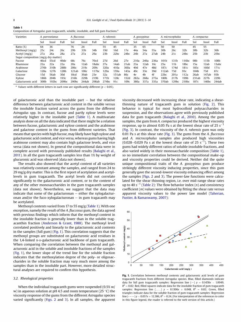
0
10
20
30
40
50
60
0 50 100 150 200 250 300 350 400 450
Met
hoxy
l con
tent
(m
g/g)
Galacturonic acid (mg/g )
Fig. 1. Correlation between methoxyl contents and galacturonic acid levels of gumtragacanth fractions from different Astragalus species. Blue, filled diamonds indicatedata for full gum tragacanth samples: Regression line (d) y ¼ 0.1458x � 1.0049,R2 ¼ 0.82; Red, filled squares indicate data for the insoluble fraction of gum tragacanthsamples: Regression line (..) y ¼ 0.1306x þ 6.048, R2 ¼ 0.82; Green, filledtriangles indicate data for the soluble fraction of gum tragacanth samples: Regressionline (- - - -) y ¼ 0.057x þ 12.386, R2 ¼ 0.24. (For interpretation of the references to colorin this figure legend, the reader is referred to the web version of this article.)
Table 1Composition of Astragalus gum tragacanth, soluble, insoluble, and full gum fractions.a
Varieties A. parrowianus A. fluccosus A. rahensis A. gossypinus A. microcephalus A. compactus
Sol Insol Full Sol Insol Full Sol Insol Full Sol Insol Full Sol Insol Full Sol Insol Full
Ratio (%) 64 36 76 24 55 45 35 65 50 50 45 55Methoxyl (mg/g) 25c 24c 26c 29b 33b 34b 19d 16d 17e 44a 54a 55a 30b 26c 32b 30b 32b 36bAcetyl (mg/g) 27a 26a 28a 27a 27a 29a 23b 22bc 24bc 24b 27a 27ab 24b 21c 24bc 25b 23b 25bcSugar composition (mg/g dry matter)Fucose 46cd 55cd 49de 68c 76c 70cd 27d 26d 27e 210a 249a 236a 101b 131b 116bc 98b 115b 108bRhamnose 22a 22a 22a 20a 12ab 18abc 27a 14ab 21ab 23a 12ab 16c 25a 11b 18bc 15a 12ab 13abcArabinose 276b 310b 288b 206c 219c 209c 320a 410a 360a 49d 47e 48d 187c 174d 181c 185c 160d 171cGalactose 128a 127b 128b 121ab 106c 117bc 129a 159a 143a 49c 33e 39d 112ab 73d 93c 104b 73d 87cGlucose 15d 56ab 30d 18cd 39ab 23e 32a 151ab 86c 4e 4b 4f 22bc 201a 112a 26ab 147ab 93bXylose 186b 204b 193c 216b 229b 219b 171b 128c 152d 282a 268a 273a 180b 217b 199b 231ab 227b 229bGalacturonic acid 300b 192bc 209bc 290bc 244ab 298ab 274bc 91c 109c 380a 337a 335a 370ab 129bc 218bc 187c 146bc 244ab
a Values with different letters in each row are significantly different (p < 0.05).
H.A. Gavlighi et al. / Food Hydrocolloids 31 (2013) 5e148
of galacturonic acid than the insoluble part e but the relativedifference between galacturonic acid content in the soluble versusthe insoluble fraction varied across the gums from the differentAstragalus spp. In contrast, fucose and partly xylose levels wererelatively higher in the insoluble part (Table 1). A multivariateanalysis done on all the data indicated that there might be a relationbetween fucose, galacturonic and xylose content and the arabinoseand galactose content in the gums from different varieties. Thatmeans that specieswithhigh fucose,may likely havehighxylose andgalacturonic acid content, and vice versa,whereas gumshaving higharabinose content may also contain high galactose levels, and viceversa (data not shown). In general the compositional data were incomplete accord with previously published results (Balaghi et al.,2011). In all the gum tragacanth samples less than 1% by weight ofglucuronic acid was observed (data not shown).
The results also showed that the acetyl content of all varietieswas relatively constant among the samples, and ranged from 24 to29mg/g dry matter. This is the first report of acetylation and acetyl-levels in gum tragacanth. The acetyl levels did not correlatesignificantly to the galacturonic acid content, or to the content ofany of the other monosaccharides in the gum tragacanth samples(data not shown). Nevertheless, we suggest that the data mayindicate that some of the galacturonan e either the xylogalactur-onan and/or the fuco-xylogalacturonan e in gum tragacanth maybe acetylated.
Methoxyl contents varied from17 to 55mg/g (Table 1).With oneexception, namely the result of the A. fluccosus gum, the data agreedwith previous findings which inform that the methoxyl content inthe insoluble fraction is generally lower than in the soluble trag-acanthin fraction (Anderson & Grant, 1988). The methoxyl levelcorrelated positively and linearly to the galacturonic acid contentsin the samples (full gum) (Fig. 1). This correlation suggests that themethoxyl groups are substituted on galacturonic acid residues inthe 1,4-linked a-D-galacturonic acid backbone of gum tragacanth.When comparing the correlation between the methoxyl and gal-acturonic acid in the soluble and insoluble fractions of the samples(Fig. 1), the lower slope of the trend line for the soluble fractionindicates that the methoxylation degree of the poly- or oligosac-charides in the soluble fraction may vary much more among thesamples than in the insoluble part. However, more detailed struc-tural analyses are required to confirm this hypothesis.
3.2. Rheological properties
When the individual tragacanth gums were suspended (0.5% w/w) in aqueous solution at pH 4.5 and room temperature (25 �C) theviscosity response of the gums from the different Astragalus speciesvaried significantly (Figs. 2 and 3). In all samples, the apparent
viscosity decreased with increasing shear rate, indicating a shear-thinning nature of tragacanth gum in solution (Fig. 2). Thisbehavior is typical for most hydrocolloid polysaccharides insuspension, and the observations agree with previously publisheddata for gum tragacanth (Balaghi et al., 2010). Among the gumsamples, the gum from A. compactus produced the highest viscosityresponse, up to almost 0.05 Pa s at the lowest shear rate of 25 s�1
(Fig. 3). In contrast, the viscosity of the A. rahensis gum was only0.01 Pa s at this shear rate (Fig. 3). The gums from the A. fluccosusand A. microcephalus samples also produced high viscosities(0.028e0.029 Pa s at the lowest shear rate of 25 s�1). These twogums had widely different ratios of soluble:insoluble fractions, andalso varied widely in their monosaccharide composition (Table 1),so no immediate correlation between the compositional make-upand viscosity properties could be derived. Neither did the quiteunique compositional traits of the A. gossypinus gum producestrikingly different viscosity inducing properties, since this gumgenerally gave the second-lowest viscosity enhancing effect amongthe samples (Figs. 2 and 3). The power-law functions were calcu-lated for the shear thinning region, i.e. over mid-range shear ratesup to 40 s�1 (Table 2): The flow behavior index (n) and consistencycoefficient (m) values were obtained by fitting the shear rate versusapparent viscosity values to the power law model (Taherian,Fustier, & Ramaswamy, 2007):

1
10
100
1 10 100 1000
App
aren
t vi
scos
ity
(mP
a· s
)
Shear rate (1/s)
C
F
M
P
G
R
Fig. 2. Flow curves of apparent viscosity versus shear rate of gum tragacanthsuspensions, 0.5% w/v gum tragacanth, at pH 4.5. C: Orange, filled circles, A. compactusgum. F: Red, filled squares A. fluccosus gum. M: Turquoise star points A. microcephalusgum. P: Green, filled triangles, A. parrowianus gum. G: Blue, filled diamonds,A. gossypinus gum. R: Purple crosses, A. rahensis gum. (For interpretation of thereferences to color in this figure legend, the reader is referred to the web version of thisarticle.)
H.A. Gavlighi et al. / Food Hydrocolloids 31 (2013) 5e14 9
ha ¼ m _gðn�1Þ
where ha is the apparent viscosity (Pa s), m is the consistencycoefficient (Pa sn), and _g is the shear rate (s�1), and n is the flowbehavior index (dimensionless). The power law modeling fittednicely for all samples, producing R2 values of w0.99, indicatinga genuine power law dependence of the apparent viscosity inresponse to the shear rate (Table 2).
Fig. 3. Comparison of viscosity values of gum tragacanth species (0.5% w/v gumtragacanth) in aqueous solutions at pH 4.5 at different, selected shear rates.
When comparing the m and n values across the different emul-sions and tragacanth gum solutions prepared with the six differenttragacanth gum species, them-values were found to be consistentlyhigher in the emulsions than in the gum solutions (Table 2). Thisfinding was of course in complete accordance with the observationthat the viscosity was always higher in the emulsions than in thegum solutions (Figs. 2e5). The nwas found to be lower than 1 for alltragacanth gum solutions as well as for all the emulsions (Table 2);this value of less than 1 confirmed the shear thinning behavior ofthe samples. The emulsion prepared with the A. fluccosus gum hadthe highest consistency coefficient (m), 0.34 Pa sn, among thesamples. This result was in relatively good accordancewith the gumsolution viscosity measurements since the A. fluccosus gumwas thegum giving the second highest viscosity response (Fig. 3), with theviscosity response approaching that of the A. compactus gum at highshear rates. The n for the A. fluccosus sample was also the highestamong the gum solutions, but in the emulsion samples theA. compactus gumproduced the highest n of 0.68 (Table 2), howeverfor the emulsions, the n-values were all in the range 0.5e0.68, theonly exception being the emulsion with the A. rahensis gum, whichhad a lower n of 0.35 (Table 2).
The viscosities of the emulsions were considerably higher thanthe gum tragacanth solutions, but the overall flow behavior in theemulsions with gum tragacanth added was quite similar to thebehavior observed in the gum tragacanth solutions (Fig. 4). Theviscosity of the model emulsion without gum tragacanth added(4.4% w/w rapeseed oil, 0.22% w/wWPI, pH 4.5) was very low at allshear rates (control, Fig. 5). These results are similar to those ob-tainedwith xanthan gum added to emulsions (Dickinson & Galazka,1991). In the emulsions, again, the A. compactus gum induced thehighest viscosity reaching a viscosity response of 0.09 Pa s at thelowest shear rate of 25 s�1. The pattern followed that observed forthe gums in suspension, so the lowest viscosity response wasobserved for the A. rahensis gum, which was only 0.02 Pa s at thisshear rate (Fig. 5). The medium-to-high range viscosities contrib-uted by the A. fluccosus and A. microcephalus samples increasedfrom just below 0.03 Pa s in the aqueous suspension system to0.075 Pa s in the emulsion system at the lowest shear rate of 25 s�1
(Fig. 5). In general, these viscosity increases did not correlate to thelevels or ratios of the insoluble (nor soluble) gum tragacanth frac-tions, so it was not possible to relate the viscosity responses directlyto the swelling ability of the insoluble gum tragacanth fraction.
Rather, the viscosity response may be related to certain struc-tural traits of the tragacanth gum, such as polysaccharide chainlength, branching pattern, substitutions, differences in back-bonebond structures or other factors, that are not revealed by thecompositional analysis, but which may impact formation of interand intra molecular interactions between the gum tragacanthpolysaccharides, the whey protein, and the emulsion droplets(Azarikia & Abbasi, 2010). The magnitude of the viscosity responseand the rheological behavior of the tragacanth gums in emulsionsagree with data for xanthan gum behavior in emulsions publishedby Papalamprou, Makri, Kiosseoglou, and Doxastakis (2005).
3.3. Particle size distribution of emulsions
The emulsions with gums from A. rahensis and A. fluccosus,respectively, had the smallest average particle size of w12 mm,measured as D[3,2], and the emulsion with the A. gossypinus gumhad the largest D[3,2] ofw38 mm (Fig. 6). The ANOVA indicated thatthe differences in particle size were statistically significant(p < 0.05). Without gum tragacanth added, the emulsion produceda monomodal particle size distribution with an average D[3,2] ofw7 mm (and D[4,3] ofw10 mm) (Fig. 7). Addition of gum tragacanthaffected the droplet size distribution to become bimodal (Fig. 7).

Table 2Power-law parameters for six species of Iranian gum tragacanth and emulsions (25 �C).a
Varieties A. parrowianus A. fluccosus A. rahensis A. gossypinus A. microcephalus A. compactus
n m R2 n m R2 n m R2 n m R2 n m R2 n m R2
Emulsions 0.61 0.17 0.99 0.53 0.34 0.99 0.35 0.16 0.99 0.57 0.15 0.99 0.64 0.24 0.99 0.68 0.25 0.99Tragacanth gum solutions 0.60 0.08 0.98 0.76 0.06 0.99 0.3 0.09 0.99 0.4 0.1 0.99 0.65 0.08 0.99 0.55 0.19 0.99
a Power-law parameters m: Pa sn, n: dimensionless; R2: Correlation coefficient for the power law function.
H.A. Gavlighi et al. / Food Hydrocolloids 31 (2013) 5e1410
The tragacanth gum stabilization of protein-emulsified emul-sions is probably a result of two mechanisms: Firstly, formation ofnon-covalent proteine(gum) polysaccharide complexes, andsecondly viscosity increase. It is well-recognized that emulsionstabilization bywhey proteinsmay be improved by polysaccharides(and particularly so at pH near the isoelectric point). For pectin-b-lactoglobulin complexes it has been shown that this effect occurswhen the protein associates to the polysaccharide, and soluble,charged complexes are formed, which have an overall charge as thepolysaccharide (Sperber, Schols, Stuart, Norde, & Voragen, 2009).The soluble complex is thus stabilized by electrostatic repulsion.From model-studies with dextranewhey protein conjugates itappears that the polysaccharide effect is mainly due to enhancedsteric stabilization provided by the bulkiness of the hydrophilicpolysaccharides that provides a stabilizing charged layer around theprotein stabilized oil droplets which protects the protein stabilizedoil droplets against flocculation under conditions where electro-static stabilization is less favorable (Akhtar & Dickinson, 2003).
The observation that the emulsion exhibiting the larger D[3,2](A. gossypinus) did not exhibit the largest viscosity despite havingthe highest galacturonic acid content (Table 1) is in accordancewith a recently published observation which showed that themedian diameter of particles of tragacanth gum dispersions variedin response to the ratio of the soluble to the insoluble gum fraction:When the same gums as used in the present study were comparedin solution the A. gossypinus sample produced the largest particlesize and there was no direct relationship between the size ofparticles, gum composition, and viscosity (Balaghi et al., 2010).
Most of previous studies in which polysaccharides (typicallypectin) were used for stabilizing protein-emulsified emulsions havereported that a stable system will have a monomodal distributionwith a small average particle size (e.g.D[4,3]< 2 mm)while unstable
1
10
100
1 10 100 1000
App
aren
t vi
scos
ity
(mP
a·s)
Shear rate (1/s)
C
F
M
P
G
R
Fig. 4. Flow curves of apparent viscosity vs. shear rate of emulsions with different gumtragacanth additions at pH 4.5. C: Orange, filled circles, A. compactus gum. F: Red, filledsquares A. fluccosus gum. M: Turquoise star points A. microcephalus gum. P: Green,filled triangles, A. parrowianus gum. G: Blue, filled diamonds, A. gossypinus gum.R: Purple crosses, A. rahensis gum. (For interpretation of the references to color in thisfigure legend, the reader is referred to the web version of this article.)
emulsions will have relatively large particle sizes with D[4,3]values>20 mm(Lucey, Tamehana, Singh,&Munro,1999). Luceyet al.(1999) also reported that higher stability is associated with moreuniformity of particle size. However, pectin does not have a fractionwith functionality like bassorin in its composition. Therefore, wepresume that the second peak (with higher values for diameter sizeassessed as D[3,2]) (Fig. 7) is not related to oil droplets, but may beelicited by the gum fraction alone (illustrated in Fig. 7 only for thegum from A. rahensis as an example). Hence, in our opinion the firstpeak (Fig. 7) is due to a very large amount of small emulsion dropletshaving a cover layer of whey protein and tragacanthin and thesecond one represent the presence of small amounts of very largeparticles of bassorin (Gorji, Mohammadifar, & Ezzatpanah, 2011).We also presume that bassorinmay be responsible for the increasedviscosity of the continuous phase in the emulsion.
When reported on a volume basis as a volume base mean, D[4,3], the particle size diameter data of the gum tragacanth stabi-lized emulsions ranged from w100e200 mm (Table 3). The volumebase evaluation also allowed calculation of the volume made up bythe smallest particles (calculated from the average area of the firstpeak in the bimodal, volumetric particle size distribution curve)(Table 3). The D[4,3] data thus corroborated the interpretation ofthe D[3,2] data (Fig. 6), namely that the particle size of theA. gossypinus was biggest because the volumetric fraction contrib-uted by the small particle size peak was significantly lower in the
Fig. 5. Comparison of viscosity responses at selected shear rates of different tragacanthgums when added individually to emulsion samples (4.4% w/w oil, 0.22% w/w WPI,0.5% w/v gum tragacanth, pH 4.5).
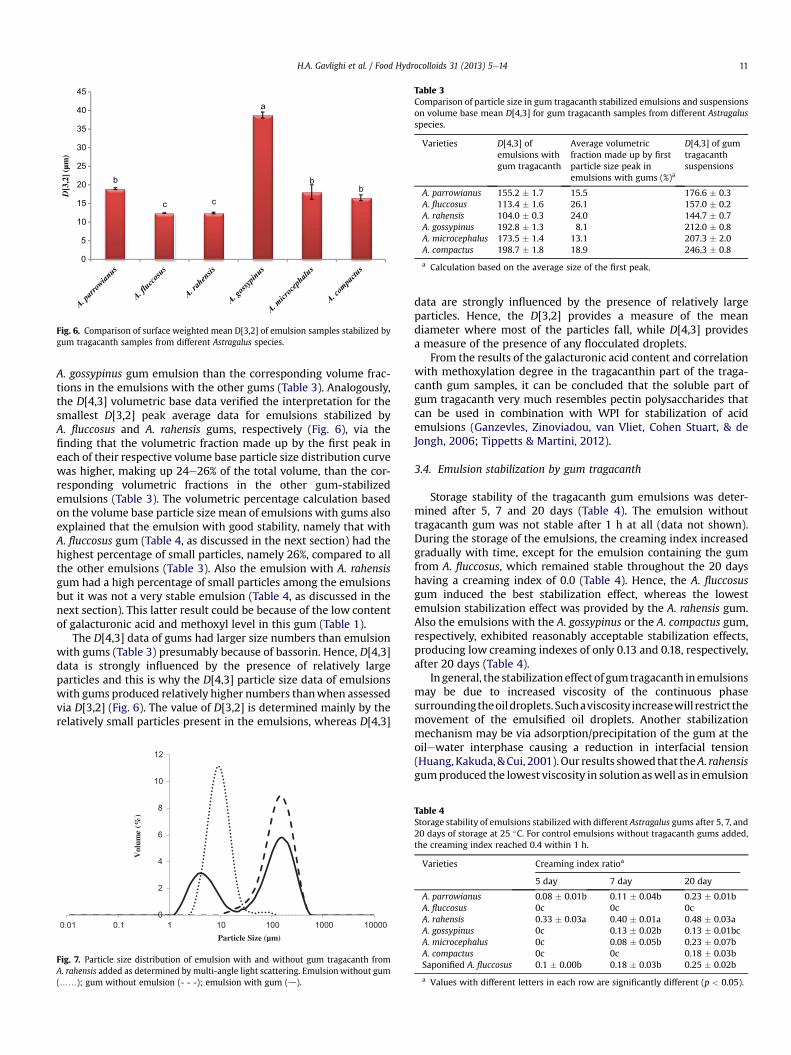
b
c c
a
bb
0
5
10
15
20
25
30
35
40
45
D[3
,2] (
µm)
Fig. 6. Comparison of surface weighted mean D[3,2] of emulsion samples stabilized bygum tragacanth samples from different Astragalus species.
Table 3Comparison of particle size in gum tragacanth stabilized emulsions and suspensionson volume base mean D[4,3] for gum tragacanth samples from different Astragalusspecies.
Varieties D[4,3] ofemulsions withgum tragacanth
Average volumetricfraction made up by firstparticle size peak inemulsions with gums (%)a
D[4,3] of gumtragacanthsuspensions
A. parrowianus 155.2 � 1.7 15.5 176.6 � 0.3A. fluccosus 113.4 � 1.6 26.1 157.0 � 0.2A. rahensis 104.0 � 0.3 24.0 144.7 � 0.7A. gossypinus 192.8 � 1.3 8.1 212.0 � 0.8A. microcephalus 173.5 � 1.4 13.1 207.3 � 2.0A. compactus 198.7 � 1.8 18.9 246.3 � 0.8
a Calculation based on the average size of the first peak.
H.A. Gavlighi et al. / Food Hydrocolloids 31 (2013) 5e14 11
A. gossypinus gum emulsion than the corresponding volume frac-tions in the emulsions with the other gums (Table 3). Analogously,the D[4,3] volumetric base data verified the interpretation for thesmallest D[3,2] peak average data for emulsions stabilized byA. fluccosus and A. rahensis gums, respectively (Fig. 6), via thefinding that the volumetric fraction made up by the first peak ineach of their respective volume base particle size distribution curvewas higher, making up 24e26% of the total volume, than the cor-responding volumetric fractions in the other gum-stabilizedemulsions (Table 3). The volumetric percentage calculation basedon the volume base particle size mean of emulsions with gums alsoexplained that the emulsion with good stability, namely that withA. fluccosus gum (Table 4, as discussed in the next section) had thehighest percentage of small particles, namely 26%, compared to allthe other emulsions (Table 3). Also the emulsion with A. rahensisgum had a high percentage of small particles among the emulsionsbut it was not a very stable emulsion (Table 4, as discussed in thenext section). This latter result could be because of the low contentof galacturonic acid and methoxyl level in this gum (Table 1).
The D[4,3] data of gums had larger size numbers than emulsionwith gums (Table 3) presumably because of bassorin. Hence, D[4,3]data is strongly influenced by the presence of relatively largeparticles and this is why the D[4,3] particle size data of emulsionswith gums produced relatively higher numbers thanwhen assessedvia D[3,2] (Fig. 6). The value of D[3,2] is determined mainly by therelatively small particles present in the emulsions, whereas D[4,3]
0
2
4
6
8
10
12
0.01 0.1 1 10 100 1000 10000
Vol
ume
(%)
Particle Size (µm)
Fig. 7. Particle size distribution of emulsion with and without gum tragacanth fromA. rahensis added as determined by multi-angle light scattering. Emulsionwithout gum(..); gum without emulsion (- - -); emulsion with gum (d).
data are strongly influenced by the presence of relatively largeparticles. Hence, the D[3,2] provides a measure of the meandiameter where most of the particles fall, while D[4,3] providesa measure of the presence of any flocculated droplets.
From the results of the galacturonic acid content and correlationwith methoxylation degree in the tragacanthin part of the traga-canth gum samples, it can be concluded that the soluble part ofgum tragacanth very much resembles pectin polysaccharides thatcan be used in combination with WPI for stabilization of acidemulsions (Ganzevles, Zinoviadou, van Vliet, Cohen Stuart, & deJongh, 2006; Tippetts & Martini, 2012).
3.4. Emulsion stabilization by gum tragacanth
Storage stability of the tragacanth gum emulsions was deter-mined after 5, 7 and 20 days (Table 4). The emulsion withouttragacanth gum was not stable after 1 h at all (data not shown).During the storage of the emulsions, the creaming index increasedgradually with time, except for the emulsion containing the gumfrom A. fluccosus, which remained stable throughout the 20 dayshaving a creaming index of 0.0 (Table 4). Hence, the A. fluccosusgum induced the best stabilization effect, whereas the lowestemulsion stabilization effect was provided by the A. rahensis gum.Also the emulsions with the A. gossypinus or the A. compactus gum,respectively, exhibited reasonably acceptable stabilization effects,producing low creaming indexes of only 0.13 and 0.18, respectively,after 20 days (Table 4).
In general, the stabilizationeffect of gumtragacanth in emulsionsmay be due to increased viscosity of the continuous phasesurroundingtheoildroplets. Suchaviscosity increasewill restrict themovement of the emulsified oil droplets. Another stabilizationmechanism may be via adsorption/precipitation of the gum at theoilewater interphase causing a reduction in interfacial tension(Huang, Kakuda,&Cui, 2001). Our results showed that theA. rahensisgumproduced the lowest viscosity in solution aswell as in emulsion
Table 4Storage stability of emulsions stabilizedwith different Astragalus gums after 5, 7, and20 days of storage at 25 �C. For control emulsions without tragacanth gums added,the creaming index reached 0.4 within 1 h.
Varieties Creaming index ratioa
5 day 7 day 20 day
A. parrowianus 0.08 � 0.01b 0.11 � 0.04b 0.23 � 0.01bA. fluccosus 0c 0c 0cA. rahensis 0.33 � 0.03a 0.40 � 0.01a 0.48 � 0.03aA. gossypinus 0c 0.13 � 0.02b 0.13 � 0.01bcA. microcephalus 0c 0.08 � 0.05b 0.23 � 0.07bA. compactus 0c 0c 0.18 � 0.03bSaponified A. fluccosus 0.1 � 0.00b 0.18 � 0.03b 0.25 � 0.02b
a Values with different letters in each row are significantly different (p < 0.05).
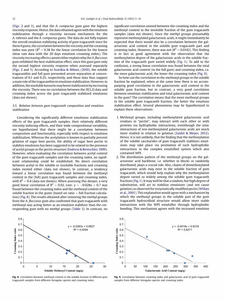
H.A. Gavlighi et al. / Food Hydrocolloids 31 (2013) 5e1412
(Figs. 2 and 3), and that the A. compactus gum gave the highestviscosity response.Hence, thedataobtainedagreewiththeemulsionstabilization through a viscosity increase mechanism for theA. rahensis and the A. compactus gums. The data do not fully explainthe overall emulsion stabilizing activity of gum tragacanth since forthese6gums, thecorrelationbetweentheviscosityandthecreamingindex was poor (R2 ¼ 0.38 for the linear correlation for the lowestshear rate data with the 20 days emulsion creaming index). Theviscosity increasingeffects cannot either explainwhy theA.fluccosusgum exhibited the best stabilization effect, since this gum gave onlythe second highest viscosity response when assessed separately(Figs. 2 and 3). According to Azarikia & Abbasi, (2010), the solubletragacanthin and full gum prevented serum separation at concen-trations of 0.1 and 0.2%, respectively, and these data thus supportamain roleof the tragacanthin inemulsionstabilization.However, inaddition, the insolublebassorinmaybooststabilizationbyincreasingthe viscosity. There was no correlation between the D[3,2] data andcreaming index across the gum tragacanth stabilized emulsions(data not shown).
3.5. Relation between gum tragacanth composition and emulsionstabilization
Considering the significantly different emulsions stabilizationeffects of the gum tragacanth samples, their relatively differentviscosity inducing effects, and their wide compositional variability,we hypothesized that there might be a correlation betweencomposition and functionality, especially with respect to emulsionstabilization. Whereas the acetylation is known to partially preventgelation of sugar beet pectin, the ability of sugar beet pectin tostabilize emulsions has been suggested to be related to the presenceof acetyl groups in the pectin structure (Endress & Rentschler,1999).However, when evaluating the correlation between acetyl contentof the gum tragacanth samples and the creaming index, no signif-icant relationship could be established. No direct correlationbetween acetyl in the soluble or insoluble fractions and creamingindex existed either (data not shown). In contrast, a tendencytoward a linear correlation was found between the methoxylcontent in the (full) gum tragacanth samples and creaming index,with R2 ¼ 0.4 (data not shown). When assessing this deeper, a verygood linear correlation of R2 ¼ 0.92, Line: y ¼ �0.028x þ 0.7 wasfound between the creaming index and themethoxyl content of thesoluble fraction in the gums (based on ratio � full fraction calcula-tion) (Fig. 8). The results obtained after removing themethyl groupsfrom the A. fluccosus gum also confirmed that gum tragacanth withmethoxyl was acting better as an emulsion stabilizer than the cor-responding gum with no methyl groups (Table 3). In contrast, no
y = -0.0283x + 0.6927R² = 0.9294
0
0.1
0.2
0.3
0.4
0.5
0.6
0.7
0.8
0 5 10 15 20 25 30
Cre
amin
g In
dex
rati
o
Soluble Methoxyl Content (mg/g)
Fig. 8. Correlation between methoxyl content in the soluble fraction of different gumtragacanth samples from different Astragalus species and creaming index.
significant correlation existed between the creaming index and themethoxyl content in the insoluble fraction of the gum tragacanthsamples (data not shown). Since the methyl groups presumablyrepresentmethoxylated galacturonic acids, it might immediately beexpected that there would also be a correlation between the gal-acturonic acid content in the soluble gum tragacanth part andcreaming index. However, there was not (R2 ¼ 0.0142). This findingis in fact in good agreement with the observation that themethoxylation degree of the galacturonic acids in the soluble frac-tion of the tragacanth gum varied widely (Fig. 1). To add to theconfusion, a strong linear correlation was found between the totalgalacturonic acid content (in the full gum) and creaming index, i.e.the more galacturonic acid, the lower the creaming index (Fig. 9).
So how can the correlation to themethoxyl groups in the solublefraction be explained, when at the same time there is no accom-panying good correlation to the galacturonic acid content in thesoluble gum fraction, but in contrast, a very good correlationbetween emulsion stabilization and total galacturonic acid contentin the gum? The correlation means that the more methoxyl groupsin the soluble gum tragacanth fraction, the better the emulsionstabilization effect. Several phenomena may be hypothesized toexplain these observations:
1. Methoxyl groups, including methoxylated galacturonic acidresidues in “pectin”, may interact with each other or withproteins via hydrophobic interactions, eventhough the ionicinteractions of non-methoxylated galacturonic acids are muchmore studied in relation to gelation (Zaidel & Meyer, 2012).Hence, it is not unlikely, that the finding that themethoxylationof the soluble saccharides of gum tragacanth stabilizes emul-sions may take place via promotion of such hydrophobicinteractions in the complex emulsified system which alsocontained WPI.
2. The distribution pattern of the methoxyl groups on the gal-acturonic acid backbone, i.e. whether in blocks or randomlydistributed, plays a crucial role. Also, chains of demethoxylatedgalacturonic acids may exist in the soluble fraction of gumtragacanth, which would help explain why the methoxylationdegree varied so widely among the soluble gum tragacanthfractions (Fig.1). Itmaywell be that a random,but highdegreeofsubstitution, will act to stabilize emulsions (and not causegelation) as observed for enzymaticallymodifiedpectin (Willatset al., 2001). This explanationwould agreewith amechanismbywhich the methoxyl groups in the soluble part of the gumtragacanth hydrocolloid structure would allow more stableinteractions with the WPI emulsifier through hydrophobicbonding. This mechanism agrees with the increased emulsion
y = -0.0018x + 0.6316R² = 0.8271
0
0.1
0.2
0.3
0.4
0.5
0.6
0.7
0.8
0 50 100 150 200 250 300 350 400
Cre
amin
g In
dex
rati
o
Galacturonic Acid Content (mg/g)
Fig. 9. Correlation between creaming index and galacturonic acid of gum tragacanthsamples from different Astragalus species and creaming index.
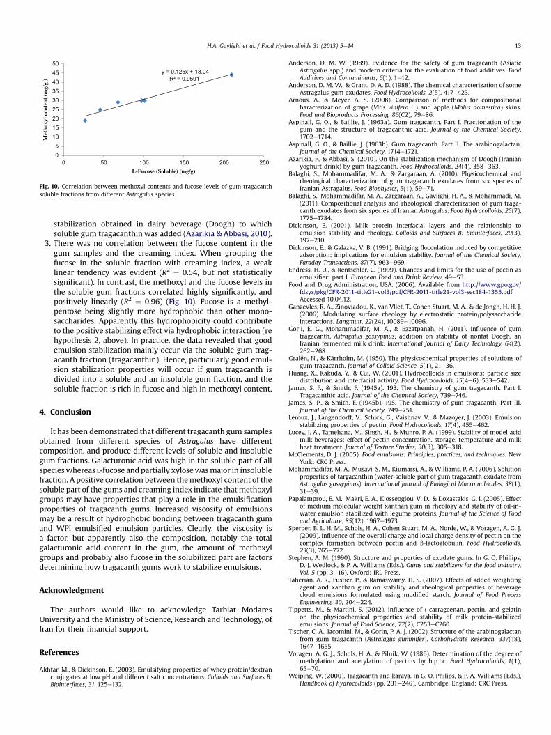
y = 0.125x + 18.04R² = 0.9591
05
101520253035404550
0 50 100 150 200 250
Met
hoxy
l con
tent
(m
g/g
)
L-Fucose (Soluble) (mg/g)
Fig. 10. Correlation between methoxyl contents and fucose levels of gum tragacanthsoluble fractions from different Astragalus species.
H.A. Gavlighi et al. / Food Hydrocolloids 31 (2013) 5e14 13
stabilization obtained in dairy beverage (Doogh) to whichsoluble gum tragacanthin was added (Azarikia & Abbasi, 2010).
3. There was no correlation between the fucose content in thegum samples and the creaming index. When grouping thefucose in the soluble fraction with creaming index, a weaklinear tendency was evident (R2 ¼ 0.54, but not statisticallysignificant). In contrast, the methoxyl and the fucose levels inthe soluble gum fractions correlated highly significantly, andpositively linearly (R2 ¼ 0.96) (Fig. 10). Fucose is a methyl-pentose being slightly more hydrophobic than other mono-saccharides. Apparently this hydrophobicity could contributeto the positive stabilizing effect via hydrophobic interaction (rehypothesis 2, above). In practice, the data revealed that goodemulsion stabilization mainly occur via the soluble gum trag-acanth fraction (tragacanthin). Hence, particularly good emul-sion stabilization properties will occur if gum tragacanth isdivided into a soluble and an insoluble gum fraction, and thesoluble fraction is rich in fucose and high in methoxyl content.
4. Conclusion
It has been demonstrated that different tragacanth gum samplesobtained from different species of Astragalus have differentcomposition, and produce different levels of soluble and insolublegum fractions. Galacturonic acid was high in the soluble part of allspecieswhereas L-fucose and partially xylosewasmajor in insolublefraction. A positive correlation between themethoxyl content of thesoluble part of the gums and creaming index indicate thatmethoxylgroups may have properties that play a role in the emulsificationproperties of tragacanth gums. Increased viscosity of emulsionsmay be a result of hydrophobic bonding between tragacanth gumand WPI emulsified emulsion particles. Clearly, the viscosity isa factor, but apparently also the composition, notably the totalgalacturonic acid content in the gum, the amount of methoxylgroups and probably also fucose in the solubilized part are factorsdetermining how tragacanth gums work to stabilize emulsions.
Acknowledgment
The authors would like to acknowledge Tarbiat ModaresUniversity and the Ministry of Science, Research and Technology, ofIran for their financial support.
References
Akhtar, M., & Dickinson, E. (2003). Emulsifying properties of whey protein/dextranconjugates at low pH and different salt concentrations. Colloids and Surfaces B:Biointerfaces, 31, 125e132.
Anderson, D. M. W. (1989). Evidence for the safety of gum tragacanth (AsiaticAstragalus spp.) and modern criteria for the evaluation of food additives. FoodAdditives and Contaminants, 6(1), 1e12.
Anderson, D. M. W., & Grant, D. A. D. (1988). The chemical characterization of someAstragalus gum exudates. Food Hydrocolloids, 2(5), 417e423.
Arnous, A., & Meyer, A. S. (2008). Comparison of methods for compositionalharacterization of grape (Vitis vinifera L.) and apple (Malus domestica) skins.Food and Bioproducts Processing, 86(C2), 79e86.
Aspinall, G. O., & Baillie, J. (1963a). Gum tragacanth. Part I. Fractionation of thegum and the structure of tragacanthic acid. Journal of the Chemical Society,1702e1714.
Aspinall, G. O., & Baillie, J. (1963b). Gum tragacanth. Part II. The arabinogalactan.Journal of the Chemical Society, 1714e1721.
Azarikia, F., & Abbasi, S. (2010). On the stabilization mechanism of Doogh (Iranianyoghurt drink) by gum tragacanth. Food Hydrocolloids, 24(4), 358e363.
Balaghi, S., Mohammadifar, M. A., & Zargaraan, A. (2010). Physicochemical andrheological characterization of gum tragacanth exudates from six species ofIranian Astragalus. Food Biophysics, 5(1), 59e71.
Balaghi, S., Mohammadifar, M. A., Zargaraan, A., Gavlighi, H. A., & Mohammadi, M.(2011). Compositional analysis and rheological characterization of gum traga-canth exudates from six species of Iranian Astragalus. Food Hydrocolloids, 25(7),1775e1784.
Dickinson, E. (2001). Milk protein interfacial layers and the relationship toemulsion stability and rheology. Colloids and Surfaces B: Biointerfaces, 20(3),197e210.
Dickinson, E., & Galazka, V. B. (1991). Bridging flocculation induced by competitiveadsorption: implications for emulsion stability. Journal of the Chemical Society,Faraday Transactions, 87(7), 963e969.
Endress, H. U., & Rentschler, C. (1999). Chances and limits for the use of pectin asemulsifier: part I. European Food and Drink Review, 49e53.
Food and Drug Administration, USA. (2006). Available from http://www.gpo.gov/fdsys/pkg/CFR-2011-title21-vol3/pdf/CFR-2011-title21-vol3-sec184-1355.pdfAccessed 10.04.12.
Ganzevles, R. A., Zinoviadou, K., van Vliet, T., Cohen Stuart, M. A., & de Jongh, H. H. J.(2006). Modulating surface rheology by electrostatic protein/polysaccharideinteractions. Langmuir, 22(24), 10089e10096.
Gorji, E. G., Mohammadifar, M. A., & Ezzatpanah, H. (2011). Influence of gumtragacanth, Astragalus gossypinus, addition on stability of nonfat Doogh, anIranian fermented milk drink. International Journal of Dairy Technology, 64(2),262e268.
Gralén, N., & Kärrholm, M. (1950). The physicochemical properties of solutions ofgum tragacanth. Journal of Colloid Science, 5(1), 21e36.
Huang, X., Kakuda, Y., & Cui, W. (2001). Hydrocolloids in emulsions: particle sizedistribution and interfacial activity. Food Hydrocolloids, 15(4e6), 533e542.
James, S. P., & Smith, F. (1945a). 193. The chemistry of gum tragacanth. Part I.Tragacanthic acid. Journal of the Chemical Society, 739e746.
James, S. P., & Smith, F. (1945b). 195. The chemistry of gum tragacanth. Part III.Journal of the Chemical Society, 749e751.
Leroux, J., Langendorff, V., Schick, G., Vaishnav, V., & Mazoyer, J. (2003). Emulsionstabilizing properties of pectin. Food Hydrocolloids, 17(4), 455e462.
Lucey, J. A., Tamehana, M., Singh, H., & Munro, P. A. (1999). Stability of model acidmilk beverages: effect of pectin concentration, storage, temperature and milkheat treatment. Journal of Texture Studies, 30(3), 305e318.
McClements, D. J. (2005). Food emulsions: Principles, practices, and techniques. NewYork: CRC Press.
Mohammadifar, M. A., Musavi, S. M., Kiumarsi, A., & Williams, P. A. (2006). Solutionproperties of targacanthin (water-soluble part of gum tragacanth exudate fromAstragalus gossypinus). International Journal of Biological Macromolecules, 38(1),31e39.
Papalamprou, E. M., Makri, E. A., Kiosseoglou, V. D., & Doxastakis, G. I. (2005). Effectof medium molecular weight xanthan gum in rheology and stability of oil-in-water emulsion stabilized with legume proteins. Journal of the Science of Foodand Agriculture, 85(12), 1967e1973.
Sperber, B. L. H. M., Schols, H. A., Cohen Stuart, M. A., Norde, W., & Voragen, A. G. J.(2009). Influence of the overall charge and local charge density of pectin on thecomplex formation between pectin and b-lactoglobulin. Food Hydrocolloids,23(3), 765e772.
Stephen, A. M. (1990). Structure and properties of exudate gums. In G. O. Phillips,D. J. Wedlock, & P. A. Williams (Eds.). Gums and stabilizers for the food industry,Vol. 5 (pp. 3e16). Oxford: IRL Press.
Taherian, A. R., Fustier, P., & Ramaswamy, H. S. (2007). Effects of added weightingagent and xanthan gum on stability and rheological properties of beveragecloud emulsions formulated using modified starch. Journal of Food ProcessEngineering, 30, 204e224.
Tippetts, M., & Martini, S. (2012). Influence of i-carrageenan, pectin, and gelatinon the physicochemical properties and stability of milk protein-stabilizedemulsions. Journal of Food Science, 77(2), C253eC260.
Tischer, C. A., Iacomini, M., & Gorin, P. A. J. (2002). Structure of the arabinogalactanfrom gum tragacanth (Astralagus gummifer). Carbohydrate Research, 337(18),1647e1655.
Voragen, A. G. J., Schols, H. A., & Pilnik, W. (1986). Determination of the degree ofmethylation and acetylation of pectins by h.p.l.c. Food Hydrocolloids, 1(1),65e70.
Weiping, W. (2000). Tragacanth and karaya. In G. O. Philips, & P. A. Williams (Eds.),Handbook of hydrocolloids (pp. 231e246). Cambridge, England: CRC Press.

H.A. Gavlighi et al. / Food Hydrocolloids 31 (2013) 5e1414
Whistler, R. L. (1993). Exudate gums. In R. L. Whistler, & J. N. BeMiller (Eds.), Industrialgums, polysaccharidesand theirderivatives (pp. 309e339). SanDiego:AcademicPress.
Willats, W. G. T., Orfila, C., Limberg, G., Buchholt, H. C., van Alebeek, G.-J. W. M.,Voragen, A. G. J., et al. (2001). Modulation of the degree and pattern of methyl-esterification of pectic homo-galacturonan in plant cell walls. Journal of Bio-logical Chemistry, 276(22), 19404e19413.
Zaidel, D. N. A., Chronakis, I. S., & Meyer, A. S. (2013). Stabilization of oil-in-wateremulsions by enzyme catalyzed oxidative gelation of sugar beet pectin. FoodHydrocolloids, 30, 19e25.
Zaidel, D. N. A., & Meyer, A. S. (2012). Biocatalytic cross-linking of pectic poly-saccharides for designed food functionality: structures, mechanisms, andreactions. Biocatalysis and Agricultural Biotechnology, 1, 207e219.

40
4. Prebiotic
This chapter explains assess effect of different molecular weight of tragacanth gum produced via enzymatic process on
growth of pure culture to get potential prebiotic examination (Paper II).
The concept of prebiotics was introduced in 1995 by Gibson and Roberfoid as an alternative approach to the modulation
of the gut microbiota (Glenn R. Gibson & Roberfroid, 1995). This definition of Prebiotic was later revised to: a
selectively fermented component that causes specific changes, in both concentration and activity of gastrointestinal
microbiota. These changes confer well-being and health benefits on the host (G.R. Gibson, Probert, Van Loo, Rastall, &
Roberfroid, 2004). Currently, the target genera are lactobacilli and bifidobacteria; however, prebiotic success has
primarily been achieved with bifidobacteria. This may be due to the fact that more bifidobacteria usually reside in the
human colon than lactobacilli and they exhibit a preference for oligosaccharides (Brownawell, et al., 2012). In addition,
worldwide interest in prebiotics have been increasing extensively both as food ingredients and pharmacological
supplements, since they have beneficial properties for human health. Thus, they can be interesting and useful
ingredients in the development of novel functional foods.
4.1 Source and production of prebiotics
Traditional dietary sources of prebiotics include: soybeans, bananas, garlic, barley, onion, Jerusalem artichoke tuber,
wheat, asparagus, rye, and chicory root. There are several types of common prebiotics; inulins, fructo -oligosaccharides
(FOS), galacto-oligosaccharides (GOS), soya-oligosaccharides, xylo-oligosaccharides, pyrodextrins, isomalto-
oligosaccharides and lactulose used in human nutrition. The majority of studies carried out to date have focused on
inulin, FOS and GOS (Macfarlane, Steed, & Macfarlane, 2008).
Prebiotic can be produced by the transgalactosylation activity of the respective carbohydrate by both chemical and
enzymatic methods by using various raw materials such as lactose, sucrose (Adamczak, Charubin, & Bednarski, 2009),
starch (Zhang, et al., 2010) and xylan (Panesar, Kumari, & Panesar, 2012). Recently, advances in the enzyme
technology have being used to synthesize novel oligosaccharides. By enzymatic synthesis oligosaccharides can be
produced in large scale. Production of oligosaccharide via using specific extraction methods and specific enzyme from
plant cell wall polysaccharide as a novel source of potential prebiotics has been started. Arabinogalacto-
oligosaccharides can be made from soybeans by endogalactanases, arabino-oligosaccharides can be made from sugar
beet by endoarabinanases,rhamnogalacturono-oligosaccharides can be made from apple by rhamnogalacturonases,
arabinoxylo-oligosaccharides can be made from wheat by xylanases and galacturono-oligosaccharides can be made
from polygalacturonic acid by endogalacturonases (Olano‐Martin, Gibson, & Rastall, 2002; Oosterveld, Beldman, &
Voragen, 2002; Van Laere, Hartemink, Bosveld, Schols, & Voragen, 2000). Also, Pectic oligosaccharides have been
observed to have bifidogenic, prebiotic properties (Olano-Martin, Gibson, & Rastall, 2002). Differences in structure,
such as changes in size and the presence of branch(s) and differences in the complexity of the substituents, can cause
significant changes in the prebiotic properties of oligosaccharides (Holck, et al., 2011b; Kabel, Kortenoeven, Schols, &
Voragen, 2002).In addition, prebiotic oligosaccharides may confer health benefits is via their inhibiting the adherence
of bacteria to epithelial cells (Figure4.1) (Shoaf, Mulvey, Armstrong, & Hutkins, 2006).

41
On the other hand, recently, exudate gums with arabinogalactan structure such as gum Arabic has been considered as a
very well tolerated dietary fiber with bifidogenic properties believed to benefit intestinal health (Meance, 2004).
From this view, Tragacanth gum made up of fucoxylogalactronan and arabinogalactan in the structure has a unique
properties. From one side has an arabinogalactan that can be hydrolysis by arabinanas and already mentioned has a
bifidogenic properties and from another side has a highly fucosylated that can be use as an anti adhesion properties.
Figure4.1 Mechanisms of prebiotic action against pathogens. adapted from (Charalampopoulos & Rastall, 2009).
4.2. Enzymatic depolymerization of gum Tragacanth: Prebiotic, bifidogenic potential of low
molecular weight oligosaccharides
Paper II: Enzymatic depolymerization of gum Tragacanth: Prebiotic, bifidogenic potential of low molecular weight
oligosaccharides
Hassan Ahmadi Gavlighi, Malwina Michalak, Anne S. Meyer and J. Dalgaard Mikkelsen, Submitted to Journal of
Agricultural and Food Chemistry
See page
4.3 Significance of study
From previous finding based on chemical composition of six different composition of gum tragacanth, Astragalus
gossypinus had specific composition (High fucose). On the other hand, enzyme technology with membrane technology
has benefic in large scale production of oligosaccharides. Also, because of interesting area to produce novel ingredient
with health beneficial is increasing in the world. Hence, the hypothesis of the present work was that gum tragacanth
with highly fucose-substituted xylo-galacturonans could be a new natural source of (food grade) prebiotics. Secondly,
It could be possible to produce different molecular weight with different composition from tragacanth gum with enzyme
and membrane separation. The aim of this work, therefore, was threefold: A. To investigate whether pectinolytic
depolymerization of tragacanth gum could reduce the viscosity of the gum in solution and allow the separation of the
42
products into different molecular sizes via membrane filtration. B: To investigate whether the resulting fuco-xylo
oligosaccharides (FXO) from this gum had potential prebiotic activity. C: To evaluate whether the molecular size
and/or structure (type of linkages) of the enzymatically produced gum tragacanth polysaccharides would influence their
prebiotic potential.
4.4 Experimental considerations
Tragacanth gum has α-1,4-D galacturonan linkages as a backbone with xylose side chain some of which are substituted
at O-3 with β-D-xylopyranosyl units and some of these being terminated with D-gal or L-fuc (Phillips & Williams,
2009). So, pectinase enzyme (Pectinex® BE Color enzyme) has been chosen for hydrolysis in 50 °C. In the first step,
continuously enzymatic reaction with 10 kDa cut off membrane was done and then separation completed with 2 kDa .
To examine efficiency of membrane separation molecular size with HPSEC .To examine structure of oligomers
produced by enzyme, linkage analysis and HPAEC have been done.
For evaluation of prebiotic activity of different enzymatic production, the bacterial strains were Bifidobacterium longum
subsp. longum (Danisco Global Culture Collection DGCC 232), Bifidobacterium longum subsp. infantis (DGCC 233),
Bifidobacterium longum subsp. infantis (DGCC 1497), Bifidobacterium longum subsp. infantis (DGCC 2238),
Lactobacillus acidophilus (NCFM, ATCC 700396), Bifidobacterium longum subsp. longum (Bl-05, DGCC 9917),
Bifidobacterium lactis (HN019, DGCC2013), and Clostridium perfringens (ATCC 13124). Growth of pure cultures was
determined as a function of OD600 and time. Galactan from potato (Megazyme International LTD, Bray, Co. Wicklow,
Ireland) was used as an established prebiotic standard control (Mäkeläinen, 2010;Michalak, et al., 2012).
4.5 Highlights
Enzymatic hydrolysis was shown that after 10 min viscosity of solution was decrease and indicating the
depolymerization of the sample. The monosaccharide composition of each fraction of membrane separation of product
was different. Also, the yield of highly fucose content products was higher than others (HAG3 > 10 kDa (~67 % w/w).
The smaller fractions, HAG1 and HAG2, became very rich in arabinose and galactose. The enzymatic process thus
seemed to cleave the pectin-resembling, polygalacturonic acid parts of the gum tragacanth polysaccharide backbone(s),
leaving the suspected xylo-galacturonan, and especially the fuco-xylo-galacturonan stretches in the gum tragacanth
intact. Hence, the enzymatic deconstruction using the pectinase can produce different molecular with significantly
different composition.
The linkage analysis of each fraction was confirmed the structure of tragacanth which has backbone of galacturonic acid
with α (1,4) -linked galacturonic acid with side chain of xylose and 2-linked xylose. Also fucose was terminally linked
to xylose. Furthermore, highly content of arabinose and galactose content in the structure can approve arabinogalactan
exists in the tragacanth gum structure.
The growth of bacteria in different fractions was significantly higher in the small molecular size compared on high
molecular size. Although, fraction higher than 10 kDa (HAG3) was less metabolized by strain but had almost similar
activity compared with galactan and there was not significantly different (P<0.05) between two substrates (Figure 5.1).

43
Also, Different composition of each fraction and linkage are also one of factors that could be effect on growth of
bacteria such as HAG1 which had high galactose may degraded simpler than by bifidobacteria because they are
generally capable of expressing β-galactosidase (EC 3.2.1.23), α-galactosidase (EC 3.2.1.22), endo-β-1,6-galactanase
(EC 3.2.1.164), exo-β-1,3-galactosidase (EC 3.2.1.145), α-1,3-galactosidase (EC 3.2.1.-) and endo-β-1,4-galactanase
(EC 3.2.1.89) (Michalak, et al., 2012).
In summary, enzymatic process with membrane separation can make different molecular size of oligosaccharides from
tragacanth gum in large scale. Also, results from bacteria growth has shown that tragacanth can be use as potential
prebiotic ingredient but further investigation is needed to approve prebiotic effect.
Figure5.1 Differential growth of bacterial strains on enzyme catalyzed (Pectinex BE Colour) degradation products from
gum tragacanth (please see text for details) against potato galactan used as control.
-150
-100
-50
0
50
100
150
200
250
300
350
HAG1 HAG2 HAG3 Galactan
B.longum 232
B.infantis 233
B.infantis 1497
B.infantis 2238
B.lactis
L.acidophilus
B.longum Bl-05
Cl.perfringens
44
Paper II
Enzymatic depolymerization of gum Tragacanth: Prebiotic, bifidogenic
potential of low molecular weight oligosaccharides
1
Enzymatic depolymerization of gum Tragacanth: Prebiotic, bifidogenic potential of low
molecular weight oligosaccharides
Authors
Hassan Ahmadi Gavlighi1,2, Malwina Michalak1, Anne S. Meyer1,*, J. Dalgaard Mikkelsen1
Address
1 Center for Bioprocess Engineering, Department of Chemical and Biochemical Engineering, Technical University of
Denmark (DTU), DK-2800 Kgs. Lyngby, Denmark
2 Department of Food Science and Technology, Faculty of Agriculture, Tarbiat Modares University, P. O. Box 14115-
336, Tehran, Iran
*Corresponding author: Tel. +45 4525 2800; Fax: +45 4588 2258; e-mail: [email protected]

2
Abstract
Gum tragacanth derived from the plant “goat’s horn” (Astragalus sp.) has a long history of use as a stabilizing,
viscosity-enhancing agent in food emulsions. The gum contains pectinaceous arabinogalactans and fucose-substituted
xylogalacturonans. In this work, gum tragacanth from Astragalus gossypinus was enzymatically depolymerized using
Aspergillus niger pectinases (Pectinex® BE Colour). The enzymatically degraded products were divided into three
molecular weight fractions via membrane separation: HAG1 < 2 kDa; 2 kDa < HAG2 < 10 kDa; HAG3 > 10 kDa.
Compositional and linkage analyses showed that these three fractions also varied with respect to composition and
structural elements: HAG1 and HAG2 were enriched in arabinose, galactose, and galacturonic acid, but low in fucose
and xylose; whereas HAG3 was high in (terminal) xylose, fucose and 1,4-bonded galacturonic acid, but low in
arabinose and galactose. The prebiotic potential of the three enzymatically produced gum tragacanth fractions was
evaluated via growth assessment on seven different probiotic strains in single culture fermentations on: Bifidobacterium
longum subsp. longum (2 strains), B. longum subsp. infantis (3 strains), Lactobacillus acidophilus, B. lactis, and on one
pathogenic strain of Clostridium perfringens. The fractions HAG1 and HAG2 consistently promoted higher growth of
the probiotic strains than HAG3, especially of the three B. longum subsp. infantis strains, and the growth promotion on
HAG1 and HAG2 was better than that on galactan (control). HAG3 completely inhibited the growth of the Cl.
perfringens strain. Tragacanth gum is thus a potential source of prebiotic carbohydrates that exert no viscosity effects
and which may find use as natural functional food ingredients.
Keywords: Gum tragacanth, molecular size, prebiotic potential, enzymatic modification, Bifidobacterium longum
subsp. infantis.

3
Introduction
There is an increasing interest in the development and identification of new carbohydrates having prebiotic effects.
Prebiotic carbohydrates are defined as selectively fermented compounds that cause specific probiotic changes in the
gastrointestinal microbiota (currently mainly understood as stimulating the growth of lactobacilli and bifidobacteria),
which in turn confer benefits upon well-being and health of the host (1,2). The beneficial growth stimulation of specific
probiotic colonic bacteria is generally explained by the capability of these bacteria to cleave the glycosidic linkages in
the prebiotic carbohydrates. A primary example of natural prebiotics is the group of human milk oligosaccharides,
which constitutes a structurally diverse family of galactose-, glucose, N-acetyl-glucosamine, sialic acid, and fucose-
substituted lactose-based structures containing different types of glycosidic linkages, and which are almost unique to
human breast milk. Human milk oligosaccharides were thus originally recognized as being responsible for the “bifidus
factor” of human milk, and involved in the relationship between the intestinal bacteria and lower incidences of
infectious diarrhea in breast-fed infants(3) Galacto-oligosaccharide (GOS) and fructo-oligosaccharide (FOS) are two
main types of oligosaccharides that are recognized as prebiotics and which have been tested in human trials and which
are already available in the market (4,5). Foods containing these compounds are generally categorized as functional
foods (6).
Gum tragacanth is an exudate from the stem of the bush like plant “goat's-horn”, Astragalus species, and is one of the
few natural plant sources of L-fucose-substituted polysaccharides. The gum has been used commercially for well over
2000 years, is approved for food use in both the US and Europe (7, 8), and is widely used as an emulsifier and thickener
in emulsion systems in different food, pharmaceutical and cosmetic applications (9).
Gum tragacanth is made up of highly substituted, heterogeneous hydrophilic polysaccharides containing L-arabinose,
D-galactose, D-glucose, D-xylose, L-fucose, L-rhamnose, and D-galacturonic acid, and confers high viscosity when in
aqueous solution (10). When solubilized in water, the gum is usually categorized in a “soluble” fraction, tragacanthin,
and an “insoluble” fraction, bassorin, respectively (11). The water-soluble tragacanthin fraction appears to resemble
pectin, and contains linear chains of galacturonic acid (probably 1,4-α-linked) and arabinogalactan structures (12) as
well as fuco-xylogalacturonans, whereas bassorin is believed to be mainly composed of a mixture of xylo- and fuco-
xylo-substituted polysaccharides (11). Terminal, fucosyl residues are also found in some human milk oligosaccharide
structures (as α1-2, α1-3, and α1-4 substitutions), hence gum tragacanth saccharides could have prebiotic effects mainly
because of the L-fucose-substituted polysaccharides. However, the high viscosity inducing effects of gum tragacanth
partly prevent unfolding of this potential, because the viscosity means that only low levels are used, since inclusion of

4
even very low levels of gum tragacanth in food products changes the physico-chemical properties of the products
significantly.
Recently, enzyme technology has been used to produce various potentially prebiotic oligo- and polysaccharides from
plant fiber polysaccharides for food applications (13,14,15). In particular, the combination of enzymatic
depolymerization of various natural polysaccharides and membrane reactor separation has proven useful for lowering
the viscosity of hydrocolloid polysaccharides and for obtaining prebiotic compounds of different molecular size and
structural composition (6,16,17). In these previous studies it has been shown that both the molecular size and the
structural make-up of the resulting poly- and oligosaccharides have a significant influence on their potential prebiotic
bacterial growth promoting properties (13,14,18).
The hypothesis driving the present work was that gum tragacanth, notably via its content of highly fucose-substituted
xylo-galacturonans, could be a new, natural source of putatively food grade prebiotics. A second hypothesis was that
the gum tragacanth could be processed into differently sized poly- and oligosaccharide fractions via enzymatic
treatment and membrane purification.
The aim of this work, therefore, was threefold: A. To investigate whether pectinase catalyzed depolymerization of
tragacanth gum could reduce the viscosity of the gum in solution, and allow separation of the products into different
molecular sizes via membrane filtration. B: To investigate whether the resulting fuco-xylooligosaccharides (FXO) from
this gum had potential prebiotic activity. C: To evaluate whether the molecular size and/or structure and type of
linkages of the enzymatically produced gum tragacanth polysaccharides would influence their prebiotic potential.
Materials and Methods
Materials. The gum tragacanth sample, Astragalus gossypinus, was selected for this work because of its particularly
high fucose content (8). The dried raw gum was ground using an OBH Nordica coffee mill (OBH Nordica A/S,
Copenhagen, Denmark) to pass through a 500 µm sieve. Dextran standards with molecular weight of 10, 40, and 110
kDa were from Pharmacia (Uppsala, Sweden). Pullulan standard with molecular weight of 1.3 kDa and 400 kDa, and L-
fucose, L-rhamnose, D-arabinose, D-galactose, D-glucose, D-xylose, and D-galacturonic acid were obtained from
Sigma-Aldrich Chemical Co. (St. Louis, MO, USA). Trifluoroacetic acid (TFA) (99%) was from Merck (Darmstadt,
Germany). Sodium hydroxide (NaOH) standard solution HPLC grade was from Sigma–Aldrich Chemical Co. (St.
Louis, MO, USA).

5
Enzyme. Pectinex® BE Colour, 3600 MOE units/mL, pH optimum 3.5-4.0, temperature optimum 45-55 °C, was
obtained from Novozymes A/S (Bagsværd, Denmark). This enzyme preparation is derived from Aspergillus niger, and
mainly contains pectin lyase and polygalactronase activity, and is approved as a processing aid for industrial food
applications (19).
Enzymatic digestion and membrane separation of gum Tragacanth. Tragacanth gum (10 g/L) was dissolved in
deionized water (4 L, milliQ water) during stirring for 2 h at room temperature (22 °C). After heating the tragacanth
gum solution to 50 °C, using a Sartorius Biostat heating cap set-up, 10 mL Pectinex® BE Colour enzyme (equivalent to
addition of 9000 MOE units/L) was added, and after 2 h of reaction, when the viscosity of the solution had dropped
from 1.2 Pa·s to 0.57 Pa·s, continuous filtration of the enzymatically treated gum tragacanth solution was initiated by
using a cross-flow Hydrosart 10 kDa membrane (Millipore, Sartorius). This filtration was continued overnight (18 h).
Then, the retentate was heated to 100 °C for 10 min to halt the enzyme reaction. This retentate (~1 L) was defined as
HAG3 > 10 kDa. The permeate (~3 L), i.e. the enzymatically degraded product, was then separated further using
another cross-flow Hydrosart membrane set-up with a 2 kDa membrane; this second filtration was stopped after
collection of ~1.5 L of permeate, and this permeate was defined as HAG1, and the retentate remaining was defined as
HAG2. Finally, the three collected fractions: HAG1 < 2 kDa; 2 kDa < HAG2 < 10 kDa; and HAG3 > 10 kDa, were
freeze dried and stored frozen at -20 °C until further analysis and microbial growth testing.
Compositional analysis. The monosaccharide composition of the A. gossypinus gum tragacanth starting material and
of the membrane separated products from the enzymatic reaction were determined after TFA hydrolysis (2 M, 2 h, 121
°C) (20) by use an ICS3000 ion chromatography system equipped with a CarboPacTM PA20 (3 mm × 150 mm)
analytical column (Dionex Corp., Sunnyvale, CA), using an elution programme described previously (8).
Molecular Weight Distribution (HPSEC). The lyophilized fractions from the membrane filtration and the gum
tragacanth starting material were dissolved in 0.1 M sodium acetate buffer (NaOAc) (pH = 6), filtered through a 0.20
µm syringe-tip nylon membrane filter (VWR International, USA), separated for high performance size exclusion
chromatography on a Shodex OHpak SB-806 HQ (8.0 mm × 300 mm) column (Showa Denko KK, Kawasaki, Japan),
and eluted with 0.1 M NaOAc (pH = 6) at a flow rate of 0.5 mL/min at 30 °C on a system consisting of a P680 HPLC
pump, an ASI-100 automated sample injector using a refractive index detector Shodex RI-101 (Showa Denko KK,
Kawasaki, Japan). The injection volume was 25µl. The column was calibrated with Pharmacia Dextran (T10, T40, and
T110 kDa) and pullulan (1.3kDa and 400 kDa) as standards (21).

6
Linkage Analysis. The samples (2 mg) were permethylated by the method of Ciukanu and Kerek (22) involving
treatment with sodium hydroxide and methyl iodide in dry DMSO. For glycosyl linkage analysis, after permethylation,
the samples were depolymerized, reduced, and acetylated. Finally, the resulting, partially methylated alditol acetates
(PMAAs) were analyzed by gas chromatography-mass spectrometry (GC-MS) (23) on a an Agilent6890N GC
interfaced to a 5975B MSD (mass selective detector, electron impact ionization mode); separation was performed on a
30 m RTX 2330 bonded phase fused silica capillary column.
Bacterial strains and growth conditions for single culture fermentations
The bacterial strains were Bifidobacterium longum subsp. longum (Danisco Global Culture Collection DGCC 232), B.
longum subsp. infantis (DGCC 233), B. longum subsp. infantis (DGCC 1497), B. longum subsp. infantis (DGCC 2238),
Lactobacillus acidophilus (NCFM, ATCC 700396), B. longum subsp. longum (Bl-05, DGCC 9917), B. lactis (HN019,
DGCC2013), and Clostridium perfringens (ATCC 13124). These strains were incubated in mono-cultures at 1% (v/v)
cell suspensions with each of the individual substrates at a final concentration of 1% (w/v) in a glucose-free MRS-
medium base (de Man, Rogosa and Sharpe medium without glucose) in designated Micro titer plates in a Bioscreen® C
system (Labsystems, Helsinki, Finland), and incubated anaerobically as described previously (24). Prior to the growth
tests, the dissolved substrate solutions were sterilized (in 10% w/v solutions) by UV-radiation for 3 minutes. Anaerobic
growth was measured by monitoring the increase of the biomass by optical density measurement at 600 nm (OD600)
using Biolink® software (Labsystems) according to the protocol detailed in (24). The bacterial growth was determined
as a function of OD600 and time. The baseline growth in the glucose-free MRS medium without addition of
carbohydrates was used as the blank control and the growth obtained was subtracted from the growth data obtained in
the presence of substrate. Galactan from potato (Megazyme International LTD, Bray, Co. Wicklow, Ireland) was used
as an established prebiotic standard control. The experiments were done in three replicates for each strain and
carbohydrate substrate. Data are given as mean values ± standard error.
Statistical and data analysis
Reported data are given as average values determined after a minimum of duplicate determination. Data were analyzed
by one-way analysis of variance (one-way ANOVA): Comparisons of mean values were calculated via 95% confidence
intervals and compared as Tukey-Kramer intervals calculated from pooled standard deviations (Minitab Statistical
Software, Addison-Wesley, and Reading, MA).
Results and Discussion
Enzymatically produced gum tragacanth polysaccharides

7
Modification of the selected, highly fucosylated, gum tragacanth with the Pectinex enzyme preparation was established
to produce potentially prebiotic poly- and oligosaccharides having different molecular size. The enzyme treatment
caused a sharp (50%) decrease in viscosity within 10 min. (data not shown), indicating the depolymerization of the gum
tragacanth sample. The enzymatic reaction products we separated by membranes with 10 and 2 kDa to get three
fractions (yields given in parenthesis as dry matter weight/weight of the starting material): HAG1 < 2 kDa (yield ~10 %
w/w); 2 kDa < HAG2 < 10 kDa (~23% w/w); HAG3 > 10 kDa (~67 % w/w). HPSEC analyses confirmed the
depolymerization of the gum into different molecular weight fractions, and revealed that the average size of the high
molecular weight fraction population, HAG3, was approximately 110 kDa (Fig. 1). By a rough estimate, the HAG3
fraction thus contained branched polysaccharides having degrees of polymerization (DP) ~600-650 monosaccharides;
the HAG2 polysaccharides were DP ~12-60; whereas the HAG1 fraction contained oligomers of maximum 10-12 DP.
The HPSEC also indicated that there were no free monosaccharides in any of the three fractions (Fig. 1), and this was
also confirmed by HPAEC analysis (data not shown). The monosaccharide composition of the three fractions turned out
to vary significantly, and to differ from the starting material (Table 1). The data confirmed the dominance of
galacturonic acid, xylose, and fucose in the starting material (Table 1). The high molecular weight fraction (HAG3) in
essence got enriched in fucose during the enzymatic depolymerization, with the final product containing 290 mg
fucose/g dry matter. This fraction, HAG3, was also rich in galacturonic acid and xylose, ~380 and 320 mg/g,
respectively (Table 1). This result agrees with the expected attack pattern of the pectinases, since the pectinases are not
able to attack the substituted fuco-xylo-galacturonic acid structures. Apparently, the enzymatic degradation thus
depleted the gum tragacanth polysaccharides in rhamnose, arabinose, galactose, and glucose, which is in accord with
previous findings that have indicated that, in addition to polygalacturonase and pectin lyase, the enzyme preparation
contains side-activities that can catalyze the cleavage of several different kinds of bonds in complex plant cell wall
structures (19). Analogously, the smaller fractions, HAG1 and HAG2, became very rich in arabinose and galactose;
hence, the arabinose content was 293 mg/g in HAG1, 125 mg/g in HAG2, and 27 mg/g in HAG3, but HAG1 and HAG2
had significantly lower fucose and xylose contents than HAG3. The level of galacturonic acid was high in all three
fractions, ranging from 196-380 mg/g, although lowest in the HAG1 (196 mg/g) (Table 1). The enzymatic process thus
seemed to cleave the pectin-resembling, polygalacturonic acid parts of the gum tragacanth polysaccharide backbone(s),
apparently leaving the suspected xylo-galacturonan, and especially the fuco-xylo-galacturonan stretches in the gum
tragacanth intact, i.e. left in the HAG3 fraction. Hence, the enzymatic deconstruction using the pectinase in

8
combination with membrane separation produced three significantly different gum tragacanth polysaccharide fractions
with respect to both molecular size and composition.
Linkage analysis
The linkage analysis confirmed the dominance of terminally linked fucose and (1,),2-linked xylose, as well as
terminally linked xylose in HAG3 (Table 2). Taken together with the high galacturonic acid content (Table 1), these
findings underscored that HAG3 contained the typical fuco-xylo-galacturonan structural elements of gum tragacanth
(Figure 2), first discovered by Aspinall and Bailile (25). The low detection of galacturonic acid linkages is due to the
linkage analysis method. In general the frequency of the gum tragacanth hallmark fucose- and xylose-linkages were
lower in HAG2, whereas the (1,),4-galactose linkages, and notably the (1,),2,4, rhamnose-linkages were higher in
HAG2 than in HAG3. The presence of these latter linkages in HAG2 appeared to indicate enrichment of
rhamnogalacturonan 1-galactan-like structures in the HAG2, a trend which became more evident in HAG1. In HAG1,
the frequency of the 1,4-linked galactose bonds was dominating (Table 2); we presume that these bonds are due to the
presence of β1,4-linked galactan structural elements; e.g. β-Galp-(1→4)-[β-Galp-(1→4)]2-Galp, β-Galp-(1→4)-β-Galp-
(1→4)-Galp, that presumably correspond to arabinogalactan-derived galactan, which is in agreement with previous
interpretations (12). In addition to the possible presence of β-bonded glucan- and galactan structures, e.g. β-Glcp-
(1→4)-[β-Galp-(1→4)]2-Galp structures, that have been proposed previously to be present in gum tragacanth (12), the
presence of 4-glucose linkages in all three fractions, HAG1, 2, and 3, also indicated the presence of 1,4-β-glucan
structures and/or starch oligomers, which is in accordance with the comprehension that gum tragacanth may contain
some cellulose and starch elements (26). With regard to the high arabinose levels in HAG1 and HAG2 (Table 1), the
linkage analysis supported that the gum fractions contained mainly 1,5 and terminally linked arabinoses, which is in
accordance with the presence of side-chain structures with α-Araf and Arap in arabinogalactan, and short stretches of
1,5-α-bonded arabinan. The results thus indicated that the lower molecular weight oligosaccharides were rich in
arabino-galactan and also contained short galacturonic acid stretches.
Growth of pure strains on tragacanth fractions
The growth of the strains in single cultures varied for all three gum tragacanth fractions, HAG1, HAG2, and HAG3, and
the growth responses also differed from those on pure galactan used as control (Fig. 3). The highest growth response
was observed for B.longum subsp. infantis (DGCC 2238) on the HAG1 fraction <2 kDa (Fig. 3). There was a
significantly increasing growth in response (p < 0.05) to lowering of the molecular weight of the gum tragacanth
samples for both of the B. longum subsp. longum strains (DGCC 232 and B1-05, DGCC 9917), for the B. longum subsp.

9
infantis strain DGCC 2238, and the B. lactis strain, whereas the growth of the B. longum subsp. infantis strain DGCC
233 was equally good on HAG1 and HAG2, but still lower on HAG3 (Fig 3.). The growth profile results for the HAG2
gum tragacanth fraction, which had molecular size of 2-10 kDa, were similar to those obtained on HAG1. The growth
of the B. longum subsp. longum strain B1-05, DGCC 9917 was suppressed by the HAG3 as well as the galactan (Fig.
3).
The HAG1 and HAG2 thus generally produced higher growth responses than galactan, the only exception being for L.
acidophilus which grew equally well on all three gum tragacanth fractions and on galactan (Fig. 3). Although the HAG3
fraction, having molecular weight higher than 10 kDa was less metabolized by the majority of the strains, the growth
responses obtained for HAG3 were the same as those on galactan, i.e. there were no significantly different effects (P >
0.05) between these two substrates, except for the Cl. perfringens growth which was suppressed by HAG3. Hence, Cl.
perfringens used as a pathogenic control did not grow at all on the HAG3 fraction, but was able to grow (and grew
equally well) on the HAG1 and HAG2 fractions, and on the galactan.
Discussion
The stimulation by prebiotic carbohydrates of specific, probiotic, colonic bacteria is explained by the capability of these
bacteria to degrade the glycosidic bonds in the putative, prebiotic carbohydrates, and metabolize at least some of the
saccharide components. This growth stimulation may provide a selective advantage when competing with other
bacterial species in the mixed bacterial community prevailing in the human colon. The glycosidic bonds, the molecular
size, as well as the monosaccharide composition of the prebiotic carbohydrates thus define the effect and the eventual
relative increase in beneficial bacteria, including certain Bifidobacterium and Lactobacillus species. Gum tragacanth,
contains some unique substituted saccharides, notably the higly substituted fuco-xylo-galacturonan and the
arabinogalactan structures. As well, the long use of gum tragacanth in foods, albeit in low concentrations due to the
marked viscosity effects, would indicate that the human gut microflora might be able to “recognize” this natural gum,
and possess genes encoding enzymes that can degrade the structural elements in this gum. The sequencing of the
genome of the B. longum subsp. infantis (ATCC15697), considered as “the archetypical” human milk oligosaccharide
utilizing bacterium, has thus shown that genome possesses a large (43 kbp) gene cluster within which several specific
glycosyl hydrolase encoding genes are located, including a fucosidase encoding gene (27). It has also recently been
demonstrated that B. longum subsp. infantis (ATCC15697) will proliferate on human milk oligosaccharides (28), and
mass spectrometry-based glycoprofiling of this oligosaccharide consumption has revealed that B. longum subsp. infantis
appears to exhibit a specific preference for fucosylated oligosaccharides (28), although, as yet, no specific relations
10
between the microbial consumption, the saccharide structures, linkages, and e.g. fucosyl-substitutions have been proven
unequivocally.
In the present work, in order to provide for a measureable response, and to test the hypothesis that gum tragacanth
saccharides might be prebiotic, the viscosity of the gum had to be lowered in order to enable the supply of higher
dosages of the gum saccharides. Pectinase assisted degradation provided a route to retain the unique fuco-xylo-
galacturonans as well as the xylo-galacturonans, and partly the available arabinogalactans, while at the same time cause
a lowering of the viscosity. The data obtained confirmed that the gum tragacanth could be processed into differently
sized poly- and oligosaccharide fractions via the enzymatic treatment and membrane purification. The size of the
tragacanth gum oligomers had an impact upon the growth of prebiotic bacteria, whereby a lower molecular size
(HAG1), of an estimated DP of 10-12, as well as the HAG2, of DP ~12-60, co-inciding with the compounds being
richer in arabino-galactans, were supporting a much higher
growth than the significantly higher molecular weight fuco-xylo-galacturonans (HAG3) (Fig. 3).
The putative fucosidase activity of the selected B. longum subsp. infantis was expected to be induced and allow these
bacteria to grow on the HAG3. However, the highly fucose-substituted xylo-galacturonans did not support the growth
of prebiotics to the extent expected, but despite the relatively modest growth, the three different B. longum subsp.
infantis strains responded significantly differently, with the B. longum subsp. infantis strain DGCC 1497 growing
significantly less (P<0.05) than the two other infantis strains, but significantly more (P<0.05) than both of the tested B.
longum subsp. longum strains (Fig. 3). We ascribe the general low growth on HAG3 as most likely being due to the
large molecular size of these polysaccharides (Fig. 1), but the differential response of the B. longum subsp. infantis
strains may be related to differences in the expression of fucosidase activity by these strains, whereas the lack of
response of the B. longum subsp. longum growth may indicate a deficiency fucosidase encoding genes. Nevertheless,
the results clearly confirmed that the molecular size, structure, and type of linkages of the enzymatically produced gum
tragacanth polysaccharides impacted their prebiotic potential. In conclusion, the results showed that enzymatically
produced, oligomeric structures from gum tragacanth had potential prebiotic activity. The data are a first step indicating
that gum tragacanth may be a source of functional poly- and oligosaccharides that may exert beneficial bioactivity as
prebiotics. The eventual application of enzymatically hydrolyzed gum tragacanth oligomers in foods would be a new,
value-added application of a classic food gum.

11
Acknowledgement. The authors would like to acknowledge Tarbiat Modares University and the Ministry of
Science, Research and Technology of Iran for their financial support.
Funding sources. This work was also partially financed by the Danish Strategic Research Council’s Committee on
Food and Health (FøSu, Project “Biological Production of Dietary Fibers and Prebiotics”, no. 2101-06-0067).
12
References
1. Gibson, G. R.; Roberfroid, M. B. Dietary modulation of the human colonic microbiota: introducing the concept
of prebiotics. J. Nutr. 1995, 125, 1401-1412.
2. Roberfroid, M. Prebiotics: the concept revisited. J. Nutr. 2007, 137, 830S-837S.
3. Bode, L. Human milk oligosaccharides: Every baby needs a sugar mama. Glycobiol. 2012, 22, 1147-1162.
4. Gopal, P. K.; Sullivan, P. A.; Smart, J. B. Utilisation of galacto-oligosaccharides as selective substrates for
growth by lactic acid bacteria including Bifidobacterium lactis DR10 and Lactobacillus rhamnosus DR20. Int.
Dairy J. 2001, 11, 19-25.
5. Roberfroid, M. B.; Van Loo, J. A. E.; Gibson, G. R. The bifidogenic nature of chicory inulin and its hydrolysis
products. J. Nutr. 1998, 128, 11-19.
6. Mountzouris, K. C.; Gilmour, S. G.; Rastall, R. A. Continuous production of oligodextrans via controlled
hydrolysis of dextran in an enzyme membrane reactor. J. Food Sci. 2002, 67, 1767-1771.
7. Food and Drug Administration, USA. Available from http://www.gpo.gov/fdsys/pkg/CFR-2011-title21-
vol3/pdf/CFR-2011-title21-vol3-sec184-1355.pdf (accessed November 06, 2012).
8. Gavlighi, H. A.; Meyer, A. S.; Zaidel, D. N. A.; Mohammadifar, M. A.; Mikkelsen, J. D. Stabilization of
emulsions by gum tragacanth (Astragalus spp.) correlates to the galacturonic acid content and methoxylation
degree of the gum. Food Hydrocolloids 2013, 31, 5-14.
9. Whistler, R. L. Industrial gums: polysaccharides and their derivatives. Academic Pr: 1993.
10. Balaghi, S.; Mohammadifar, M. A.; Zargaraan, A. Physicochemical and rheological characterization of gum
tragacanth exudates from six species of Iranian Astragalus. Food Biophys. 2010, 5, 59-71.
11. Balaghi, S.; Mohammadifar, M. A.; Zargaraan, A.; Gavlighi, H. A.; Mohammadi, M. Compositional analysis
and rheological characterization of gum tragacanth exudates from six species of Iranian. Food Hydrocolloids
2011, 25, 1775-1784.
12. Tischer, C. A.; Iacomini, M.; Gorin, P. A. J. Structure of the arabinogalactan from gum tragacanth (Astralagus
gummifer). Carbohydr. Res. 2002, 337, 1647-1655.
13. Al-Tamimi, M.; Palframan, R.; Cooper, J.; Gibson, G.; Rastall, R. In vitro fermentation of sugar beet arabinan
and arabino-oligosaccharides by the human gut microflora. J. Appl. Microbiol. 2006, 100, 407-414.
14. Thomassen, L. V.; Vigsnæs, L. K.; Licht, T. R.; Mikkelsen, J. D.; Meyer, A. S. Maximal release of highly
bifidogenic soluble dietary fibers from industrial potato pulp by minimal enzymatic treatment. Appl. Microbiol.
Biotechnol. 2011, 90, 873-884.
15. Michalak, M.; Thomassen, L. V.; Roytio, H.; Ouwehand, A. C.; Meyer, A. S.; Mikkelsen, J. D. Expression and
characterization of an endo-1, 4-β-galactanase from Emericella nidulans in Pichia pastoris for enzymatic
design of potentially prebiotic oligosaccharides from potato galactans. Enzyme Microb. Technol. 2011, 50,
121-129.
16. Olano-Martin, E.; Mountzouris, K. C.; Gibson, G. R.; Rastall, R. A. Continuous production of pectic
oligosaccharides in an enzyme membrane reactor. J. Food Sci. 2001, 66, 966-971.
13
17. Pinelo, M.; Jonsson, G.; Meyer, A. S. Membrane technology for purification of enzymatically produced
oligosaccharides: Molecular and operational features affecting performance. Sep. Purif. Technol. 2009, 70, 1-
11.
18. Holck, J.; Hjernø, K.; Lorentzen, A.; Vigsnæs, L. K.; Hemmingsen, L.; Licht, T. R.; Mikkelsen, J. D.; Meyer,
A. S. Tailored enzymatic production of oligosaccharides from sugar beet pectin and evidence of differential
effects of a single DP chain length difference on human faecal microbiota composition after in vitro
fermentation. Process Biochem. 2011, 46, 1039-1049.
19. Arnous, A.; Meyer, A. S. Discriminated release of phenolic substances from red wine grape skins (Vitis
vinifera L.) by multicomponent enzymes treatment. Biochem. Eng. J. 2010, 49, 68-77.
20. Arnous, A.; Meyer, A. S. Comparison of methods for compositional characterization of grape ( Vitis vinifera
L.) and apple ( Malus domestica) skins. Food Bioprod. Process. 2008, 86, 79-86.
21. Rasmussen, L. E.; Meyer, A. S. Size exclusion chromatography for the quantitative profiling of the enzyme-
catalyzed hydrolysis of xylo-oligosaccharides. J. Agric. Food Chem. 2009, 58, 762-769.
22. Ciucanu, I.; Kerek, F. A simple and rapid method for the permethylation of carbohydrates. Carbohydr. Res.
1984, 131, 209-217.
23. York, W. S.; Darvill, A. G.; McNeil, M.; Stevenson, T. T.; Albersheim, P. Isolation and characterization of
plant cell walls and cell wall components. In Methods Enzymol., Arthur Weissbach, H. W., Ed. Academic
Press: 1986; Vol. 118, pp 3-40.
24. Mäkeläinen, H.; Saarinen, M.; Stowell, J.; Rautonen, N.; Ouwehand, A. Xylo-oligo-saccharides and lactitol
promote the growth of Bifidobacterium lactis and Lactobacillus species in pure cultures. Benef. Microbes
2010, 1, 139-148.
25. Aspinall, G. O.; Baillie, J. Gum tragacanth. Part I. Fractionation of the gum and the structure of tragacanthic
acid. J. Chem. Soc. 1963, 1702-1714.
26. Glicksman, M. Utilization of natural polysaccharide gums in the food industry. In Adv. Food Res., C.O.
Chichester, E. M. M.; Stewart, G. F., Eds. Academic Press: 1963; Vol. 11, pp 109-200.
27. Sela, D.; Chapman, J.; Adeuya, A.; Kim, J.; Chen, F.; Whitehead, T.; Lapidus, A.; Rokhsar, D.; Lebrilla, C.;
German, J. The genome sequence of Bifidobacterium longum subsp. infantis reveals adaptations for milk
utilization within the infant microbiome. Proc. Natl. Acad. Sci. U.S.A. 2008, 105, 18964-18969.
28. Marcobal, A.; Barboza, M.; Froehlich, J. W.; Block, D. E.; German, J. B.; Lebrilla, C. B.; Mills, D. A.
Consumption of human milk oligosaccharides by gut-related microbes. J. Agric. Food Chem. 2010, 58, 5334-
5340.

14
Figure Legends
Fig. 1. Molecular size profiles of gum tragacanth samples of of different molecular size: HAG1 < 2 kDa; 2 kDa <
HAG2 < 10 kDa; and HAG3 > 10 kDa, obtained after enzymatic modification and membrane separation. Tra Control
indicates the track of the original gum tragacanth starting material.
Fig. 2. Structural motif of a putative fuco-xylo-galacturonan structural element (with galacto-xylo-substitution) present
in gum tragacanth and presumed to be elevated in the HAG 3 fraction after enzymatic treatment.
Fig. 3. Differential growth of bacterial strains on enzymatically produced gum tragacanth samples of different
molecular size fractions: HAG1 < 2 kDa; 2 kDa < HAG2 < 10 kDa; and HAG3 > 10 kDa, obtained after enzymatic
modification and membrane separation, against potato galactan used as control. Growth responses for the 4 substrates
are shown for a substrate concentration 10 g/L for all bacterial strains. Data are given as average values of 3 growth
assay replicates and shown ± S.D. For the one-way ANOVA of the bacterial growth the pooled S.D.s were: B.longum
subsp. longum (DGCC 232): 14.11, B. longum subsp. infantis (DGCC 233): 19.46, B. longum subsp. infantis (DGCC
1497): 10.67, B. longum subsp. infantis (DGCC 2238): 15.15, L. acidophilus (NCFM, ATCC 700396): 21.03, B.
longum subsp. longum (Bl-05, DGCC 9917): 19.46, B. lactis (HN019, DGCC2013): 17.99, and Cl. perfringens (ATCC
13124): 30.72. For the total ANOVA (level: 32) the pooled S.D. was: 18.75).
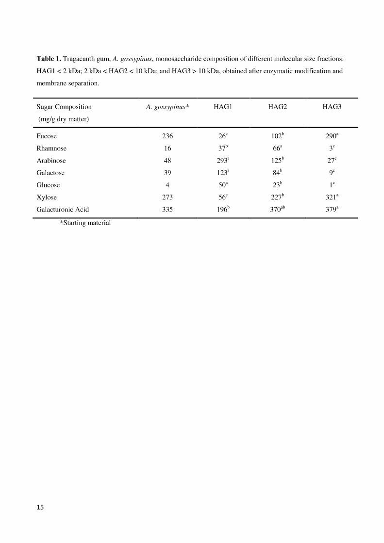
15
Table 1. Tragacanth gum, A. gossypinus, monosaccharide composition of different molecular size fractions:
HAG1 < 2 kDa; 2 kDa < HAG2 < 10 kDa; and HAG3 > 10 kDa, obtained after enzymatic modification and
membrane separation.
Sugar Composition
(mg/g dry matter)
A. gossypinus* HAG1 HAG2 HAG3
Fucose 236 26c 102b 290a
Rhamnose 16 37b 66a 3c
Arabinose 48 293a 125b 27c
Galactose 39 123a 84b 9c
Glucose 4 50a 23b 1c
Xylose 273 56c 227b 321a
Galacturonic Acid 335 196b 370ab 379a
*Starting material

16
Table 2. Glycosidic linkage analysis of different gum tragacanth fractions of different molecular size: HAG1 < 2 kDa;
2 kDa < HAG2 < 10 kDa; and HAG3 > 10 kDa, obtained after enzymatic modification and membrane separation.
Values given in parenthesis indicate the relative abundance of the type of bond for the particular monosaccharide
(quantified from peak area in the glycosidic linkage chromatogrpahy profile).
Monosaccharide HAG1 HAG2 HAG3
Linkage type % peak area % peak area % peak area
L-Fucose
t-Fucp - 14.4(80.4) 41.6(88.1)
1,3-Fucp - 0.8(4.5) 1.2(2.6)
1,4-Fucp - 0.7(3.9) 1.1(2.3)
1,2-Fucp - 1.7(9.5) 2.8(5.9)
1,3,4-Fucp - 0.3(1.7) 0.5(1.1)
L-Rhamnose
t-Rhap - 0.2(1.4) -
1,2,4 Rhap 2.3(100) 6.9(48.6) -
1,4 Rhap - 7.1(50) -
L-Arabinose t-Araf 0.4(5.8) 0.8(53.3) 1.2(100)
1,5 Araf 6.5(94.2) 0.7(46.7) -
D-Galactose
t-Galp 3.7(5.6) 3.7(20.2) 1.6(88.9)
1,4-Galp 58.8(88.7) 13.5(73.8) 0.2(11.1)
1,6 Galp - 0.8(4.4) -
1,4,6 Galp 3.8(5.7) 0.3(1.6) -
D-Glucose
t-Glup 2.8(18.6) 2.6(40.6) 1.1(47.8)
1,4-Glup 8.5(56.3) 3.5(54.7) 1.2(52.2)
1,2,4-Glup 3.8(25.1) 0.3(4.7) -
D-Xylose
t-Xylp - 5.4(22.6) 7.4(15.8)
1,2,-Xylp - 18.5(77.4) 38.3(81.7)
1,2,3-Xylp - - 1.2(2.5)
D-Galacturonic acid t-GalAp 0.2(2.9) 2.8(24.6) 0.2(20)
1,4-GalA 6.6(97.1) 8.5(74.6) 0.8(80)
1,4,6-GalA - 0.1(0.9) -

17
FIGURE 1
0.00
10.00
20.00
30.00
40.00
50.00
60.00
70.00
5.00 7.50 10.00 12.50 15.00 17.50 20.00 22.50 25.00 27.50 30.00
Tra Control
HAG1
HAG2
HAG3
1.3 kDa
10 kDa
40 kDa
110 kDa
400 kDa
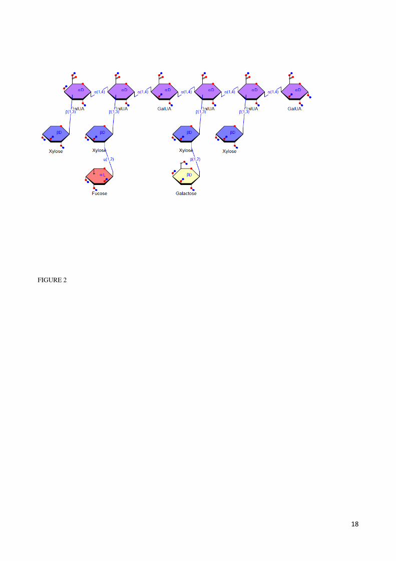
18
FIGURE 2

19
FIGURE 3
-150
-100
-50
0
50
100
150
200
250
300
350
HAG1 HAG2 HAG3 Galactan
B.longum 232
B.infantis 233
B.infantis 1497
B.infantis 2238
B.lactis
L.acidophilus
B.longum Bl-05
Cl.perfringens

64
Paper IV
Tragacanth gum: Functionality and new Prebiotics Potential

1
5. Tragacanth gum: Functionality and new Prebiotics Potential
This chapter is included review paper that explains mechanism of emulsion stabilization by gum tragacanth and
explores the enzymatic production of different fractions of gum tragacanth and evaluates their prebiotic activity
potential (Paper IV)
Authors
Hassan Ahmadi Gavlighi1,2, Anne S. Meyer1,*, J. Dalgaard Mikkelsen1
Address
1 Center for Bioprocess Engineering, Department of Chemical and Biochemical Engineering, Technical University of
Denmark (DTU), DK-2800 Kgs. Lyngby, Denmark
2 Department of Food Science and Technology, Faculty of Agriculture, Tarbiat Modares University, P. O. Box 14115-
336, Tehran, Iran
*Corresponding author: Tel. +45 4525 2800; Fax: +45 4588 2258; e-mail: [email protected]
2
Abstract
Gum tragacanth is a plant derived hydrocolloid that can increase viscosity and stabilize emulsions and which has a long
history of use in food, pharma, and cosmetics. The gum is mainly produced in the Middle East, and permitted for food
use in the US as well as in Europe (E-number E413). The gum is made up of heterogeneous hydrophilic
polysaccharides, which contain various highly substituted pectin-like structural elements. Enzymatically produced low-
molecular weight fractions of gum tragacanth exhibit potential prebiotic activity by promoting growth in vitro of
Bifidobacterium longum subsp. infantis strains. These findings may lead to new uses of this gum as a starting point for
enzymatic production of value-added prebiotic compounds for functional foods.
Keywords: Gum tragacanth, viscosity, prebiotic, Bifidogenic, enzymatic depolymerisation.
Introduction
The natural plant exudate gum tragacanth is obtained from the stem of the bush like plant “goat's-horn”, Astragalus
species, and the gum is permitted for use in foods in both the US and in Europe. Gum tragacanth is made up of
complex, highly branched, heterogeneous hydrophilic polysaccharides that contain L-arabinose, D-galactose, D-
glucose, D-xylose, L-fucose, L-rhamnose, and D-galacturonic acid (1, 2). The gum saccharides form complexes with
salts, notably salts with divalent cations, such as Ca2+ salts (3). Tragacanthin is the water-soluble fraction, that confers
high viscosity when suspended in water, and bassorin is the insoluble part which swells in water to form a gel (1).
Tragacanthin appears to resemble pectin, and contains linear chains of galacturonic acid (probably 1,4-α-linked) rich in
xylose with varying levels of fucose, the latter depending on the plant species from which the gum is obtained (4). Gum
tragacanth rich in xylose and galacturonic acid may thus contain xylogalacturonan, whereas gum samples having high
fucose, xylose, and galacturonic acid levels, may mainly contain fuco-xylogalacturonan, in addition to highly branched
of arabinogalactan structures (1,5). Gum tragacanth has both emulsifying and stabilizing properties in emulsions, and is
considered as being “bi-functional” by having both the capacity to facilitate emulsification as well as providing
stabilization of the emulsion after its formation (6). The structure and composition of the gum are important issues in
relation to emulsion stabilizing properties (4).
On the other hand, there is an increasing interest in the development and identification of new carbohydrates with
prebiotic effects that selectively stimulate the growth or activity of certain beneficial intestinal bacteria (currently

3
lactobacilli and bifidiobacteria), which in turn may confer benefits upon well-being and health of the host (7). Due to
its high viscosity inducing effects, it is not possible to include substantial levels, i.e. sufficient “dosages”, of gum
tragacanth into foods to elicit a possible prebiotic effect. However, targeted enzymatic depolymerization has previously
proven useful as a technique to decrease the viscosity, by making the lower molecule size of natural, highly viscose
polysaccharides and hydrocolloids. Especially in combination with membrane separation, prebiotic compounds of
different molecular size and structural composition can be obtained enzymatically from various natural, viscous
polysaccharides (8-10).
Gum tragacanth could be another source of such production of prebiotics dietary fibers due to the unique sugar
composition and structure encompassing both arabinogalactan and fucoxylogalacturonan polymers. The presumed
presence of α1,2-linked fucosyl moieties in the structure may confer unique bifidogenic properties just as it is known
that terminal, fucosyl residues found in some human milk oligosaccharide structures (as α1-2, α1-3, and α1-4
substitutions) support the growth of Bifidobacterium longum subsp. infantis, an organism considered as desirable in the
infant-gut microflora (11). Exudates gums with arabinogalactan structure such as gum Arabic has for example been
considered as a very well tolerated dietary fiber with bifidogenic properties believed to benefit intestinal health (12).
The objectives of this review are: Firstly, to examine the mechanism of emulsion stabilization by gum tragacanth,
including an investigation of the correlation between gum structure and composition with stabilization of protein based
emulsions. Secondly, to explore the enzymatic production of different fractions of gum tragacanth and evaluate their
prebiotic activity potential.
Functional properties, stability and application
The use of gum tragacanth in foods has been approved as GRAS in the US for more than 50 years at a level of 0.2–
1.3% and in Europe, where gum tragacanth has an E-number (E413) on the list of additives approved by the Scientific
Committee for Food of the European Community (13, 14) . In spite of the availability of alternative materials,
tragacanth gum has superior functional properties (viscosity and emulsification) compared to other gums and it is fairly
stable over a wide pH range down to extremely acidic conditions at about pH 2 (15). Gum tragacanth is therefore used
as the primary substitute in a number of applications and the gum has thus been used for many years as a stabilizer,
thickener, emulsifier and suspending agent in food, pharmaceutical, and cosmetic products, as well as in technical
applications based on its high viscosity at low concentration. The gum has unusually high stability to heat and acidity
and exhibits effective emulsifying properties (6). Evaluation of the flow behavior of six species of Iranian gum

4
tragacanth dispersions confirmed very high viscosity inducing properties and has shown that gum tragacanth exhibits
shear-thinning behavior in aqueous dispersion. Depending on the species of the plant origin of gum, the viscosity of
1.5% (weight/weight) solutions may typically range widely, e.g. reported from ~1.5 up to 35 Pa · s (1,4).
It also is pourable and has a creamy mouth feel and good flavor-release properties (16). Gum tragacanth is used in the
food industry in citrus oil emulsions (17), salad dressings, condiments, sauces, bakery emulsions, oil and flavour
emulsions, bakery fruit-based fillings and toppings (give a shiny, clear appearance and a creamy texture) (18),
confectionery, soft drinks, jellies, desserts, ice creams (provide texture to the product), flavors, spices (15).
Mechanism of tragacanth gum stabilization
There have been different studies that have analyzed the stabilizing effect of gum tragacanth in different emulsion
systems. Yokoyama, Srinivasan (19) have shown that the stabilizing effect of gum tragacanth was a result of the steric
repulsion force and the stability can be controlled by changing pH. On the other hand, the ability of tragacanth gum to
stabilize beverage emulsions could be due to residual surface activity in addition to enhancement of emulsion viscosity
(20).
Considering the relation between gum tragacanth composition and emulsion stabilization, our own recent findings have
shown a highly significant correlation between the creaming index and the methoxyl content of the soluble fraction in
the gums. Also, a strong correlation was found between the total galacturonic acid content (in the full gum) and
creaming index, indicating that it is not the galacturonic acid alone can stabilize the emulsions (4).
In summary, tragacanth gum stabilization of protein-emulsified emulsions is probably a result of two mechanisms:
Firstly, formation of non-covalent protein–(gum) polysaccharide complexes via electrostatic interaction mainly by
galacturonic acid content in the soluble tragacanthin part of the gum, and secondly, the viscosity increase induced by
the insoluble fraction (bassorin) (Figure 1).
Potential prebiotic effect of tragacanth gum
The concept of prebiotics was introduced in 1995 by Gibson and Roberfoid as an alternative approach to the modulation
of the gut microbiota mainly lactobacilli and bifidobacteria (7). From the new definition of prebiotic (updated in 2007)
the criteria of a prebiotic is: ‘’1- resistance to gastrointestinal acidity 2- fermented by intestinal microflora 3- selective
stimulate of the growth or/activity of intestinal bacteria’’ (21). Until now, different prebiotic compounds have been
produced for industrial application especially, fructo -oligosaccharides (FOS), galacto-oligosaccharides (GOS) and a
main approach is via enzymatic processing (22-24). Hence, production of novel ingredients with potential prebiotic

5
activity from many different types of plant cell wall polysaccharides has been reported. Arabinogalacto-
oligosaccharides can be made from soybeans by endo-galactanases, arabino-oligosaccharides can be made from sugar
beet pulp by pectinases and other plant-cell wall degrading enzymes, rhamnogalacturono-oligosaccharides can be made
from apple by rhamnogalacturonases, arabinoxylo-oligosaccharides can be made from wheat by xylanases and
galacturono-oligosaccharides can be made from polygalacturonic acid by endo-galacturonases (25-27). Also,
differences in structure, such as changes in size and the presence of branch(s) and differences in the complexity of the
substituent can cause significant changes in the prebiotic properties of oligosaccharides (28, 29).
Tragacanth gum is one of exudates gum that is naturally and commercially available and which enzymatically
depolymerised exerts potential prebiotic effects by promoting growth of Bifidobacteria in single culture fermetnations
(Figure 2). In this study, gum tragacanth was hydrolysed by pectinolytic enzymes and purified into three fractions
using membrane separation, which gave rise to production of different molecular size oligosaccharides (Figure 3).
Interestingly, in addition to different molecular sizes also differences in composition and structures were achieved with
this process. To assess the prebiotic potential of the products, different pure cultures of beneficial and pathogen
bacteria were chosen.The results showed that, the beneficial bacteria, notably gut-related Bifidobacteria spp. grew
significantly better on the small molecular size structures as compared on the larger gum tragacanth molecules. Smaller
molecular size of plant polysaccharides such as potato galactan oligomers have previously been shown to support the
growth of bifidobacteria better than high molecular weight polysaccharides (30) .However, the high molecular weight
fraction (with high fucose content) inhibited the growth of Cl. perfringens (ATCC 13124). L. acidophilus grew equally
well on all the gum tragacanth fractions as well as on the galactan (. In general, the growth of the putatively beneficial
bifidobacteria including both B. longum subsp. Infantis and B.longum subsp. longum bacteria were significantly
higher on gum tragacanth oligosaccharides than on galactan used as a control for established prebiotic activity (Figure
2).
Conclusion
• Tragacanth gum is permitted for food use and can stabilize different types of emulsion formulations, but is
notably efficient as a viscosity enhancer and stabilizer in acidic solutions
• Different tragacanth gum samples obtained from different species of Astragalus have different composition,
and produce different levels of soluble and insoluble gum fractions
• Emulsion stabilization of the gum is related to the gum composition and structure, and mainly galacturonic
acid content and degree of esterification are important

6
• low molecular size oligosaccharides produced enzymatically has higher potential prebiotic activity than longer
chain gum saccharides
• Tragacanth gum can be a new source for development of innovative functional foods with health claims
Acknowledgement. The authors would like to acknowledge Tarbiat Modares University and the Ministry of
Science, Research and Technology of Iran for their financial support.

7
References
1. S. Balaghi, M.A. Mohammadifar et al., Food Hydrocolloids., 25, pp. 1775-1784 (2011).
2. G. Aspinall, J. Baillie, J. Chem. Soc., pp. 1702-1714 (1963).
3. S. Balaghi, M.A. Mohammadifar et al., Food Biophys., 5, pp. 59-71 (2010).
4. H.A. Gavlighi, A.S. Meyer et al., Food Hydrocolloids., 31, pp. 5-14 (2013).
5. C.A. Tischer, M. Iacomini et al., Carbohydr Res., 18, pp. 1647-1655 (2002).
6. W.Weiping, G. Phillips et al., Handbook of hydrocolloids, CRC Press, (2000).
7. G.R. Gibson, M.B. Roberfroid, J Nutr., 125, pp. 1401-1412 (1995).
8. K.C. Mountzouris, S.G. Gilmour et al., J Food Sci., 67, pp. 1767-1771 (2002).
9. L.V. Thomassen, D.M. Larsen et al., Enzyme Microb. Technol., 49, pp. 289-297 (2011).
10. M. Pinelo, G. Jonsson et al., Sep Purif Technol., 70, pp. 1-11 (2009).
11. G.M. Ruiz-Palacios, L.E. Cervantes et al., J Biol Chem., 278, pp. 14112-14120 (2003).
12. S. Meance. Agro Food Ind. Hi-Tech., 15, pp. 24-29 (2004).
13. Food and Drug Administration, USA. Available from http://www.gpo.gov/fdsys/pkg/CFR-2011-title21-
vol3/pdf/CFR-2011-title21-vol3-sec184-1355.pdf (accessed November 06, 2012).
14. D.M.W. Anderson, M.M.E. Bridgeman, Phytochemistry., 24, pp. 2301-2304 (1985).
15. G.O. Phillips, P.A. Williams, Handbook of Hydrocolloids (2nd Edition), Woodhead Publishing, (2009).
16. G. Levy, T. Schwarz, J Am Pharmaceut Assoc., 47, pp. 451-454 (1958).
17. A.R. Taherian, P. Fustier et al., Int. J. Food Prop., 11, pp. 24-43 (2008).
18. J.D. Dziesak, Food Technol., 45, pp. 8-20 (1991).
19. A. Yokoyama, K.R. Srinivasan et al., J. Colloid Interface Sci., 126, pp. 141-149 (1988).
20. E. Rezvani, G. Schleining et al., LWT-Food Sci. Technol., 48, pp. 82-88 (2012).
21. M. Roberfroid, J Nutr., 137, pp. 830s-837s (2007).
22. M. Adamczak, D. Charubin et al., Chem. Pap., pp. 111-116 (2009).
23. P.S. Panesar, S. Kumari et al., Crit. Rev. Biotechnol., pp. 1-20 (2012).
24. L. Zhang, Y. Su et al., Bioresour. Technol., 23, pp. 9144-9149 (2010).
25. E. Olano-Martin, G.R. Gibson et al., J. Appl. Microbiol., 93, pp. 505-511 (2002).
26. J. Concha Olmos, M.E. Zúñiga Hansen., Chem. Eng. J., 192, pp. 29-361 (2012).
27. M. Martínez Sabajanes, Y. Remedios et al., Int. J. Food Sci. Technol., 47, pp. 747–754 (2012)

8
28. J. Holck, K. Hjernø et al., Process Biochem., 46, pp. 1039-1049 (2011).
29. M.A. Kabel, L. Kortenoeven et al., J. Agric. Food Chem., 50, pp. 6205-6210 (2002).
30. M. Michalak, L.V. Thomassen et al., Enzyme Microb. Technol., 50, pp. 121-129 (2012).

9
Figure 1. The scheme of stabilization mechanism of protein-emulsified (WPI indicating whey protein isolate) emulsions
by Gum Tragacanth.

10
Figure 2. Differential growth of bacterial strains on enzyme catalyzed pectolytic enzyme degradation products from gum tragacanth against potato galactan used as control. Growth responses for the 4 substrates is shown for a substrate concentration 10 g/L for all bacterial strains. Data are given as average values of 3 growth assay replicates and shown ± S.D.
0
50
100
150
200
250
300
350
Are
a
un
der
th
e g
row
th c
urv
e (O
.D×
min
)
HAG1
HAG2
HAG3
Galactan

11
Figure 3. Schematic flow sheet for the enzymatic hydrolysis process of tragacanth gum and membrane separation steps
Tragacanth Gum
Solution
Membrane separation
Pectin Lyase
Polygalacturonas
Permeate <10KDa
Membrane separation
Permeate <2 KDa
Retentate 2-10KDa
Retentate >10KDa
Prebiotic Activity test
76
6. Conclusion
The carbohydrate compositional and methoxyl and acetyl contents of six different species of Astragalus to acquire
different gum tragacanth were examined. This work is prerequisite to assess effect of structural composition on
mechanisms of effect of this type of gums for application in different area mainly food application. Also, production of
new products with functional properties was achieved via enzymatic modification.
The hypothesis of paper I and III were to get knowledge of physicochemical properties of different species of
Astragalus and find relationship between the gum composition and stabilization of emulsion. The results presented that
the sugar-compositional analysis of gum tragacanth from different species of Astragalus by HPAEC-PAD were
different values for each of the sugar components (arabinose, xylose, galactose, glucose, fucose, galacturonic acid and
traces of rhamnose); therefore, the sugar composition of Astragalus gum exudates is strongly species-dependent and the
functional properties of the gums are greatly influenced by their sugar compositions. Galacturonic acid was high in the
soluble part of all species whereas L-fucose and partially xylose was major in insoluble fraction. The species with high
fucose, may likely have high xylose and galacturonic acid content, and vice versa, whereas gums having high arabinose
content may also contain high galactose levels, and vice versa.
The results in paper I showed that positive correlation between the methoxyl content of the soluble part of the gums
content and creaming index indicate that methoxyl groups may have properties that play a role in the emulsification
properties of tragacanth gums. Also, galacturonic acid of gum tragacanth samples from different Astragalus species was
correlated with creaming index. Hence, particularly good emulsion stabilization properties will occur if gum tragacanth
is divided into a soluble and an insoluble gum fraction, and the soluble fraction is rich in fucose and high in methoxyl
content. Evaluating of the correlation between acetyl content of the gum tragacanth samples and the creaming index
shown no significant relationship could be established.
In all samples prepared from gum tragacanth, the apparent viscosity decreased with increasing shear rate, indicating a
shear-thinning nature of tragacanth gum in solution.
In general as we discussed in paper IV, the factors for determining how tragacanth gums work to stabilize emulsions
were may hydrophobic bonding between tragacanth gum and WPI emulsified emulsion particles with increasing
viscosity of emulsion, but apparently also the composition, notably the total galacturonic acid content in the gum, the
amount of methoxyl groups and probably also fucose in the solubilized part.
Consequently, our finding confirmed that hypothesis that all exudates tragacanth gum had different composition and
functional properties and any research carried out on gum tragacanth or any industrial application of this gum without
respect to the plant species will lead to misleading results.
Nowadays, producing ingredient with health beneficial or food ingredient (Functional foods) has been interested and
demanded. Thus, finding and making new source with high stability in heating and acidic condition and lower viscosity
to enable the supply of higher dosages of the gum saccharides for food application is essential. The results from

77
composition of different gum shown that A. gossypinus has specific monosaccharide composition mainly high level of
fucose (Paper I and III).
The next hypothesis was to firstly, examine possibility of producing different molecular size of gum tragacanth and
secondly, evaluate prebiotic effect of these fractions. So, our finding shown that enzymatic process integrated with
membrane separation can produce different molecular size of gum tragacanth with different carbohydrate profiles
(paper II). Small molecular size mainly composed of arabinose and galactose whereas high molecular size was high
fucose and xylose.
Linkage analysis of each fraction had good information to elucidate structure of gum tragacanth in more detail. In
HAG1 and HAG3, the frequency of the 1,4-linked galactose bonds was dominating; we presume that these bonds are
due to the presence of β-1,4-linked galactan structural elements. The dominance of terminally linked fucose and (1,),2-
linked xylose, as well as terminally linked xylose in HAG3 with the high galacturonic acid content underscored that
HAG3 contained the typical fuco-xylogalacturonan structural elements of gum tragacanth. These findings confirmed
that it is possible to produce low viscosity of tragacanth gum with different composition for some application that
viscosity is problem.
The glycosidic bonds, the molecular size, as well as the monosaccharide composition of the prebiotic carbohydrates
thus define the effect and the eventual relative increase in beneficial bacteria, including certain Bifidobacterium and
Lactobacillus species. Higher molecular size inhibited of growth of pathogenic clostridia and growth better on the
lactobacillus spp. whereas, lower molecular size grew better than higher size on bifidobacteria spp. Also, The linkage
analysis confirmed the dominance of terminally linked fucose and (1,),2-linked xylose in HAG3 structure and this
similarity with human milk oligosaccharides that contain 2-Fucoylated lactose can be one of the source of producing of
human milk oligosaccharides. Thus our illustrations confirmed potential bioactivity of gum tragacanth that contained
some unique substituted saccharides, notably the highly substituted fuco-xylo-galacturonan and the arabinogalactan
structures after depolymerization especially in lower molecular weight. Although, higher molecular weight may be use
as antimicrobial ingredient because of inhibition of clostridia.
In summary, the data are a first step indicating that gum tragacanth may be a source of functional poly- and
oligosaccharides that may exert beneficial bioactivity as prebiotics. The eventual application of enzymatically
hydrolyzed gum tragacanth oligomers in foods would be a new, value-added application of a classic food gum.
6.1 Future perspectives
Compositional and physicochemical properties information was established and will use in food industrial application
based on interaction and effect on the formulation. Although, there is need more information about how interaction of
arabinogalactan and xylogalactronan in the gum tragacanth structure. The distribution pattern of the methoxyl groups on
the galacturonic acid backbone, i.e. whether in blocks or randomly distributed, plays a crucial role. Also, chains of de-
methoxylated galacturonic acids may exist in the soluble fraction of gum tragacanth, which would help explain why the
methoxylation degree varied so widely among the soluble gum tragacanth fractions .It may well be that a random, but
high degree of substitution, will act to stabilize emulsions (and not cause gelation) as observed for enzymatically
modified pectin.

78
Knowledge about the structural make up of tragacanth gum is important for tailoring of enzymatic process. So,
development of hydrolyzing methods using mono active enzyme and produce of new functional properties of gum is
one of the goals in the future work. Also, a physicochemical property of high fraction (HAG3) of gum tragacanth is
needed to find application in industry.
At present work we assessed potential prebiotics in pure culture so; there is a severe lack of knowledge about the
specific carbohydrate structural effects and about the interactions among different “probiotic” and pathogenic strains in
vivo. Any data on this would provide a significantly improved foundation for tailoring of carbohydrate structures for gut
health. Therefore, examination of different fraction of gum tragacanth in human gut flora is needed to establishment of
fractions as prebiotic compound.
At present, prebiotic carbohydrates purification represents the most expensive operation in food manufacturing
involving these types of compounds, accounting for around 90% of costs of food production. Current commercial
processes for carbohydrate's purification are usually based on chromatographic processes involving ion-exchange resins
or activated charcoal. Also, membrane processes, such as nanofiltration, seem to show potential for large scale
carbohydrate mixtures separation. Therefore, purification of oligosaccharide using new economical technique such as
supercritical fluid extraction is important issue to examine effect of specific oligomers with different DP on prebiotic
effects.
Evaluation of antimicrobial properties of six species is another interesting area mainly to extend shelf life of food
products with natural ingredient.
Also, the variety in composition is one of the approaches that could be modified via enzymatic process and test
prebiotic effect. Other application in formulation of nano-emulsions or nano-liposomes is need to explore.
Modification of larger fraction of tragacanth gum (HAG3) with special enzyme such as xylogalacturonan hydrolysis
and evaluation of bioactivity of different compound is interesting mainly in prebiotic area.
79
7. References
Adamczak, M., Charubin, D., & Bednarski, W. (2009). Influence of reaction medium composition on enzymatic
synthesis of galactooligosaccharides and lactulose from lactose concentrates prepared from whey permeate. Chemical Papers, 63(2), 111-116.
Akhtar, M., & Dickinson, E. (2003). Emulsifying properties of whey protein–dextran conjugates at low pH and
different salt concentrations. Colloids and Surfaces B: Biointerfaces, 31(1–4), 125-132. Al-Tamimi, M., Palframan, R., Cooper, J., Gibson, G., & Rastall, R. (2006). In vitro fermentation of sugar beet arabinan
and arabino-oligosaccharides by the human gut microflora. Journal of Applied Microbiology, 100(2), 407-414. Anderson, D. M. W., & Bridgeman, M. M. E. (1985). The composition of the proteinaceous polysaccharides exuded by
astragalus microcephalus, A. Gummifer and A. Kurdicus—The sources of turkish gum tragacanth. Phytochemistry, 24(10), 2301-2304.
Anderson, D. M. W., & Grant, D. A. D. (1988). The chemical characterization of some Astragalus gum exudates. Food
Hydrocolloids, 2(5), 417-423. Arnous, A., & Meyer, A. S. (2008). Comparison of methods for compositional characterization of grape (Vitis vinifera
L.) and apple (Malus domestica) skins. Food and Bioproducts Processing, 86(C2), 79-86. Aspinall, G. O., & Baillie, J. (1963). 318. Gum tragacanth. Part I. Fractionation of the gum and the structure of
tragacanthic acid. Journal of the Chemical Society (Resumed), 1702-1714. Azarikia, F., & Abbasi, S. (2010). On the stabilization mechanism of Doogh (Iranian yoghurt drink) by gum tragacanth.
Food Hydrocolloids, 24(4), 358-363. Balaghi, S., Mohammadifar, M. A., & Zargaraan, A. (2010). Physicochemical and rheological characterization of gum
tragacanth exudates from six species of Iranian Astragalus. Food Biophysics, 5(1), 59-71. Balaghi, S., Mohammadifar, M. A., Zargaraan, A., Gavlighi, H. A., & Mohammadi, M. (2011). Compositional analysis
and rheological characterization of gum tragacanth exudates from six species of Iranian Astragalus. Food
Hydrocolloids, 25(7), 1775-1784. Brownawell, A. M., Caers, W., Gibson, G. R., Kendall, C. W. C., Lewis, K. D., Ringel, Y., & Slavin, J. L. (2012).
Prebiotics and the health benefits of fiber: current regulatory status, future research, and goals. The Journal of
Nutrition, 142(5), 962-974. Chanamai, R., & McClements, D. J. (2001). Depletion Flocculation of Beverage Emulsions by Gum Arabic and
Modified Starch. Journal of Food Science, 66(3), 457-463. Charalampopoulos, D., & Rastall, R. A. (2009). Prebiotics and probiotics science and technology. New York: Springer
Verlag. Chiewchan, N., Phungamngoen, C., & Siriwattanayothin, S. (2006). Effect of homogenizing pressure and sterilizing
condition on quality of canned high fat coconut milk. Journal of Food Engineering, 73(1), 38-44.
Ciucanu, I., & Kerek, F. (1984). A simple and rapid method for the permethylation of carbohydrates. Carbohydrate
Research, 131(2), 209-217. Cutié, S. S., & Martin, S. J. (1995). Size-exclusion chromatography of cross-linked superabsorbent polymers. Journal
of Applied Polymer Science, 55(4), 605-609. Damodaran, S., Parkin, K. L., & Fennema, O. R. (2008). Fennema's food chemistry (4th ed. / edited by Srinivasan
Damodaran, Kirk Parkin, and Owen R. Fennema. ed.). Boca Raton ; London: CRC Press.

80
Demetriades, K., Coupland, J. N., & McClements, D. J. (1997). Physicochemical Properties of Whey Protein-Stabilized Emulsions as affected by Heating and Ionic Strength. Journal of Food Science, 62(3), 462-467.
Denkova, P. S., Tcholakova, S., Denkov, N. D., Danov, K. D., Campbell, B., Shawl, C., & Kim, D. (2004). Evaluation
of the precision of drop-size determination in oil/water emulsions by low-resolution NMR spectroscopy. Langmuir, 20(26), 11402-11413.
Dickinson, E. (1992). An introduction to food colloids: Oxford University Press. Dickinson, E. (1997). Properties of Emulsions Stabilized with Milk Proteins: Overview of Some Recent Developments.
Journal of Dairy Science, 80(10), 2607-2619. Dickinson, E. (2001). Milk protein interfacial layers and the relationship to emulsion stability and rheology. Colloids
and Surfaces B: Biointerfaces, 20(3), 197-210.
Dickinson, E. (2008). Interfacial structure and stability of food emulsions as affected by protein-polysaccharide
interactions. Soft Matter, 4(5), 932-942.
Dickinson, E. (2009). Hydrocolloids as emulsifiers and emulsion stabilizers. Food Hydrocolloids, 23(6), 1473-1482.
Dickinson, E., & McClements, D. J. (1996). Advances in food colloids: Blackie Academic & Professional Glasgow.
Dickinson, E., Murray, B. S., Stainsby, G., & Anderson, D. M. W. (1988). Surface activity and emulsifying behaviour of some Acacia gums. Food Hydrocolloids, 2(6), 477-490.
Elias,H-G. (1992) Makromoleküle. Band2, Technologie. Hüthig & Wepf Verlag, Basel, Heidelberg, New York
FAO (1995) Gums, resins and latexes of plant origin. (Non-wood forest products 6). FAO, Rome Food and Drug Administration, USA. (2006). Available from http://www.gpo.gov/fdsys/pkg/CFR-2011-title21-
vol3/pdf/CFR-2011-title21-vol3-sec184-1355.pdf Accessed 10.04.12.
Garti, N. (1999). HYDROCOLLOIDS AS EMULSIFYING AGENTS FOR OIL-IN-WATER EMULSIONS. Journal
of Dispersion Science and Technology, 20(1-2), 327-355. Gavlighi, H. A., Meyer, A. S., Zaidel, D. N. A., Mohammadifar, M. A., & Mikkelsen, J. D. (2013). Stabilization of
emulsions by gum tragacanth (Astragalus spp.) correlates to the galacturonic acid content and methoxylation degree of the gum. Food Hydrocolloids, 31(1), 5-14.
Gibson, G. R., Probert, H. M., Van Loo, J., Rastall, R. A., & Roberfroid, M. B. (2004). Dietary modulation of the
human colonic microbiota: updating the concept of prebiotics. Nutr Res Rev, 17(2), 259-275. Gibson, G. R., & Roberfroid, M. B. (1995). Dietary modulation of the human colonic microbiota: introducing the
concept of prebiotics.The Journal of Nutrition, 125(6), 1401-1412.
Gorji, E. G., Mohammadifar, M. A., & Ezzatpanah, H. (2011). Influence of gum tragacanth, Astragalus gossypinus, addition on stability of nonfat Doogh, an Iranian fermented milk drink. International Journal of Dairy
Technology, 64(2), 262-268. Guzey, D., & McClements, D. J. (2006). Formation, stability and properties of multilayer emulsions for application in
the food industry. Advances in Colloid and Interface Science, 128–130(0), 227-248. Holck, J., Hjernø, K., Lorentzen, A., Vigsnæs, L. K., Hemmingsen, L., Licht, T. R., Mikkelsen, J. D., & Meyer, A. S.
(2011b). Tailored enzymatic production of oligosaccharides from sugar beet pectin and evidence of differential effects of a single DP chain length difference on human faecal microbiota composition after< i> in vitro</i> fermentation. Process Biochemistry, 46(5), 1039-1049.

81
Home, D. S. (1996). Protein-stabilized emulsions. Current Opinion in Colloid & Interface Science, 1(6), 752-758. Kabel, M. A., Kortenoeven, L., Schols, H. A., & Voragen, A. G. J. (2002). In vitro fermentability of differently
substituted xylo-oligosaccharides. Journal of agricultural and food chemistry, 50(21), 6205-6210. karaya, T. a., Phillips, G. O., & Williams, P. A. (2000). Handbook of hydrocolloids. Boca Raton, Fla.: CRC Press ;
Cambridge : Woodhead. Lapasin, R., & Pricl, S. (1995). Rheology of polysaccharide systems. Rheology of industrial polysaccharides: Theory
and applications. Glasgow: Blackie, 250-494. Leroux, J., Langendorff, V., Schick, G., Vaishnav, V., & Mazoyer, J. (2003). Emulsion stabilizing properties of pectin.
Food Hydrocolloids, 17(4), 455-462.
Levy, G., & Schwarz, T. W. (1958). Tragacanth solutions I. The relation of method of preparation to the viscosity and
stability. Journal of the American Pharmaceutical Association, 47(6), 451-454. Lo, T. C. T., Kang, M. W., Wang, B. C., & Chang, C. A. (2007). Glycosyl linkage characteristics and classifications of
exo-polysaccharides of some regionally different strains of Lentinula edodes by amplified fragment length polymorphism assay and cluster analysis. Analytica Chimica Acta, 592(2), 146-153.
Macfarlane, G. T., Steed, H., & Macfarlane, S. (2008). Bacterial metabolism and health-related effects of galacto-
oligosaccharides and other prebiotics. Journal of Applied Microbiology, 104(2), 305-344. Macosko, C. W., & Larson, R. G. (1994). Rheology: principles, measurements, and applications. Mäkeläinen, H., Saarinen, M., Stowell, J., Rautonen, N., & Ouwehand, A. (2010). Xylo-oligosaccharides and lactitol
promote the growth of Bifidobacterium lactis and Lactobacillus species in pure cultures. Beneficial Microbes,
1(2), 139-148. Michalak, M., Thomassen, L. V., Roytio, H., Ouwehand, A. C., Meyer, A. S., & Mikkelsen, J. D. (2012). Expression
and characterization of an endo-1,4-β-galactanase from Emericella nidulans in Pichia pastoris for enzymatic design of potentially prebiotic oligosaccharides from potato galactans. Enzyme and Microbial Technology,
50(2), 121-129.
McClements, D. J. (2004). Context and background. In Food emulsions: principles, practices, and techniques: CRC Press.
Meance, S. (2004). Acacia gum (Fibregum™), a very well tolerated specific natural prebiotic having a wide range of
food applications-Part 1. Agro Food Industry Hi-Tech, 15(1), 24-29. Miri, T. (2011). Viscosity and Oscillatory Rheology. In Practical Food Rheology (pp. 7-28): Wiley-Blackwell. Mohammadifar, M., Musavi, S., & Williams, P. (2007). Study of complex coacervation between β-lactoglobulin and
tragacanthin (soluble part of gum tragacanth). Milchwissenschaft, 62(4), 389-392. Mohebbi, M., Amiryousefi, M. R., Hasanpour, N., & Ansarifar, E. (2012). Employing an intelligence model and
sensitivity analysis to investigate some physicochemical properties of coated bell pepper during storage. International Journal of Food Science & Technology, 47(2), 299-305.
Nasirian, R., Vaziri, A., Safekordi, A. A., & Ardjmand, M. (2010). Use of Gum Tragacanth in reduction of dairy cream
fat. Milchwissenschaft-Milk Science International, 65(1), 49-52. Norton, I. T., Spyropoulos, F., & Cox, P. (2011). Practical Food Rheology - An Interpretive Approach. In: John Wiley
& Sons.

82
Olano-Martin, E., Gibson, G. R., & Rastall, R. A. (2002). Comparison of the in vitro bifidogenic properties of pectins and pectic-oligosaccharides. Journal of Applied Microbiology, 93(3), 505-511.
Oosterveld, A., Beldman, G., & Voragen, A. G. J. (2002). Enzymatic modification of pectic polysaccharides obtained
from sugar beet pulp. Carbohydrate Ppolymers, 48(1), 73-81. Panesar, P. S., Kumari, S., & Panesar, R. (2012). Biotechnological approaches for the production of prebiotics and their
potential applications. Critical Reviews in Biotechnology(00), 1-20. Phillips, G. O., & Williams, P. A. (2009). Handbook of Hydrocolloids (2nd Edition). In: Woodhead Publishing. Quemada, D., & Berli, C. (2002). Energy of interaction in colloids and its implications in rheological modeling.
Advances in Colloid and Interface Science, 98(1), 51-85. Ramachandran, S., Chen, S., & Etzler, F. (1999). Rheological characterization of hydroxypropylcellulose gels. Drug
Development and Industrial Pharmacy, 25(2), 153-161. Rao, M. A. (2007). Introduction: Food Rheology and Structure
Rheology of Fluid and Semisolid Foods. In (pp. 1-26): Springer US. Rasmussen, L. E., & Meyer, A. S. (2009). Size exclusion chromatography for the quantitative profiling of the enzyme-
catalyzed hydrolysis of xylo-oligosaccharides. Journal of Agricultural and Food Chemistry, 58(2), 762-769. Rezvani, E., Schleining, G., & Taherian, A. R. (2012). Assessment of physical and mechanical properties of orange oil-
in-water beverage emulsions using response surface methodology. Lwt-Food Science and Technology, 48(1), 82-88.
Roberfroid, M. (2007). Prebiotics: the concept revisited. The Journal of Nutrition, 137(3), 830S-837S. Schwarz, T. W., Levy, G., & Kawagoe, H. H. (1958). Tragacanth solutions III. The effect of pH on the stability.
Journal of the American Pharmaceutical Association, 47(10), 695-696. Shoaf, K., Mulvey, G. L., Armstrong, G. D., & Hutkins, R. W. (2006). Prebiotic galactooligosaccharides reduce
adherence of enteropathogenic Escherichia coli to tissue culture cells. Infection and immunity, 74(12), 6920-6928.
Sperber, B. L. H. M., Schols, H. A., Cohen Stuart, M. A., Norde, W., & Voragen, A. G. J. (2009). Influence of the
overall charge and local charge density of pectin on the complex formation between pectin and β-lactoglobulin. Food Hydrocolloids, 23(3), 765-772.
Steffe, J. F. (1996). Rheological methods in food process engineering: Freeman Press. Stephen, A. M. (1990). Structure and properties of exudate gums. In G. O. Phillips, D. J. Wedlock, & P. A.Williams
(Eds.). Gums and Stabilizers for the Food Industry, Vol. 5, (pp 3-16). Oxford, IRL Press.
Taherian, A. R., Fustier, P., & Ramaswamy, H. S. (2008). Steady and dynamic shear rheological properties, and stability of non-flocculated and flocculated beverage cloud emulsions. International Journal of Food
Properties, 11(1), 24-43. Tischer, C. A., Iacomini, M., & Gorin, P. A. J. (2002). Structure of the arabinogalactan from gum tragacanth
(Astralagus gummifer). Carbohydrate Research, 337(18), 1647-1655. Van Laere, K. M. J., Hartemink, R., Bosveld, M., Schols, H. A., & Voragen, A. G. J. (2000). Fermentation of plant cell
wall derived polysaccharides and their corresponding oligosaccharides by intestinal bacteria. Journal of
Agricultural and Food Chemistry, 48(5), 1644-1652.
83
Verbeken, D., Dierckx, S., & Dewettinck, K. (2003). Exudate gums: occurrence, production, and applications. Applied
Microbiology and Biotechnology, 63(1), 10-21. Weiping, W. (2000). Tragacanth and karaya. In G. O. Philips, & P. A. Williams (Ed.), Handbook of hydrocolloids (pp.
231-246): Cambridge, England: CRC Press. Whistler, R. L. (1993). Exudate gums. In R. L. Whistler & J. N. BeMiller (Eds.), Industrial gums, polysaccharides and
their derivatives (pp. 309–339): Academic Press, San Diego. Ye, A., & Singh, H. (2000). Influence of calcium chloride addition on the properties of emulsions stabilized by whey
protein concentrate. Food Hydrocolloids, 14(4), 337-346. Yokoyama, A., Srinivasan, K. R., & Fogler, H. S. (1988). Stabilization mechanism of colloidal suspensions by gum
tragacanth: the influence of pH on stability. Journal of Colloid and Interface Science, 126(1), 141-149. York, W. S., Darvill, A. G., McNeil, M., Stevenson, T. T., & Albersheim, P. (1986). Isolation and characterization of
plant cell walls and cell wall components. Methods in Enzymology, 118, 3-40. Zaidel, D. N. A., & Meyer, A. S. (2012). Biocatalytic cross-linking of pectic polysaccharides for designed food
functionality: Structures, mechanisms, and reactions. Biocatalysis and Agricultural Biotechnology,1, 207-219.
Zhang, L., Su, Y., Zheng, Y., Jiang, Z., Shi, J., Zhu, Y., & Jiang, Y. (2010). Sandwich-structured enzyme membrane reactor for efficient conversion of maltose into isomaltooligosaccharides. Bioresource Technology, 101(23), 9144-9149.