Traditional uses, phytochemistry and pharmacological - Scholars
24
Available online at www.scholarsresearchlibrary.com Scholars Research Library Der Pharmacia Lettre, 2011, 3(2): 141-164 (http://scholarsresearchlibrary.com/archive.html) ISSN 0975-5071 USA CODEN: DPLEB4 141 Scholar Research Library Traditional uses, phytochemistry and pharmacological properties of Moringa oleifera plant: An overview Garima Mishra 1* , Pradeep Singh 1 , Ramesh Verma 1 , Sunil Kumar 1 , Saurabh Srivastav 1 , K. K. Jha 1 and R. L. Khosa 2 1 Department of Pharmacognosy, Teerthanker Mahaveer College of Pharmacy, Teerthanker Mahaveer University, Moradabad, Uttar Pradesh, India, 2 Deptt. of Pharmacy, Bharat Institute of Technology, Partapur Bypass, Delhi Road, Meerut ______________________________________________________________________________ ABSTRACT Moringa oleifera, an important medicinal plant is one of the most widely cultivated species of the family Moringaceae. It is highly valued from time immemorial because of its vast medicinal properties. The present article provides all necessary information regarding its phytochemical investigations, pharmacological actions and medicinal properties like anemia, anxiety, asthma, blackheads, blood impurities, bronchitis, catarrh, chest congestion, cholera, conjunctivitis, cough, diarrhoea, eye and ear infections, fever, abnormal blood pressure, pain in joints, scurvy, semen deficiency, headaches and tuberculosis. It gives an account of all the data and reports which have been appeared to prove its medicinal and nutritional importance. Its utility as a non- food product has also been extensively described. Every part of Moringa is said to have beneficial properties that can serve humanity so the whole plant can be extensively studied for further research aspects. Keywords: Moringa oleifera, Drumstick tree, Horse-radish tree, cardiovascular, anti- inflammatory, antimicrobial. _____________________________________________________________________________ INTRODUCTION In the last few decades there has been an exponential growth in the field of herbal medicine. It is getting popularized in developing and developed countries owing to its natural origin and lesser side effects [1]. Herbal drugs constitute a major share of all the officially recognized systems of health in India viz. Ayurveda, Yoga, Unani, Siddha, Homeopathy and Naturopathy, except
Transcript of Traditional uses, phytochemistry and pharmacological - Scholars
Available online at www.scholarsresearchlibrary.com
Scholars Research Library
Der Pharmacia Lettre, 2011, 3(2): 141-164
(http://scholarsresearchlibrary.com/archive.html)
ISSN 0975-5071
USA CODEN: DPLEB4
141
Scholar Research Library
Traditional uses, phytochemistry and pharmacological properties of Moringa oleifera plant: An overview
Garima Mishra 1*, Pradeep Singh1, Ramesh Verma1, Sunil Kumar1, Saurabh Srivastav1, K.
K. Jha1 and R. L. Khosa2
1Department of Pharmacognosy, Teerthanker Mahaveer College of Pharmacy, Teerthanker
Mahaveer University, Moradabad, Uttar Pradesh, India, 2Deptt. of Pharmacy, Bharat Institute of Technology, Partapur Bypass, Delhi Road, Meerut
______________________________________________________________________________ ABSTRACT Moringa oleifera, an important medicinal plant is one of the most widely cultivated species of the family Moringaceae. It is highly valued from time immemorial because of its vast medicinal properties. The present article provides all necessary information regarding its phytochemical investigations, pharmacological actions and medicinal properties like anemia, anxiety, asthma, blackheads, blood impurities, bronchitis, catarrh, chest congestion, cholera, conjunctivitis, cough, diarrhoea, eye and ear infections, fever, abnormal blood pressure, pain in joints, scurvy, semen deficiency, headaches and tuberculosis. It gives an account of all the data and reports which have been appeared to prove its medicinal and nutritional importance. Its utility as a non-food product has also been extensively described. Every part of Moringa is said to have beneficial properties that can serve humanity so the whole plant can be extensively studied for further research aspects. Keywords: Moringa oleifera, Drumstick tree, Horse-radish tree, cardiovascular, anti-inflammatory, antimicrobial. _____________________________________________________________________________
INTRODUCTION
In the last few decades there has been an exponential growth in the field of herbal medicine. It is getting popularized in developing and developed countries owing to its natural origin and lesser side effects [1]. Herbal drugs constitute a major share of all the officially recognized systems of health in India viz. Ayurveda, Yoga, Unani, Siddha, Homeopathy and Naturopathy, except

Garima Mishra et al Der Pharmacia Lettre, 2011, 3(2):141-164 ______________________________________________________________________________
142
Scholar Research Library
Allopathy. More than 70% of India's 1.1 billion populations still use these non-allopathic systems of medicine [2]. In many developing countries, a large proportion of the population relies on traditional practitioners and their armamentarium of medicinal plants in order to meet health care needs. Although modern medicines may exist side-by-side with such traditional practice, herbal medicines have often maintained their popularity for historical and cultural reasons. Such products have become more widely available commercially, especially in developed countries. Use of herbal medicines in developed countries has expanded sharply in the latter half of the twentieth century. In India, herbal drugs are an integral part of The Indian System of Medicine (Ayurveda) which is an ancient and mainstream system [3]. The evaluation of various plant products according to their traditional uses and medicinal value based on their therapeutic efficacy leads to the discovery of newer and recent drugs for treating various ailments. This fact forms the basis for the development of new drugs from various plant sources. One of such plants of medicinal value is Moringa olifera, belonging to the family Moringaceae, commonly known as ‘sahajan’ in Hindi, Horse radish in English. It is a small, fast, growing, evergreen, or deciduous tree that usually grows upto 10 or 12 m in height. It is distributed among Sub Himalayan Tracts, Assam, Bengal and Peninsular India [4]. Various properties are attributed to it like antispasmodic, diuretic, expectorant and abortifacient [5]. TAXONOMIC CLASSIFICATION Kingdom - Plantae Sub kingdom - Tracheobionta Super Division - Spermatophyta Division - Magnoliophyta Class - Magnoliopsida Subclass - Dilleniidae Order - Capparales Family - Moringaceae Genus - Moringa Species - Oleifera BOTANICAL DESCRIPTION Synonyms Latin - Moringa oleifera Sanskrit - Subhanjana Hindi - Saguna, Sainjna Gujarati - Suragavo Tamil - Morigkai Telugu - Mulaga, Munaga Malayalam - Murinna, Sigru Punjabi - Sainjna, Soanjna Unani - Sahajan Ayurvedic - Akshiva, Haritashaaka, Raktaka, Tikshnagandhaa Arabian - Rawag French - Moringe à graine ailée, Morungue Spanish - Ángela, Ben, Moringa Portuguese - Moringa, Moringueiro Chinese - La ken English - Drumstick tree, Horseradish tree, Ben tree
Garima Mishra et al Der Pharmacia Lettre, 2011, 3(2):141-164 ______________________________________________________________________________
143
Scholar Research Library
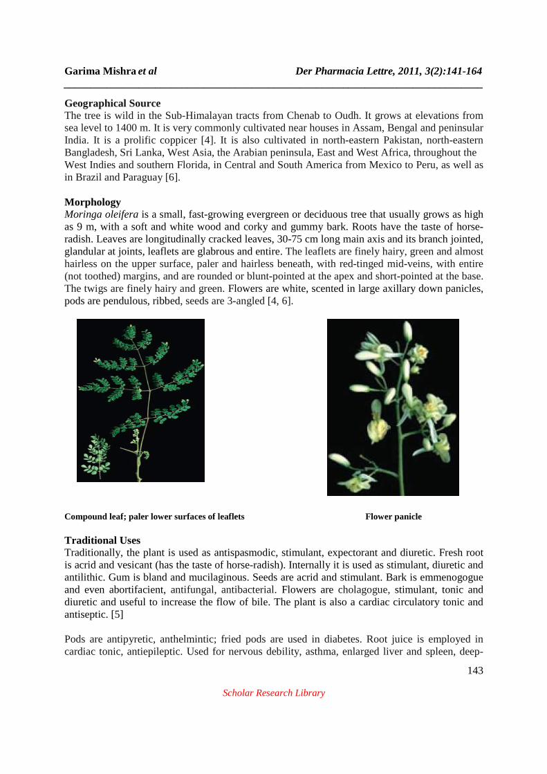
Geographical Source The tree is wild in the Sub-Himalayan tracts from Chenab to Oudh. It grows at elevations from sea level to 1400 m. It is very commonly cultivated near houses in Assam, Bengal and peninsular India. It is a prolific coppicer [4]. It is also cultivated in north-eastern Pakistan, north-eastern Bangladesh, Sri Lanka, West Asia, the Arabian peninsula, East and West Africa, throughout the West Indies and southern Florida, in Central and South America from Mexico to Peru, as well as in Brazil and Paraguay [6]. Morphology Moringa oleifera is a small, fast-growing evergreen or deciduous tree that usually grows as high as 9 m, with a soft and white wood and corky and gummy bark. Roots have the taste of horse-radish. Leaves are longitudinally cracked leaves, 30-75 cm long main axis and its branch jointed, glandular at joints, leaflets are glabrous and entire. The leaflets are finely hairy, green and almost hairless on the upper surface, paler and hairless beneath, with red-tinged mid-veins, with entire (not toothed) margins, and are rounded or blunt-pointed at the apex and short-pointed at the base. The twigs are finely hairy and green. Flowers are white, scented in large axillary down panicles, pods are pendulous, ribbed, seeds are 3-angled [4, 6].
Compound leaf; paler lower surfaces of leaflets Flower panicle Traditional Uses Traditionally, the plant is used as antispasmodic, stimulant, expectorant and diuretic. Fresh root is acrid and vesicant (has the taste of horse-radish). Internally it is used as stimulant, diuretic and antilithic. Gum is bland and mucilaginous. Seeds are acrid and stimulant. Bark is emmenogogue and even abortifacient, antifungal, antibacterial. Flowers are cholagogue, stimulant, tonic and diuretic and useful to increase the flow of bile. The plant is also a cardiac circulatory tonic and antiseptic. [5] Pods are antipyretic, anthelmintic; fried pods are used in diabetes. Root juice is employed in cardiac tonic, antiepileptic. Used for nervous debility, asthma, enlarged liver and spleen, deep-

Garima Mishra et al Der Pharmacia Lettre, 2011, 3(2):141-164 ______________________________________________________________________________
144
Scholar Research Library
seated inflammation and as diuretic in calculus affection. Decoction is used as a gargle in hoarseness and sore throat. Root and fruit are antiparalytic. Leaf juice is used in hiccough (emetic in high doses); cooked leaves are given in influenza and catarrhal affections. Root-bark is used as antiviral, anti-inflammatory, analgesic. Stem-bark and flowers are hypoglyceamic. Infusion of seed is anti-inflammatory, antispasmodic and diuretic, also given in venereal diseases. Along with other therapeutic applications, The Ayurvedic Pharmacopoeia of India indicated the use of the dried root bark in goitre, glycosuria and lipid disorders (also dried seeds), and leaf, seed, root bark and stem bark in internal abscess, piles. [7] Dosage : Leaf—10–20ml. juice; root bark—2–5 g powder; stem bark—2–5g powder; seed—5–10 g powder. Leaf, flower, fruit, seed, bark, root—1–3 g powder; 50–100 ml decoction. [8, 9]. Phytochemistry U. Eilert et al. (1981) reported the isolation of 4(alpha-L-Rhamnosyloxy) benzyl isothiocyanate form seeds of Moringa oleifera [10]. I.M. Villasenor et al. (1989) reported certain biosynthetically and chemically related compounds from roasted seeds of Moringa oleifera. Their structures have been elucidated by spectral analysis as 4(-L-rhamnosyloxy) phenylacetonitrile, 4-hydroxyphenylacetonitrile, and 4-hydroxyphenyl-acetamide [11, 12]. S. Faizi et al. (1994) reported the isolation of two nitrile glycosides from the ethanolic extracts of Moringa oleifera leaves, niazirin and niazirinin and three mustard oil glycosides, 4-[(4'-O-acetyl-alpha-L-rhamnosyloxy)benzyl]isothiocyanate, niaziminin A, and niaziminin B. Niazirinin is a new compound. Niaziminins A and B have previously been obtained from the left extract as a mixture, while 4-[(4'-O-acetyl-alpha-L-rhamnosyloxy)benzyl]isothiocyanate is new from this source. Structural determination was accomplished by means of spectroscopic methods including appropriate 2D nmr experiments and chemical reactions. This is the first report of the isolation of nitriles, an isothiocyanate, and thiocarbamates from the same plant species [13]. S. Faizi et al. (1995) isolated six new and three synthetically known glycosides from the leaves of Moringa oleifera, employing a bioassay-directed isolation method on the ethanolic extract. Most of these compounds, bearing thiocarbamate, carbamate or nitrile groups, are fully acetylated glycosides, which are very rare in nature. Elucidation of the structures was made using chemical and spectroscopic methods, including 2D NMR techniques [14]. S. Faizi et al. (1998) isolated two new compounds from the ethanolic extract of whole pods of Moringa oliefera, O-[2'-hydroxy-3'-(2"-heptenyloxy)]-propyl undecanoate and O-ethyl-4-[(alpha-L-rhamnosyloxy)-benzyl] carbamate along with the known substances like methyl p-hydroxybenzoate and beta-sitosterol [15]. A.Murakami et al. (1998) isolated niaziminin, a thiocarbamate from the leaves of Moringa oleifera [16]. A.P. Guevara et al. (1999) investigated the ethanolic extracts of the seeds of Moringa oleifera and isolated a new compound O-ethyl-4-(alpha-L-rhamnosyloxy)benzyl carbamate together with seven known compounds, 4(alpha-L-rhamnosyloxy)-benzyl isothiocyanate, niazimicin, niazirin, beta-sitosterol, glycerol-1-(9-octadecanoate), 3-O-(6'-O-
Garima Mishra et al Der Pharmacia Lettre, 2011, 3(2):141-164 ______________________________________________________________________________
145
Scholar Research Library
oleoyl-beta-D-glucopyranosyl)-beta-sitosterol and beta-sitosterol-3-O-beta-D-glucopyranoside [17]. R.N. Bennett et al. (2003) isolated various glucosinolates and phenolic compounds from various parts of Moringa oleifera. The seeds only contained 4-(alpha-l-rhamnopyranosyloxy)-benzylglucosinolate at high concentrations. Roots of Moringa oleifera contains high concentrations of both 4-(alpha-l-rhamnopyranosyloxy)-benzylglucosinolate and benzyl glucosinolate. Leaves of the plant contains 4-(alpha-l-rhamnopyranosyloxy)-benzylglucosinolate and three monoacetyl isomers of this glucosinolate. Only 4-(alpha-l-rhamnopyranosyloxy)-benzylglucosinolate was detected in Moringa oleifera bark tissue. The leaves also contains quercetin-3-O-glucoside and quercetin-3-O-(6''-malonyl-glucoside), and lower amounts of kaempferol-3-O-glucoside and kaempferol-3-O-(6''-malonyl-glucoside), 3-caffeoylquinic acid and 5-caffeoylquinic acid [18]. P. Siddhuraju & K. Becker (2003) reported quercetin and kaempferol from the ethanolic extracts of freeze-dried leaves of Moringa oliefera [19]. F. Nikkon et al. (2003) isolated aglycone of Deoxy-Niazimicine which is characterized as N-benzyl; S-ethyl thioformate from the chloroform extract of Moringa oleifera roots barks [20]. Phytochemical studies on Moringa oleifera by M. Ndong et al. (2007) revealed major polyphenols such as quercetin glucosides, rutin, kaempferol glycosides and chlorogenic acids in Moringa oleifera powder by HPLC analysis [21]. L.O. Manguro & P. Lemmen (2007) reported the isolation of five flavonol glycosides characterised as kaempferide 3-O-(2'',3''-diacetylglucoside), kaempferide 3-O-(2''-O-galloylrhamnoside), kaempferide 3-O-(2''-O-galloylrutinoside)-7-O-alpha-rhamnoside, kaempferol 3-O-[beta-glucosyl-(1 → 2)]-[alpha-rhamnosyl-(1 → 6)]-beta-glucoside-7-O-alpharhamnoside and kaempferol 3-O-[alpha-rhamnosyl-(1 → 2)]-[alpha-rhamnosyl-(1 →4)]-betaglucoside-7-O-alpha-rhamnoside together with benzoic acid 4-O-beta-glucoside, benzoic acid 4-O-alpha-rhamnosyl-(1 → 2)-beta-glucoside and benzaldehyde 4-O-beta-glucoside have been isolated from methanolic extract of Moringa oleifera leaves. Also obtained from the same extract were known compounds, kaempferol 3-O-alpha-rhamnoside, kaempferol, syringic acid, gallic acid, rutin and quercetin 3-O-beta-glucoside. Their structures were determined using spectroscopic methods as well as comparison with data from known compounds [22]. S.K. Roy et al. (2007) isolated a water-soluble polysaccharide from the aqueous extract of pods of Moringa oleifera. The polysaccharide contains d-galactose, 6-O-Me-D-galactose, D-galacturonic acid, l-arabinose, and l-rhamnose in a molar ratio of 1:1:1:1 [23]. F. Anwar & U. Rashid (2007) reported various sterols, tocopherols and fatty acids present in the seeds and seed oil from the n-hexane extract. Among the sterols, stigmasterol has the highest percentage (18.8%), whereas among the tocopherols, α – tocopherol was present in high amount (140.5 mg/kg) [24].

Garima Mishra et al Der Pharmacia Lettre, 2011, 3(2):141-164 ______________________________________________________________________________
146
Scholar Research Library
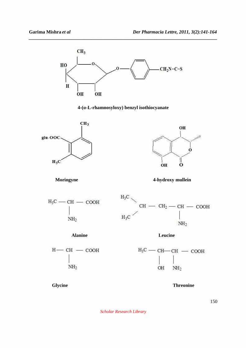
K. Shanker et al. (2007) isolated nitrile glycosides (niaziridin & niazirin) from the leaves, pods and bark of Moringa oleifera by reverse phase HPLC [25]. Hueih-Min Chen et al. (2007) reported fourty four compounds from the essential oil isolated from the leaves of Moringa oleifera by GC-MS analysis [26]. D. Yammuenart et al. (2008) isolated seven compounds, β-sitosterol-3-O-β-D-glucopyranoside, β-sitosterol, linoleic sitosteroate, linoleic acid, 1,2,3-triolein, a mixture of 1,3-dilinoleoyl-2-olein, 1,3-dioleoyl-2-linolein and 1,2,3-trilinolein and isothiocyanatomethylbenzene from the ethylene chloride extract of Moringa oleifera [27]. K.V. Sashidhara et al. (2009) from the roots of Moringa oleifera isolated and characterized aurantiamide acetate 4 and 1, 3-dibenzyl urea 5. Both these compounds were isolated for the first time from this genus [28]. A.O. Ogunbinu et al. (2009) isolated monoterpenoid compounds (81.8%) from the essential oil of Moringa oleifera extracted by hydrodistillation and analysed by GC and GC-MS. The oil consists of alpha-phellandrene with highest percentage (25.2%) along with p-cymene (24.9%) [29]. B.N. Singh et al. (2009) reported presence of gallic acid, chlorogenic acid, ellagic acid, ferulic acid, kaempferol, quercetin and vanillin from the aqueous extracts of leaves, fruits and seeds of Moringa oliefera. All compounds were analyzed by HPLC and MS/MS techniques [30]. A.R. Verma et al. (2009) reported presence of phenolic acids like gallic acid, chlorogenic acid, ellagic acid, ferulic acid and flavonoids like kaempferol, quercetin and rutin from the leaves of Moringa oleifera by HPLC techniques [31]. R.E. Renitta et al. (2009) investigated various phytochemicals present in the leaves, seeds and flowers of ethanolic extract of Moringa oleifera by GC-MS. The leaves contain fifteen components. The major compounds were hexadecanoic acid, Ethyl palmitate, Palmitic acid ethyl ester, 2, 6-Dimethyl-1, 7-octadiene-3-ol, 4-Hexadecen-6-yne, 2-hexanone, 3-cyclohexyliden-4-ethyl - E2- Dodecenylacetate, Hi-oleic safflower oil, Safflower oil . From the seeds, the major compounds were Roridin E, Veridiflorol, 9-Octadecenoic acid. From the flowers, 9- Octadecen – 1- ol, cis - 9 – Octadecen – 1 –ol, Oleol, Satol, Ocenol, Sipo, Decanoic acid, Dodecanal were identified as major compounds [32]. The seeds also contain Moringyne, 4-(α-L-rhamnosyloxy) benzyl isothiocyanate & several amino acids. [33, 34, 35, 36]. The roots also contain benzyl isothiocyanate. The plant also contains antibacterial principles such as spirochin and pterygospermin, which are effective against both gram negative and gram positive bacteria [37]. The gum contains aldotriouronic acid which is obtained from the acid hydrolysis of gum and is characterized as O-(β-D-glucopyranosyluronic acid) (1→6)-β-D-galactopyranosyl (1→6)-D-galactose. The leaves contain aspartic acid, glutamic acid, glycine, threonine, alanine, valine, leucine, isoleucine, histidine, lysine, phenylalanine, tryptophan, cysteine and methionine [38]. The stem contains 4-hydroxy mellein, vanillin, octacosonoic acid, β-sitosterol and β-sitosterone [38] & Kaempferol-3-rutinoside was identified in flowers [34].

Garima Mishra et al Der Pharmacia Lettre, 2011, 3(2):141-164 ______________________________________________________________________________
147
Scholar Research Library
1, 2, 3 - triolein 1,3 – dilinoleoyl-2-olein 1, 3-dioleoyl-2-linolein Linoleic acid Isothiocyanatomethylbenzene 4-(4'-O-acetyl-α-L-rhamnopyranosyloxy) benzyl isothiocyanate Benzyl isothiocyanate R=H → β-sitosterol R= β-D-glucopyranosyl → β-sitosterol-3-O-β-D-glucopyranoside

Garima Mishra et al Der Pharmacia Lettre, 2011, 3(2):141-164 ______________________________________________________________________________
148
Scholar Research Library
Linoleic sitosteroate 1, 2, 3-trilinolein 4-(α-L-rhamnopyranosyloxy) benzyl isothiocyanate Niazimicin
4-(α-L-rhamnopyranosyloxy ) benzyl glucosinolate

Garima Mishra et al Der Pharmacia Lettre, 2011, 3(2):141-164 ______________________________________________________________________________
149
Scholar Research Library
Aglycone of Deoxy-Niazimicine (N-benzyl, S-ethyl thioformate)
Niaziridin
Niazirin
Pterygospermin
Garima Mishra et al Der Pharmacia Lettre, 2011, 3(2):141-164 ______________________________________________________________________________
150
Scholar Research Library
4-(α-L-rhamnosyloxy) benzyl isothiocyanate
Moringyne 4-hydroxy mullein
Alanine Leucine
Glycine Threonine

Garima Mishra et al Der Pharmacia Lettre, 2011, 3(2):141-164 ______________________________________________________________________________
151
Scholar Research Library
Aspartic Acid Valine
Tryptophan Glutamic Acid Structures of some phytoconstituents isolated from Moringa oleifera Pharmacological Actions Antimicrobial Activity A. Cáceres et al. (1991) reported antimicrobial activities of Moringa oleifera leaves, roots, bark and seeds in vitro against bacteria, yeast, dermatophytes and helminths by a disk-diffusion method. The fresh leaf juice and aqueous extracts from the seeds inhibit the growth of Pseudomonas aeruginosa and Staphylococcus aureus. No activity was demonstrated against other pathogenic Gram-positive and Gram-negative bacteria and Candida albicans [39]. M.O. Nwosu and J.I. Okafor (1995) reported antifungal activity of Moringa oleifera against seven pathogenic fungi using the broth dilution and agar plate methods. Extracts from the plant were effective against Trichophyton rubrum and T. mentagraphytes [40]. V. Spiliotis et al. (1997) reported antimicrobial activity from various varieties of Moringa oleifera seeds. All the varieties were investigated against Bacillus cereus, Candida albicans, Streptococcus faecalis, Staphylococcus aureus, Staphylococcus epidermidis, Bacillus subtilis, Pseudomonas aeruginosa, E.coli and Aspergillus niger [41]. M. Umar Dahot (1998) investigated antimicrobial activity from three fractions of Moringa oleifera leaves which were obtained by Sephadex G-25 column chromatography. Single bands of these fractions were detected on Polyacrylamide SDS gel electrophoresis. An antibacterial action of small protein/peptide was tested against E. coli Kl. aerogenes, Kl. pneumoniae, S.aureus, and B. subtilis. Fraction 1, 2 and 3 showed strong inhibitory activity against E. coli, S. aureus and B

Garima Mishra et al Der Pharmacia Lettre, 2011, 3(2):141-164 ______________________________________________________________________________
152
Scholar Research Library
subtilis but clear zone of inhibition was also noted against K1. aerogenes with peptide 1. Fraction 2 showed significant zone of inhibition against Aspergillus niger [42]. F. Nikkon et al. (2003) reported antimicrobial activity of aglycone of Deoxy-Niazimicine which is characterized as N-benzyl, S-ethyl thioformate from the chloroform extract of Moringa oleifera roots barks. The compound shows antibacterial and antifungal activities against Shigella boydii, Shigella dysenteriae and Staphylococcus aureus [20]. Khesorn Nantachit (2006) reported antibacterial activity from the methanolic crude extract and purified dichloromethane extract of Moringa oleifera capsules. The activity was also determined from isolated parts from column chromatography of Moringa oleifera capsules by agar-well diffusion. The methanolic crude extract showed no activity against Staphylococcus aureus, Escherichia coli, Pseudomonas aeruginosa and Klebsiella pneumoniae. The purified dichloromethane extract and isolated parts from column chromatography showed antibacterial activity against these bacteria [43]. J. H. Doughari et al. (2007) reported antibacterial activity from the aqueous, acetone and ethanolic extracts of the leaves of Moringa oleifera. Of the three solvents used, ethanolic extract of the plant demonstrated the highest activity, while the aqueous extract showed the least activity at 100 mg/ml. The activities of the plant extracts were comparable to those of antibiotics, ciprofloxacin, cotrimoxazole and chloramphenicol [44]. Hueih-Min Chen et al. (2007) reported in vitro antifungal activity from the ethanolic extracts of the leaves of Moringa oleifera against against dermatophytes such as Trichophyton rubrum, Trichophyton mentagrophytes, Epidermophyton Xoccosum, and Microsporum canis.[26] Amer Jamil et al. (2008) evaluated antimicrobial activity from the seeds of Moringa oleifera. The seed extracts of Moringa oleifera were assayed for the evaluation of antimicrobial activity against bacterial (Pasturella multocida, Escherichia coli, Bacillus subtilis and Staphlocuccus aureus) and fungal (Fusarium solani and Rhizopus solani) strains. The zones of growth inhibition showed greater sensitivity against the bacterial strains as compared to the fungal strains. Minimum inhibitory concentrations (MIC) extracts revealed that Pasturella multocida and Bacillus subtilis were most sensitive strains [45]. M. F. Alam et al. (2009) investigated antibacterial activity of leaf juice and extracts of Moringa oleifera using disc diffusion and minimum inhibitory concentration (MIC) determination method against human pathogenic bacteria. The fresh leaf juice (10 µl), powder from fresh leaf juice, cold water extract of fresh leaf, 1175 µg disc-1, displayed a potential antibacterial activity against all the tested four Gramnegative bacteria (Shigella shinga, Pseudomonas aeruginosa, Shigella sonnei and Pseudomonas spp.) and six Gram-positive bacteria (Staphylococcus aureus, Bacillus cereus, Streptococcus-B- haemolytica, Bacillus subtilis, Sarcina lutea and Bacillus megaterium). However, ethanol extract (1175 µg) of fresh leaves exhibited inhibitory effect against all the tested Gram-negative bacteria and Gram-positive bacteria except in S. aureus and Streptococcus-B- haemolytica [46].
Garima Mishra et al Der Pharmacia Lettre, 2011, 3(2):141-164 ______________________________________________________________________________
153
Scholar Research Library
R.E. Renitta et al. (2009) reported antimicrobial activity from the ethanolic extract of leaves, seeds and flowers of Moringa oleifera against microorganisms like Escherichia coli, Klebsiella pneumoniae, Enterobacter spp, Proteus mirabilis, Pseudomonas aeroginosa, Salmonella typhi A, Staphylococcus aureus, Streptococcus and Candida albicans [32]. T.R. Prashith Kekuda et al. (2010) investigated antibacterial and antifungal efficacy of steam distillate of Moringa oleifera. Among bacteria tested, more inhibition was observed in case of E. coli followed by S. aureus, K. pneumoniae, P. aeruginosa and B. subtilis. Inhibition of fungi was observed as reduced colony diameter in plates poisoned with distillate as compared to control plates. More inhibition of A. niger was observed followed by A. oryzae, A. terreus and A. nidulans [47]. Anti-inflammatory Activity A Cáceres et al. (1992) reported anti-inflammatory activity from the hot water infusions of flowers, leaves, roots, seeds and stalks or bark of Moringa oleifera using carrageenan-induced hind paw edema in rats [48]. B. Medhi et al. (1996) evaluated anti-inflammatory activity of methanol extract of Moringa oleifera root bark using carrageenin induced paw edema in mice as animal model [49]. M. Ndiaye et al. (2002) tested anti-inflammatory action of an aqueous extract of root of Moringa oleifera in rats using indomethacin (10 mg/kg) as standard drug and oedema was induced in the rat-paw by subcutaneous injection of carrageenin. At a dose of 750 mg/kg the Moringa oleifera treatment significantly inhibited the development of oedema at 1, 3 and 5 hours (reduction by 53.5, 44.6 and 51.1% respectively) [50]. S.G. Mahajan et al. (2007) investigated anti – inflammatory activity from the ethanolic extract of seeds of Moringa oleifera. The extract was pharmacologically evaluated against immune-mediated inflammatory responses in toluene diisocyanate (TDI as antigen)-induced asthma in Wistar rats [51]. S.G. Mahajan et al. (2007) reported anti-arthritic activity of ethanolic extract of seeds of Moringa oleifera Lam. in adjuvant-induced arthritis in adult female Wistar rats [52]. K.V. Sashidhara et al. (2009) from the roots of Moringa oleifera isolated and characterized aurantiamide acetate 4 and 1,3-dibenzyl urea 5. Isolated compounds inhibited the production of TNF-alpha and IL-2. [28] S.G. Mahajan et al. (2009) evaluated anti-inflammatory activity from the n-butanol extract of the seeds of Moringa oleifera against ovalbumin-induced airway inflammation in guinea pigs [53]. The crude methanol extract of root by oral administration inhibits carrageenan-induced rat paw oedema in a dose dependent manner [54].

Garima Mishra et al Der Pharmacia Lettre, 2011, 3(2):141-164 ______________________________________________________________________________
154
Scholar Research Library
Antioxidant Activity R. Bharali et al. (2003) investigated the antioxidants effects of hydro-alcoholic extract of Moringa oleifera at doses of 125 mg/kg bodyweight and 250 mg/ kg body weight for 7 and 14 days, respectively, were investigated with reference to drug metabolising Phase I (Cytochrome b(5) and Cytochrome p(450) ) and Phase II (Glutathione-S- transferase) enzymes, anti-oxidant enzymes, glutathione content and lipid peroxidation in the liver of 6-8 week old female Swiss albino mice [55]. N. Ashok Kumar & L. Pari (2003) investigated antioxidant potential of Moringa oleifera on hepatic marker enzymes, lipid peroxidation, and antioxidants. The antioxidant property was investigated during antitubercular drug (isoniazid, rifampicin, and pyrazinamide)-induced toxicity in rats. Enhanced hepatic marker enzymes and lipid peroxidation of antitubercular drug treatment was accompanied by a significant decrease in the levels of vitamin C, reduced glutathione, superoxide dismutase, catalase, glutathione peroxidase, and glutathione S-transferase. Administration of Moringa oleifera extract and silymarin significantly decreased hepatic marker enzymes and lipid peroxidation with a simultaneous increase in the level of antioxidants [56]. P. Siddhuraju & K. Becker (2003) reported antioxidant and radical scavenging property from water, aqueous methanol, and aqueous ethanol extracts of freeze-dried leaves of Moringa oleifera. All leaf extracts were capable of scavenging peroxyl and superoxyl radicals. Overall, both methanol (80%) and ethanol (70%) were found to be the best solvents for the extraction of antioxidant compounds from Moringa leaves [19]. M. Bajpai et al. (2005) reported antioxidant activity from the leaves of Moringa oleifera. The antioxidant property was found due to presence of kaempferol [57]. P. Chumark et al. (2008) reported free radical scavenging activity of Moringa oleifera using 1,1-diphenyl-2-picrylhydrazyl radicals (DPPH).The investigation reported that in scavenging DPPH radicals the extract and Trolox had IC(50) of 78.15+/-0.92 and 2.14+/-0.12microg/ml respectively [58]. S .Sreelatha & P.R. Padma (2009) evaluated antioxidant effects of Moringa oleifera leaf extracts, which were tested in two stages of maturity using standard in vitro models. The successive aqueous extract of Moringa oleifera exhibited strong scavenging effect on 2, 2-diphenyl-2-picryl hydrazyl (DPPH) free radical, superoxide, nitric oxide radical and inhibition of lipid per oxidation. The free radical scavenging effect of Moringa oleifera leaf extract was comparable with that of the reference antioxidants [59]. B.Sultana et al. (2009) investigated antioxidant activity from the hydro alcoholic and aqueous extracts of leaves and roots of Moringa oleifera using two extraction techniques (shaking and reflux) [60]. B.N.Singh et al. (2009) reported antioxidant potential of aqueous extracts of leaf, fruit and seed of Moringa oleifera [30]. A.R. Verma et al. (2009) tested in vitro and in vivo antioxidant properties of different fractions of leaves of Moringa oleifera using different in vitro systems, such as beta-Carotene bleaching, reducing power, DPPH/superoxide/hydroxyl radical
Garima Mishra et al Der Pharmacia Lettre, 2011, 3(2):141-164 ______________________________________________________________________________
155
Scholar Research Library
scavenging, ferrous ion chelation and lipid peroxidation. On the basis of in vitro antioxidant properties polyphenolic fraction of M. oleifera leaves (MOEF) was chosen as the potent fraction and used for the DNA nicking and in vivo antioxidant properties [31]. Anticancer Activity A.Murakami et al. (1998) reported antitumor activity from the leaves of Moringa oleifera. The antitumor potential was due the presence of three known thiocarbamate (TC) and isothiocyanate (ITC)-related compounds which are acting as inhibitors of tumor promoter teleocidin B-4-induced Epstein-Barr virus (EBV) activation in Raji cells. Interestingly, only niaziminin among 10 TCs including 8 synthetic ones showed considerable inhibition against EBV activation. On the other hand, among the ITC-related compounds, naturally occurring 4-[(4'-O-acetyl-alpha-L-rhamnosyloxy)benzyl]ITC and commercially available allyl- and benzyl-ITC significantly inhibited activation, suggesting that the isothiocyano group is a critical structural factor for antitumor activity [16]. A.P. Guevara et al. (1999) investigated anticancer activity from the ethanolic extract of seeds of Moringa oleifera. The said extract was found to contain various antitumor compounds like O-ethyl-4-(alpha-L-rhamnosyloxy)benzyl carbamate which was a new compound, together with seven known compounds, 4(alpha-L-rhamnosyloxy)-benzyl isothiocyanate, niazimicin, niazirin, beta-sitosterol, glycerol-1-(9-octadecanoate), 3-O-(6'-O-oleoyl-beta-D-glucopyranosyl)-beta-sitosterol and beta-sitosterol-3-O-beta-D-glucopyranoside. Their potential antitumor promoting activity was performed using an in vitro assay which tested their inhibitory effects on Epstein-Barr virus-early antigen (EBV-EA) activation in Raji cells induced by the tumor promoter, 12-O-tetradecanoyl-phorbol-13-acetate (TPA) [17]. R. Bharali et al. (2003) investigated chemomodulatory effect of hydro-alcoholic extract of Moringa oleifera. The chemopreventive efficacy of the extract was evaluated in a two stage model of 7, 12 - dimethylbenzanthracene induced skin papillomagenesis in mice [55]. L.V. Costa-Lotufo et al. (2005) reported anticancer activity of Moringa oleifera. The extracts were tested for cytotoxicity by using the brine shrimp lethality assay, sea urchin eggs assay, hemolysis assay and MTT assay using tumor cell lines [61]. M.V.S Parvathy and A. Umamaheshwari (2007) investigated cytotoxic effect of various extracts of leaves of Moringa oleifera. The cytotoxic efficacy was evaluated on human multiple myeloma cell lines. Of the organic extracts, methanolic extracts of Moringa leaves showed least viability at highest dose [62]. Antifertility Activity A.O. Prakash et al. (1987) investigated antifertility activity from the aqueous extract of Moringa oleifera roots. The effect of aqueous extract has been studied on histoarchitecture of the uterus during pre and post-implantation stages in rats [63]. S. Shukla et al. (1988) reported anti-implantation activity from the aqueous extract of Moringa oleifera in female reproductive organs of cyclic rats and also antifertility activity from the

Garima Mishra et al Der Pharmacia Lettre, 2011, 3(2):141-164 ______________________________________________________________________________
156
Scholar Research Library
aqueous extract of the roots of the plant. Oral administration of extract progressively increased the uterine wet weight of bilaterally ovariectomized rats. This estrogenic activity was supported by stimulation of uterine histo-architecture. When the extract was given conjointly with estradiol dipropionate (EDP), there was a successive reduction in the uterine wet weight when compared to the gain with EDP alone and uterine histological structures were also inhibited [64, 65]. S. Shukla et al. (1989) investigated antifertility effect of aqueous extract of Moringa oleifera roots was studied histologically on the genital tract of ovariectomized rats in the presence and absence of estradiol dipropionate and progesterone. Administration of the extract itself stimulated the uterine histoarchitecture as revealed by increases in the height of luminal epithelium, well developed glands, loose stroma and rich vascularity. The cervix showed metaplastic changes in the epithelium with marked keratinization. In the vagina, cornification was very prominent, rugae increased and stroma was loose. Conjoint administration of the extract with estradiol showed a synergistic action, and an inhibition was observed when administered conjointly with progesterone [66]. D. Nath et al. (1992) investigated antifertility property from the aqueous and 90% ethanol extracts of the plant in rats orally dosed for 10 days after insemination with special reference to effects on foetal development. Leaf extracts of Moringa oleifera were 100% abortive at doses equivalent to 175 mg/kg of starting dry material [67]. Hepatoprotective Activity U.K. Mazumder et al. (1999) investigated hematological along with hepatorenal functions of methanolic extract of Moringa oleifera roots. Doses of the crude extract (CE) on liver and kidney functions and hematological parameters in mice were studied. No alteration in hematological and biochemical parameters at low and moderate dose level of daily and low dose level of weekly treatment of the extract was observed. However, the extract at moderate dose level in weekly treatment changed serum aminotransferase and plasma cholesterol levels significantly. High dose in addition to the above parameters changed total bilirubin, non protein nitrogen, blood urea and plasma protein. High dose of daily treatment and moderate and high dose of weekly treatment of CE increased WBC count and decreased clotting time significantly [68]. L. Pari & N.A. Kumar (2002) evaluated hepatoprotective effect of ethanolic extract leaves of Moringa oleifera on liver damage induced by antitubercular drugs such as isoniazid (INH), rifampicin (RMP), and pyrazinamide (PZA) in rats. Oral administration of the extract showed a significant protective action made evident by its effect on the levels of glutamic oxaloacetic transaminase (aspartate aminotransferase), glutamic pyruvic transaminase (alanine aminotransferase), alkaline phosphatase, and bilirubin in the serum; lipids, and lipid peroxidation levels in liver. This observation was supplemented by histopathological examination of liver sections [69]. A.A. Hamza (2007) reported hepatoprotective action of Moringa oleifera seeds against Diclofenac (DIC)-induced hepatic toxicity in male albino rats. Administration of DIC at 150 mg/kg developed acute hepatic damage, as demonstrated by increased serum alanine aminotransferase (ALT) activity and histopathological changes. In addition, DIC treatment
Garima Mishra et al Der Pharmacia Lettre, 2011, 3(2):141-164 ______________________________________________________________________________
157
Scholar Research Library
resulted in an increase in the hepatic malonialdehyde level and depletion in total antioxidant capacity, reduced glutathione content, catalase, and superoxide dismutase activities. Treatment with herbal extracts for 30 days before DIC treatment significantly ameliorated the indices of hepatotoxicity induced by DIC [70]. S. Fakurazi et al. (2008) reported hepatoprotective action of Moringa oleifera against acetaminophen induced liver injury in Sprague-Dawley rats using silymarin as standard drug. The hepatoprotective activity of Moringa extract was observed following significant histopathological analysis and reduction of the level of alanine aminotransferase (ALT), aspartate aminotransferase (AST) and alkaline phosphatase (ALP) in groups pretreated with Moringa compared to those treated with Acetaminophen alone. Meanwhile, the level of glutathione (GSH) was found to be restored in Moringa treated animals compared to the groups treated with Acetaminophen alone [71]. A.A. Hamza (2010) evaluated the effect of Moringa oleifera seed extract on liver fibrosis. Liver fibrosis was induced by the oral administration of 20% carbon tetrachloride (CCl4). Simultaneously, M.oleifera seed extract (1g/kg) was orally administered daily. The administration of Moringa seed extract decreased the CCl4-induced elevation of serum aminotransferase activities and globulin level. The elevations of hepatic hydroxyproline content and myeloperoxidase activity were also reduced by Moringa treatment. Furthermore, the immunohistochemical study showed that Moringa markedly reduced the numbers of smooth muscle alpha-actin-positive cells and the accumulation of collagens I and III in liver [72]. Cardiovascular Activity S. Faizi et al. (1994) reported the isolation of two nitrile glycosides from the ethanolic extracts of Moringa oleifera leaves, niazirin and niazirinin and three mustard oil glycosides. Compounds such as 4-[(4'-O-acetyl-alpha-L-rhamnosyloxy) benzyl]isothiocyanate, niaziminin A and B showed hypotensive activity [13]. S. Faizi et al. (1995) reported six new and three synthetically known glycosides from the ethanolic extract of the leaves of Moringa oleifera. Most of these compounds, bearing thiocarbamate, carbamate or nitrile groups, are fully acetylated glycosides, Thiocarbamates showed hypotensive activity [14]. S. Faizi et al. (1998) isolated thiocarbamate and isothiocyanate glycosides. These compounds showed promising hypotensive activity [15]. S. Ghasi et al. (2000) investigated hypocholesterolemic effect of crude leaf extract of Moringa oleifera. The effect on the serum cholesterol was statistically significant. No significant effect on serum total protein was observed. However, the crude extract increased serum albumin by 15.22% [73]. M. Ndong et al. (2007) tested preventive effects of Moringa oleifera on hyperlipidemia caused by iron deficiency in male Wistar rats [74].

Garima Mishra et al Der Pharmacia Lettre, 2011, 3(2):141-164 ______________________________________________________________________________
158
Scholar Research Library
N. Ara et al. (2008) reported comparative effects of ethanolic extracts of leaves of Moringa oleifera with atenolol on serum cholesterol level, serum triglyceride level, blood glucose level, heart weight and body weight of adrenaline induced rats. Above mentioned biochemical parameters were established and the relationship between them was measured. The Moringa oleifera leaves extract made significant changes in each cardiovascular parameter after proper investigation [75]. P. Chumark et al. (2008) reported antiatherosclerotic and hypolipidaemic activity of Moringa oleifera leaves using simvastatin as standard drug. The extract significantly prolonged the lag-time of conjugated diene (CD) formation and inhibited thiobarbituric acid reactive substances (TBARS) formation in both in vitro and ex vivo experiments in a dose-dependent manner. In hypercholesterol-fed rabbits, it significantly lowered the cholesterol levels and reduced the atherosclerotic plaque formation to about 50 and 86%, respectively [58]. M. Nandave et al. (2009) investigated cardioprotective effect of lyophilized hydroalcoholic extract of Moringa oleifera in the isoproterenol (ISP)-induced model of myocardial infarction in male wistar albino rats. Chronic treatment with Moringa resulted in significant favorable modulation of the biochemical enzymes (superoxide dismutase, catalase, glutathione peroxidase, lactate dehydrogenase, and creatine kinase-MB) but failed to demonstrate any significant effect on reduced glutathione compared to the ISP control group. Moringa treatment significantly prevented the rise in lipid peroxidation in myocardial tissue [76]. Anti-ulcer Activity S. Debnath & D. Guha (2007) reported anti-ulcer effect of aqueous extract of Moringa oleifera leaves on adult holtzman albino rats of either sex using ondansetron as standard drug [77]. Analgesic, Antipyretic & Wound Healing Activity B. Medhi et al. (1996) investigated methanolic extract of Moringa oleifera root bark in mice using acetic acid-induced writhing method for analgesic activity [49]. B.S. Rathi et al. (2006) evaluated wound healing property from the aqueous extract of leaves of Moringa oleifera in male Swiss albino mice. Significant increase in wound closure rate, skin-breaking strength, granuloma breaking strength, hydroxyproline content, granuloma dry weight and decrease in scar area was observed [78]. V.I. Hukkeri et al. (2006) reported antipyretic and wound healing activity from the ethanolic and ethyl acetate extracts of Moringa oleifera leaves. The ethanolic and ethyl acetate extracts of seeds showed significant antipyretic activity in rats, whereas ethyl acetate extract of dried leaves showed significant wound healing activity (10% extracts in the form of ointment) on excision, incision and dead space (granuloma) wound models in rats [79]. Anti-diabetic Activity K. Suzuki et al. (2007) reported anti-diabetic effect of leaves of Moringa oleifera on glucose tolerance in Goto-Kakizaki and Wistar rats. Moringa significantly decreased the blood glucose in Wistar rats. The area under the curve of changes in the blood glucose was significantly higher in

Garima Mishra et al Der Pharmacia Lettre, 2011, 3(2):141-164 ______________________________________________________________________________
159
Scholar Research Library
the Goto-Kakizaki rats. The action of MO was greater in Goto-Kakizaki rats than in Wistar rats [80]. D. Jaiswal et al. (2009) reported anti-diabetic activity of aqueous extract of Moringa oleifera leaves on glycemic control, haemoglobin, total protein, urine sugar, urine protein and body weight [81]. Diuretic & Antiurolithiatic Activity A Cáceres et al. (1992) reported diuretic activity from hot water infusions of flowers, leaves, roots, seeds and stalks or bark of Moringa oleifera. The extracts of Moringa were administered orally in rats and diuretic activity is determined by urine output in metabolic cages [48]. R.V. Karadi et al. (2006) investigated antiurolithiatic activity from the aqueous and alcoholic extract of Moringa oleifera root-wood on calcium oxalate urolithiasis in male Wistar albino rats. Oral administration of aqueous and alcoholic extract of Moringa oleifera significantly reduced the elevated urinary oxalate, showing a regulatory action on endogenous oxalate synthesis. The increased deposition of stone forming constituents in the kidneys of calculogenic rats was also significantly lowered by curative and preventive treatment using aqueous and alcoholic extracts [82]. R.V. Karadi et al. (2008) reported antiurolithiatic property from the aqueous and alcoholic extract of the root bark of Moringa oleifera. Both the extracts significantly lowered the urinary excretion and kidney retention levels of oxalate, calcium and phosphate. Moreover, elevated serum levels of urea nitrogen, creatinine and uric acid were significantly reduced by the extracts [83]. CNS Activity K. Ray et al. (2003) investigated anticonvulsant activity from the aqueous extract of Moringa oleifera roots. The effect was studied on penicillin (PCN) induced convulsion, locomotor behaviour, brain serotonin (5-HTT), dopamine (DA) and norepinephrine (NE) level in Holtzman strain adult albino rats [84]. R. Ganguly & D. Guha (2008) evaluated ethanolic extract of Moringa oleifera leaves in alteration of brain monoamines (norepinephrine, dopamine and serotonin) & EEG wave pattern in Alzheimer's disease in rats. Treatment with Moringa oleifera leaf extract restores the monoamine levels of brain regions to near control levels [85]. .Local Anaesthetic Activity B. Medhi et al. (1996) investigated methanolic extract of Moringa oleifera root bark in frog and guinea pig models for local anaesthetic activity. The local anaesthetic activity was seen in both animal models [49]. Anti-allergic Activity S.G. Mahajan & A.A. Mehta (2007) reported inhibitory action of ethanolic extract Moringa oleifera seeds on systemic and local anaphylaxis. The potential anti-anaphylactic effect of extract

Garima Mishra et al Der Pharmacia Lettre, 2011, 3(2):141-164 ______________________________________________________________________________
160
Scholar Research Library
was studied in a mouse model of Compound 48/80-induced systemic anaphylactic shock. Passive cutaneous anaphylaxis activated by anti IgE-antibody was also used to assess the effect of extract [86]. Anthelmintic Activity T. Rastogi et al. (2009) reported anthelmintic activity of Moringa oleifera. Ethanolic extracts of Moringa oleifera were taken for anthelmintic activity against Indian earthworm Pheritima posthuma. Various concentrations of extract were tested and results were expressed in terms of time for paralysis and time for death of worms. Piperazine citrate (10 mg/ml) was used as a reference standard and distilled water as a control group [87].
CONCLUSION
For decades natural products have been a source of drugs and drug leads. Natural products provide the inspiration for a variety of strategies used in the diversity-oriented synthesis of novel small molecule libraries. An increasing body of evidence supports the effectiveness of these strategies for identifying new biologically active molecules. Moringa oleifera, an important medicinal plant is one of the most widely cultivated species of the family Moringaceae. Various parts of the plant have been used for human medication. Insight of literature review on this plant clearly explained its various traditional uses as antispasmodic, stimulant, expectorant etc. Bark is emmenogogue and even abortifacient, antifungal, antibacterial. Flowers are cholagogue, stimulant, tonic and diuretic. Root-bark is used as antiviral, anti-inflammatory, analgesic. Pharmacologically reported activities includes antimicrobial, anti-inflammatory, antioxidant, anticancer, antifertility, hepatoprotective, cardiovascular, antiulcer, analgesic, wound healing, anticonvulsant, antiallergic and anthelmintic activities. Phytochemically, it is known as rich source of glycosides, phenols, sterols, flavanol glycosides present in Moringa oleifera might be medicinally important and/or nutritionally valuable. the ethanolic extracts of Moringa oleifera leaves, niazirin and niazirinin and three mustard oil glycosides, 4-[(4'-O-acetyl-alpha-L-rhamnosyloxy)benzyl]isothiocyanate, niaziminin A, and niaziminin B. The seeds only contained 4-(alpha-l-rhamnopyranosyloxy)-benzylglucosinolate at high concentrations. Roots of Moringa oleifera contains high concentrations of both 4-(alpha-l-rhamnopyranosyloxy)-benzylglucosinolate and benzyl glucosinolate. Leaves of the plant contains 4-(alpha-l-rhamnopyranosyloxy)-benzylglucosinolate and three monoacetyl isomers of this glucosinolate and various amino acids like aspartic acid, glutamic acid, glycine, threonine, alanine, valine, leucine, isoleucine, histidine, lysine, phenylalanine, tryptophan, cysteine, methionine. The present review summarizes some important pharmacological studies on Moringa oleifera and phytochemical investigations and isolated principles from them, which can be investigated further to achieve lead molecules in the search of novel herbal drugs. Acknowledgement The authors are thankful to Hon’ble Chancellor, Shri Suresh Jain, Teerthanker Mahaveer University, Moradabad, India, for providing literature survey facility to carry out the work.
Garima Mishra et al Der Pharmacia Lettre, 2011, 3(2):141-164 ______________________________________________________________________________
161
Scholar Research Library
REFERENCES
[1] U. N. Brahmachari. Current Science, 2001, 81(1), 15-16. [2] A.D.B. Vaidya, T.P.A. Devasagayam. Journal of Clinical Biochemistry and Nutrition, (2007), 41, 1-11. [3] J. Rai. JK Science, 2005, 7(3), 2005, 180. [4] R.K. Gupta. Medicinal & Aromatic Plants. CBS publishers & distributors, 2010, 151-152. [5] K.M. Nadkarni. Indian Materia Medica. Bombay Popular Prakashan, 2009, Vol.I, 811-816. [6] A. Roloff, H. Weisgerber, U. Lang, B. Stimm. Enzyklopädie der Holzgewächse, Handbuch und Atlas der Dendrologie. 2009, 1-8. [7] C.P. Khare. Indian medicinal plants. Springer, 2007, 422-423. [8] The Ayurvedic pharmacopoeia of India, Departmaent of Indian Sysytemsof Medicine & Homeopathy New Delhi, Part –I, Vol.II, 155-157. [9] The Ayurvedic pharmacopoeia of India, Departmaent of Indian Sysytemsof Medicine & Homeopathy New Delhi, Part –I, Vol.IV, 124-131. [10] U. Eilert, B. Wolters, A. Nahrstedt. Planta Medica, 1981, 42(5), 55-61. [11] I.M. Villasenor, C.Y. Lim-Sylianco, F. Dayrit. Mutat Res. 1989, 224(2), 209-212. [12] Ram P. Rastogi, B.N. Mehrotra. Compendium of Indian Medicinal plants. Central Drug Research Institute, Lucknow and National institute of science communication and information resources, New Delhi, Vol.V, 2004, 551-552. [13] S. Faizi, B.S. Siddiqui, R. Saleem, S. Siddiqui, K. Aftab, A.H. Gilani. J Nat Prod., 1994, 57(9):1256-1261. [14] S. Faizi, B.S. Siddiqui, R. Saleem, S. Siddiqui, K. Aftab, A.H. Gilani. Phytochemistry. 1995, 38(4), 957-963. [15] S. Faizi, B.S. Siddiqui, R. Saleem, S. Siddiqui, K. Aftab, A.H. Gilani. Planta Medica, 1998, 64(3), 225-228. [16] A. Murakami, Y. Kitazono, S. Jiwajinda, K. Koshimizu, H. Ohigashi. Planta Medica, 1998, 64(4):319-323. [17] A.P Guevara., C. Vargas, H. Sakurai, Y. Fujiwara, K. Hashimoto, T. Maoka, M. Kozuka, Y. Ito, H. Tokuda, H. Nishino. Mutat Res. 1999, 440(2), 181-188. [18] R.N. Bennett, F.A. Mellon, N. Foidl, J.H. Pratt, M.S. Dupont, L.Perkins, P.A. Kroon. J Agric Food Chem. 2003, 51(12), 3546-3553. [19] P. Siddhuraju, K. Becker. J Agric Food Chem. 2003, 51(8), 2144-2155. [20] F. Nikkon, Z.A. Saud, M.H. Rahman, M.E. Haque. Pakistan Journal of Biological Sciences, 2003, 6(22), 1888-1890. [21] M. Ndong, M. Uehara, S. Katsumata, K. Suzuki. J Clin Biochem Nutr. 2007, 40(3), 229-233. [22] L.O. Manguro, P.Lemmen. Nat Prod Res. 2007, 21(1), 56-68. [23] S.K. Roy, K. Chandra, K. Ghosh, S. Mondal, D. Maiti, A.K. Ojha, S. Mondal, I. Chakraborty, S.S. Islam. Carbohydr Res. 2007, 342(16), 2380-2389. [24] F. Anwar, Umer Rashid, Pak. J. Bot., 2007, 39(5), 1443-1453. [25] K. Shanker, M.M. Gupta, S.K. Srivastava, D.U. Bawankule, A. Pal, S.P.S. Khanuja, Food Chemistry, 2007, 105, 376–382. [26] P.H. Chuang, C.W. Lee, J.Y. Chou, M. Murugan, B.J. Shieh, H.M.Chen. Bioresource Technology, 2007, 98, 232–236.

Garima Mishra et al Der Pharmacia Lettre, 2011, 3(2):141-164 ______________________________________________________________________________
162
Scholar Research Library
[27] D. Yammuenart,W. Chavasiri, K. Pongrapeeporn. The Science Forum, 2008, 80 - 81. [28] K.V. Sashidhara , J.N. Rosaiah, E. Tyagi, R. Shukla, R. Raghubir, S.M. Rajendran. Eur J Med Chem., 2009, 44(1), 432-436. [29] A.O. Ogunbinu, G. Flamini, P.L. Cioni, M.A. Adebayo, I.A. Ogunwande. Nat Prod Commun. 2009, 4(4), 573-578. [30] B.N. Singh, B.R. Singh, R.L. Singh, D. Prakash, R. Dhakarey, G. Upadhyay, H.B. Singh. Food Chem Toxicol. 2009, 1109-1116. [31] A.R. Verma, M. Vijayakumar, C.S. Mathela, C.V. Rao. Food Chem Toxicol. 2009, 47(9), 2196-3001. [32] P. Nepolean, J. Anitha, R.E. Renitta. Current biotica, 2009, 3(1), 33-39. [33] Gupta A.K. Quality Standards of Indian Medicinal Plants, Indian Council of Medical Research, New Delhi, Vol. 1, 2008, 130-135. [34] Ram P. Rastogi, B.N. Mehrotra. Compendium of Indian Medicinal plants. Central Drug Research Institute, Lucknow and National institute of science communication and information resources, New Delhi, Vol.IV, 2004, 484. [35] Ram P. Rastogi, B.N. Mehrotra. Compendium of Indian Medicinal plants. Central Drug Research Institute, Lucknow and National institute of science communication and information resources, New Delhi, Vol.III, 2004, 434. [36] Ram P. Rastogi, B.N. Mehrotra. Compendium of Indian Medicinal plants. Central Drug Research Institute, Lucknow and National institute of science communication and information resources, New Delhi, Vol.I, 2004, 280. [37] Anonymous. The Wealth of India - Raw Materials, Council of Scientific & Industrial Research, New Delhi, 2005, 158-160. [38] Ram P. Rastogi, B.N. Mehrotra. Compendium of Indian Medicinal plants. Central Drug Research Institute, Lucknow and National institute of science communication and information resources, New Delhi, Vol.II, 2006, 468. [39] A. Cáceres, O. Cabrera, O. Morales, P. Mollinedo, P. Mendia. Journal of Ethnopharmacology, 1991, 33(3), 213-216. [40] M.O. Nwosu, J.I. Okafor. Mycoses, 1995, 38(5-6), 191-195. [41] 41. V. Spiliotis, S. Lalas, V. Gergis, V. Dourtoglou. Pharm Pharmacol Lett., 1997, 7(4), 39-40. [42] M. Umar Dahot. Journal of Islamic Academy of Sciences, 1998, 11(1), 27-32. [43] K. Nantachit. CMU Journal, 2006, 5(3), 365-368. [44] J. H Doughari, M. S. Pukuma, N. De. African Journal of Biotechnology, 2007, 6(19), 2212-2215. [45] Raheela Jabeen, Muhammad Shahid, Amer Jamil, Muhammad Ashraf. Pak. J. Bot., 2008, 40(4), 1349-1358. [46] M.M. Rahman, M. M. Islam Sheikh, S. A. Sharmin, M. S. Islam, A. Rahman, M. Mizanur Rahman, M. F. Alam. CMU Journal, 2009, 8(2), 219-228. [47] T.R. Prashith Kekuda N. Mallikarjun, D. Swathi , K.V. Nayana ,Meera B Aiyar, T.R. Rohini. J. Pharm. Sci. & Res., 2010, 2(1), 34-37. [48] A. Cáceres, A. Saravia, S. Rizzo, L. Zabala, E. De Leon, F. Nave. Journal of Ethnopharmacology, 1992, 36(3), 233-237. [49] B. Medhi, H.N. Khanikor, L.C. Lahon, P. Mohan, C.C. Barua. International journal of Pharmacognosy, 1996, 34(3), 207-212.

Garima Mishra et al Der Pharmacia Lettre, 2011, 3(2):141-164 ______________________________________________________________________________
163
Scholar Research Library
[50] M. Ndiaye, A.M. Dieye, F. Mariko, A.Tall, A. Sall Diallo, B. Faye. Dakar Med. 2002, 47(2), 210-212. [51] S.G. Mahajan, R.G. Mali, A.A. Mehta. Journal of Immunotoxicology, 2007, 4(2), 85-96. [52] S.G. Mahajan, R.G. Mali, A.A. Mehta. Journal of Immunotoxicology, 2007, 4(1):39-47. [53] S.G. Mahajan, A. Banerjee, B.F. Chauhan, H. Padh, M. Nivsarkar, A.A. Mehta. International Journal of Toxicology, 2009, 28(6), 519-527. [54] Anonymous. The Wealth of India - Raw Materials, Council of Scientific & Industrial Research, New Delhi, 2005, 200-201. [55] R. Bharali, J. Tabassum, M.R. Azad. Asian Pac J Cancer Prev., 2003, 4(2), 131-139. [56] N.A. Kumar, L. Pari. J Med Food., 2003, 6(3), 255-259. [57] M. Bajpai, A. Pande, S.K. Tewari, D. Prakash. Int J Food Sci Nutr., 2005, 56(4), 287-291. [58] P. Chumark, P. Khunawat, Y. Sanvarinda, S. Phornchirasilp, N.P. Morales, L. Phivthong-Ngam, P. Ratanachamnong, S. Srisawat, K.U. Pongrapeeporn. Journal of Ethnopharmacology, 2008, 116(3), 439-446. [59] S. Sreelatha, P.R. Padma. Plant Foods Hum Nutr., 2009, 64(4), 303-311. [60] B. Sultana, F. Anwar, M. Ashraf. Molecules, 2009, 14(6):2167-2180. [61] L.V. Costa-Lotufo, M.T. Khan, A. Ather, D.V. Wilke, P.C. Jimenez, C. Pessoa, ME de Moraes, MO de Moraes. Journal of Ethnopharmacology, 2005, 99(1), 21-30. [62] M.V.S Parvathy, A. Umamaheshwari. Trends in Medical Research, 2007, 2(1), 44-50. [63] A.O. Prakash, S. Pathak, S. Shukla, R. Mathur. Acta Eur Fertil., 1987, 18(2), 129-135. [64] S. Shukla, R. Mathur, A.O. Prakash. Acta Eur Fertil. 1988, 19(4), 225-232. [65] S. Shukla, R. Mathur, A.O. Prakash. Journal of Ethnopharmacology, 1988, 22(1), 51-62. [66] S. Shukla, R. Mathur, A.O. Prakash. Journal of Ethnopharmacology, 1989, 25(3), 249-261. [67] D. Nath, N. Sethi, R.K. Singh, A.K. Jain. Journal of Ethnopharmacology 1992, 36(2), 147-154. [68] U.K. Mazumder, M. Gupta, S. Chakrabarti, D. Pal. Indian Journal of Experimental Biology, 1999, 37(6), 612-614. [69] L. Pari, N.A. Kumar. J Med Food., 2002, 5(3), 171-177. [70] A.A. Hamza. American Journal of Pharmacology and Toxicology, 2007, 2(2), 80-88. [71] S. Fakurazi, I. Hairuszah, U. Nanthini. Food Chem Toxicol. 2008, 46(8):2611-2615. [72] A.A. Hamza. Food Chem Toxicol., 2010, 48(1), 345-355. [73] S. Ghasi, E. Nwobodo, J.O. Ofili. Journal of Ethnopharmacology, 2000, 69(1), 21-25. [74] M. Ndong, M. Uehara, S. Katsumata, S. Sato, K. Suzuki. Biosci Biotechnol Biochem., 2007, 71(8), 1826-1833. [75] N. Ara, M. Rashid, M. S. Amran. Saudi Journal of Biological Sciences, 2008, 15(2), 253-258. [76] M. Nandave, S.K. Ojha, S. Joshi, S. Kumari, D.S. Arya. J Med Food., 2009, 12(1), 47-55. [77] S. Debnath, D. Guha. Indian Journal of Experimental Biology, 2007, 45, 726-731. [78] B.S. Rathi, S.L. Bodhankar, A.M. Baheti. Indian Journal of Experimental Biology, 44, 2006, 898-901. [79] V.I. Hukkeri, C.V. Nagathan, R.V. Karadi, B.S. Patil. Indian J Pharm Sci., 2006, 68, 124-126. [80] M. Ndong, M. Uehara, S. Katsumata, K. Suzuki. J. Clin. Biochem. Nutr., 2007, 40, 229–233.

Garima Mishra et al Der Pharmacia Lettre, 2011, 3(2):141-164 ______________________________________________________________________________
164
Scholar Research Library
[81] D. Jaiswal, P. Kumar Rai, A. Kumar, S.Mehta, G. Watal. Journal of Ethnopharmacology, 2009, 123(3), 392-396. [82] R.V. Karadi, N.B. Gadge, K.R. Alagawadi, R.V. Savadi. Journal of Ethnopharmacology, 2006, 105(1-2), 306-311. [83] R.V. Karadi, M.B. Palkar, E.N. Gaviraj, N.B. Gadge, V.S. Mannur, K.R. Alagawadi. Pharmaceutical Biology, 2008, 46(12), 861-865. [84] K. Ray, R. Hazra, D. Guha. Indian Journal of Experimental Biology, 2003, 41(11), 1279-1284. [85] R. Ganguly, D. Guha. Indian J Med Res., 2008, 128(6), 744-751. [86] S.G. Mahajan, A.A.Mehta. J Immunotoxicol. 2007, 4(4), 287-294. [87] T. Rastogi, V. Bhutda, K. Moon, P.B. Aswar, S.S. Khadabadi. Asian J. Research Chem. 2009, 2(2), 181-182.