TRACING THE CRIMINAL Part nine: Criminals in spiral form Institute for microbiology shows
45
TRACING THE CRIMINAL Part nine: Criminals in spiral form Institute for microbiology shows
-
Upload
aubrey-walton -
Category
Documents
-
view
215 -
download
1
Transcript of TRACING THE CRIMINAL Part nine: Criminals in spiral form Institute for microbiology shows

TRACING THE CRIMINAL
Part nine:Criminals in spiral form
Institute for microbiology shows
Survey of topicsClinical characteristics of spiral bacteria
Microbiological characteristics & dg. of spirochets

Clinical characteristics of
spiral bacteria

Story one• Roseanne Pinkspot started to have pink spots on
her body. She thought, that probably… Oh yes, several weeks ago she participated on a girl scout camp and several times during the camp she had a tick.
• Her GP sent her to children infection clinic, and experienced infection disease expert confirmed, that most likely it is the disease that Roseanne supposed. For sure, she took serum for antibody detection…

www.borrelia.de
www.med.sc.edu
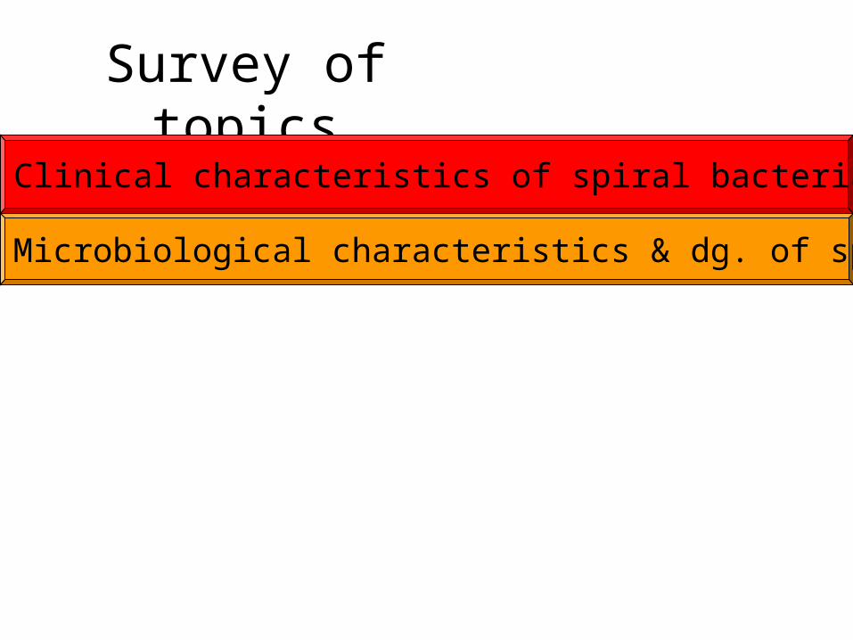
Erythema migrans• This is a picture of Erythema migrans of student M. M.,
who kindly agreed to let it for use in education
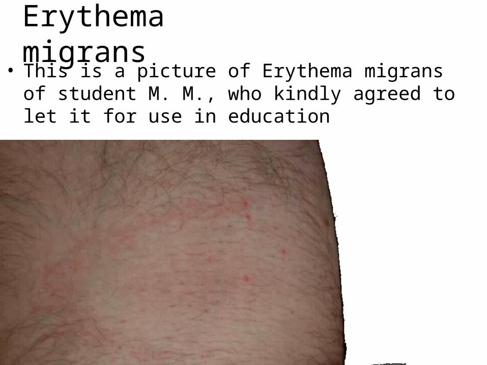
The causative agent was• Borrelia afzelii, one of borrelias, causing Lyme
disease and belonging to the group Borrelia burgdorgeri sensu lato (= „broad sense of meaning“)
• This species „in broad sense“ is divided into several genomospecies. The most important are B. garinii, B. afzelii and B. burgdorferi sensu stricto
• While in the USA mostly the third of them is common and joint symptomatology is common, in Europe two first borrelias are more common, and the typical disease is neuroborreliosis
• Besides Lyme diseases there exist other species causing recurrent fever (B. duttoni, B. recurrentis)
http://www.pasteur.fr
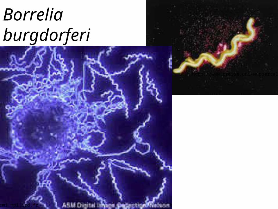
Borrelia burgdorferi
http://www.presse.uni-wuppertal.de
gemi.mpl.ird.fr
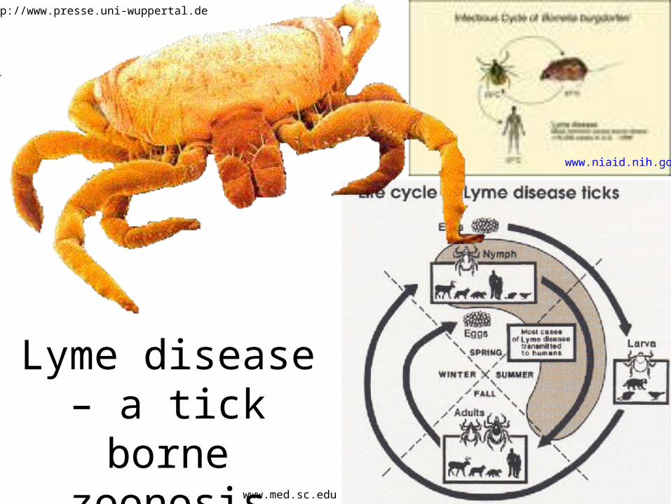
Lyme disease – a tick borne
zoonosis
http://www.presse.uni-wuppertal.de
www.niaid.nih.gov
www.med.sc.edu
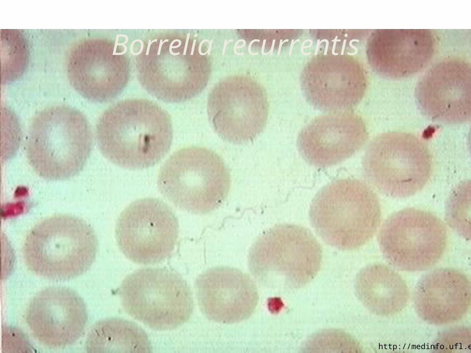
Borrelia recurrentis
http://medinfo.ufl.edu

Story two (virtual, but basis is from a real story)
• When Phyllis found, that she really needs pervitin, and more and more, she decided to earn money by her own body.
• When the client paid more, she went with him without a preservative, she used contraception and she felt more OK
• Then she fell in love and decided to have a child. She stopped the contraception and was happy. Helmut will be a good father…
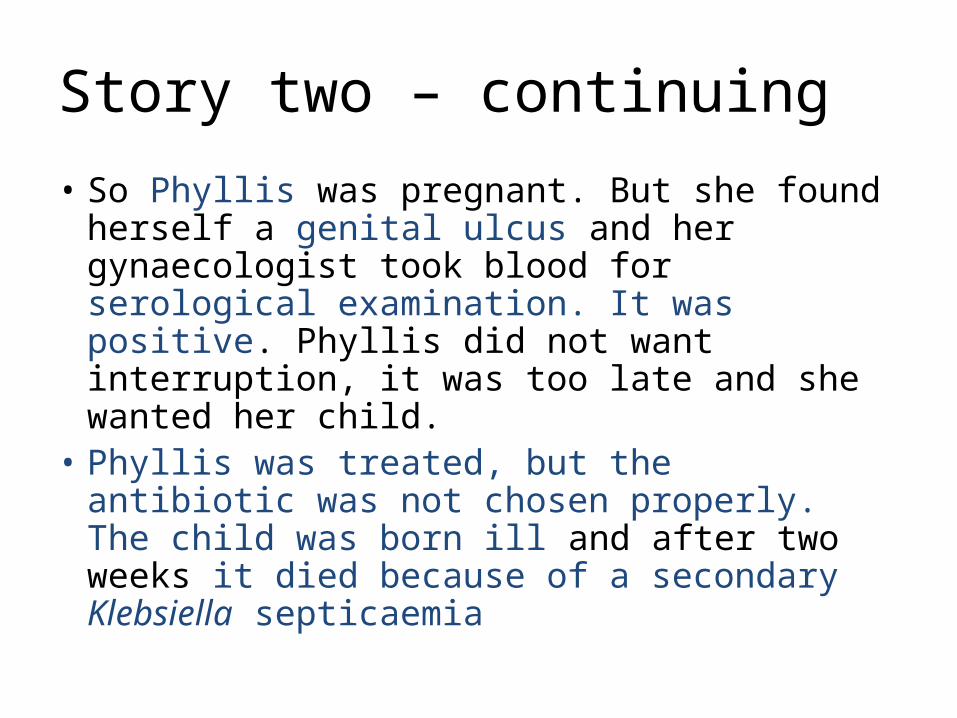
Story two – continuing
• So Phyllis was pregnant. But she found herself a genital ulcus and her gynaecologist took blood for serological examination. It was positive. Phyllis did not want interruption, it was too late and she wanted her child.
• Phyllis was treated, but the antibiotic was not chosen properly. The child was born ill and after two weeks it died because of a secondary Klebsiella septicaemia
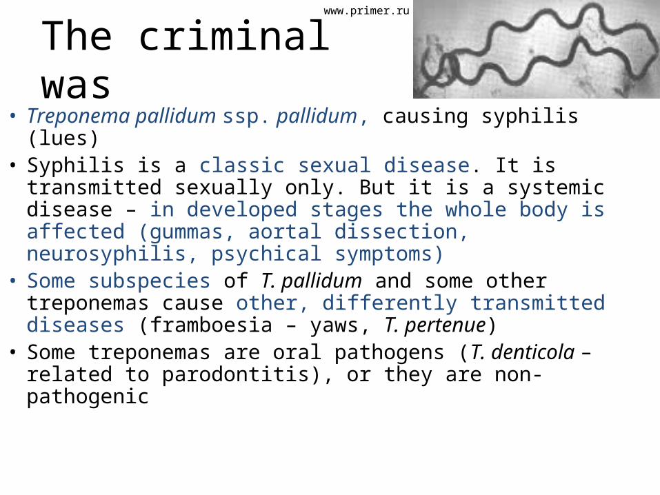
The criminal was• Treponema pallidum ssp. pallidum, causing syphilis
(lues)• Syphilis is a classic sexual disease. It is transmitted
sexually only. But it is a systemic disease – in developed stages the whole body is affected (gummas, aortal dissection, neurosyphilis, psychical symptoms)
• Some subspecies of T. pallidum and some other treponemas cause other, differently transmitted diseases (framboesia – yaws, T. pertenue)
• Some treponemas are oral pathogens (T. denticola – related to parodontitis), or they are non-pathogenic
www.primer.ru
uhavax.hartford.edu
http://nl.wikipedia.org
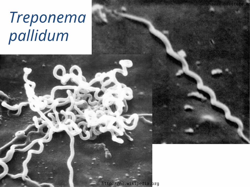
Treponema pallidum
www.geocities.com
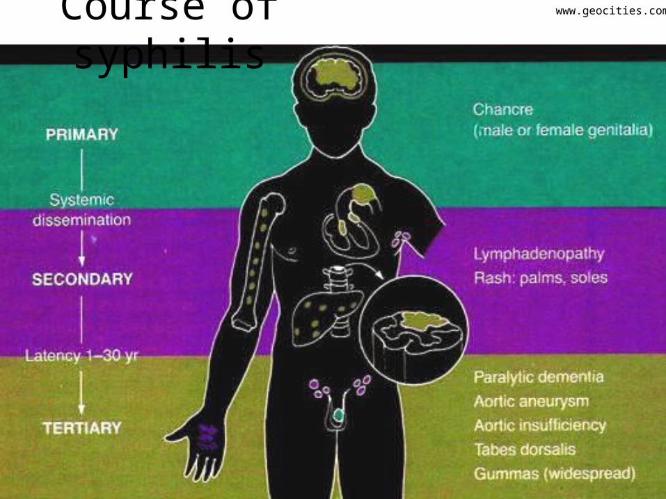
Course of syphilis

Course of syphilis
primary syphilis („chancre“)
secondary syphilis
uhavax.hartford.edu (2×)
www.common-place.org
www.geocities.com
www.nlm.nih.gov
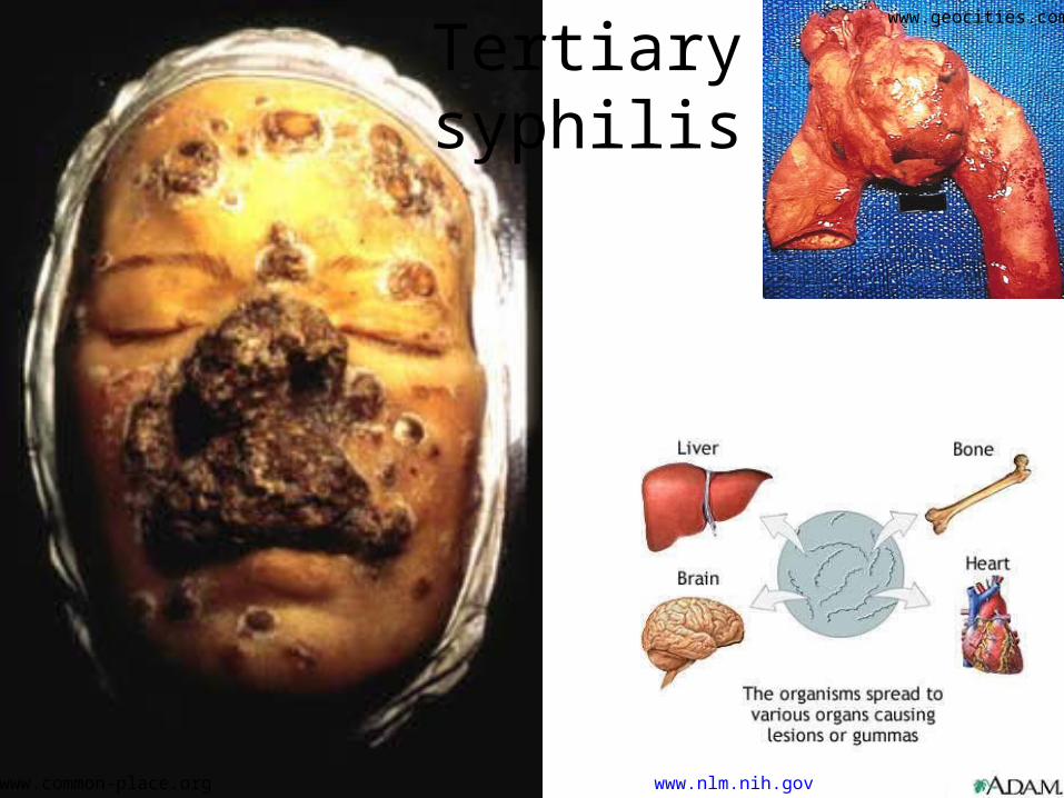
Tertiary syphilis
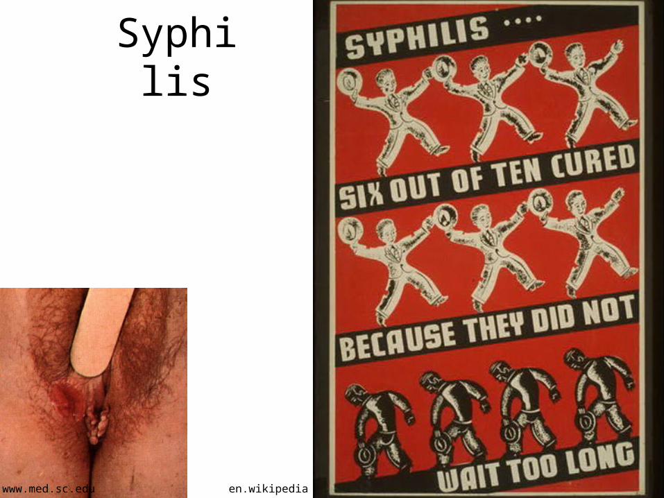
Syphilis
www.med.sc.edu en.wikipedia.org

Yaws (framboesia)
www.med.sc.edu

Story three
• Mr. Ratter was an employee of NWPS Ltd. (Nowhere Water Pipes and Sewage)
• His job was sewage cleaning. He knew all sewage corridors. He also knew rat habits, he liked rats and he understood them.
• Nevertheless, once there was some misunderstanding between him and the leader of rat group and Mr. Ratter was bitten to his leg.
• Some time after this, Mr. Ratter was hospitalized with icterus and bleeding…
Kidney with the corresponding
disease
www.med.sc.edu

This is not Mr. Ratter, but his Venezuelan colleague with a similar fate…
http://www.ceniap.gov.ve
The disease is caused by…• Leptospira interrogans ser. Icterohemorrhagiae• Formerly individual serovars of Leptospira were
considered to be individual species, now all pathogenic ones are taken as a part of species Leptospira interrogans (second species Leptospira biflexa is non-pathogenic)
• Symptomatology varies, from „flu-typhoid“ symptoms of serovar Grippotyphosa (field fever, canefield fever) to jaundice and bleeding (Weil disease, as in Mr. Ratter) in serovar Ictero-hemorragiae.
• (At least these two serovars are quite simple for remembering, try to remember at least them )

Microbiologic characteristics and diagnostics
of spirochets
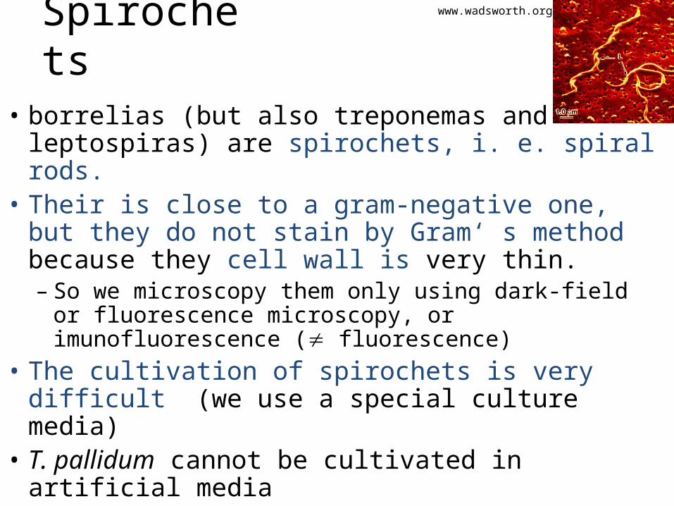
Spirochets
• borrelias (but also treponemas and leptospiras) are spirochets, i. e. spiral rods.
• Their is close to a gram-negative one, but they do not stain by Gram‘ s method because they cell wall is very thin.– So we microscopy them only using dark-field or
fluorescence microscopy, or imunofluorescence ( fluorescence)
• The cultivation of spirochets is very difficult (we use a special culture media)
• T. pallidum cannot be cultivated in artificial media
www.wadsworth.org
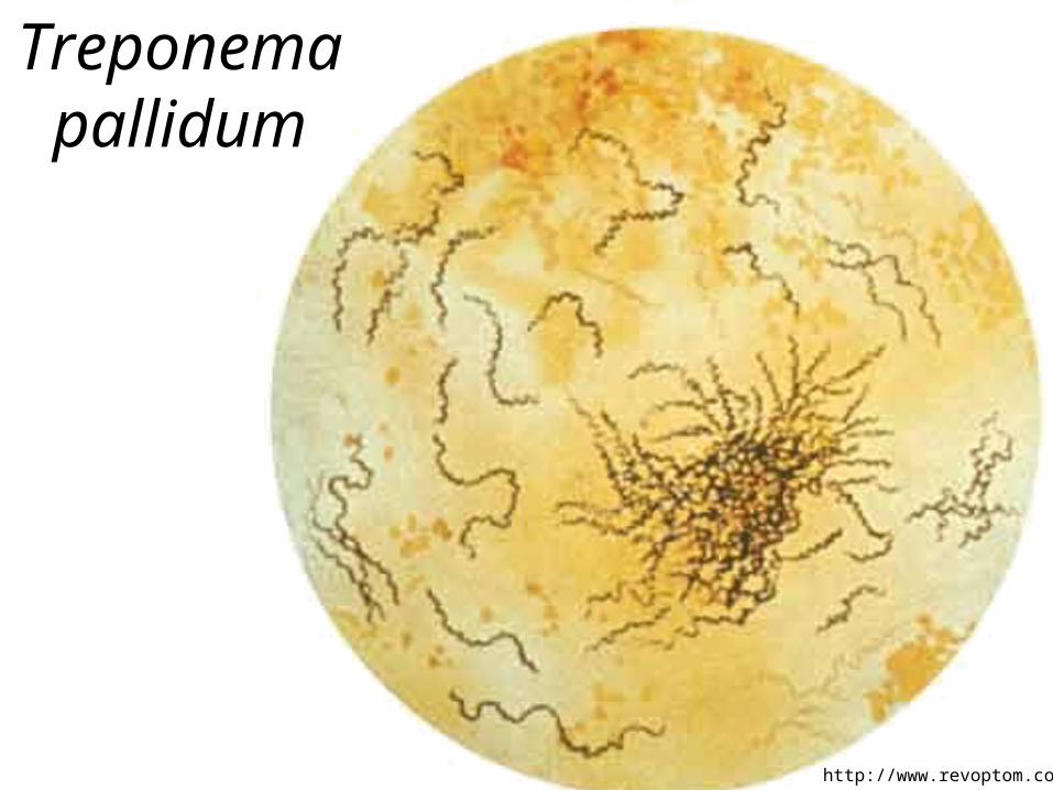
Treponema pallidum
http://www.revoptom.com
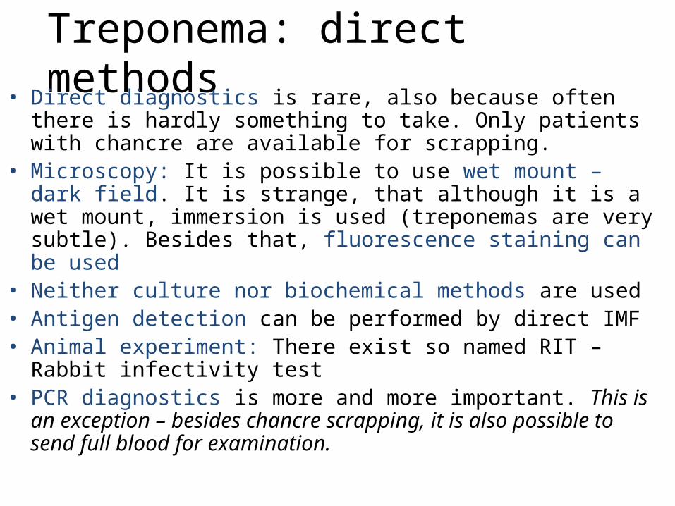
Treponema: direct methods• Direct diagnostics is rare, also because often there is hardly
something to take. Only patients with chancre are available for scrapping.
• Microscopy: It is possible to use wet mount – dark field. It is strange, that although it is a wet mount, immersion is used (treponemas are very subtle). Besides that, fluorescence staining can be used
• Neither culture nor biochemical methods are used• Antigen detection can be performed by direct IMF• Animal experiment: There exist so named RIT – Rabbit infectivity
test• PCR diagnostics is more and more important. This is an exception –
besides chancre scrapping, it is also possible to send full blood for examination.
www.primer.ru
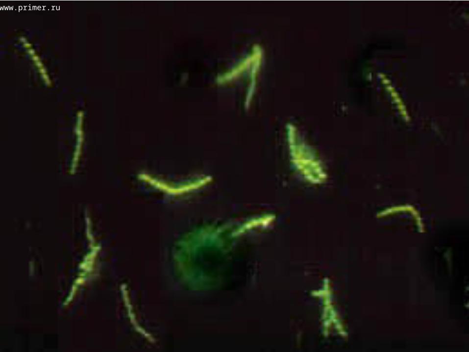
Direct syphilis diagnostics – survey
• RIT – Rabbit infectivity test. For ethical reasons, but also as it is too much work, the RIT is minimized today.
• Dark field – shining Treponema pallidum is observed against the dark field
• Direct IMF – another direct, but difficult method
• PCR – also from blood

New Zealand Rabbit used for RITwww.rockinjawrabbits.com
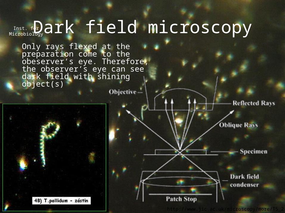
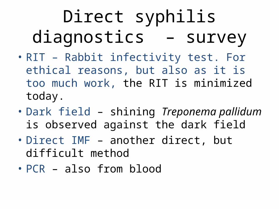
Dark field microscopyOnly rays flexed at the preparation come to the obeserver‘s eye. Therefore, the observer‘s eye can see dark field with shining object(s)
Inst. for Microbiology
http://www.jic.ac.uk/microscopy/more/T5_2.htm
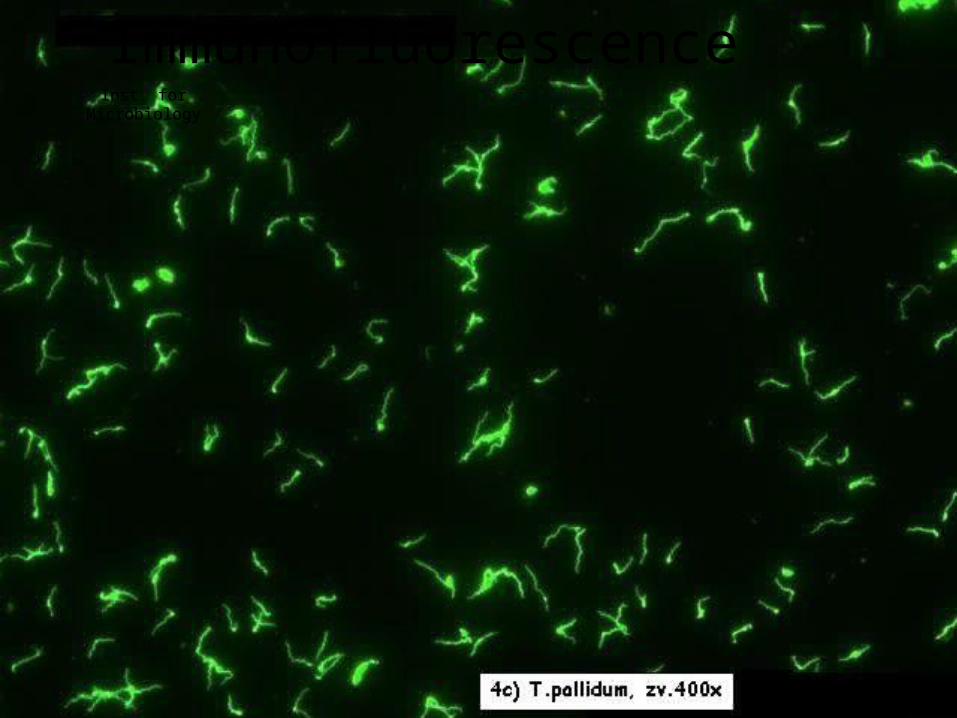
ImmunofluorescenceInst. for Microbiology

Treponema: indirect methods• We use non-treponema tests, which usually plays the
role of antigen cardiolipin from bovine heart, and treponema tests, where we have a real antigen from Treponema pallidum
• Diagnostics is composed of screening and confirmation. We confirm everything that was positive or at least borderline at screening, in reasonable cases even negative results.
• Screening usually consists of a non-treponema and a treponema test
• Confirmation is performed by highly specific treponema tests
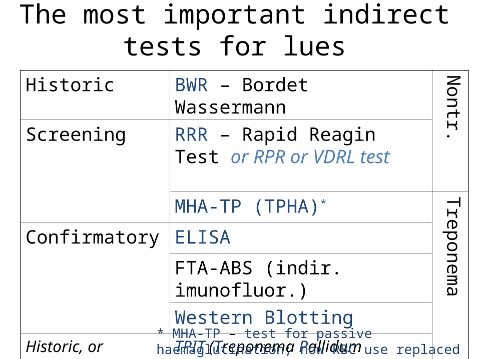
The most important indirect tests for lues
Historic BWR – Bordet Wassermann
Nontr.Screening RRR – Rapid Reagin Test or RPR
or VDRL test
MHA-TP (TPHA)*
Treponema
Confirmatory ELISA
FTA-ABS (indir. imunofluor.)
Western BlottingHistoric, or superconfirmation
TPIT (Treponema Pallidum Imobilisation Test) = Nelson
* MHA-TP – test for passive haemaglutination; now RBC use replaced by polycelluslose
RRR and TPHA
• In RRR, the well with turbidity is positive (it looks like the positive control). It is necessary shake the panel, otherwise the reaction would not be visible.
• TPHA is an agglutination on carrier (RBC). A „potato shaped formation“ is positive, a dense dot is negative
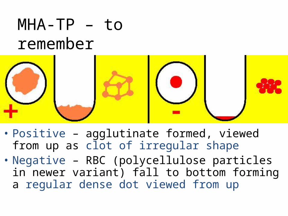
MHA-TP – to remember
• Positive – agglutinate formed, viewed from up as clot of irregular shape
• Negative – RBC (polycellulose particles in newer variant) fall to bottom forming a regular dense dot viewed from up

TPHA – reading:
RRR – reading: turbidity = positive, no turbidity = negative
Inst. for Microbiology

Indications for confirmation• Screening reactions are performed always, when
somebody is to be tested for syphilis (including e. g. pregnant women that are not at all supposed to be positive). Screening reactions are usually performed only qualitatively or semiquantitatively (although it would not be a problem to do them quantitatively)
• Indication for confirmation is:– any positive or at least borderline result in RRR and/or MHA-TP
reaction, OR– presence of suspicious lesions on body, or anamnesis of risky
sexual intercourse – here even in case of negativity of both reactions
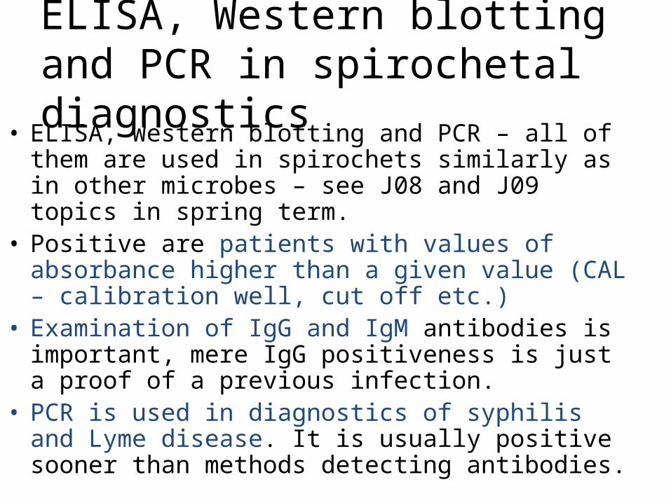
ELISA, Western blotting and PCR in spirochetal diagnostics
• ELISA, Western blotting and PCR – all of them are used in spirochets similarly as in other microbes – see J08 and J09 topics in spring term.
• Positive are patients with values of absorbance higher than a given value (CAL – calibration well, cut off etc.)
• Examination of IgG and IgM antibodies is important, mere IgG positiveness is just a proof of a previous infection.
• PCR is used in diagnostics of syphilis and Lyme disease. It is usually positive sooner than methods detecting antibodies.
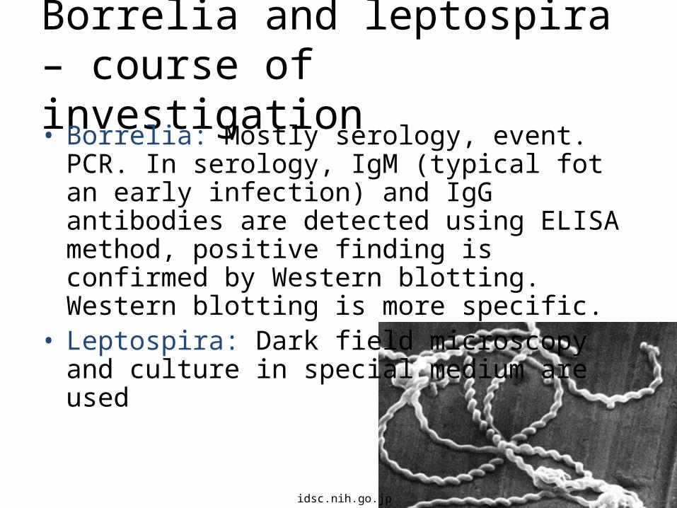
Borrelia and leptospira – course of investigation• Borrelia: Mostly serology, event. PCR. In serology,
IgM (typical fot an early infection) and IgG antibodies are detected using ELISA method, positive finding is confirmed by Western blotting. Western blotting is more specific.
• Leptospira: Dark field microscopy and culture in special medium are used
idsc.nih.go.jp
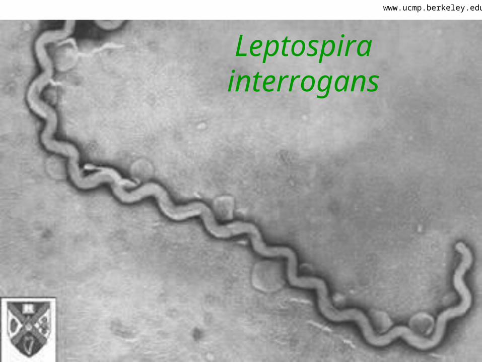
Leptospira interrogans
www.ucmp.berkeley.edu
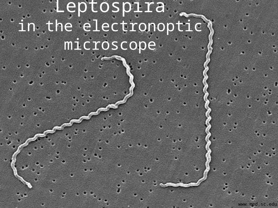
Leptospirain the electronoptic microscope
www.med.sc.edu

Leptospiral diagnostics
• Microscopy of leptospira
Inst. for Microbiology
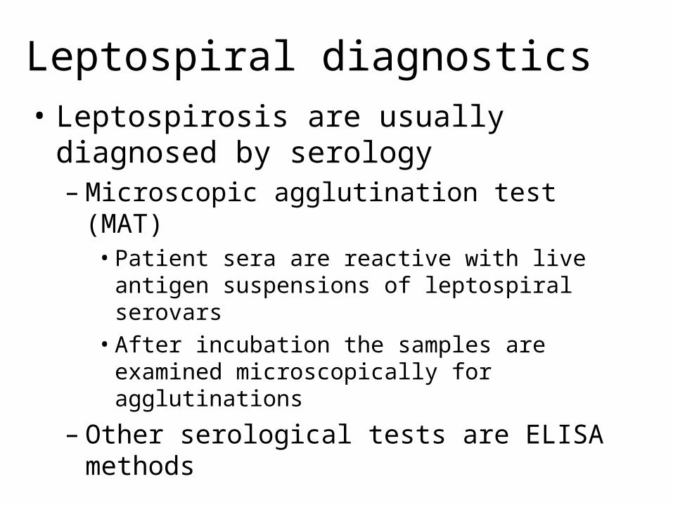
Leptospiral diagnostics• Leptospirosis are usually diagnosed by serology
– Microscopic agglutination test (MAT)• Patient sera are reactive with live antigen suspensions of
leptospiral serovars• After incubation the samples are examined
microscopically for agglutinations– Other serological tests are ELISA methods

More diagnostic opportunities in leptospira (latex agglutination) 4× www.thailabonline.com
The End
www.asci.org/artikel754.html
Jody Rasch: Leptospira 60" x 70" – acrylic on canvas