Toxoplasma gondii Infection Suppresses House Dust Mite Extract … · 2015-09-03 · Toxoplasma...
8
© Copyright The Korean Academy of Asthma, Allergy and Clinical Immunology • The Korean Academy of Pediatric Allergy and Respiratory Disease 557 http://e-aair.org INTRODUCTION A significant increase in the prevalence of allergic diseasesis becoming an important public health problem in industrial- ized countries. 1-3 Recent epidemiological studies havesuggest- ed that a steady decline in exposure to viral, bacterial, and par- asitic infection is associated with an increase in allergic disor- ders. 4,5 That is, reduced exposure to microbes and their prod- ucts in childhood may lead to a failure to development of ap- propriate immune regulation. Although several experimental studies have consistently shown that parasite infections or par- asite-derived products help induction a Th1-biased immunity and prevent the induction of the Th2 system that causes aller- gic disease, 6-11 the mechanism of how parasite infection pre- vents the development of allergic disorder remains unclear. Parasites employ various strategies to evade effective host im- mune response that thwart infection. Although immune eva- sion has been developed to favor parasite establishment within the host, some particular immune-escaping strategies might, quite paradoxically, be also beneficial for the host. 12 Regulatory B cells (Bregs), regulatory T cells (Tregs), and alternatively activat- ed macrophages have been identified as the key components of the immune regulatory network functioning during helminth infections. 13-15 These immune regulatory cells expand during parasite infection and may prevent the development of unrelat- ed immune-driven pathology, such as allergy and autoimmune diseases. 14,16 Typically, B cells function as antibody-producing cells, but Toxoplasma gondii Infection Suppresses House Dust Mite Extract- Induced Atopic Dermatitis in NC/Nga Mice Young-Il Jeong, Sung-Hee Hong, Shin-Hyeong Cho, Won-Ja Lee, Sang-Eun Lee* Division of Malaria & Parasitic Disease, Korea National Institute of Health, Cheongwon-gun, Chungbuk, Korea This is an Open Access article distributed under the terms of the Creative Commons Attribution Non-Commercial License (http://creativecommons.org/licenses/by-nc/3.0/) which permits unrestricted non-commercial use, distribution, and reproduction in any medium, provided the original work is properly cited. Purpose: Toxoplasma gondii ( T. gondii ) is an obligate intracellular protozoan parasite that infects humans and animals via congenital or postnatal routes, and it is found worldwide. Modulation of the immune system by parasite infection is proposed to suppress allergic inflammation. Growing evidences have shown that interleukin (IL)-10-producing regulatory B cells (Bregs) and CD4 + CD25 + FoxP3 + regulatory T cells (T regs) induced by parasite infection play a critical role in allergic or autoimmune diseases because these cells regulate negatively cellular immune responses and inflamma- tion. Currently, the role of IL-10-producing regulatory B cells in host immune response during T. gondii infection is unknown. In this study, we investi- gate whether T. gondii infection can suppress the development of unrelated atopic dermatitis (AD)-like lesions. Methods: AD is a chronically relaps- ing inflammatory skin disease accompanied by severe itching; for this, we used NC/Nga mice, a well-known experimental model of systemic AD. Repeated exposure to Dermatophagoides farinae crude extract (DfE), known as a major environmental allergen, evokes AD-like skin lesions in NC/ Nga mice under specific pathogen-free conditions. NC/Nga mice were intraperitoneally infected with 10 cysts of T. gondii . Results: T. gondii infec- tion significantly ameliorated AD-like skin lesions in NC/Nga mice. The subpopulation of Bregs and T regs in the AD mice was expanded in the course of T. gondii infection. In addition, T. gondii infection inhibited Th2 and enhanced Th1 immune response in the DfE-treated AD mice. Conclusions: We have experimentally demonstrated for the first time that T. gondii infection ameliorated AD-like skin lesions in a mouse model of AD. Our study could in part explain the mechanisms of how parasite infection prevents the development of allergic disorder. Therefore, these immunemechanisms induced by T. gondii infection may be beneficial for the host in terms of reduced risk of allergic immune reactions. Key Words: Atopic dermatitis; Toxoplasma; regulatory B-Cells; regulatory T-Cells Correspondence to: Sang-Eun Lee, DVM, PhD, Division of Malaria & Parasitic Diseases, Korea National Institute of Health, Korea Centers for Disease Control & Prevention, 187 Osongsaengmyeong 2-ro, Cheongwon-gun, Chungbuk 363-951, Korea. Tel: +82-43-719-8525; Fax: +82-43-719-8559; E-mail: [email protected] Received: October 22, 2014; Revised: December 29, 2014; Accepted: January 26, 2015 • This research was supported by the Korea Centers for Diseases Control and Prevention (grant # 4845-300-210-13, 2012). • There are no financial or other issues that might lead to conflict of interest. Original Article Allergy Asthma Immunol Res. 2015 November;7(6):557-564. http://dx.doi.org/10.4168/aair.2015.7.6.557 pISSN 2092-7355 • eISSN 2092-7363
Transcript of Toxoplasma gondii Infection Suppresses House Dust Mite Extract … · 2015-09-03 · Toxoplasma...
© Copyright The Korean Academy of Asthma, Allergy and Clinical Immunology • The Korean Academy of Pediatric Allergy and Respiratory Disease 557http://e-aair.org
INTRODUCTION
A significant increase in the prevalence of allergic diseasesis becoming an important public health problem in industrial-ized countries.1-3 Recent epidemiological studies havesuggest-ed that a steady decline in exposure to viral, bacterial, and par-asitic infection is associated with an increase in allergic disor-ders.4,5 That is, reduced exposure to microbes and their prod-ucts in childhood may lead to a failure to development of ap-propriate immune regulation. Although several experimental studies have consistently shown that parasite infections or par-asite-derived products help induction a Th1-biased immunity and prevent the induction of the Th2 system that causes aller-gic disease,6-11 the mechanism of how parasite infection pre-vents the development of allergic disorder remains unclear.
Parasites employ various strategies to evade effective host im-mune response that thwart infection. Although immune eva-sion has been developed to favor parasite establishment within the host, some particular immune-escaping strategies might,
quite paradoxically, be also beneficial for the host.12 Regulatory B cells (Bregs), regulatory T cells (Tregs), and alternatively activat-ed macrophages have been identified as the key components of the immune regulatory network functioning during helminth infections.13-15 These immune regulatory cells expand during parasite infection and may prevent the development of unrelat-ed immune-driven pathology, such as allergy and autoimmune diseases.14,16
Typically, B cells function as antibody-producing cells, but
Toxoplasma gondii Infection Suppresses House Dust Mite Extract-Induced Atopic Dermatitis in NC/Nga MiceYoung-Il Jeong, Sung-Hee Hong, Shin-Hyeong Cho, Won-Ja Lee, Sang-Eun Lee*
Division of Malaria & Parasitic Disease, Korea National Institute of Health, Cheongwon-gun, Chungbuk, Korea
This is an Open Access article distributed under the terms of the Creative Commons Attribution Non-Commercial License (http://creativecommons.org/licenses/by-nc/3.0/) which permits unrestricted non-commercial use, distribution, and reproduction in any medium, provided the original work is properly cited.
Purpose: Toxoplasma gondii (T. gondii) is an obligate intracellular protozoan parasite that infects humans and animals via congenital or postnatal routes, and it is found worldwide. Modulation of the immune system by parasite infection is proposed to suppress allergic inflammation. Growing evidences have shown that interleukin (IL)-10-producing regulatory B cells (Bregs) and CD4+CD25+FoxP3+ regulatory T cells (Tregs) induced by parasite infection play a critical role in allergic or autoimmune diseases because these cells regulate negatively cellular immune responses and inflamma-tion. Currently, the role of IL-10-producing regulatory B cells in host immune response during T. gondii infection is unknown. In this study, we investi-gate whether T. gondii infection can suppress the development of unrelated atopic dermatitis (AD)-like lesions. Methods: AD is a chronically relaps-ing inflammatory skin disease accompanied by severe itching; for this, we used NC/Nga mice, a well-known experimental model of systemic AD. Repeated exposure to Dermatophagoides farinae crude extract (DfE), known as a major environmental allergen, evokes AD-like skin lesions in NC/Nga mice under specific pathogen-free conditions. NC/Nga mice were intraperitoneally infected with 10 cysts of T. gondii. Results: T. gondii infec-tion significantly ameliorated AD-like skin lesions in NC/Nga mice. The subpopulation of Bregs and Tregs in the AD mice was expanded in the course of T. gondii infection. In addition, T. gondii infection inhibited Th2 and enhanced Th1 immune response in the DfE-treated AD mice. Conclusions: We have experimentally demonstrated for the first time that T. gondii infection ameliorated AD-like skin lesions in a mouse model of AD. Our study could in part explain the mechanisms of how parasite infection prevents the development of allergic disorder. Therefore, these immunemechanisms induced by T. gondii infection may be beneficial for the host in terms of reduced risk of allergic immune reactions.
Key Words: Atopic dermatitis; Toxoplasma; regulatory B-Cells; regulatory T-Cells
Correspondence to: Sang-Eun Lee, DVM, PhD, Division of Malaria & Parasitic Diseases, Korea National Institute of Health, Korea Centers for Disease Control & Prevention, 187 Osongsaengmyeong 2-ro, Cheongwon-gun, Chungbuk 363-951, Korea.Tel: +82-43-719-8525; Fax: +82-43-719-8559; E-mail: [email protected]: October 22, 2014; Revised: December 29, 2014; Accepted: January 26, 2015• This research was supported by the Korea Centers for Diseases Control and Prevention (grant # 4845-300-210-13, 2012).
•There are no financial or other issues that might lead to conflict of interest.
Original ArticleAllergy Asthma Immunol Res. 2015 November;7(6):557-564.
http://dx.doi.org/10.4168/aair.2015.7.6.557pISSN 2092-7355 • eISSN 2092-7363

Jeong et al.
Allergy Asthma Immunol Res. 2015 November;7(6):557-564. http://dx.doi.org/10.4168/aair.2015.7.6.557
Volume 7, Number 6, November 2015
558 http://e-aair.org
they are also involved in other immune mechanisms, including cytokine and chemokine secretion and antigen presentation. In addition, B cells have been shown to participate in the induc-tion of immune tolerance and suppression of inflammation, suggesting the existence of IL-10-producing Bregs.16-18 The IL-10-producing subset of Bregs was first identified to play a critical role in limiting disease severity in allergic inflammatory and autoimmune conditions.13,19,20 In recent studies, it has also been found that these Bregs are significantly induced during parasite infection.6,9,13,21 Several studies have demonstrated that in para-site infections, IL-10-producing Bregs are stimulated as part of the parasite-induced host immune response beneficial for the infection.9,21 Interestingly, IL-10-producing Bregs induced by par-asite infection have been shown to suppress allergic inflamma-tory and autoimmune diseases.9,13,22
Tregs also play essential role in limiting potentially harmful im-mune-mediated pathology as the negative regulators of the T cell immune response.23 The well-characterized Treg cell type is the CD4+CD25+ T cell expressing the transcription factor FoxP3. Tregs may reduce injurious host inflammatory and immune re-sponses as well as Th2 responses, such as eosinophil activation required to kill parasites.24
Toxoplasma gondii (T. gondii) is a worldwide-distributed in-tracellular protozoan parasite thatinfectsa variety of warm-blooded mammals and causes one of the most common chron-ic parasitic infections in humans: approximately one-third of the world population carries T. gondii.25 Toxoplasmosis is usu-ally clinically asymptomatic in healthy individuals but can lead to severe complications in pregnant women and immunocom-promised patients.
In animal models, various helminths alleviate the symptoms of experimental allergic and autoimmune diseases via the in-duction of IL-10-producing Bregs and Tregs.6,26-28 In addition, a re-cent study demonstrated that activated Tregs during T. gondii in-fection contribute to suppression of the development of allergic airway inflammation.29 However, the role of IL-10-producing Bregs induced by T. gondii infection in unrelated immune-driven pathology, such as allergic inflammation, is poorly understood, and there have been no published data on immunomodulation by T. gondii infection in atopic dermatitis (AD) animal models. AD is a chronically relapsing inflammatory skin disease accom-panied by severe itching; we used NC/Nga mice, a well-known experimental model of systemic AD, because of the similarity between clinical symptoms displayed in these mice and AD in humans. Models based on NC/Nga mice are thought to provide important information about AD. Repeated exposure to Der-matophagoides farinaecrude extract (DfE), known as a major environmental allergen, evokes AD-like skin lesions in NC/Nga mice under specific pathogen-free conditions.30,31
The aim of this study was to examine whether T. gondii infec-tion renders mice less susceptible to AD in a mice model and to investigate the influence of T. gondii infection on the expansion
of immune regulatory cells, particularly IL-10-producing CD19+ Bregs and CD4+CD25+FoxP3+ Treg.
MATERIALS AND METHODS
Animals Specific pathogen-free 6-week-old female NC/Nga mice were
purchased from Japan SLC, Inc. (Tokyo, Japan) and housed in a specific pathogen-free facility in individually ventilated and fil-tered cages. The animal protocol used in this study was re-viewed and approved based on ethical procedures and scientif-ic care by the Korea Centers for Disease Control & Prevention-Institutional Animal care and Use Committee (KCDC-IACUC; Approval Number KCDC-12-039-2A). All animal care and pro-tocols were performed in accordance with the guideline for the Care and Use of Laboratory Animals of Korean Centers for Dis-ease Control.
Experimental animal model of AD and parasitic infectionAD-like skin lesions were induced in 7-week-old female NC/
Nga mice using DfE. Ointment containing the components of crude DfE was purchased from Biostir Inc. (Tokyo, Japan). For AD development, the hair on the upper back was shaved, and 200 μL of 4% (w/v) sodium dodecyl sulfate was applied to the shaved dorsal skin and both surfaces of each ear for barrier dis-ruption. After 2 hours, the back skin and both ears of NC/Nga mice were repeatedly treated with 50 mg of ointment contain-ing crude DfE 3 times per week for 4 weeks. Cysts of the T. gon-dii ME49 strain were obtained from the brains of chronically in-fected mice. NC/Nga mice were infected with 10 cysts intraper-itoneally at 7 days before or after the first application of DfE (Fig. 1A). All mice were sacrificed at the end of the experiment.
Antibodies and flow cytometry analysisFor flow cytometry analysis of IL-10-producing CD19+ Bregs,
cells in splenocytes were analyzed for the expression of surface markers using the following monoclonal antibodies: fluorescein isothiocyanate (FITC)-conjugated anti-CD19 (clone 1D3; BD Biosciences, San Jose, CA, USA) and phycoerythrin (PE)-conju-gated anti-IL-10 (clone JES5-16E3; eBioscience, San Diego, CA, USA). For subsequent IL-10intracellular staining, the Cytofix/Cytoperm kit (BD Biosciences) was used according to the man-ufacturer’s protocol (BD Biosciences). For the analysis of CD4+
CD25+FoxP3+ Treg cells, the mouse Treg-staining kit was used ac-cording to the manufacturer’s instructions (eBioscience). The stained cells were analyzed by 3-5 color flow cytometry using a FACSversa flow cytometer (BD Biosciences). The FlowJo soft-ware (Tree Star Inc., OR, USA) was used to analyze flow cytome-try data. To determine background staining, non-reactive iso-type-matched control monoclonal antibodies (eBioscience) were used and gated to exclude ≥98% of non-reactive cells.
T. gondii Infection Inhibits Atopic Dermatitis
Allergy Asthma Immunol Res. 2015 November;7(6):557-564. http://dx.doi.org/10.4168/aair.2015.7.6.557
AAIR
559http://e-aair.org
Evaluation of dermatitis severityThe severity of dermatitis was determined macroscopically
based on the presence of erythema/hemorrhage, edema, exco-riation/erosion, and dryness/scaling and evaluated according to the severity index scoring system. Skin lesions on the back and both ears were scored based on the presence of clinical symptoms: no symptoms=0, mild=1, moderate=2, and se-vere=3. The total clinical skin severity score was defined as the sum of the individual scores.
Measurement of total serum IgE, IgG1, and IgG2a by ELISA Blood samples were collected from the heart, and isolated se-
rum samples were stored at -70˚C until quantitative analysis for Ig subclasses. The concentration of total IgE, IgG1, and IgG2a in mouse serum was measured using the mouse ELISA kit (eBio-science) according to the manufacturer’s instructions.
mRNA quantification of cytokines in skin lesionsThe expression of cytokine, chemokine, and prostaglandin-
endoperoxide synthase 2 (COX-2) mRNAin skin lesions was de-termined by RT-PCR. Total RNA was isolated according to the manufacturer’s protocol (Qiagen, Valencia, CA, USA and used to generate cDNA by reverse transcription. PCR was performed with primers specific for each cytokine, chemokine, and COX-2 (Table 1), and cDNA as templates. The amplified products were analyzed by automated capillary electrophoresis (QIAxcel Ad-vance system; Qiagen Irvine, CA, USA) and β-actin expression was used as the internal control.
Histological analysisDorsal skin from the back and ear lesions was resected, fixed
in 10% formalin solution, embedded into paraffin, sectioned, stained with hematoxylin-eosin solution, and examined by light microscopy. Mast cells and eosinophils were stained with
Day 7
Tg+DfE
DfE
DfE+Tg
Day 14 Day 21 Day 28
Application with DfE three times per week
T. gondill InfectionT. gondill Infection
-7 0 7 28
A
B C
Derm
atiti
s sco
re
10
8
6
4
2
0
Days after first application of DfE
0 7 14 21 26
ControlDfETg infection+DfEDfE+Tg infection
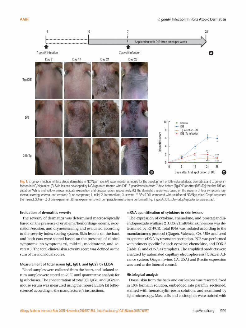
Fig. 1. T. gondii infection inhibits atopic dermatitis in NC/Nga mice. (A) Experimental schedule for the development of DfE-induced atopic dermatitis and T. gondii in-fection in NC/Nga mice. (B) Skin lesions developed by NC/Nga mice treated with DfE. T. gondii was injected 7 days before (Tg+DfE) or after (DfE+Tg) the first DfE ap-plication. White and yellow arrows indicate excoriation and desquamation, respectively. (C) The dermatitis score was based on the severity of four symptoms (ery-thema, scarring, edema, and erosion): 0, no symptoms; 1, mild; 2, intermediate; 3, severe. ***P<0.001 compared with uninfected NC/Nga mice. Graph represent the mean±SD (n=5) of one experiment (three experiments with comparable results were performed). Tg, T. gondii; DfE, Dermatophagoides farinae extract.
***
*** ***
***
Jeong et al.
Allergy Asthma Immunol Res. 2015 November;7(6):557-564. http://dx.doi.org/10.4168/aair.2015.7.6.557
Volume 7, Number 6, November 2015
560 http://e-aair.org
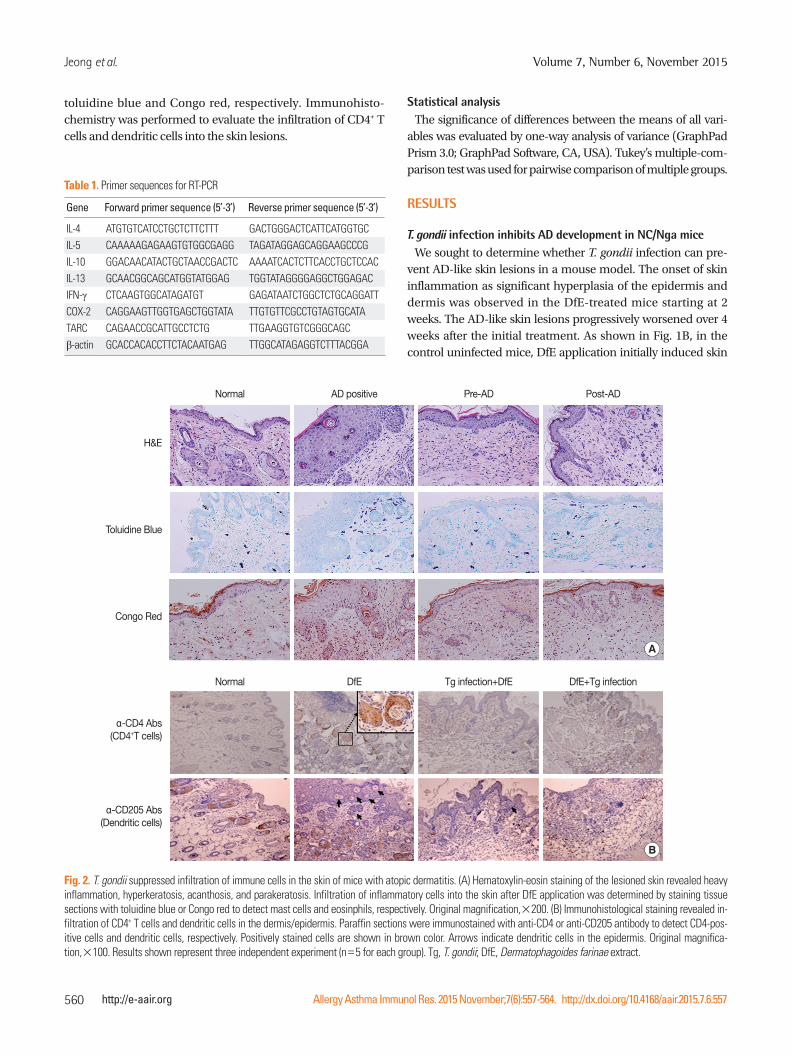
toluidine blue and Congo red, respectively. Immunohisto-chemistry was performed to evaluate the infiltration of CD4+ T cells and dendritic cells into the skin lesions.
Normal
Normal
AD positive
DfE
Pre-AD
Tg infection+DfE
Post-AD
DfE+Tg infection
H&E
α-CD4 Abs(CD4+T cells)
Toluidine Blue
α-CD205 Abs(Dendritic cells)
Congo Red
Fig. 2. T. gondii suppressed infiltration of immune cells in the skin of mice with atopic dermatitis. (A) Hematoxylin-eosin staining of the lesioned skin revealed heavy inflammation, hyperkeratosis, acanthosis, and parakeratosis. Infiltration of inflammatory cells into the skin after DfE application was determined by staining tissue sections with toluidine blue or Congo red to detect mast cells and eosinphils, respectively. Original magnification,×200. (B) Immunohistological staining revealed in-filtration of CD4+ T cells and dendritic cells in the dermis/epidermis. Paraffin sections were immunostained with anti-CD4 or anti-CD205 antibody to detect CD4-pos-itive cells and dendritic cells, respectively. Positively stained cells are shown in brown color. Arrows indicate dendritic cells in the epidermis. Original magnifica-tion,×100. Results shown represent three independent experiment (n=5 for each group). Tg, T. gondii; DfE, Dermatophagoides farinae extract.
B
A
Statistical analysisThe significance of differences between the means of all vari-
ables was evaluated by one-way analysis of variance (GraphPad Prism 3.0; GraphPad Software, CA, USA). Tukey’s multiple-com-parison test was used for pairwise comparison of multiple groups.
RESULTS
T. gondii infection inhibits AD development in NC/Nga miceWe sought to determine whether T. gondii infection can pre-
vent AD-like skin lesions in a mouse model. The onset of skin inflammation as significant hyperplasia of the epidermis and dermis was observed in the DfE-treated mice starting at 2 weeks. The AD-like skin lesions progressively worsened over 4 weeks after the initial treatment. As shown in Fig. 1B, in the control uninfected mice, DfE application initially induced skin
Table 1. Primer sequences for RT-PCR
Gene Forward primer sequence (5’-3’) Reverse primer sequence (5’-3’)
IL-4 ATGTGTCATCCTGCTCTTCTTT GACTGGGACTCATTCATGGTGCIL-5 CAAAAAGAGAAGTGTGGCGAGG TAGATAGGAGCAGGAAGCCCGIL-10 GGACAACATACTGCTAACCGACTC AAAATCACTCTTCACCTGCTCCACIL-13 GCAACGGCAGCATGGTATGGAG TGGTATAGGGGAGGCTGGAGACIFN-γ CTCAAGTGGCATAGATGT GAGATAATCTGGCTCTGCAGGATTCOX-2 CAGGAAGTTGGTGAGCTGGTATA TTGTGTTCGCCTGTAGTGCATATARC CAGAACCGCATTGCCTCTG TTGAAGGTGTCGGGCAGCβ-actin GCACCACACCTTCTACAATGAG TTGGCATAGAGGTCTTTACGGA
T. gondii Infection Inhibits Atopic Dermatitis
Allergy Asthma Immunol Res. 2015 November;7(6):557-564. http://dx.doi.org/10.4168/aair.2015.7.6.557
AAIR
561http://e-aair.org
**
IgE
(ng/
mL)
7,500
5,000
2,500
0DfE Tg+DfE DfE+Tg
******
IgG1
(μL/
mL)
4,000
3,000
2,000
1,000
0DfE Tg+DfE DfE+Tg
******
IgG2
a (μ
g/m
L)
3,000
2,000
1,000
0DfE Tg+DfE DfE+Tg
***
A
B C
Control DfE Tg+DfE DfE+Tg
IL-4
TARC
IL-5
IFN-γ
IL-13
IL-10
β-actin
COX-2
Rela
tive
inte
ncity
/ β-actin
0.25
0.20
0.15
0.10
0.05
0
IL-4 IL-13 TARC IL-10IL-5 COX-2 IFN-γ
******
*** ***
*** ******
***
***
***
*
*
* *
*
*
*
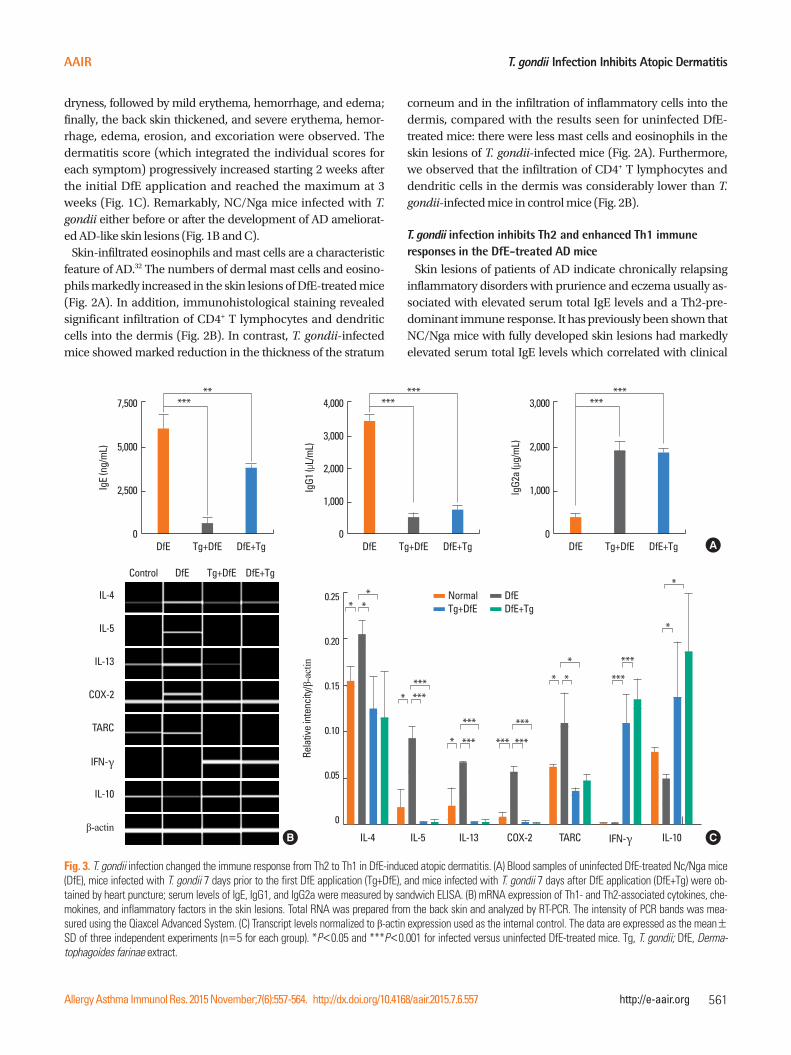
Fig. 3. T. gondii infection changed the immune response from Th2 to Th1 in DfE-induced atopic dermatitis. (A) Blood samples of uninfected DfE-treated Nc/Nga mice (DfE), mice infected with T. gondii 7 days prior to the first DfE application (Tg+DfE), and mice infected with T. gondii 7 days after DfE application (DfE+Tg) were ob-tained by heart puncture; serum levels of IgE, IgG1, and IgG2a were measured by sandwich ELISA. (B) mRNA expression of Th1- and Th2-associated cytokines, che-mokines, and inflammatory factors in the skin lesions. Total RNA was prepared from the back skin and analyzed by RT-PCR. The intensity of PCR bands was mea-sured using the Qiaxcel Advanced System. (C) Transcript levels normalized to β-actin expression used as the internal control. The data are expressed as the mean±SD of three independent experiments (n=5 for each group). *P<0.05 and ***P<0.001 for infected versus uninfected DfE-treated mice. Tg, T. gondii; DfE, Derma-tophagoides farinae extract.
NormalTg+DfE
DfEDfE+Tg
dryness, followed by mild erythema, hemorrhage, and edema; finally, the back skin thickened, and severe erythema, hemor-rhage, edema, erosion, and excoriation were observed. The dermatitis score (which integrated the individual scores for each symptom) progressively increased starting 2 weeks after the initial DfE application and reached the maximum at 3 weeks (Fig. 1C). Remarkably, NC/Nga mice infected with T. gondii either before or after the development of AD ameliorat-ed AD-like skin lesions (Fig. 1B and C).
Skin-infiltrated eosinophils and mast cells are a characteristic feature of AD.32 The numbers of dermal mast cells and eosino-phils markedly increased in the skin lesions of DfE-treated mice (Fig. 2A). In addition, immunohistological staining revealed significant infiltration of CD4+ T lymphocytes and dendritic cells into the dermis (Fig. 2B). In contrast, T. gondii-infected mice showed marked reduction in the thickness of the stratum
corneum and in the infiltration of inflammatory cells into the dermis, compared with the results seen for uninfected DfE-treated mice: there were less mast cells and eosinophils in the skin lesions of T. gondii-infected mice (Fig. 2A). Furthermore, we observed that the infiltration of CD4+ T lymphocytes and dendritic cells in the dermis was considerably lower than T. gondii-infected mice in control mice (Fig. 2B).
T. gondii infection inhibits Th2 and enhanced Th1 immune responses in the DfE-treated AD mice
Skin lesions of patients of AD indicate chronically relapsing inflammatory disorders with prurience and eczema usually as-sociated with elevated serum total IgE levels and a Th2-pre-dominant immune response. It has previously been shown that NC/Nga mice with fully developed skin lesions had markedly elevated serum total IgE levels which correlated with clinical
Jeong et al.
Allergy Asthma Immunol Res. 2015 November;7(6):557-564. http://dx.doi.org/10.4168/aair.2015.7.6.557
Volume 7, Number 6, November 2015
562 http://e-aair.org
severity of dermatitis.33 Consistent with these results, we ob-served vigorous production of total IgE and IgG1 in DfE-treated NC/Nga mice (6,065.5±910.7 and 3,419±147.4 μg/mL, respec-tively) (Fig. 3A). We also examined whether T. gondii infection before or after the development of skin lesions affect serum lev-els of the Ig subclasses, including IgE, IgG1, and IgG2a. Our re-sults indicate that the generation of IgE and IgG1 in T. gondii-infected NC/Nga mice was clearly inhibited (IgE: 588.4±795.9 and 3,455.4±743.6 ng/mL, P<0.001 and 0.05, respectively; IgG1: 470±369.9 and 759.4±477.9 μg/mL, P<0.001, respec-tively), whereas IgG2a levels markedly increased (1,943±296.5 and 1,854.1±162.6 μg/mL, respectively) compared to the con-trol group (299.5±131.1 ng/mL, P<0.001) (Fig. 3A).
Next, we examined the mRNA expression of Th1- and Th2-as-sociated cytokines, chemokines, and inflammatory factors in the skin lesions. The mRNA expression of Th2-type cytokines, including IL-4, IL-5, and IL-13, significantly increased in the DfE-treated NC/Nga mice. The infiltration of inflammatory cells into inflammation sites is dependent on the local production of chemokines with leukocyte chemoattractant activity.31 TARC/CCL17 functions as a selective chemoattractant that contributes to local recruitment and migration of Th2 cells expressing CC
CD19
IL-1
0
2.25% 10.3% 16.1%
P<0.001
% o
f IL-
10+ CD
19+
B ce
lls
15.0
12.5
10.0
7.5
5.0
2.5
0DfE Tg+DfE DfE+Tg
P<0.05
A
CD25
FoxP
3
5.92% 4.05% 16.5%
P<0.05
% o
f CD4
+ CD25
+ FoxP
3+
T ce
lls
20
15
10
5
0DfE Tg+DfE DfE+Tg
ns
B
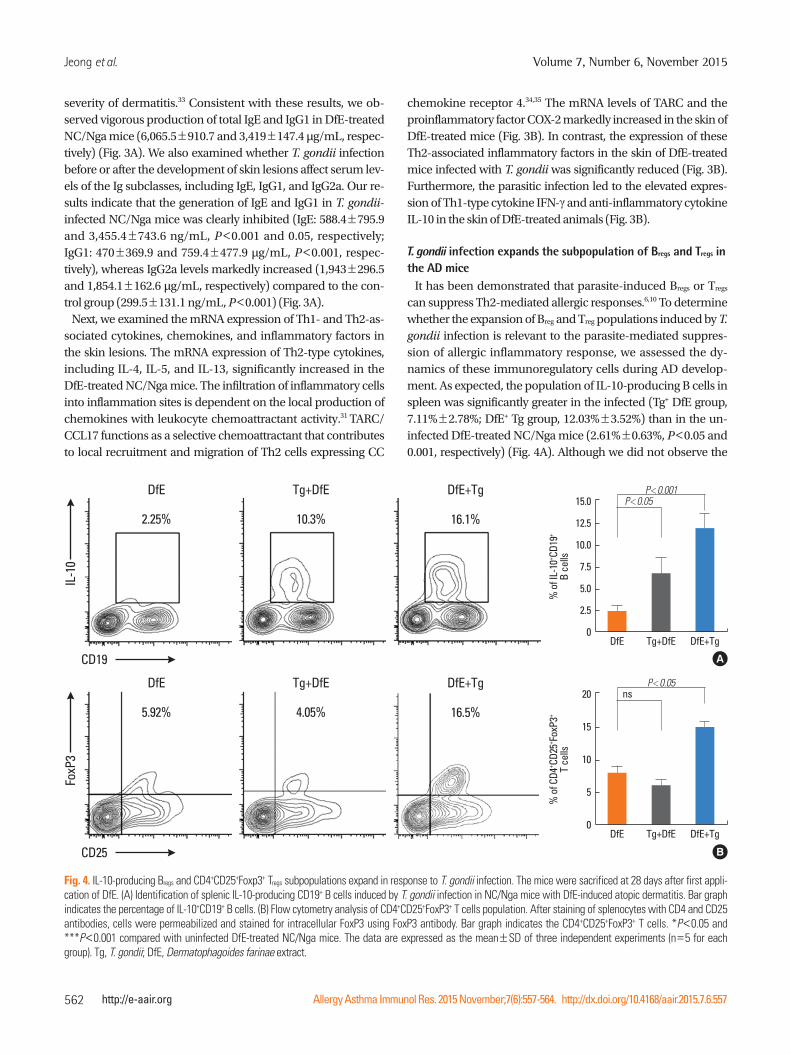
Fig. 4. IL-10-producing Bregs and CD4+CD25+Foxp3+ Tregs subpopulations expand in response to T. gondii infection. The mice were sacrificed at 28 days after first appli-cation of DfE. (A) Identification of splenic IL-10-producing CD19+ B cells induced by T. gondii infection in NC/Nga mice with DfE-induced atopic dermatitis. Bar graph indicates the percentage of IL-10+CD19+ B cells. (B) Flow cytometry analysis of CD4+CD25+FoxP3+ T cells population. After staining of splenocytes with CD4 and CD25 antibodies, cells were permeabilized and stained for intracellular FoxP3 using FoxP3 antibody. Bar graph indicates the CD4+CD25+FoxP3+ T cells. *P<0.05 and ***P<0.001 compared with uninfected DfE-treated NC/Nga mice. The data are expressed as the mean±SD of three independent experiments (n=5 for each group). Tg, T. gondii; DfE, Dermatophagoides farinae extract.
DfE Tg+DfE DfE+Tg
DfE Tg+DfE DfE+Tg
chemokine receptor 4.34,35 The mRNA levels of TARC and the proinflammatory factor COX-2 markedly increased in the skin of DfE-treated mice (Fig. 3B). In contrast, the expression of these Th2-associated inflammatory factors in the skin of DfE-treated mice infected with T. gondii was significantly reduced (Fig. 3B). Furthermore, the parasitic infection led to the elevated expres-sion of Th1-type cytokine IFN-γ and anti-inflammatory cytokine IL-10 in the skin of DfE-treated animals (Fig. 3B).
T. gondii infection expands the subpopulation of Bregs and Tregs in the AD mice
It has been demonstrated that parasite-induced Bregs or Tregs can suppress Th2-mediated allergic responses.6,10 To determine whether the expansion of Breg and Treg populations induced by T. gondii infection is relevant to the parasite-mediated suppres-sion of allergic inflammatory response, we assessed the dy-namics of these immunoregulatory cells during AD develop-ment. As expected, the population of IL-10-producing B cells in spleen was significantly greater in the infected (Tg+ DfE group, 7.11%±2.78%; DfE+ Tg group, 12.03%±3.52%) than in the un-infected DfE-treated NC/Nga mice (2.61%±0.63%, P<0.05 and 0.001, respectively) (Fig. 4A). Although we did not observe the
T. gondii Infection Inhibits Atopic Dermatitis
Allergy Asthma Immunol Res. 2015 November;7(6):557-564. http://dx.doi.org/10.4168/aair.2015.7.6.557
AAIR
563http://e-aair.org
increase in CD4+CD2+FpxP3+Tregs in the mice infected with T. gondii prior to the emergence of skin lesions, the animals in-fected with the parasite after AD development displayed re-markable increase of Tregs (15.03%±1.4% ) compared with the uninfected control mice (8.52%±2.27%, P<0.05) (Fig. 4B).
DISCUSSION
Recent studies in animal models have demonstrated that chronic helminth infections are strongly associated with a re-duced prevalence of inflammatory disorders, including allergic diseases.6,13,27 It has also been shown that parasitic infection may prevent the development of allergic and autoimmune diseases by inducing IL-10-producing B cells and IL-10-dependent mechanisms, suggesting suppressive function of IL-10-produc-ing Breg cells in allergic diseases.13,17,26,36,37 NC/Nga mice treated with DfE are used as a sensitive animal model for human AD. Most of the population of AD patients has high titers of specific IgE to Dermatophagoides mites.31,37 In this study, we examined whether T. gondii infection can inhibit allergic inflammatory re-sponse in AD using NC/Nga mice as an experimental AD mod-el. T. gondii infection rendered protection against AD-related pathogenic mechanisms. Histological analysis demonstrated reduced infiltration of inflammatory cells including eosinophils, mast cells, CD4+ T cells, and dendritic cells, into DfE-induced skin lesions of T. gondii-infected NC/Nga mice. Moreover, the high levels of IgE and IgG1 production as well as elevated ex-pression of Th2-associated inflammatory factors observed in AD mice, were significantly decreased in T. gondii-infected AD-like NC/Nga mice. Interestingly, we observed an increase in se-rum IgG2a and IFN-γ expression in the skin in T. gondii-infect-ed AD-like mice, suggesting that the infection switched the im-mune response from the Th2 to the Th1 type.
Recent studies have shown that helminthic infections in-creased IL-10-producing B cells, and that adoptive transfer of these cells from infected mice protected recipient mice against experimentally induced allergic inflammatory disease.6,13 In-deed, a recent clinical study involving helminth-infected pa-tients with multiple sclerosis also highlighted a role of IL-10-producing B cells in ameliorating disease symptoms.36 Amu et al.6 have demonstrated that Bregs induced by Schistosoma mansoni infection prevent and reverse allergic airway inflam-mation via Tregs, suggesting that immunoregulatory Bregs and Tregs play an important role in parasite-mediated protection against allergic conditions. Consistent with this notion, our findings reveal that IL-10-producing Bregs and Tregs induced by T. gondii infection can effectively modulate allergic inflammatory responses.
In conclusion, our results demonstrate that T. gondii up-regu-lates IL-10-producing Bregs and CD4+CD25+FoxP3+ Tregs, which can protect against the development of severe AD. These im-mune mechanisms induced by T. gondii infection may be ben-
eficial for the host in terms of reduced risk of allergic immune reactions and provide a basis for the development of novel therapeutic strategies to treat chronic AD.
REFERENCE
1. Asher MI, Montefort S, Björkstén B, Lai CK, Strachan DP, Weiland SK, et al. Worldwide time trends in the prevalence of symptoms of asthma, allergic rhinoconjunctivitis, and eczema in childhood: ISAAC Phases One and Three repeat multicountry cross-sectional surveys. Lancet 2006;368:733-43.
2. Ring J, Krämer U, Schäfer T, Behrendt H. Why are allergies increas-ing? Curr Opin Immunol 2001;13:701-8.
3. Wills-Karp M, Santeliz J, Karp CL. The germless theory of allergic disease: revisiting the hygiene hypothesis. Nat Rev Immunol 2001;1:69-75.
4. Gereda JE, Leung DY, Thatayatikom A, Streib JE, Price MR, Klin-nert MD, et al. Relation between house-dust endotoxin exposure, type 1 T-cell development, and allergen sensitisation in infants at high risk of asthma. Lancet 2000;355:1680-3.
5. Garn H, Renz H. Epidemiological and immunological evidence for the hygiene hypothesis. Immunobiology 2007;212:441-52.
6. Amu S, Saunders SP, Kronenberg M, Mangan NE, Atzberger A, Fal-lon PG. Regulatory B cells prevent and reverse allergic airway in-flammation via FoxP3-positive T regulatory cells in a murine mod-el. J Allergy Clin Immunol 2010;125:1114-24.e8.
7. Jeong YI, Kim SH, Ju JW, Cho SH, Lee WJ, Park JW, et al. Clonorchis sinensis-derived total protein attenuates airway inflammation in murine asthma model by inducing regulatory T cells and modulat-ing dendritic cell functions. Biochem Biophys Res Commun 2011;407:793-800.
8. Jeong YI, Kim YJ, Ju JW, Hong SH, Lee MR, Cho SH, et al. Identifica-tion of anti-allergic effect of Clonorchis sinensis-derived protein venom allergen-like proteins (CsVAL). Biochem Biophys Res Com-mun 2014;445:549-55.
9. Ronet C, Hauyon-La Torre Y, Revaz-Breton M, Mastelic B, Tacchi-ni-Cottier F, Louis J, et al. Regulatory B cells shape the development of Th2 immune responses in BALB/c mice infected with Leishma-nia major through IL-10 production. J Immunol 2010;184:886-94.
10. Wilson MS, Taylor MD, Balic A, Finney CA, Lamb JR, Maizels RM. Suppression of allergic airway inflammation by helminth-induced regulatory T cells. J Exp Med 2005;202:1199-212.
11. Kishi C, Amano H, Suzue K, Ishikawa O. Plasmodium berghei in-fection ameliorates atopic dermatitis-like skin lesions in NC/Nga mice. Allergy 2014;69:1412-9.
12. Zaccone P, Fehervari Z, Phillips JM, Dunne DW, Cooke A. Parasitic worms and inflammatory diseases. Parasite Immunol 2006;28:515-23.
13. Hussaarts L, van der Vlugt LE, Yazdanbakhsh M, Smits HH. Regu-latory B-cell induction by helminths: implications for allergic dis-ease. J Allergy Clin Immunol 2011;128:733-9.
14. Taylor MD, van der Werf N, Maizels RM. T cells in helminth infec-tion: the regulators and the regulated. Trends Immunol 2012;33:181-9.
15. Gordon S, Martinez FO. Alternative activation of macrophages: mechanism and functions. Immunity 2010;32:593-604.
16. Mizoguchi A, Mizoguchi E, Takedatsu H, Blumberg RS, Bhan AK. Chronic intestinal inflammatory condition generates IL-10-pro-
Jeong et al.
Allergy Asthma Immunol Res. 2015 November;7(6):557-564. http://dx.doi.org/10.4168/aair.2015.7.6.557
Volume 7, Number 6, November 2015
564 http://e-aair.org
ducing regulatory B cell subset characterized by CD1d upregula-tion. Immunity 2002;16:219-30.
17. Fillatreau S, Sweenie CH, McGeachy MJ, Gray D, Anderton SM. B cells regulate autoimmunity by provision of IL-10. Nat Immunol 2002;3:944-50.
18. Lund FE. Cytokine-producing B lymphocytes-key regulators of im-munity. Curr Opin Immunol 2008;20:332-8.
19. Lee MK, Lee WY, Yong SJ, Shin KC, Lee SN, Lee SJ, et al. A case of autoimmune progesterone dermatitis misdiagnosed as allergic contact dermatitis. Allergy Asthma Immunol Res 2011;3:141-4.
20. Noh G, Lee JH. Regulatory B cells and allergic diseases. Allergy Asthma Immunol Res 2011;3:168-77.
21. Jeong YI, Hong SH, Cho SH, Lee WJ, Lee SE. Induction of IL-10-producing CD1d(high)CD5+ regulatory B cells following Babe-sia microti-infection. PLoS One 2012;7:e46553.
22. Wilson MS, Taylor MD, O’Gorman MT, Balic A, Barr TA, Filbey K, et al. Helminth-induced CD19+CD23hi B cells modulate experi-mental allergic and autoimmune inflammation. Eur J Immunol 2010;40:1682-96.
23. Lee JC, Hayman E, Pegram HJ, Santos E, Heller G, Sadelain M, et al. In vivo inhibition of human CD19-targeted effector T cells by natu-ral T regulatory cells in a xenotransplant murine model of B cell malignancy. Cancer Res 2011;71:2871-81.
24. Velavan TP, Ojurongbe O. Regulatory T cells and parasites. J Biomed Biotechnol 2011;2011:520940.
25. Elmore SA, Jones JL, Conrad PA, Patton S, Lindsay DS, Dubey JP. Toxoplasma gondii: epidemiology, feline clinical aspects, and pre-vention. Trends Parasitol 2010;26:190-6.
26. Mangan NE, Fallon RE, Smith P, van Rooijen N, McKenzie AN, Fal-lon PG. Helminth infection protects mice from anaphylaxis via IL-10-producing B cells. J Immunol 2004;173:6346-56.
27. Smits HH, Hammad H, van Nimwegen M, Soullie T, Willart MA, Lievers E, et al. Protective effect of Schistosoma mansoni infection on allergic airway inflammation depends on the intensity and chronicity of infection. J Allergy Clin Immunol 2007;120:932-40.
28. Girgis NM, Gundra UM, Loke P. Immune regulation during hel-minth infections. PLoS Pathog 2013;9:e1003250.
29. Fenoy I, Giovannoni M, Batalla E, Martin V, Frank FM, Piazzon I, et al. Toxoplasma gondii infection blocks the development of allergic air-way inflammation in BALB/c mice. Clin Exp Immunol 2009;155:275-84.
30. Matsuoka H, Maki N, Yoshida S, Arai M, Wang J, Oikawa Y, et al. A mouse model of the atopic eczema/dermatitis syndrome by re-peated application of a crude extract of house-dust mite Derma-tophagoides farinae. Allergy 2003;58:139-45.
31. Oshio T, Sasaki Y, Funakoshi-Tago M, Aizu-Yokota E, Sonoda Y, Matsuoka H, et al. Dermatophagoides farinae extract induces se-vere atopic dermatitis in NC/Nga mice, which is effectively sup-pressed by the administration of tacrolimus ointment. Int Immu-nopharmacol 2009;9:403-11.
32. Matsui K, Nishikawa A. Percutaneous application of peptidoglycan from Staphylococcus aureus induces an increase in mast cell num-bers in the dermis of mice. Clin Exp Allergy 2005;35:382-7.
33. Matsuda H, Watanabe N, Geba GP, Sperl J, Tsudzuki M, Hiroi J, et al. Development of atopic dermatitis-like skin lesion with IgE hy-perproduction in NC/Nga mice. Int Immunol 1997;9:461-6.
34. Hirata H, Arima M, Cheng G, Honda K, Fukushima F, Yoshida N, et al. Production of TARC and MDC by naive T cells in asthmatic pa-tients. J Clin Immunol 2003;23:34-45.
35. Jahnz-Rozyk K, Targowski T, Paluchowska E, Owczarek W, Kuchar-czyk A. Serum thymus and activation-regulated chemokine, mac-rophage-derived chemokine and eotaxin as markers of severity of atopic dermatitis. Allergy 2005;60:685-8.
36. Correale J, Farez M, Razzitte G. Helminth infections associated with multiple sclerosis induce regulatory B cells. Ann Neurol 2008;64:187-99.
37. Yanaba K, Bouaziz JD, Haas KM, Poe JC, Fujimoto M, Tedder TF. A regulatory B cell subset with a unique CD1dhiCD5+ phenotype controls T cell-dependent inflammatory responses. Immunity 2008;28:639-50.