Toxicity Study of Pittosporum ochrosiaefolium Bojer ...
9
Journal of Plant Sciences 2015; 3(6): 349-357 Published online December 9, 2015 (http://www.sciencepublishinggroup.com/j/jps) doi: 10.11648/j.jps.20150306.19 ISSN: 2331-0723 (Print); ISSN: 2331-0731 (Online) Toxicity Study of Pittosporum ochrosiaefolium Bojer (Pittosporaceae) a Medicinal Plant of Madagascar Maholy Pricille Ratsimiebo 1 , David Ramanitrahasimbola 2 , Clara Fredeline Rajemiarimoelisoa 2 Zoarilala Rinah Razafindrakoto 3 , Hanitra Ranjana Randrianarivo 1 , Danielle Aurore Doll Rakoto 1 , Victor Louis Jeannoda 1 1 Laboratory of Applied Biochemistry to Medical Sciences, Fundamental and Applied Biochemistry Department, Faculty of Sciences, University of Antananarivo, Antananarivo, Madagascar 2 Department of Pharmacy, Faculty of Medicine, University of Antananarivo, Antananarivo, Madagascar 3 Malagasy Institute for Applied Research (IMRA), Antananarivo, Madagascar Email address: [email protected] (M. P. Ratsimiebo), [email protected] (D. Ramanitrahasimbola), [email protected] (Z. R. Razafindrakoto), [email protected] (C. F. Rajemiarimoelisoa), [email protected] (D. A. D. Rakoto), [email protected] (H. R. Randrianarivo), [email protected] (V. L. Jeannoda) To cite this article: Maholy Pricille Ratsimiebo, David Ramanitrahasimbola, Clara Fredeline Rajemiarimoelisoa, Zoarilala Rinah Razafindrakoto, Hanitra Ranjana Randrianarivo, Danielle Aurore Doll Rakoto, Victor Louis Jeannoda. Toxicity Study of Pittosporum ochrosiaefolium Bojer (Pittosporaceae) a Medicinal Plant of Madagascar. Journal of Plant Sciences. Vol. 3, No. 6, 2015, pp. 349-357. doi: 10.11648/j.jps.20150306.19 Abstract: The present work aimed to assess the leaf toxicity of Pittosporum ochrosiaefolium Bojer, a well-known medicinal plant endemic to Madagascar. Leaf methanolic extract (LME), obtained after successive extractions by hexan and methanol, was tested in vivo on warm and cold-blooded animals and in vitro on isolated atria of guinea-pig. LME was toxic to mice with a LD 50 of about 46.69 mg/kg of body weight by intraperitoneal route. It induced mainly nervous disorders (body fasciculation, clonic convulsions), respiratory troubles (reduction of respiration frequency and cyanosis) and diarrheas. By intraperitoneal route, LME (46.69 mg/kg) caused histopathological lesions in lungs, liver, kidneys, small and large intestines but had no effects on brain, heart and stomach. Vascular congestion, inflammatory infiltrates, edema and necrosis were frequently observed. LME had a positive inotropic effect but no significant chronotropic one on isolated atria. It did not alter renal and hepatic functions at 21.24 mg/kg. It was highly toxic to the frog Ptychadena mascareniensis (LC 50 of 13.51 µg/mL) and the fish Cyprinus carpio (LC 50 of 8.2 µg/mL). It was also toxic to mosquito larvae Culex quinquefasciatus and Aedes albopictus with LC 50 of 720 ppm and 910 ppm respectively. Different chemical compound groups were found in LME but only saponins proved to be toxic. Under certain conditions, P. ochrosiaefolium might be exploited as source of pesticides or therapeutic molecules. Keywords: Pittosporum ochrosiaefolium, Leaf Methanolic Extract, Saponins, Toxicity, Histopathological Lesions, Pharmacological Activity 1. Introduction This study was undertaken to investigate the toxic effect of leaf methanolic extract (LME) of Pittosporum ochrosiaefolium (Pittosporaceae), an endemic species to Madagascar used in traditional medicine. It was the continuation of our preliminary investigations on the toxicity of Malagasy Pittosporum species [1, 2]. The Pittosporum genus, originating from East Asia, is one of the 9 genera belonging to the Pittosporaceae family. It comprises about 160 species growing wild in tropical and subtropical regions [3]. It is often cultivated as ornamental plants in the Mediterranean region [4]. Pittosporum has other uses. Its timber is used in marquetry. Some species are employed as a fish poison [5-7]. Different species are widely used as medicinal plants for the treatment of various diseases. P. floribundum parts are used against skin diseases and itches [7]. In high doses, its bark acts as narcotic used as an antidote to snake poison, general weakness and also as a stimulant [8]. Australian Aborigines use P. phylliraeoides to treat a variety of conditions. An infusion of the leaves, seeds, fruit pulp or
Transcript of Toxicity Study of Pittosporum ochrosiaefolium Bojer ...

Journal of Plant Sciences 2015; 3(6): 349-357
Published online December 9, 2015 (http://www.sciencepublishinggroup.com/j/jps)
doi: 10.11648/j.jps.20150306.19
ISSN: 2331-0723 (Print); ISSN: 2331-0731 (Online)
Toxicity Study of Pittosporum ochrosiaefolium Bojer (Pittosporaceae) a Medicinal Plant of Madagascar
Maholy Pricille Ratsimiebo1, David Ramanitrahasimbola
2, Clara Fredeline Rajemiarimoelisoa
2
Zoarilala Rinah Razafindrakoto3, Hanitra Ranjana Randrianarivo
1, Danielle Aurore Doll Rakoto
1,
Victor Louis Jeannoda1
1Laboratory of Applied Biochemistry to Medical Sciences, Fundamental and Applied Biochemistry Department, Faculty of Sciences,
University of Antananarivo, Antananarivo, Madagascar 2Department of Pharmacy, Faculty of Medicine, University of Antananarivo, Antananarivo, Madagascar 3Malagasy Institute for Applied Research (IMRA), Antananarivo, Madagascar
Email address: [email protected] (M. P. Ratsimiebo), [email protected] (D. Ramanitrahasimbola),
[email protected] (Z. R. Razafindrakoto), [email protected] (C. F. Rajemiarimoelisoa),
[email protected] (D. A. D. Rakoto), [email protected] (H. R. Randrianarivo), [email protected] (V. L. Jeannoda)
To cite this article: Maholy Pricille Ratsimiebo, David Ramanitrahasimbola, Clara Fredeline Rajemiarimoelisoa, Zoarilala Rinah Razafindrakoto, Hanitra
Ranjana Randrianarivo, Danielle Aurore Doll Rakoto, Victor Louis Jeannoda. Toxicity Study of Pittosporum ochrosiaefolium Bojer
(Pittosporaceae) a Medicinal Plant of Madagascar. Journal of Plant Sciences. Vol. 3, No. 6, 2015, pp. 349-357.
doi: 10.11648/j.jps.20150306.19
Abstract: The present work aimed to assess the leaf toxicity of Pittosporum ochrosiaefolium Bojer, a well-known medicinal
plant endemic to Madagascar. Leaf methanolic extract (LME), obtained after successive extractions by hexan and methanol, was
tested in vivo on warm and cold-blooded animals and in vitro on isolated atria of guinea-pig. LME was toxic to mice with a LD50
of about 46.69 mg/kg of body weight by intraperitoneal route. It induced mainly nervous disorders (body fasciculation, clonic
convulsions), respiratory troubles (reduction of respiration frequency and cyanosis) and diarrheas. By intraperitoneal route, LME
(46.69 mg/kg) caused histopathological lesions in lungs, liver, kidneys, small and large intestines but had no effects on brain, heart
and stomach. Vascular congestion, inflammatory infiltrates, edema and necrosis were frequently observed. LME had a positive
inotropic effect but no significant chronotropic one on isolated atria. It did not alter renal and hepatic functions at 21.24 mg/kg. It
was highly toxic to the frog Ptychadena mascareniensis (LC50 of 13.51 µg/mL) and the fish Cyprinus carpio (LC50 of 8.2 µg/mL).
It was also toxic to mosquito larvae Culex quinquefasciatus and Aedes albopictus with LC50 of 720 ppm and 910 ppm
respectively. Different chemical compound groups were found in LME but only saponins proved to be toxic. Under certain
conditions, P. ochrosiaefolium might be exploited as source of pesticides or therapeutic molecules.
Keywords: Pittosporum ochrosiaefolium, Leaf Methanolic Extract, Saponins, Toxicity, Histopathological Lesions,
Pharmacological Activity
1. Introduction
This study was undertaken to investigate the toxic effect of
leaf methanolic extract (LME) of Pittosporum
ochrosiaefolium (Pittosporaceae), an endemic species to
Madagascar used in traditional medicine. It was the
continuation of our preliminary investigations on the toxicity
of Malagasy Pittosporum species [1, 2].
The Pittosporum genus, originating from East Asia, is one
of the 9 genera belonging to the Pittosporaceae family. It
comprises about 160 species growing wild in tropical and
subtropical regions [3]. It is often cultivated as ornamental
plants in the Mediterranean region [4].
Pittosporum has other uses. Its timber is used in
marquetry. Some species are employed as a fish poison [5-7].
Different species are widely used as medicinal plants for
the treatment of various diseases. P. floribundum parts are
used against skin diseases and itches [7]. In high doses, its
bark acts as narcotic used as an antidote to snake poison,
general weakness and also as a stimulant [8]. Australian
Aborigines use P. phylliraeoides to treat a variety of
conditions. An infusion of the leaves, seeds, fruit pulp or

350 Maholy Pricille Ratsimiebo et al.: Toxicity Study of Pittosporum ochrosiaefolium
Bojer (Pittosporaceae) a Medicinal Plant of Madagascar
wood is utilized to treat bruises, muscle aches, sprains and
cramps. P. phylliraeoides infusions are drunk to treat cough
and cold and to induce lactation [9]. A decoction of fruit is
used both externally and by ingestion to treat eczema and
pruritus [10]. In south of India, P. tetraspermum is an
expectorant and possesses febrifuge and narcotic properties.
It is used to cure chronic bronchitis, leprosy and skin
diseases. The paste of the root bark is applied to
inflammatory and rheumatoid swelling [11]. In Azores
archipelago, infusion of P. undulatum fruits in alcohol or
vinegar is employed for its anti-inflammatory activity [12].
In South Africa and Kenya, P. manii is used for the treatment
of fever, malaria, inflammation, stomachache and as an
antidote for insect bites [13, 14]. Some Pittosporum species
displayed an antidote activity for snake poisoning [7].
Several active compounds were isolated from Pittosporum.
Triterpene saponins were found in P. senacia
(senaciapittosides A and B) [15], P. viridiflorum
(Pittoviridoside) [16], P. verticillatum [17], P. manii [18].
Sesquiterpene glycosides were isolated from P. undulatum
(undulatumosides A and B) [19] and P. viridiflorum [20].
Iridoid glycosides (6α-hydroxygeniposide) were obtained
from P. glabratum [21], carotenoids (tobiraxanthins) from P.
tobira [22]. Essential oil was extracted from P. undulatum
[23], P. neelgherrense and P. viridilum [24].
In addition, a number of Pittosporum species were the
subject of pharmacological studies. Methanolic extract from
the bark of P. manii showed activity against Plasmodium
falciparum strains [18]. Antimicrobial and antifungal
activities of several species such as P. tobira [25], P.
floribundum [7]. P. neelgherrense [26], P. undulatum [23], P.
viridulum [24], P. tetraspermum [27] were demonstrated.
Larvicidal activity of P. tobira was reported [28]. Anti-
inflammatory activity of P. tetraspermum [11] and P.
undulatum [19] was revealed. The methanol and aqueous
extracts of stem bark from P. dasycaulon showed antioxidant
activities [29]. Cytotoxicity properties of P. verticillatum
[17], P. venulosum [30] and P. tobira [22] were reported.
In Madagascar, Pittosporum is represented by 11 species,
9 of which are endemic. Different species are widely used as
medicinal plants. They have anti-inflammatory, antimicrobial
and antispasmodic activities [31, 32].
P. ochrosiaefolium Bojer var madagascariense Danguy
Cufod is a shrub or tree up to 10 m high growing in
rainforest, in hot and humid areas of the eastern part of
Madagascar.
The leaf infusion is employed for the treatment of
gonnorhea. The bark decoction is a vermifuge in moderate
doses. The stem and leaf infusion serve to calm bellyache
and chewed leaf is an antidote for poisonous spider bites [31,
32]. It is also used against cough and rash. In North East of
Madagascar, bark decoction is recommended to fight against
tiredness, back pain and urinating difficulty [33]. In
Ranomafana, according to traditional healers, P.
ochrosiaefolium is used to heal wounds.
The present work mainly focused on the assessment and
characterization of the LME toxic effects on animals and the
chemical nature of the compound(s) responsible of its
toxicity. Its effects on disease vector insects were also
investigated.
2. Experimental
2.1. Plant Materials
Leaves were harvested in Ranomafana (Region of
Vatovavy-Fitovinany, 400 km South East of Antananarivo),
in February 2013. Voucher specimens of P. ochrosiaefolium
were deposited in the herbarium of Plant Biology and
Ecology Department of the Faculty of Sciences of the
University of Antananarivo.
2.2. Animals
OF-1 strain Albino mice (Mus musculus), weighing 25±2
g, came from the Pasteur Institute of Madagascar breeding
farm.
Male or female Guinea pigs weighing 300±50 g were used.
They were purchased from local farmers and kept at least a
week in the IMRA animal house before use. Those rodents
were housed under standard environmental conditions and
fed with standard rodent diet and water ad libitum.
Fishes (Cyprinus carpio), Royal strain, 2-4 cm size, were
provided by a fish farmer.
Apode frog tadpoles (Ptychadena mascareniensis) were
harvested from the ponds in the vicinity of the Antananarivo
University site.
Fishes and tadpoles were allowed to acclimatize to the
aquarium conditions for three days after their arrival in
laboratory.
The mosquito larvae (Culex quinquefasciatus, Aedes
albopictus) were furnished by Entomology department of
Pasteur Institute of Madagascar (IPM).
One day old chicks (Gallus gallus domesticus), Hubbard
classic strain, were provided by poultry farmer.
2.3. Leaf Methanolic Extract Preparation
The dried leaves of P. ochrosiaefolium were ground into
powder. Leaf powder (100 g) was extracted with hexan until
complete discoloration. The colorless powder was extracted
with 3x1000 mL of methanol for 24 h under stirring. The
filtrate was evaporated under reduced pressure to yield dry
extract powders. The water solution of this leaf powder was
named LME.
2.4. Extraction of the Crude Saponins
The extraction of the crude saponins was performed as
following. LME residue was dissolved in methanol. A
mixture of acetone-ether (v/v) was added dropwise to the
resulting solution placed in ice-bath until no more crude
saponins precipitated. Precipitate was dissolved in distilled
water and constituted the crude saponin fraction. The
supernatant was evaporated to dryness. Dry residue
dissolved in distilled water constituted the non saponosidic

Journal of Plant Sciences 2015; 3(6): 349-357 351
fraction. The toxicity activity of both fractions was assessed
on mice.
2.5. Phytochemical Screening
The detection reactions of chemical groups on LME were
carried out according to the methods of Fong et al, 1997 [34]
and Marini-Bettolo et al, 1981 [35].
2.6. Acute Toxicity Tests
Toxic effects on mice and chicks were evaluated by
intraperitoneal route and oral route. By intraperitoneal route,
LME was injected at a volume of 0.25 mL per 25 g of body
weight while by oral route, it was given by gavage by means
of a curved distal end needle at the rate of 0.25 mL per 25 g
of body weight.
Subcutaneous route was employed to assess LME toxicity
on mice (0.25 mL per 25±2g of body weight).
2.6.1. Behavioral Observations of Intoxicated Mice
All changes in intoxicated mice behavior were observed
during 24 h after LME administration, continuously for the
first 4 h and then every hour. Changes in general behavior and
other physiological activities or death were noted [36, 37].
2.6.2. LD50 Assessment on Mice
The toxicity study was carried out using 40 female mice.
The animals were randomly distributed into 8 groups of 5
animals: one control group and seven treated groups.
Animals were provided with food and water ad libitum.
Seven different doses (from 42.48 to 60.24 mg/kg body
weight) of LME were intraperitoneally injected. The control
group received physiological serum.
The LD50 (24 h) of the extract was determined by
calculation and graphical methods of Reed and Muench,
1938 [38] and Boyd et al, 1968 [39].
2.6.3. LC50 Assessment on Cold Blooded Animals
The LC50 (24 h) of LME was determined on fishes, frog
tadpoles and mosquito larvae. Eleven groups of five animals
(fishes or frog tadpoles) were placed each in 500 mL
crystallizer containing 250 mL of spring water. Ten groups
were treated with 10 different concentrations of LME and the
last one served as control.
For the mosquitoes, 25 larvae were placed in 25 mL of
distilled water. Different concentrations of LME were added
in the medium. One group without extract is used as control.
The tests were performed in triplicate.
2.7. Anatomopathological Study
Three groups of 10 mice were used. Animals of group 1
were injected with 46.49 mg/kg, a dose corresponding to
LD50, then sacrificed 3, 9, 24, 48 h after treatment. Animals
of group 2 were treated orally with a single daily dose of
42.48 mg/kg (LD0 dose) for 10 days. At the end of
experiences, the animals were sacrificed. Animals of group 3
served as control.
Brain, lungs, heart, stomach, liver, kidneys, large and
small intestines were harvested afterwards and preserved in
formol 10%.
The preparation of the organ sections for histopathological
examinations was carried out using a classical method
including the following steps: body fixation, inclusion, organ
sections, preparation by microtomy, glass slide mounting,
staining and microscope examination [40].
2.8. Study of LME Cardiac Effect
The effect of LME on the chronotropy and inotropy of the
guinea pig isolated atria was assessed utilizing the method
described in previous paper [41].
Increasing concentrations of LME from 12.5 to 50 µg/mL
were tested on the same animals. The responses of atria were
recorded at 1, 3 and 5 min after each injection. The number
of beats per min and the amplitude of contraction were
measured and percentage of stimulation or inhibition was
calculated according to the parameter controls.
2.9. Study of LME Effect on the Renal and Hepatic
Functions
This study consisted in the daily administration of a sub-
lethal dose of the extract during 30 days and the assessment
of the biochemical parameters to evaluate hepatic and renal
function after treatment in mice. After exposure to a few
possible toxic substances, there will be changes in body
weight gain which would reflect toxicity [42].
The animals were weighed and divided into two groups of
five animals each. They were treated daily at a fixed time in
the morning by gavage of 10 mL/kg of distilled water for the
control animals and 10 mL/kg of the extract prepared in
distilled water for the treated animals. The tested LME dose
was 21.24 mg/kg body-weight. This dose corresponded to the
1/2 of LD0 by intraperitoneal route. The animals were then
weighed every day, from the start until the end of the
treatment, to note any weight variation. On the 31st day, the
animals were anesthetized by chloroform inhalation. Their
blood was collected on the retro-orbital.
Mean plasma levels of ALAT, ASAT and creatinine from
control animals were statistically compared with those of the
extract treated animals.
2.10. Statistical Analysis
The results of LC50 assessment were treated using the
method of statistical analysis ANOVA (Graphpad prism 5
software). Those of the pharmacological study were
expressed as mean ± standard error mean (SEM). Significant
differences were determined using a Student’s t test and the
differences were considered significant if p<0.05 [43].
3. Results and Analysis
3.1. LME Extraction Yields
Extraction of the dried leaves of P. ochrosiaefolium gave a
green colored extract with a yield of 20.59%.

352 Maholy Pricille Ratsimiebo et al.: Toxicity Study of Pittosporum ochrosiaefolium
Bojer (Pittosporaceae) a Medicinal Plant of Madagascar
3.2. Major Chemical Groups in LME
The results of phytochemical screening of LME revealed
the presence of saponins, triterpenes, unsaturated sterols,
alkaloids, flavonoids, iridoïds, tannins and polyphenols
(Table 1).
3.3. Acute Toxicity of LME on Mice
3.3.1. Behavior Studies
The signs developed by intoxicated animals were various.
The signs of the nervous system disorders were developed by
hypoactivity, exophthalmos, fasciculation, paralysis of the
hind legs and clonic convulsions. The respiratory systems
attacks were manifested by reduction of respiration
frequency, cyanosis etc. Other disorders as diarrhea were
noted.
3.3.2. Influence of Administration Route
The influence of the administration route on LME effect
was shown in Table 2. The intraperitoneal route was by far
the most efficient. At doses about 52 times higher than the
intraperitoneal LD100 (60.24 mg/kg), no mortality was
observed by subcutaneous and oral routes.
3.3.3. LD50 Values
The LD50 of P. ochrosiaefolium value was assessed
between 46.24 mg/kg and 47.15 mg/kg.
Table 1. Phytochemical screening of leaf preparations of P. ochrosiaefolium.
Chemical groups Tests Results
powder LME
Alkaloids
Mayer + +
Wagner + +
Dragendorff + +
Flavonoids Willstätter + +
Iridoïds Hot HCl - -
Leucoanthocyanins Bate-Smith - -
Saponins Froth test + +
Steroids Liebermann-Burchard
- -
Triterpenes + +
Unsaturated sterols + +
Cardenolides - -
Cyanogenic glycoside Grignard - -
Insaturated lactones Kedde - -
Coumarin - -
Tannins and polyphenols
Gelatin 1% - -
Gelatin-salt 10% + +
FeCl3 + +
Quinones Borntrager - -
Table 2. Effect of LME on mice according to administration route.
DOSE (mg/kg) Mortality rate (%)
Intraperitoneal Subcutaneous Oral
60.24 100 0 0
463.68 100 0 0
3000 100 0 0
3500 100 100 30
4000 100 100 100
3.4. Anatomopathological Studies
The macroscopic analysis of the organs did not show any
significant changes in aspect, size, color or texture when
compared with the control group.
Brain, heart and stomach appeared normal. Three hours
after LME injection, vascular exudative inflammation
appeared as vascular congestion in lungs, liver, kidneys and
large intestine. Inflammatory infiltrates of variable density
consisting of neutrophil polymorphonuclears were visible.
No histologic lesion was observed in small intestine.
Hemorrhagic foci with the presence or not of hemosiderin
pigments appeared in purifier organs (lungs, liver and
kidneys) after 9 h of intoxication. Edematous and necrotic
foci were also noted in liver. Hemorrhagic areas or
edematous foci and residual inflammatory cells were found at
24 h after injection. A starting tissue repair in inflammation
target organs with capillary hyperplasia (small and large
intestines) appeared after 48 h.
As examples of organ injuries, lesions in lungs, liver,
kidney, small and large intestines, which were more visible
after at 9 h of poisoning were shown in Fig. 1.
Lesions by oral intoxication were generally the same as by
intraperitoneal route after 3 h of intoxication. However, some
lesions were not observed.
3.5. Effects of LME on Three Major Physiological
Functions
3.5.1. Effects of LME on the Cardiac Contraction in
Guinea-Pig
On the one hand, compared to the magnitude of the
contraction control, the perfusion of LME at a concentration
of 25 µg/mL significantly increased auricular force of
contraction at 3rd
and 5th
minute (p<0.05) of recording (Fig.
2). At 50 µg/mL, it produced a positive inotropic effect time-
dependent. On the other hand, up to 50 µg/mL, no significant
effect was recorded on the auricular frequency of contraction
(p>0.05).
3.5.2. Effects of LME on the Renal and Hepatic Functions
in Mice
(i). Effects of LME on the Body Weight
The mean weekly body weight gain of control and daily
treated mice with LME during 30 days was presented in
Table 3.
The body weight of all treated animals increased. The
weight gain was even higher in treated animals.
(ii). Effects of LME on the Biochemical Parameters
The impact of a sub-chronic intoxication by LME (21.24
mg/kg) on hepatic and renal function was assessed by any
rate change of respectively transaminases (ASAT and ALAT)
and creatinine. No significant change of the rate of the 3
biochemical parameters (p>0.05) was observed under the
experiment conditions (Table 4).
3.6. Effects of LME on Chicks and Cold Blooded Animals
At 60.24 mg/kg, a dose corresponding to LD100 on mice,

Journal of Plant Sciences 2015; 3(6): 349-357 353
LME had no effect on chicks by intraperitoneal and oral
routes.
LME had toxic effects on Ptychadena mascareniensis
tadpoles, Cyprinus carpio alvins and Culex quinquefasciatus
and Aedes albopictus larvae (Table 5).
Figure 1. Main lesions induced by LME on lungs, kidney, small intestine (G x10), liver and large intestine by intraperitoneal route after 9 h of exposure
(magnification factor x 40).
ED: edema; HA: hemorrhagic area; VD: vasodilatation; NP: neutrophil polymorphonuclears; NZ: necrotic zone; HS: hemosiderin pigments; II: inflammatory
infiltrate; M: mucus
Figure 2. Effect of different concentrations of LME and contact time on the number of beats per minute (a) and the amplitude of contraction (b) and of the
isolated guinea pig atria (n=3); *: p<0.05; **: p<0.02; ***: p<0.01.
354 Maholy Pricille Ratsimiebo et al.: Toxicity Study of Pittosporum ochrosiaefolium
Bojer (Pittosporaceae) a Medicinal Plant of Madagascar
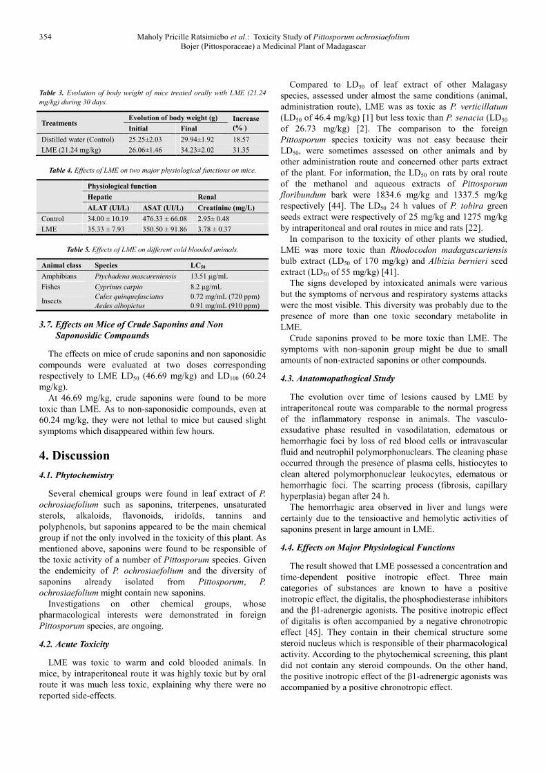
Table 3. Evolution of body weight of mice treated orally with LME (21.24
mg/kg) during 30 days.
Treatments Evolution of body weight (g) Increase
(% ) Initial Final
Distilled water (Control) 25.25±2.03 29.94±1.92 18.57
LME (21.24 mg/kg) 26.06±1.46 34.23±2.02 31.35
Table 4. Effects of LME on two major physiological functions on mice.
Physiological function
Hepatic Renal
ALAT (UI/L) ASAT (UI/L) Creatinine (mg/L)
Control 34.00 ± 10.19 476.33 ± 66.08 2.95± 0.48
LME 35.33 ± 7.93 350.50 ± 91.86 3.78 ± 0.37
Table 5. Effects of LME on different cold blooded animals.
Animal class Species LC50
Amphibians Ptychadena mascareniensis 13.51 µg/mL
Fishes Cyprinus carpio 8.2 µg/mL
Insects Culex quinquefasciatus
Aedes albopictus
0.72 mg/mL (720 ppm)
0.91 mg/mL (910 ppm)
3.7. Effects on Mice of Crude Saponins and Non
Saponosidic Compounds
The effects on mice of crude saponins and non saponosidic
compounds were evaluated at two doses corresponding
respectively to LME LD50 (46.69 mg/kg) and LD100 (60.24
mg/kg).
At 46.69 mg/kg, crude saponins were found to be more
toxic than LME. As to non-saponosidic compounds, even at
60.24 mg/kg, they were not lethal to mice but caused slight
symptoms which disappeared within few hours.
4. Discussion
4.1. Phytochemistry
Several chemical groups were found in leaf extract of P.
ochrosiaefolium such as saponins, triterpenes, unsaturated
sterols, alkaloids, flavonoids, iridoïds, tannins and
polyphenols, but saponins appeared to be the main chemical
group if not the only involved in the toxicity of this plant. As
mentioned above, saponins were found to be responsible of
the toxic activity of a number of Pittosporum species. Given
the endemicity of P. ochrosiaefolium and the diversity of
saponins already isolated from Pittosporum, P.
ochrosiaefolium might contain new saponins.
Investigations on other chemical groups, whose
pharmacological interests were demonstrated in foreign
Pittosporum species, are ongoing.
4.2. Acute Toxicity
LME was toxic to warm and cold blooded animals. In
mice, by intraperitoneal route it was highly toxic but by oral
route it was much less toxic, explaining why there were no
reported side-effects.
Compared to LD50 of leaf extract of other Malagasy
species, assessed under almost the same conditions (animal,
administration route), LME was as toxic as P. verticillatum
(LD50 of 46.4 mg/kg) [1] but less toxic than P. senacia (LD50
of 26.73 mg/kg) [2]. The comparison to the foreign
Pittosporum species toxicity was not easy because their
LD50, were sometimes assessed on other animals and by
other administration route and concerned other parts extract
of the plant. For information, the LD50 on rats by oral route
of the methanol and aqueous extracts of Pittosporum
floribundum bark were 1834.6 mg/kg and 1337.5 mg/kg
respectively [44]. The LD50 24 h values of P. tobira green
seeds extract were respectively of 25 mg/kg and 1275 mg/kg
by intraperitoneal and oral routes in mice and rats [22].
In comparison to the toxicity of other plants we studied,
LME was more toxic than Rhodocodon madagascariensis
bulb extract (LD50 of 170 mg/kg) and Albizia bernieri seed
extract (LD50 of 55 mg/kg) [41].
The signs developed by intoxicated animals were various
but the symptoms of nervous and respiratory systems attacks
were the most visible. This diversity was probably due to the
presence of more than one toxic secondary metabolite in
LME.
Crude saponins proved to be more toxic than LME. The
symptoms with non-saponin group might be due to small
amounts of non-extracted saponins or other compounds.
4.3. Anatomopathogical Study
The evolution over time of lesions caused by LME by
intraperitoneal route was comparable to the normal progress
of the inflammatory response in animals. The vasculo-
exsudative phase resulted in vasodilatation, edematous or
hemorrhagic foci by loss of red blood cells or intravascular
fluid and neutrophil polymorphonuclears. The cleaning phase
occurred through the presence of plasma cells, histiocytes to
clean altered polymorphonuclear leukocytes, edematous or
hemorrhagic foci. The scarring process (fibrosis, capillary
hyperplasia) began after 24 h.
The hemorrhagic area observed in liver and lungs were
certainly due to the tensioactive and hemolytic activities of
saponins present in large amount in LME.
4.4. Effects on Major Physiological Functions
The result showed that LME possessed a concentration and
time-dependent positive inotropic effect. Three main
categories of substances are known to have a positive
inotropic effect, the digitalis, the phosphodiesterase inhibitors
and the β1-adrenergic agonists. The positive inotropic effect
of digitalis is often accompanied by a negative chronotropic
effect [45]. They contain in their chemical structure some
steroid nucleus which is responsible of their pharmacological
activity. According to the phytochemical screening, this plant
did not contain any steroid compounds. On the other hand,
the positive inotropic effect of the β1-adrenergic agonists was
accompanied by a positive chronotropic effect.
Journal of Plant Sciences 2015; 3(6): 349-357 355
LME did not act on the chronotropy but only on inotropy,
indicating that it could not contain any molecules capable to
stimulate the beta-1 adrenergic receptors. The
pharmacological profile of the cardiac effect of this tested
extract was closer than that of the phosphodiesterase
inhibitors. Effectively, the PDE inhibitors have only a
positive inotropic effect [46]. So, its positive inotropic effect
could be due to a PDE type III inhibition mechanistic.
The body weight of the animals did not show any
significant change when compared to control group.
The serum biochemical parameters were studied to
evaluate the possible alterations in hepatic and renal
functions influenced by the extracts.
The function of renal filtration was not significantly
affected. The creatinine plasma level was statistically not
different for the group of treated animals and those of the
control group.
There were no changes in the ALAT and ASAT levels,
which revealed that LME did not affect the liver function/ or
metabolism.
Therefore, it can be inferred that LME at 21.24 mg/kg did
not affect the normal hepatic and renal functions on 30 days
treatment.
4.5. Effects of LME on Other Animals
LME toxicity was not selective since cold-blooded animals
were also found to be susceptible to its action. The fish
Cyprinus carpio was more sensitive to LME than the frog
tadpole Ptychadena mascareniensis. Compared to the
toxicity of the seed extracts of Malagasy Albizia assessed in
the same conditions [41], LME (LC50 of 13 µg/mL) was
more toxic to Ptychadena mascareniensis than the seed
methanolic extract of Albizia aurisparsa (LC50 of 60 µg/mL)
but less toxic than Albizia androyensis (LC50 of 3.56 µg/mL).
LME was more toxic to Cyprinus carpio than Albizia
aurisparsa (LC50 of 15 µg/mL) but less toxic than Albizia
tulearensis (LC50 of 2.28 µg/mL).
The high toxic effects of LME on cold-blooded animals
were probably due to saponins. The toxicity of these
compounds to cold blooded animals is well-known. It accounts
for the use of many plants containing saponins as poison
fishing in several countries. Many species of Pittosporum were
also reported to be used as fish-poisoning [5, 7].
Concerning the toxicity to mosquito larvae, LME was
compared to leaf methanolic extracts of other plants [47]. LME
(LC50 of 720 ppm) was much more efficient against Culex
quinquefasciatus than Pavonia zeylanica (Malvaceae) and
Acacia ferruginea (Leguminosae) whose LC50 were 2214.7
ppm and 5362.6 ppm respectively. On the contrary, it was less
efficient than Vitex trifolia (Verbenaceae) (LC50 of 41.41 ppm).
To Aedes albopictus, LME (LC50 of 910 ppm) was less toxic
than Annona squamosa (Annonaceae) LC50 of 20.26 ppm).
5. Conclusion
Our results showed that P. ochrosiaefolium contained
saponosides toxic to mice, fishes, tadpoles and mosquitoes.
These scientific data are necessary before exploiting any
pharmacological property of this plant. They also
demonstrated the weak toxicity of LME by oral route which
justifies the use of P. ochrosiaefolium in traditional medicine.
However, any use by injection is strongly not recommended.
Keeping in mind the various pharmacological properties
concerning the saponosides isolated from foreign
Pittosporum species and saponosides in general,
investigations are ongoing for isolating P. ochrosiaefolium
saponins and exploring their properties.
The toxicity of LME to cold blooded animals was
interesting but its rational use in pest control must take
account its non-selective effects.
Our results contributed to a better knowledge of medicinal
and poisonous plants from Madagascar.
Acknowledgment
The authors are grateful to the Pasteur Institute of
Madagascar (IPM) for providing mice for toxicity assessment,
and the mosquito larvae and the Malagasy Institute for Applied
Research (IMRA) for its technical support.
References
[1] M. Arijaona. Purification et caractérisation partielles des principes toxiques de feuilles de Pittosporum verticillatum (Pittosporaceae). Mémoire de DEA en Sciences de la vie” spécialité Biochimie, Université d’Antananarivo, (2005) p 79.
[2] V. E. Razafintsalama. Etude chimique et toxicologique des extraits toxiques de feuilles de Pittosporum senacia (Pittosporaceae). Mémoire de DEA en Sciences de la vie” spécialité Biochimie, Université d’Antananarivo, (2006) p68.
[3] G. Cufodontis. 92e Famille Pittosporacées. In: Humbert H, Gouvernement général de Madagascar, editors. Flore de Madagascar et des Comores (plantes vasculaires), Typographie Firmin-Didot et Cie, Paris; 1 (6) (1955) 33–39.
[4] A. Loukis and C. Hatziioannou. Volatile Constituents of Pittosporum tobira (Thunb.) Aiton fil Cultivated in Greece. J. Essent. Oil Res., 17 (2005) 186-187.
[5] S. N. Yoganarasimhan. Medicinal plants of India, Karnataka Interline Publishing Pv. 1st ed. Ltd., Bangalore, India, (1996).
[6] C. Thomas, Fuller and E. McClintock. Poisonous plants of California. California Natural History Guides, (1988).
[7] K. Nagamalleswari, N. Yasodamma and A. J. R. Binny. Phytochemical screening, antibacterial and antifungal studies of Pittosporum floribundum Wight & Arn. Leaf, bark, fruit and seed extracts. International Journal of Pharma and Bio Sciences, 4 (2) (2013) 464–474.
[8] K. M. Malleswari, N. Yasodamma and D. Chaithra. Acute toxicity studies of Pittosporum floribundum Wt & Arn. A herbal drug from Tirumala hills. Golden Research Thoughts, 4 (1) (2014) 2231-5063.
[9] J. Vesoul and I. E. Cock. An Examination of the Medicinal Potential of Pittosporum phylliraeoides: Toxicity, Antibacterial and Antifungal Activities. Pharmacognosy Communications, 1 (2) (2011) 7-17.

356 Maholy Pricille Ratsimiebo et al.: Toxicity Study of Pittosporum ochrosiaefolium
Bojer (Pittosporaceae) a Medicinal Plant of Madagascar
[10] E.V. Lassak and T. McCarthy. Australian medicinal plants. New Holland Publishers, Australia, (2006).
[11] P. J. Rosakutty, A. S. Roslin and S. Ignacimuthu. Anti-inflammatory and acute toxicity effects of Pittosporum tetraspermum Wight and Arn on rats. Journal of Phytology, 2 (6) (2010) 14–20.
[12] M. Botelho. Etnobotânica da ilha de São Miguel: Valorização Patrimonial e potencial económico. Master Thesis. University of Azores, (2007).
[13] C. Clarkson, V. J. Maharaj, N. R. Crouch, O. M. Grace, P. Pillay and M. G. Matsabisa. In vitro antiplasmodial activity of medicinal plants native to or naturalized in South Africa. Journal of Ethnopharmacology, 92 (2004) 177-179.
[14] C. N. Muthaura, G. M. Rukunga, S. C. Chhabra, S. A. Omar, A. N. Guantai and J. W. Gathirwa. Antimalarial activity of some plants traditionally used in Meru district of Kenya. Phytotherapy Research, 21 (2007) 860-867.
[15] J. Linnek, A. Mitaine-Offer, T. Paululat and L. Dubois M. Two new triterpenoid saponins from Pittosporum senacia Putterlick (Pittosporaceae). Magnetic Resonance in Chemistry, (2012).
[16] Y. Seo, J. M. Berger, J. Hoch, K. M. Neddermann, I. Bursuker, S. W. Mamber and D. G. I Kingston. A New Triterpene Saponin from Pittosporum viridiflorum from the Madagascar Rainforest. J. Nat. Prod., 65 (2002) 65-68.
[17] J. M. Mahenina, A-C. Mitaine-Offer, T. Miyamoto, C. Tanaka, S. Delemasure, P. Dutartre and M-A Lacaille-Dubois. New triterpenoid estersaponins from the root barks of Pittosporum verticillatum subsp. verticillatum and evaluation of cytotoxicities. Fitoterapia, 91 (2013) 231–235.
[18] K. D. Nyongbela, A. M. Lannang, G. A. Ayimele, M. N. Ngemenya, Q. Bickle and S. Efange. Isolation and identification of an antiparasitic triterpenoid estersaponin from the stem bark of Pittosporum mannii (Pittosporaceae). Asian Pacific Journal of Tropical Disease, 3 (5) (2013) 389-392.
[19] S. A. C. Mendes, T. A. Mansoor, A. Rodrigues, J. B. Armas and M. U. Ferreira. Anti-inflammatory guaiane-type sesquiterpenes from the fruits of Pittosporum undulatum. Phytochemistry, 95 (2013) 308–314.
[20] T. Nie, H. Zhao and B. Hong. Chemical Constituents of Pittosporum glabratum. Chinese Journal of Natural Medicines, 9 (3) (2011) 0180−0184.
[21] V. Ramanandraibe, M. Rakotovao, F. Frappier and M. T. Martin. 1H and 13C NMR structure determination of new sesquiterpene glycosides isolated from Pittosporum viridiflorum viridiflorum. Magn. Reson. Chem., 39 (2001) 762–764.
[22] I. D'Acquarica, M. C. Giovanni, F. Gasparrini, D. Misiti, C. D'Arrigo, N. Fagnano, D. Guarnieri, G. Iacono, G. Bifulco and R. Riccio. Isolation and structure elucidation of four new triterpenoid estersaponins from fruits of Pittosporum tobira. Tetrahedron, 58 (51) (2002) 10127–10136.
[23] J. R. Medeiros, L. B. Campos, S. C. Mendonc, L. B. Davin and N. G. Lewis. Composition and antimicrobial activity of the essential oils from invasive species of the Azores, Hedychium gardnerianum and Pittosporum undulatum. Phytochemistry, 64 (2003) 561–565.
[24] A. J. John, V. P. Karunakaran, V. George, N. S. Pradeep and M. G. Sethuraman. Constituents and antibacterial activity of the Essential oils from the leaves and fruits of Pittosporum viridulum. J. Essent. Oil Res., 19 (2007) 591–593.
[25] J. H. Oh, Y. J. Jeong, H. J. Koo, D. W. Park, S. C. Kang, H. V. B. Khoa, L. B. Le, J. H. Cho and J. Y. Lee. Antimicrobial activities against Periodontopathic bacteria of Pittosporum tobira and its active compound. Molecules, 19 (2014) 3607-3616.
[26] V. George, A. J. John, N. S. Pradeep, M. G. Sethuraman. Composition and antibacterial activity of the leaf and fruit oils of Pittosporum neelgherrense Wight and Arn. Journal of Essential Oil Research, 20 (2008) 380- 382.
[27] P. J. Rosakutty and A. S. Roslin. Isolation and characterization of an antimicrobial compound from the traditional medicinal plant Pittosporum tetraspermum Wight & Arn Int. J. Med. Arom. Plants, 2 (1) (2012) 141-150.
[28] I. Chung, S. Seo, E. Kang, W. Park and H. Moon. Larvicidal effects of the major essential oil of Pittosporum tobira against Aedes aegypti (L.). Journal of Enzyme Inhibition and Medicinal Chemistry, 25 (3) (2010) 391–393.
[29] B. Mani and T. D. Thomas. Evaluation of the antioxidant potential of Pittosporum dasycaulon Miq. Stem Bark. Food Sci. Biotechnol., 23 (2) (2014) 539-545.
[30] Q. Wang, T. Grkovic, J. Font, S. Bonham, R. H. Pouwer, C. G. Bailey, A. M. Moran, R. M. Ryan, J. E. J. Rasko, M. Jormakka, R. J. Quinn and J. Holst. Monoterpene Glycoside ESK246 from Pittosporum Targets LAT3 amino acid transport and prostate cancer cell growth. ACS Chem. Biol., 9 (2014) 1369−1376.
[31] P. Boiteau. Médecine traditionnelle et pharmacopée: précis de matière médicale malgache. Madagascar: agence de coopération culturelle et technique, (1986) p141.
[32] Z. Rabesa. Pharmacopée de l’Alaotra. Madagascar, (1986) p287.
[33] J. P. Nicolas. Santé de la famille et plantes médicinales au nord de Madagascar, (2009) p263.
[34] E. H. S. Fong, M. Tin-Wa, N. R. Farnsworth and R.H. Dobberstein. “Phytochemical Screening Methods. Rev.” Department of pharmacognosy and pharmacology, college of pharmacy, University of Illinois, (1997).
[35] G. B. Marini-Bettolo, S. Nicoletti and M. Patami. “Plant Screening by Chemical and Chromatographic Procedure under Field Conditions.” J. Chromatogr., 218 (1981) 113-217.
[36] A. M. Shah, S. K. Garg and K. M. Garg. Subacute toxicity studies on Pendimethalin in rats. Indian J. Pharm., 29 (1997) 322-324.
[37] C. Bürger, D. R. Fischer, D. A. Cordenunzzi, A. P. Batschauer de Borba., V. C. Filho and Soaresdos Santos A. R. Acute and subacute toxicity of the hydroalcoholic extract from Wedelia paludosa (Acmela brasilinsis) (Asteraceae) in mice. J. Pharm. Sci., 8 (2) (2005) 370-373.
[38] L. J. Reed and H Muench. “Simple Method of Estimating Fifty Per Cent Points.” Am. J. Hyg., 2 (1938) 493.
[39] E. M. Boyd, D. Michael and M. Abel. “The Acute Toxicity of Barium Sulfate Administered Intragastrically.” Can. Med. Assoc. J., 94 (16) (1966) 849-853.

Journal of Plant Sciences 2015; 3(6): 349-357 357
[40] J. D. Bancroft and M. Gamble. Theory and practice of histological techniques. 5th ed. London: Churchill Livingstone, (2005).
[41] H. R. Randrianarivo, A. R. Razafindrakoto, H. C. Ratsimanohatra, L. J. Randriamampianina, C. F. Rajemiarimoelisoa, L. Ramamonjisoa, D., Ramanitrahasimbola, D. A. D. Rakoto and V. L. Jeannoda. Toxic Effects of Seed Methanolic Extracts of Endemic Albizia Species (Fabaceae) from Madagascar on Animals. Journal of Life Sciences, 8 (8) (2014) 676-689.
[42] S. Teo, D. Strlig, S. Thomas, A. Hoberman, A. Kiorpes and V. Khetani. A 90 days oral gavage toxicity study of d-methylphenidate and d,l-methylphenidate in Sprague-Dawley rats. Toxicology, 79 (2002) 183–196.
[43] D Schwartz. Méthode statistique à l’usage des médecins et des
biologistes. Médecine Sciences Flammarion, 3ème édition, (1969) 41-56.
[44] N. Yasodamma, K. N. Malleswari and D. Chaithra. Anti-inflammatory activity of Pittosporum floribundum WT. & ARN. World Journal of Pharmacy and Pharmaceutical Sciences, 4 (2) (2015) 351-362.
[45] H. Brugere. Pharmacologie. (Fascicule 1). Polycopié. Ecole Nationale Vétérinaire d'Alfort, Unité Pédagogique de Physiologie et Thérapeutique, (2001-2002) p168.
[46] M. Movsesian, O. Wever-Pinzon and F. Vandeput. PDE3 inhibition in dilated cardiomyopathy. Curr Opin Pharmacol., 11 (2011) 707–13.
[47] A. Ghosh, N. Chowdhury. and G. Chandra. Plant extracts as potential mosquito larvicides. Indian J Med Res., 135 (2012) 581-598.