![Cell Segmentation: 50 Years Down the · PDF fileCell Segmentation: 50 Years Down the Road ... processing, and analysis. Consequently, comput- ... in cell biology [3–6]](https://static.fdocuments.net/doc/165x107/5aa26a647f8b9ab4208d048b/cell-segmentation-50-years-down-the-segmentation-50-years-down-the-road-processing.jpg)
Towards High Performance Cell Segmentation in ... · Towards High Performance Cell Segmentation in...
12
Towards High Performance Cell Segmentation in Multispectral Fine Needle Aspiration Cytology of Thyroid Lesions Edgar Gabriel, Vishwanath Venkatesan, and Shishir Shah University of Houston Department of Computer Science Houston, TX 77204-3010 {gabriel,venkates,shah}@cs.uh.edu Abstract. Thyroid nodule is a common cancer of the thyroid gland that affects up to 20% of the world population and approximately 50% of 60- year-old persons. Early detection and screening of the disease, especially analysis by fine needle aspiration cytology (FNAC), has led to improved diagnosis and management of the disease. Simultaneously, advances in imaging technology has enabled the rapid digitization of large volumes of FNAC specimen leading to increased interest in computer assisted diag- nosis (CAD). This has led to development of a variety of algorithms for automated analysis of FNAC images, but due to the large scale memory and computing resource requirements, has had limited success in clinical use. In this paper, we present our experiences with two parallel versions of a code used for texture-based segmentation of thyroid FNAC images, a critical first step in realizing a fully automated CAD solution. An MPI version of the code is developed to exploit distributed memory compute resources such as PC clusters. An OpenMP version is developed for the currently emerging multi-core CPU architectures, which allow for paral- lel execution on every desktop system. Experiments are performed with image sizes ranging from 512 ×512 pixels with 31 channels to 8192 ×8192 pixels with 21 channels. Each parallelization is evaluated for performance and scalability. 1 Introduction Cancer continues to remain a major health problem in the United States, with one of two men and one of three women developing cancer in their lifetime. Among various cancers, thyroid nodule is a common cancer of the thyroid gland. It has been estimated that up to 20% of the world population and approximately 50% of 60-year-old persons have palpable thyroid nodule or nodules [1]. The clin- ical spectrum ranges from the incidental, asymptotic, small, solitary nodule, in which the exclusion of cancer is a major concern, to the large, partly intratho- racic nodule that causes pressure symptoms, for which treatment is warranted regardless of cause. In spite of the growing incidences of thyroid lesions, the rate of thyroidectomies is on the decline. Early detection of the disease has been
Transcript of Towards High Performance Cell Segmentation in ... · Towards High Performance Cell Segmentation in...
Towards High Performance Cell Segmentation inMultispectral Fine Needle Aspiration Cytology
of Thyroid Lesions
Edgar Gabriel, Vishwanath Venkatesan, and Shishir Shah
University of HoustonDepartment of Computer Science
Houston, TX 77204-3010{gabriel,venkates,shah}@cs.uh.edu
Abstract. Thyroid nodule is a common cancer of the thyroid gland thataffects up to 20% of the world population and approximately 50% of 60-year-old persons. Early detection and screening of the disease, especiallyanalysis by fine needle aspiration cytology (FNAC), has led to improveddiagnosis and management of the disease. Simultaneously, advances inimaging technology has enabled the rapid digitization of large volumes ofFNAC specimen leading to increased interest in computer assisted diag-nosis (CAD). This has led to development of a variety of algorithms forautomated analysis of FNAC images, but due to the large scale memoryand computing resource requirements, has had limited success in clinicaluse. In this paper, we present our experiences with two parallel versionsof a code used for texture-based segmentation of thyroid FNAC images,a critical first step in realizing a fully automated CAD solution. An MPIversion of the code is developed to exploit distributed memory computeresources such as PC clusters. An OpenMP version is developed for thecurrently emerging multi-core CPU architectures, which allow for paral-lel execution on every desktop system. Experiments are performed withimage sizes ranging from 512×512 pixels with 31 channels to 8192×8192pixels with 21 channels. Each parallelization is evaluated for performanceand scalability.
1 Introduction
Cancer continues to remain a major health problem in the United States, withone of two men and one of three women developing cancer in their lifetime.Among various cancers, thyroid nodule is a common cancer of the thyroid gland.It has been estimated that up to 20% of the world population and approximately50% of 60-year-old persons have palpable thyroid nodule or nodules [1]. The clin-ical spectrum ranges from the incidental, asymptotic, small, solitary nodule, inwhich the exclusion of cancer is a major concern, to the large, partly intratho-racic nodule that causes pressure symptoms, for which treatment is warrantedregardless of cause. In spite of the growing incidences of thyroid lesions, therate of thyroidectomies is on the decline. Early detection of the disease has been
2
partly responsible for improved outcomes. Among screening and detection pro-cedures, fine needle aspiration (FNA) is believed to be a safe, inexpensive, andminimally invasive procedure to diagnose tumors [2]. For cytological evaluationof FNA samples, smears are appropriately prepared and stained. Typically, thestain changes the color of the cells and tissue so that examination of the smearunder standard microscopes with moderate magnification (20–40x) is sufficientfor clinical evaluation.
With the advances in imaging technology, there is considerable interest in au-tomated analysis of FNA cell smears that could help to reduce the time requiredfor manual screening and increase the detection rate of abnormalities [3]. Severalcommercial products such as ScanScope from Aperio Technologies, DX-40 fromDMetrix, Inc., and iScan from BioImagene, Inc. have been developed to auto-mate the process of digitizing microscope slides. They provide high throughputcapabilities to digitize cell smears, resulting in a stitched image of scan areas ofthe order of 1.5cm x 1.5cm in less than 5 minutes. This provides a single imageper smear that can be as large as 60,000 x 60,000 pixels under 40x magnification(resolution of 0.25µm/pixel). More recently, multispectral microscopes capableof acquiring spectral images under transmitted illumination have also been usedto digitize and analyze cell smears [4, 5]. Spectral imaging allows for the simul-taneously measurement of spectral and spatial information of a sample suchthat the measurement of the spectral response at any pixel of a two-dimensionalimage is possible. A spectral image consists of a series of images and each im-age is acquired under a narrow band wavelength of light. Studies have shownthat biological tissue exhibits unique spectra in transmission. By exploring thespectral differences in tissue pathology, many chemical and physical characteris-tics not revealed under traditional imaging systems can be realized and used toimprove the analysis efforts. Several efforts have already resulted in algorithmsfor cell segmentation, morphometric and karyometric feature analysis, as well ascomputer assisted diagnosis (CAD), with cell segmentation being the most chal-lenging step for automated systems. However, to our knowledge, most of theseefforts have been relatively limited in size due to the large data size and thecomputational bottlenecks. It is not uncommon to acquire anywhere from 5 to31 spectral channels for each sample. Considering an average size of the smearto be 1.0cm x 1.0cm, the image cube to be analyzed would be approximately8GB to 50GB in size. This creates difficulties in analyzing the entire data set ona standard desktop.
In this paper, we present our experiences with two parallel versions of a codeused for texture-based image segmentation. Our main interest here is not to de-fine the best segmentation algorithm, but to define a set of processes that wouldbe realistically required in a typical CAD system. Specifically, we use Gabor fil-ters for texture measurement and combine it with absorption computed from thespectral image stack to generate a feature vector for each pixel. K-means clus-tering is used to group pixels into different classes resulting in the segmentationof thyroid cells. An MPI version of the code has been developed to exploit dis-tributed memory compute resources such as PC clusters. An OpenMP version
3
has been developed for the currently emerging multi-core CPU architectures,which allow for parallel execution on every desktop system. The rest of the pa-per is organized as follows: section 2 provides a brief overview of multispectralmicroscopy and the digitization of thyroid lesion cell smears. The texture-basedapproach for segmentation of thyroid cells is presented in section 3. Section 4presents the parallelization strategy. The experiments performed for large scaleanalysis of entire scans of smear samples and according results are presented insection 5. Finally, the paper is concluded in section 6.
2 Multispectral Microscopy
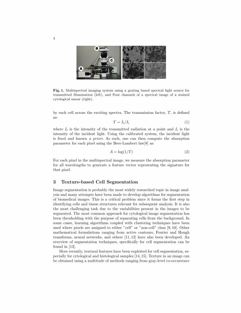
The core element of any spectral imaging system is the spectral dispersion com-ponent that separates light into its spectral components, and is coupled to atwo-dimensional (2D) optical detector such as a CCD camera, or to an arrayof photomultiplier tubes (PMT). In our system, we use a quarter-meter class,Czerny-Turner type monochromator that provides a tunable light emission spec-trum at 10nm resolution. We utilize a wavelength range from 400-700nm for thisstudy. The monochromator is connected to an Olympus BX51 upright opticalmicroscope such that the light output from the monochromator feeds in to thetransmitted light path of the microscope. This allows for the use of conventionaloptical microscopy to acquire brightfield images at desired wavelengths (trans-mitted light). An Olympus UPlanApo 40X NA 0.9 was used for imaging. ThePhotometrics SenSysTMCCD camera having 768 x 512 pixels (9x9µm) at 8-bitdigitization is used which provides for high resolution low light image acquisition.Figure 1 (left) shows the system that has been assembled. To image each sam-ple, the illumination from the monochromator was adjusted by achieving Kohlerillumination for uniform excitation of the specimen. The condenser, aperture di-aphragm, and the field stop were kept constant during measurements. Focusingwas performed at the central wavelength of 550nm to minimize the chromaticaberration at all wavelengths. The system was calibrated as per the method pro-posed in [6]. Using a stepper controlled microscope stage, multiple images wereacquired to cover the extent of the smear on the microscope slide. Resultingimages were stitched to generate a composite mosaic.
A multispectral image allows the possibility to locate, discriminate, measure,and enumerate many entities within a specimen by detecting subtle differencesamong their individual spectral signatures[7]. Clearly, different stained cells willbe spectrally distinct. However, spectral information in any cell can come fromsuch optical processes as reflection and scattering. As long as the phenomenol-ogy is based on reproducible physical reality, classification of spectrally distinctspecies can be of great utility. Figure 1 (right) shows a subset of the spectral im-age (400nm, 500nm, 600nm, and 700nm) of a Papanicolaou stained cytologicalspecimen. As seen the light absorption across various cellular constituents varyas a function of wavelength. This forms the spectral signature for each cellularentity.
To understand the spectral characteristics of biological samples, gray levelimage intensities may be used to determine the proportion of light transmitted
4
Fig. 1. Multispectral imaging system using a grating based spectral light source fortransmitted illumination (left), and Four channels of a spectral image of a stainedcytological smear (right).
by each cell across the exciting spectra. The transmission factor, T , is definedas:
T = It/Ii (1)
where It is the intensity of the transmitted radiation at a point and Ii is theintensity of the incident light. Using the calibrated system, the incident lightis fixed and known a priori. As such, one can then compute the absorptionparameter for each pixel using the Beer-Lambert law[8] as:
A = log(1/T ) (2)
For each pixel in the multispectral image, we measure the absorption parameterfor all wavelengths to generate a feature vector representing the signature forthat pixel.
3 Texture-based Cell Segmentation
Image segmentation is probably the most widely researched topic in image anal-ysis and many attempts have been made to develop algorithms for segmentationof biomedical images. This is a critical problem since it forms the first step inidentifying cells and tissue structures relevant for subsequent analysis. It is alsothe most challenging task due to the variabilities present in the images to besegmented. The most common approach for cytological image segmentation hasbeen thresholding with the purpose of separating cells from the background. Insome cases, learning algorithms coupled with clustering techniques have beenused where pixels are assigned to either ”cell” or ”non-cell” class [9, 10]. Othermathematical formulations ranging from active contours, Fourier and Houghtransforms, neural networks, and others [11, 12] have also been developed. Anoverview of segmentation techniques, specifically for cell segmentation can befound in [13].
More recently, textural features have been exploited for cell segmentation, es-pecially for cytological and histological samples [14, 15]. Texture in an image canbe obtained using a multitude of methods ranging from gray-level co-occurrence
5
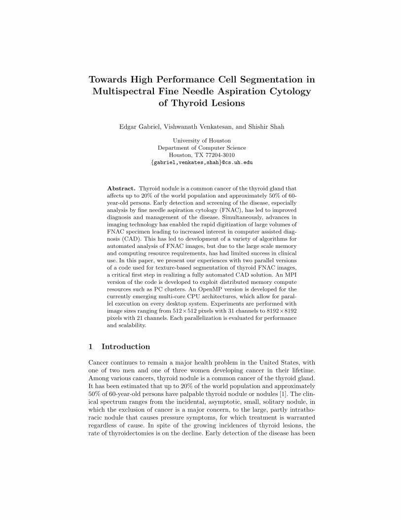
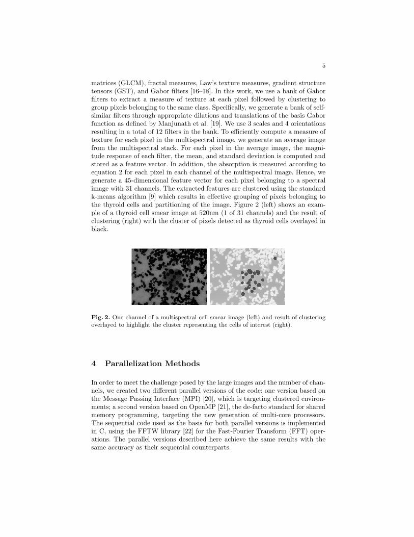
matrices (GLCM), fractal measures, Law’s texture measures, gradient structuretensors (GST), and Gabor filters [16–18]. In this work, we use a bank of Gaborfilters to extract a measure of texture at each pixel followed by clustering togroup pixels belonging to the same class. Specifically, we generate a bank of self-similar filters through appropriate dilations and translations of the basis Gaborfunction as defined by Manjunath et al. [19]. We use 3 scales and 4 orientationsresulting in a total of 12 filters in the bank. To efficiently compute a measure oftexture for each pixel in the multispectral image, we generate an average imagefrom the multispectral stack. For each pixel in the average image, the magni-tude response of each filter, the mean, and standard deviation is computed andstored as a feature vector. In addition, the absorption is measured according toequation 2 for each pixel in each channel of the multispectral image. Hence, wegenerate a 45-dimensional feature vector for each pixel belonging to a spectralimage with 31 channels. The extracted features are clustered using the standardk-means algorithm [9] which results in effective grouping of pixels belonging tothe thyroid cells and partitioning of the image. Figure 2 (left) shows an exam-ple of a thyroid cell smear image at 520nm (1 of 31 channels) and the result ofclustering (right) with the cluster of pixels detected as thyroid cells overlayed inblack.
Fig. 2. One channel of a multispectral cell smear image (left) and result of clusteringoverlayed to highlight the cluster representing the cells of interest (right).
4 Parallelization Methods
In order to meet the challenge posed by the large images and the number of chan-nels, we created two different parallel versions of the code: one version based onthe Message Passing Interface (MPI) [20], which is targeting clustered environ-ments; a second version based on OpenMP [21], the de-facto standard for sharedmemory programming, targeting the new generation of multi-core processors.The sequential code used as the basis for both parallel versions is implementedin C, using the FFTW library [22] for the Fast-Fourier Transform (FFT) oper-ations. The parallel versions described here achieve the same results with thesame accuracy as their sequential counterparts.
6
4.1 MPI parallel version
As with most MPI applications, the parallelization strategy used in this versionof the code relies on data decomposition: each process holds one part of theoverall image, and all processes execute the same code on different data items.FFTW version 2.1.5 supports MPI parallel FFT operations. However, the librarymandates a one-dimensional data decomposition, i.e. each process holds a certainnumber of rows of the overall image. The size of the image passed to the FFTWroutines have to be padded by the number of rows/columns of the Gabor filters,resulting in a slightly uneven distribution of rows for the unpadded image acrossthe processes. I/O operations are implemented using collective MPI I/O routines.
The most challenging part of the MPI version was the parallelization of thek-means clustering routine. Assuming that the number of clusters are small com-pared to the number of pixels, we replicated the information about the clusterson all processes. Each process determines locally for each pixel in its domainthe cluster whose center is closest to each pixel, and assigns that pixels to theappropriate cluster. The code also determines the number of pixels assigned toeach cluster and the weight of each cluster. Following these local calculations arethree global reduction operations, which determine the overall number of pixelsassigned to each cluster across all processes, the overall weight of each cluster,and the global error, defined as the sum of the squared distance of each pixelto the center of the closest cluster. Using these global values, each process canindependently determine the new center of each cluster for the next iteration ofthe algorithm. This iterative procedure is terminated when the error betweentwo iterations is smaller than a predefined threshold.
The code than performs a smoothing operation by comparing the clusterassigned to a particular pixel with the clusters assigned to its neighboring pixels.In the MPI version access to information owned by another process is realizedby introducing ’ghostcells’, i.e. introducing copies of information (in this casewhich cluster does a pixel belong to) owned by another process. The size of theghostcells is determined by the number of neighboring cells analyzed.
4.2 OpenMP parallel version
The OpenMP parallel version of the code also follows a data decomposition ap-proach. Each of the three main modules (convolution, clustering, smoothing)has been parallelized individually and invoked in a sequential fashion in orderto avoid nested parallelism. FFTW version 1.2.5 also supports OpenMP typethread level parallelism, which eased the parallelization of the filtering and con-volution, as it only required an additional parameter (number of threads) tobe passed to the functions. However, the initial version of this code section didnot perform well. Profiling the application with the performance analysis toolTAU [23] revealed, that OpenMP directives inserted for a loop performing thepadding of the image degraded the performance due to a large number of cachemisses between the different threads. Thus, the performance of this code sectioncould be improved significantly by not parallelizing it.
7
The k-means clustering revealed a series of other challenges. Since the codeis organized in a large while loop, the very first idea was to parallelize thisouter loop. However, OpenMP as of version 2.5 does not support parallelizationof loops with unknown number of iterations at compile time. Following a sim-ilar approach as in the MPI version, OpenMP directives were then inserted toparallelize the access to the individual pixels. Determining the number of pixelsassigned to a cluster and the weight of each cluster across all threads posed how-ever some problems, since OpenMP does not support reductions over arrays inC in the current specification. Different alternatives have been explored to over-come this limitation including introducing critical regions around the clusteringarrays, atomic updates, and locking only a particular value of the clustering data.The version leading to the best performance privatized the clustering arrays andperformed global updates by executing element-wise reduction operations.
5 Evaluation
In the following, we present the performance of the code described in the previoussection. The MPI measurements have been executed on the shark cluster at theUniversity of Houston. The cluster consists of 24 single processor, dual coreAMD Opteron nodes running at 2.2 GHz, each node equipped with 2GB ofmain memory. Nodes are connected by a 4xInfiniBand and a Gigabit Ethernetnetwork. The compute nodes have access to an NFS mounted home file system aswell as to a PVFS2 file system. The OpenMP measurements have been executedon zeola, a shared memory system consisting of eight dual core 2.6 GHz AMDOpteron processors with 64GB of main memory. We used gcc v4.2 and OpenMPI v1.2.5Tests have been executed for image sizes ranging from 512 × 512pixels with 31 spectral channels, to 1024× 1024, 2048× 2048, 4096× 4096 and8192× 8192 pixels, all using 21 spectral channels. Since the image is stored in araw, uncompressed format, the largest image analyzed was close to 1.5 GB. Eachtest presented in this paper has been repeated three times, and the minimumtime over the three runs has been used.
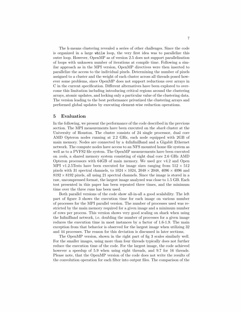
Both parallel versions of the code show all-in-all a good scalability. The leftpart of figure 3 shows the execution time for each image on various numberof processes for the MPI parallel version. The number of processes used was re-stricted by the main memory required for a given image and a minimum numberof rows per process. This version shows very good scaling on shark when usingthe InfiniBand network, i.e. doubling the number of processes for a given imagereduces the execution time in most instances by a factor of 1.6-1.9. The mainexception from that behavior is observed for the largest image when utilizing 32and 44 processes. The reason for this deviation is discussed in later sections.
The OpenMP version, shown in the right part of fig 3 scales similarly well.For the smaller images, using more than four threads typically does not furtherreduce the execution time of the code. For the largest image, the code achievedhowever a speedup of 5.9 when using eight threads, and 9.7 for 16 threads.Please note, that the OpenMP version of the code does not write the results ofthe convolution operation for each filter into output files. The comparison of the
8
execution times of the MPI and the OpenMP versions can only be performed onselected code sections.
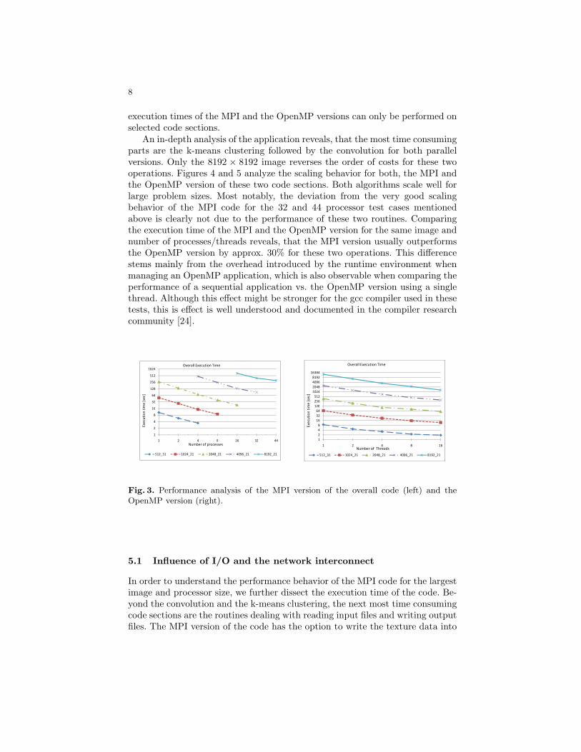
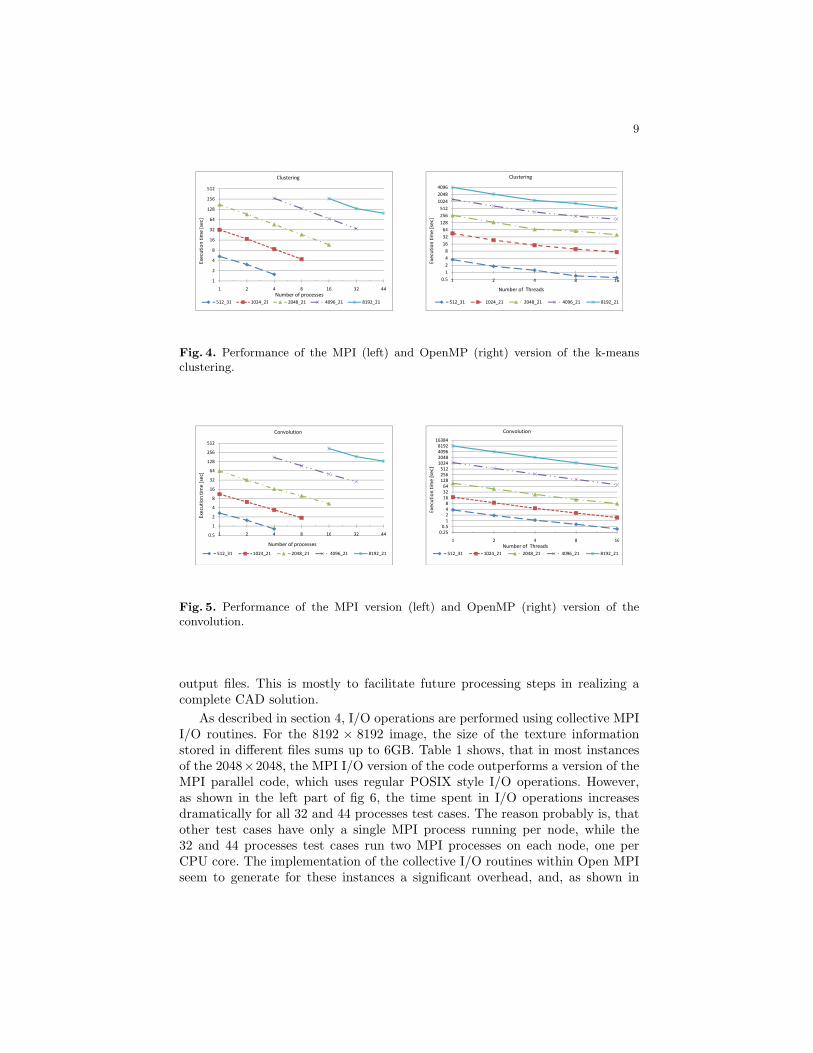
An in-depth analysis of the application reveals, that the most time consumingparts are the k-means clustering followed by the convolution for both parallelversions. Only the 8192 × 8192 image reverses the order of costs for these twooperations. Figures 4 and 5 analyze the scaling behavior for both, the MPI andthe OpenMP version of these two code sections. Both algorithms scale well forlarge problem sizes. Most notably, the deviation from the very good scalingbehavior of the MPI code for the 32 and 44 processor test cases mentionedabove is clearly not due to the performance of these two routines. Comparingthe execution time of the MPI and the OpenMP version for the same image andnumber of processes/threads reveals, that the MPI version usually outperformsthe OpenMP version by approx. 30% for these two operations. This differencestems mainly from the overhead introduced by the runtime environment whenmanaging an OpenMP application, which is also observable when comparing theperformance of a sequential application vs. the OpenMP version using a singlethread. Although this effect might be stronger for the gcc compiler used in thesetests, this is effect is well understood and documented in the compiler researchcommunity [24].
16
32
64
128
256
512
1024
tim
e [
sec]
Overall Execution Time
1
2
4
8
16
1 2 4 8 16 32 44
512_31 1024_21 2048_21 4096_21 8192_21
Number of processes
Exe
cuti
on
tim
e [
sec]
128
256
512
1024
2048
4096
8192
16384
tim
e [
sec]
Overall Execution Time
1
2
4
8
16
32
64
128
1 2 4 8 16
512_31 1024_21 2048_21 4096_21 8192_21
Number of Threads
Exe
cuti
on
tim
e [
sec]
Fig. 3. Performance analysis of the MPI version of the overall code (left) and theOpenMP version (right).
5.1 Influence of I/O and the network interconnect
In order to understand the performance behavior of the MPI code for the largestimage and processor size, we further dissect the execution time of the code. Be-yond the convolution and the k-means clustering, the next most time consumingcode sections are the routines dealing with reading input files and writing outputfiles. The MPI version of the code has the option to write the texture data into
9
32
64
128
256
512
tim
e [
sec]
Clustering
1
2
4
8
16
1 2 4 8 16 32 44
512_31 1024_21 2048_21 4096_21 8192_21
Number of processes
Exe
cuti
on
tim
e [
sec]
32
64
128
256
512
1024
2048
4096
tim
e [
sec]
Clustering
0.5
1
2
4
8
16
32
1 2 4 8 16
512_31 1024_21 2048_21 4096_21 8192_21
Number of Threads
Exe
cuti
on
tim
e [
sec]
Fig. 4. Performance of the MPI (left) and OpenMP (right) version of the k-meansclustering.
16
32
64
128
256
512
tim
e [
sec]
Convolution
0.5
1
2
4
8
16
1 2 4 8 16 32 44
512_31 1024_21 2048_21 4096_21 8192_21
Number of processes
Exe
cuti
on
tim
e [
sec]
32
64
128
256
512
1024
2048
4096
8192
16384
tim
e [
sec]
Convolution
0.25
0.5
1
2
4
8
16
32
1 2 4 8 16
512_31 1024_21 2048_21 4096_21 8192_21
Number of Threads
Exe
cuti
on
Fig. 5. Performance of the MPI version (left) and OpenMP (right) version of theconvolution.
output files. This is mostly to facilitate future processing steps in realizing acomplete CAD solution.
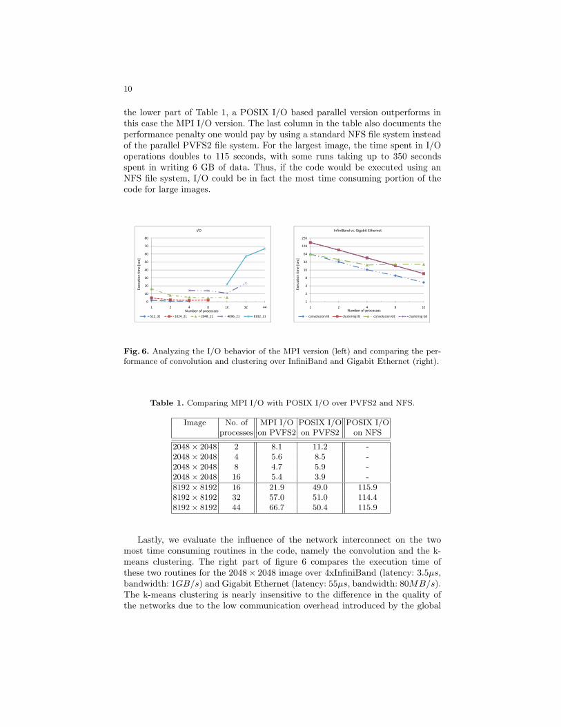
As described in section 4, I/O operations are performed using collective MPII/O routines. For the 8192 × 8192 image, the size of the texture informationstored in different files sums up to 6GB. Table 1 shows, that in most instancesof the 2048×2048, the MPI I/O version of the code outperforms a version of theMPI parallel code, which uses regular POSIX style I/O operations. However,as shown in the left part of fig 6, the time spent in I/O operations increasesdramatically for all 32 and 44 processes test cases. The reason probably is, thatother test cases have only a single MPI process running per node, while the32 and 44 processes test cases run two MPI processes on each node, one perCPU core. The implementation of the collective I/O routines within Open MPIseem to generate for these instances a significant overhead, and, as shown in
10
the lower part of Table 1, a POSIX I/O based parallel version outperforms inthis case the MPI I/O version. The last column in the table also documents theperformance penalty one would pay by using a standard NFS file system insteadof the parallel PVFS2 file system. For the largest image, the time spent in I/Ooperations doubles to 115 seconds, with some runs taking up to 350 secondsspent in writing 6 GB of data. Thus, if the code would be executed using anNFS file system, I/O could be in fact the most time consuming portion of thecode for large images.
40
50
60
70
80
tim
e [
sec]
I/O
0
10
20
30
40
1 2 4 8 16 32 44
512_31 1024_21 2048_21 4096_21 8192_21
Number of processes
Exe
cuti
on
tim
e [
sec]
16
32
64
128
256
tim
e [
sec]
InfiniBand vs. Gigabit Ethernet
1
2
4
8
16
1 2 4 8 16
convolusion IB clustering IB convolusion GE clustering GE
Number of processes
Exe
cuti
on
tim
e [
sec]
Fig. 6. Analyzing the I/O behavior of the MPI version (left) and comparing the per-formance of convolution and clustering over InfiniBand and Gigabit Ethernet (right).
Table 1. Comparing MPI I/O with POSIX I/O over PVFS2 and NFS.
Image No. of MPI I/O POSIX I/O POSIX I/Oprocesses on PVFS2 on PVFS2 on NFS
2048× 2048 2 8.1 11.2 -2048× 2048 4 5.6 8.5 -2048× 2048 8 4.7 5.9 -2048× 2048 16 5.4 3.9 -
8192× 8192 16 21.9 49.0 115.98192× 8192 32 57.0 51.0 114.48192× 8192 44 66.7 50.4 115.9
Lastly, we evaluate the influence of the network interconnect on the twomost time consuming routines in the code, namely the convolution and the k-means clustering. The right part of figure 6 compares the execution time ofthese two routines for the 2048× 2048 image over 4xInfiniBand (latency: 3.5µs,bandwidth: 1GB/s) and Gigabit Ethernet (latency: 55µs, bandwidth: 80MB/s).The k-means clustering is nearly insensitive to the difference in the quality ofthe networks due to the low communication overhead introduced by the global
11
reductions. However, the convolution, which consists of a large number of FFTs,does not scale beyond four processes on the Gigabit Ethernet network, whileit still shows a performance improvement for 16 processes for the very sametest case when using InfiniBand. This can be explained with the communicationpattern of a parallel FFT, which involves a large number of (small) messages,and therefore shows a sensitivity to the latency of a network.
6 Summary and Conclusions
This paper presented an MPI and an OpenMP parallel version of a code usedfor texture-based image segmentation applied to multispectral fine needle aspi-ration cytology of thyroid lesions. We evaluated the performance of both codeversions using a series of images of increasing sizes, with 31 and 21 spectralchannels. Both versions show good scalability all-in-all. A comparison of themost time consuming parts of the code between the two parallel versions showsa performance advantage for the MPI version. However, a detailed analysis ofthe MPI application reveals, that performance and scalability for this applica-tion strongly depends on state-of-the-art technology for the network and the filesystem. Off-the-shelf network interconnects such as Gigabit Ethernet and thepopular NFS file system clearly do not show the technical capability to keep upwith the requirements of this highly resource intensive application.
We plan to further extend this analysis by integrating additional functionalityrequired for the overall goal, such as including classifiers or comparing textureinformation of an image to a data base of known cases. Since the final goal isto analyze images of up to 20GB sizes, we also plan to make further scalabilitytests beyond the resources currently available, e.g. through a Teragrid Grant.Our experiments provide useful insights into the ability to scale and parallelizetypical image analysis algorithms. While the results presented here cannot begeneralized for all segmentation algorithms, the use of convolution and k-meansclustering is common in variety of image processing tasks, and the respectiveparallelization results of the two modules would generalize well characterized bythe available hardware.
Acknowledgments. We would like to thank Oscar Hernandez for his supportwith the OpenMP version of the code.
References
1. Hegedus, L.: The thyroid nodule. New Eng J Med. 351 (2004) 1764–17712. Gharib, H., Goellner, J.R.: Fine-needle aspiration of the thyroid: an appraisal.
Ann. Intern. Med. 118 (1993) 282–2893. Association, T.A.T.: Thyroid fine needle aspiration (FNA) and cytology. In: Con-
sensus guidelines for thyroid testing in the new millennium. Volume 6. (2003) 1–804. Shah, S., Schwartz, M.R., Mody, D.R., Scheiber-Pacht, M., Amrikachi, M.: The
role of multispectral microscopy in differentiating benign and malignant thyroid
12
nodules: A pilot study of 24 cases. In: Proceedings of the Annual Meeting of theUnited States and Canadian Academy of Pathology. (2008)
5. Feng, C., Shuzhen, C., Libo, Z.: New abnormal cervical cell detection methodof multi-spectral pap smears. Wuhan University Journal of Natural Sciences 12(2007) 476–480
6. Shah, S., Thigpen, J., Merchant, F., Castleman, K.: Photometric calibration forautomated multispectral imaging of biological samples. In Metaxas, D., Whitaker,R., Rittscher, J., Sebastian, T., eds.: Proceedings of 1st Workshop on MicroscopicImage Analysis with Applications in Biology (in conjunction with MICCAI, Copen-hagen). (2006) 27–33
7. Farkas, D., Ballou, B., Fisher, F., Fishman, D.: Microscopic and mesoscopic spec-tral bio-imaging. In: Proceedings of SPIE. Volume 2678. (1996) 200–209
8. Ornberg, R.L., Woerner, M., Edwards, D.A.: Analysis of stained objects inhistopathological sections by spectral imaging and differential absorption. J. His-tochem. Cytochem. 47 (1999) 1307–1313
9. Hartigan, J.A., Wong, M.A.: A K-means clustering algorithm. Applied Statistics28 (1979) 100–108
10. Faugeras, O.D., Pratt, W.K.: Decorrelation methods of texture feature extraction.IEEE Trans. Pattern Analysis and Machine Intelligence 2 (1980) 323–332
11. Bamford, P., Lovell, B.: Unsupervised cell nucleus segmentation with active con-tours. Signal Processing 71 (1998) 203–213
12. Kurugollu, F., Sankur, B.: Color cell image segmentation using pyramidal con-straint satisfaction neural network. In: IAPR Workshop on Machine Vision Appli-cations. (1998) 85–88
13. Ablameyko, S., Nedzved, A., Lagunovsky, D., Patsko, O., Kirillov, V.: Cell imagesegmentation: review of approaches. In: Proc. ICPR. Volume 2. (2001) 26–34
14. Ferrer-Roca, O., Gomez, J.A.P., Estevez, M.: Chromatin texture from hematoxylinstained thyroid lesions. Anal Cell Pathol. 17 (1998) 209–217
15. Yogesan, K., Jorgensen, T., Albregtsen, F., Tveter, K.J., Danielsen, H.E.: Entropy-based texture analysis of chromatin structure in advanced prostate cancer. Cy-tometry 24 (1996) 268–276
16. Laws, K.I.: Textured Image Segmentation. PhD thesis (1980)17. Tourassi, G.D., Frederick, E.D., Vittitoe, N.F., Coleman, R.E.: Fractal texture
analysis of perfusion lung scans. Comput Boimed Res 33 (2000) 161–17118. Shah, S., Aggarwal, J.K.: A Bayesian segmentation framework for textured visual
images. In: Proceedings of IEEE Conference on Computer Vision and PatternRecognition. (1997) 1014–1020
19. Manjunath, B.S., Ma, W.: Texture features for browsing and retrieval of imagedata. IEEE Transactions on Pattern Analysis and Machine Intelligence (PAMI -Special issue on Digital Libraries) 18 (1996) 837–42
20. Message Passing Interface Forum: MPI: A Message Passing Interface Standard.(1995) http://www.mpi-forum.org/.
21. Board, O.A.R.: OpenMP Application Program Interface. (2005) Version 2.5.22. Frigo, M., Johnson, S.G.: The Design and Implementation of FFTW3. Proceedings
of IEEE 93 (2005) 216–23123. Malony, A.D., Shende, S., Bell, R., Li, K., Li, L., Trebon, N.: Advances in the tau
performance system. Performance analysis and grid computing (2004) 129–14424. Huang, L., Eachempati, D., Hervey, M.W., Chapman, B.: Extending global opti-
mizations in the OpenUH compiler for OpenMP. In: Open64 Workshop at CGO2008, In Conjunction with the International Symposium on Code Generation andOptimization (CGO), Boston, MA, USA (2008)