Towards a molecular pathway for myoblast fusion in Drosophila · with high levels of Twi expression...
9
Towards a molecular pathway for myoblast fusion in Drosophila Elizabeth H. Chen 1,2 and Eric N. Olson 1 1 Department of Molecular Biology, University of Texas Southwestern Medical Center at Dallas, 6000 Harry Hines Boulevard, Dallas, TX 75390, USA 2 Department of Molecular Biology and Genetics, Johns Hopkins University School of Medicine, 725 North Wolfe Street, Baltimore, MD 21205, USA Intercellular fusion among myoblasts is required for the generation of multinucleated muscle fibers during skeletal muscle development. Recent studies in Droso- phila have shed light on the molecular mechanisms that underlie this process, and a signaling pathway that relays fusion signals from the cell membrane to the cytoskeleton has emerged. In this article, we review these recent advances and discuss how Drosophila offers a powerful model system to study myoblast fusion in vivo. Membrane fusion is one of the most fundamental processes in life. Cell–cell fusion is the most poorly understood of the three types of membrane-fusion events (intracellular fusion of organelles; virus–cell fusion and cell–cell fusion). Cell–cell fusion is crucial for the devel- opment of multicellular organisms and is required for processes as diverse as fertilization, the formation of bone and placenta, and myogenesis [1,2]. Despite the diversity of the cell types that undergo fusion, the cellular events that are involved in this process – cell recognition, adhesion and membrane merger – are common to all of these cell types, which suggests that shared molecular mechanisms might be used. Myoblast fusion, by which mononucleated myoblasts fuse to form multinucleated muscle fibers, is an essential early step during skeletal muscle differentiation. Most studies of myoblast fusion during the past three decades have been carried out in mammalian cell-culture systems in which myoblast fusion can be synchronized [3,4]. These in vitro studies have implicated several classes of protein in myoblast fusion, including cell-adhesion molecules, metalloproteases, calmodulin, protein kinases and phos- pholipases [4,5]. However, it remains to be determined whether these proteins are involved in myoblast fusion in vivo (for a review of recent advances regarding the genes that regulate mammalian myoblast fusion, see Ref. [6]). Considering the limitations of in vitro studies, an in vivo system is desirable for investigating the molecular mechanisms that underlie myoblast fusion. The fruit fly Drosophila provides an ideal paradigm for such a purpose. The somatic musculature (or larval body-wall muscle) of Drosophila is functionally equivalent to vertebrate skeletal muscle. As in vertebrates, myoblast fusion is an indispensable step during Drosophila myogenesis. Fur- thermore, the distinctive cellular changes during the fusion process, including myoblast recognition, adhesion, alignment and membrane coalescence, are morphologi- cally similar between Drosophila and vertebrates [3,4,7]. Thus, it is conceivable that the genes that are involved in myoblast fusion in Drosophila, or a portion of them at least, have evolutionarily conserved roles in vertebrate myogenesis. Despite the similarities between fly and vertebrates, the Drosophila musculature is much less complex (at most, 30 myoblasts per fiber, compared with thousands of myoblasts per fiber in vertebrates) and its development takes less time (hours, compared with days and weeks in vertebrates) [8]. These features, together with the powerful molecular and genetic tools that are available, make Drosophila a tractable system to unravel the molecular mechanisms that control myoblast fusion in vivo. In this article, we discuss the basic developmental and cell biology of myoblast fusion in Drosophila and highlight recent advances in the molecular and genetic investigations of this process. The developmental biology of myoblast fusion Primary and secondary myotubes in vertebrates Vertebrate skeletal muscles originate from the embryonic mesoderm. Skeletal muscle cells, or myoblasts, are derived from epithelial somites and are specified by the sequential actions of the paired-box transcription factor Pax-3 and the myogenic basic helix–loop–helix (bHLH) transcription factors MyoD and Myf5 [9]. The withdrawal of proliferating myoblasts from the cell cycle in response to extracellular cues is accompanied by the fusion of myoblasts to form multinucleated myotubes. The early wave of myoblast fusion produces primary myotubes that function as scaffolds for the later waves of fusion that lead to the formation of secondary and tertiary myotubes. During the final wave of embryonic myogenesis, a pool of ‘muscle satellite cells’ is formed. Some satellite cells remain quiescent for a period of time, after which they proliferate, differentiate and fuse with existing muscle fibers during exercise and injury, and in degenerative muscle diseases [10,11]. Corresponding author: Elizabeth H. Chen ([email protected]). www.sciencedirect.com 0962-8924/$ - see front matter Q 2004 Elsevier Ltd. All rights reserved. doi:10.1016/j.tcb.2004.07.008 Review TRENDS in Cell Biology Vol.14 No.8 August 2004
Transcript of Towards a molecular pathway for myoblast fusion in Drosophila · with high levels of Twi expression...
Towards a molecular pathway formyoblast fusion in DrosophilaElizabeth H. Chen1,2 and Eric N. Olson1
1Department of Molecular Biology, University of Texas Southwestern Medical Center at Dallas, 6000 Harry Hines Boulevard, Dallas,
TX 75390, USA2Department of Molecular Biology and Genetics, Johns Hopkins University School of Medicine, 725 North Wolfe Street, Baltimore,
MD 21205, USA
Intercellular fusion among myoblasts is required for the
generation of multinucleated muscle fibers during
skeletal muscle development. Recent studies in Droso-
phila have shed light on the molecular mechanisms that
underlie this process, and a signaling pathway that
relays fusion signals from the cell membrane to the
cytoskeleton has emerged. In this article, we review
these recent advances and discuss how Drosophila
offers a powerful model system to study myoblast
fusion in vivo.
Membrane fusion is one of the most fundamentalprocesses in life. Cell–cell fusion is the most poorlyunderstood of the three types of membrane-fusion events(intracellular fusion of organelles; virus–cell fusion andcell–cell fusion). Cell–cell fusion is crucial for the devel-opment of multicellular organisms and is required forprocesses as diverse as fertilization, the formation of boneand placenta, and myogenesis [1,2]. Despite the diversityof the cell types that undergo fusion, the cellular eventsthat are involved in this process – cell recognition,adhesion and membrane merger – are common to all ofthese cell types, which suggests that shared molecularmechanisms might be used.
Myoblast fusion, by which mononucleated myoblastsfuse to form multinucleated muscle fibers, is an essentialearly step during skeletal muscle differentiation. Moststudies of myoblast fusion during the past three decadeshave been carried out in mammalian cell-culture systemsin which myoblast fusion can be synchronized [3,4]. Thesein vitro studies have implicated several classes of proteinin myoblast fusion, including cell-adhesion molecules,metalloproteases, calmodulin, protein kinases and phos-pholipases [4,5]. However, it remains to be determinedwhether these proteins are involved in myoblast fusionin vivo (for a review of recent advances regarding the genesthat regulate mammalian myoblast fusion, see Ref. [6]).
Considering the limitations of in vitro studies, anin vivo system is desirable for investigating the molecularmechanisms that underlie myoblast fusion. The fruit flyDrosophila provides an ideal paradigm for such a purpose.The somatic musculature (or larval body-wall muscle) ofDrosophila is functionally equivalent to vertebrate
Corresponding author: Elizabeth H. Chen ([email protected]).
www.sciencedirect.com 0962-8924/$ - see front matter Q 2004 Elsevier Ltd. All rights reserved
skeletal muscle. As in vertebrates, myoblast fusion is anindispensable step during Drosophila myogenesis. Fur-thermore, the distinctive cellular changes during thefusion process, including myoblast recognition, adhesion,alignment and membrane coalescence, are morphologi-cally similar between Drosophila and vertebrates [3,4,7].Thus, it is conceivable that the genes that are involved inmyoblast fusion in Drosophila, or a portion of them atleast, have evolutionarily conserved roles in vertebratemyogenesis. Despite the similarities between fly andvertebrates, the Drosophila musculature is much lesscomplex (at most, 30 myoblasts per fiber, compared withthousands of myoblasts per fiber in vertebrates) and itsdevelopment takes less time (hours, compared with daysand weeks in vertebrates) [8]. These features, togetherwith the powerful molecular and genetic tools that areavailable, make Drosophila a tractable system to unravelthe molecular mechanisms that control myoblast fusionin vivo. In this article, we discuss the basic developmentaland cell biology of myoblast fusion in Drosophila andhighlight recent advances in the molecular and geneticinvestigations of this process.
The developmental biology of myoblast fusion
Primary and secondary myotubes in vertebrates
Vertebrate skeletal muscles originate from the embryonicmesoderm. Skeletal muscle cells, or myoblasts, arederived from epithelial somites and are specified by thesequential actions of the paired-box transcription factorPax-3 and the myogenic basic helix–loop–helix (bHLH)transcription factors MyoD and Myf5 [9]. The withdrawalof proliferating myoblasts from the cell cycle in response toextracellular cues is accompanied by the fusion ofmyoblasts to form multinucleated myotubes. The earlywave of myoblast fusion produces primary myotubes thatfunction as scaffolds for the later waves of fusion that leadto the formation of secondary and tertiary myotubes.During the final wave of embryonic myogenesis, a pool of‘muscle satellite cells’ is formed. Some satellite cellsremain quiescent for a period of time, after which theyproliferate, differentiate and fuse with existing musclefibers during exercise and injury, and in degenerativemuscle diseases [10,11].
Review TRENDS in Cell Biology Vol.14 No.8 August 2004
. doi:10.1016/j.tcb.2004.07.008

Review TRENDS in Cell Biology Vol.14 No.8 August 2004 453
Muscle founder cells and fusion-competent cells in
Drosophila
Based on their different behaviors during fusion, twomyoblast cell types have been revealed by studies ofDrosophila myogenesis: muscle founder cells and fusion-competent cells. Muscle founder cells function as ‘attrac-attractants’ for the surrounding fusion-competent cellsand they prefigure many properties of future musclefibers, including position, orientation, size, attachmentsites and patterns of nerve innervation [8]. Musclefounder cells are further divided into different subsets bythe expression of different ‘selector’ transcription factorssuch as Nautilus, Kruppel, S59, Apterous, Vestigial, Evenskipped and Ladybird [12,13]. The neighboring fusion-competent cells fuse with founder cells and, thereafter,adopt the same selector-gene expression profile. Initially, afounder cell fuses with one or two competent cells to formbinucleated or trinucleated muscle precursors [14].Additional rounds of fusion between these precursorsand fusion-competent cells result in the formation ofmultinucleated myotubes [14]. Thus, myoblast fusion inDrosophila occurs in two step-wise phases. Recent in vitrostudies of mammalian myoblast fusion have also revealedtwo phases of fusion: first, the fusion between a subset ofmyoblasts to form nascent myotubes and, second,additional rounds of fusion between myoblasts andnascent myotubes [6]. However, it is not clear whetherthe two-phase fusion process occurs in vivo and whether afounder-cell population exists during the first phase ofmammalian myoblast fusion.
Muscle founder cells and fusion-competent cells arespecified by a hierarchy of transcription factors duringDrosophila myogenesis [5,12,13,15] (Figure 1). Duringearly embryogenesis, the bHLH transcription factor Twist(Twi) is required to specify the embryonic mesoderm. Aftergastrulation, the mesoderm is subdivided into regions ofalternating high and low Twi expression. The domainswith high levels of Twi expression contain clusters of cellsthat express another gene, lethal of scute, that encodes abHLH transcription factor. These clusters of cells form theso-called myogenic equivalence groups. One muscleprogenitor cell from each myogenic equivalence group isthen specified by a Notch- and Delta-mediated lateralinhibition process. This single cell undergoes one round ofasymmetric cell division to generate either two musclefounder cells or one founder cell and one adult muscleprecursor. The remaining cells of the myogenic equival-ence group differentiate as fusion-competent cells. Thislater stage of myogenic differentiation also seems to becontrolled by additional transcription factors. Forexample, lame duck (lmd) [also called myoblast incompe-tent (minc) and gleeful (glee)] encodes a Gli familytranscription factor that is required for the differentiationof fusion-competent cells [16–18]. In lmd/minc-mutantembryos, there is an absence of fusion-competent cells,whereas founder cells are properly specified. Interestingly,one of the downstream target genes of lmd/minc/glee isDmef2, which encodes a MADS-box transcription factorthat is required for the differentiation of all the somatic,cardiac and visceral muscle lineages. At present, it is notclear whether other transcription factors are required for
www.sciencedirect.com
the differentiation of all muscle founder cells, as Lmd/Minc/Glee is in fusion-competent cells.
Cellular aspects of myoblast fusion
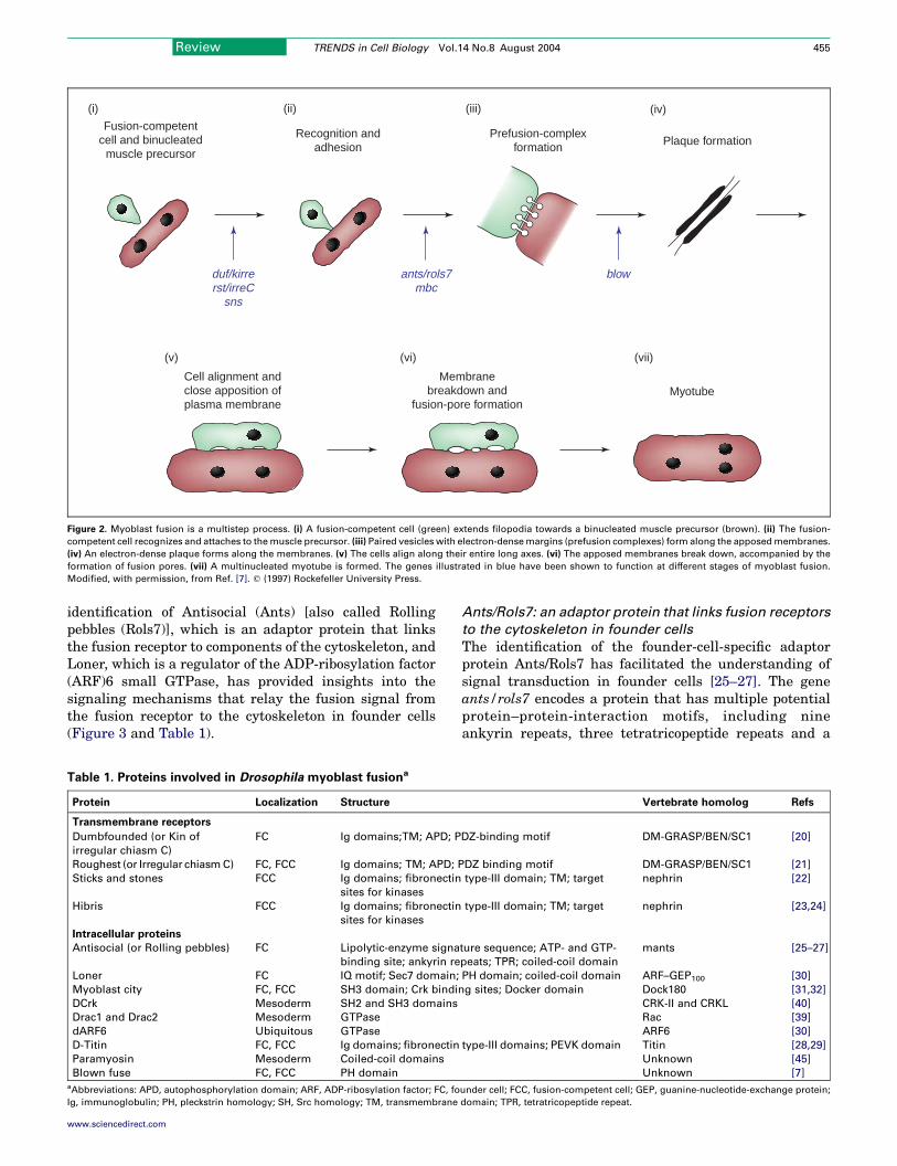
Like other types of cell–cell fusion events, myoblast fusionis a multistep process. The initial steps of cell recognitionand adhesion can be observed readily at the light-microscopy level. In Drosophila, for example, fusion-competent cells are seen to extend membrane protrusions(filopodia) towards founder cells and the tips of thefilopodia are observed to be attached to the founder-cellmembrane [19]. The electron microscopy (EM) studies ofDrosophila myoblast fusion that were carried out byDoberstein et al. are particularly informative with respectto the subcellular changes that follow the initial recog-nition and adhesion of myoblasts [7] (Figure 2). Theauthors observed paired vesicles (called prefusion com-plexes) that had electron-dense margins at the sites ofcell–cell contact. These vesicles line up with each otheracross the apposed membranes of two adhering myoblasts.The prefusion complex then resolves into electron-denseplaques between apposed myoblasts while the two cellsbecome elongated and align themselves along their longaxes. Subsequently, cytoplasmic continuity forms throughmultiple small zones (fusion pores) between the apposedplasma membranes, followed by vesiculation of theresidual membranes. Eventually, these events lead tothe formation of multinucleated myotubes.
These detailed cell biology studies of myoblast fusionhave raised many questions regarding the mechanismsthat underlie this process. How do fusion-competent cellssense the signal from founder cells for fusion? Whatmediates the attraction and adhesion between the two cellpopulations? How are fusion signals transduced to thecytoskeleton to affect its rearrangement, which is aprerequisite for cell alignment and fusion? What are thecomponents of the prefusion complex? What mediates thebreakdown of the plasma membrane and how do fusionpores form? A genetic approach to address these funda-mental questions is to isolate mutations that cause specificdefects in myoblast fusion. The identification and thefunctional characterization of the corresponding genes arebeginning to reveal a signaling cascade that transducesthe fusion signal from the cell surface to changes in thecytoskeleton during Drosophila myoblast fusion. Theserecent advances are discussed later.
The molecular biology of myoblast fusion
Myoblast recognition and adhesion: the transmembrane
receptors
The first step during myoblast fusion is the recognitionbetween muscle founder cells and fusion-competent cells.This seems to be mediated by cell-type-specific transmem-brane receptors (Figure 3 and Table 1). In founder cells,two immunoglobulin (Ig)-domain-containing cell-adhesionmolecules – Dumbfounded (Duf) [also called Kin ofIrregular chiasm C (Kirre)] and Roughest (Rst) [alsocalled Irregular chiasm C (IrreC)] – function redundantly toattract fusion-competent cells [20,21]. The deletion of bothduf and rst causes a complete block of fusion, whereas theoverexpression of either gene can attract fusion-competent

(i)
(iv)
(ii) (iii)
(v) (vi) (vii)
Muscle progenitorcells and
fusion-competent cells
Muscle foundercells and
adult precusor
Firstphase of
fusion
Secondphase of
fusion
Myogenicequivalence
groups
Myogenicfield
HighTwist
expression
Lethal ofscute
expression
Notch and Deltasignaling
Lmd/Minc/Glee
Notch andNumb
localization
Fusionreceptors
Ants/Rols7
P2
P1A
C
B
AP APAP
Figure 1. Overview of Drosophilamuscle development. (i) A stage-11 embryo showing alternating levels of Twist (Twi) expression. Cells that express high levels of Twi (dark
green) acquire a myogenic fate (ii). (iii) Clusters of cells (myogenic equivalence groups; blue) within the myogenic field express Lethal of scute. (iv) Amuscle progenitor cell
(P1 or P2) is singled out from each equivalence group by a lateral inhibition process that is mediated by Notch and Delta signaling. The remaining cells in the equivalence
group are specified to become fusion-competent cells by a process that requires the transcription factor Lmd/Minc/Glee. (v) Each progenitor cell undergoes asymmetric cell
division to produce either two founder cells (A and B) or one founder cell (C) and one adult muscle precursor (AP). Each founder cell expresses a specificmuscle-identity gene
that is also known as a selector gene. (vi) Founder cells attract surrounding fusion-competent cells to fuse with them. This is mediated by specific ‘fusion receptors’ and
downstream signaling components. The first phase of fusion yields binucleated or trinucleatedmuscle precursors. A fusion-competent cell expresses the same selector gene
after fusing with a founder cell. (vii) Muscle precursors continue to attract additional fusion-competent cells in the second phase of fusion, which requires the function of
Antisocial (Ants) [also called Rolling pebbles (Rols7)] and leads to the formation of multinucleated myotubes. Modified, with permission, from Ref. [13].
Review TRENDS in Cell Biology Vol.14 No.8 August 2004454
myoblasts to the ectopic sites of expression. In fusion-competent cells, Sticks and stones (Sns), which is also anIg-domain-containing cell-adhesion molecule, is requiredfor fusion because the loss of sns results in a lack of fusion[22]. Another fusion-competent cell-specific cell-adhesionmolecule is the paralog of Sns Hibris (Hbs) [23,24].Hbs is not essential for myoblast fusion but it seems toinhibit Sns function. The overexpression of hbs blocksmyoblast fusion, whereas the loss of hbs causes onlyminor fusion defects.
The careful examination of the cellular behavior offusion-competent cells in duf rst double-mutant or snssingle-mutant embryos revealed that these myoblasts doextend filopodia, albeit with random orientations [5,20].The failure of these filopodia to attach to founder cells isconsistent with the hypothesis that Duf, Rst and Sns arerequired for the initial recognition and adhesion betweenthe two cell populations. In addition, there is evidence thatDuf and Sns might interact directly with each other tomediate cell adhesion because cultured Drosophila cells(S2 cells) that express Duf can aggregate with Sns-expressing cells [15,23].
It remains to be determined how fusion-competent cellsare attracted to the founder cells initially. One possibility
www.sciencedirect.com
is that fusion-competent cells randomly extend filopodia tolocate the founder cells. Alternatively, fusion-competentcells might sense a kind of concentration gradient from thefounder cells and extend filopodia specifically in thatdirection. It is also unclear how the sites of fusion areselected. For example, the transmembrane protein Dufmight be localized to predetermined sites in founder cellsby intrinsic cues. Alternatively, extrinsic contacts made bythe filopodia from fusion-competent cells could have a rolein determining Duf localization in founder cells. Detailedstudies of receptor localization during the fusion processwill provide clues to the answers to these questions.
Signal transduction: from membrane to cytoskeleton
Two events occur after a fusion-competent cell makescontact with a founder cell. First, the fusion-competentcell moves towards the founder cell. Second, the fusion-competent cell aligns with the founder cell, thus juxtapos-ing the two cell membranes. These cellular events requirechanges in the actin cytoskeleton. Thus, rearrangement ofthe actin cytoskeleton in both founder cells and fusion-competent cells is a prerequisite for myoblast fusion. Howis the fusion signal transduced to the cytoskeleton to effectthe rearrangement of the cytoskeleton? The recent
Fusion-competentcell and binucleated
muscle precursor
Recognition andadhesion
Prefusion-complexformation Plaque formation
Cell alignment andclose apposition ofplasma membrane
Membranebreakdown and
fusion-pore formationMyotube
(i) (iv)(ii) (iii)
(v) (vi) (vii)
blowants/rols7mbc
duf/kirrerst/irreC
sns
Figure 2. Myoblast fusion is a multistep process. (i) A fusion-competent cell (green) extends filopodia towards a binucleated muscle precursor (brown). (ii) The fusion-
competent cell recognizes and attaches to themuscle precursor. (iii) Paired vesicles with electron-densemargins (prefusion complexes) form along the apposedmembranes.
(iv) An electron-dense plaque forms along the membranes. (v) The cells align along their entire long axes. (vi) The apposed membranes break down, accompanied by the
formation of fusion pores. (vii) A multinucleated myotube is formed. The genes illustrated in blue have been shown to function at different stages of myoblast fusion.
Modified, with permission, from Ref. [7]. q (1997) Rockefeller University Press.
Review TRENDS in Cell Biology Vol.14 No.8 August 2004 455
identification of Antisocial (Ants) [also called Rollingpebbles (Rols7)], which is an adaptor protein that linksthe fusion receptor to components of the cytoskeleton, andLoner, which is a regulator of the ADP-ribosylation factor(ARF)6 small GTPase, has provided insights into thesignaling mechanisms that relay the fusion signal fromthe fusion receptor to the cytoskeleton in founder cells(Figure 3 and Table 1).
Table 1. Proteins involved in Drosophila myoblast fusiona
Protein Localization Structure
Transmembrane receptors
Dumbfounded (or Kin of
irregular chiasm C)
FC Ig domains;TM; APD; P
Roughest (or Irregular chiasmC) FC, FCC Ig domains; TM; APD; P
Sticks and stones FCC Ig domains; fibronectin
sites for kinases
Hibris FCC Ig domains; fibronectin
sites for kinases
Intracellular proteins
Antisocial (or Rolling pebbles) FC Lipolytic-enzyme signa
binding site; ankyrin re
Loner FC IQ motif; Sec7 domain;
Myoblast city FC, FCC SH3 domain; Crk bindi
DCrk Mesoderm SH2 and SH3 domains
Drac1 and Drac2 Mesoderm GTPase
dARF6 Ubiquitous GTPase
D-Titin FC, FCC Ig domains; fibronectin
Paramyosin Mesoderm Coiled-coil domains
Blown fuse FC, FCC PH domainaAbbreviations: APD, autophosphorylation domain; ARF, ADP-ribosylation factor; FC, fo
Ig, immunoglobulin; PH, pleckstrin homology; SH, Src homology; TM, transmembrane
www.sciencedirect.com
Ants/Rols7: an adaptor protein that links fusion receptors
to the cytoskeleton in founder cells
The identification of the founder-cell-specific adaptorprotein Ants/Rols7 has facilitated the understanding ofsignal transduction in founder cells [25–27]. The geneants/rols7 encodes a protein that has multiple potentialprotein–protein-interaction motifs, including nineankyrin repeats, three tetratricopeptide repeats and a
Vertebrate homolog Refs
DZ-binding motif DM-GRASP/BEN/SC1 [20]
DZ binding motif DM-GRASP/BEN/SC1 [21]
type-III domain; TM; target nephrin [22]
type-III domain; TM; target nephrin [23,24]
ture sequence; ATP- and GTP-
peats; TPR; coiled-coil domain
mants [25–27]
PH domain; coiled-coil domain ARF–GEP100 [30]
ng sites; Docker domain Dock180 [31,32]
CRK-II and CRKL [40]
Rac [39]
ARF6 [30]
type-III domains; PEVK domain Titin [28,29]
Unknown [45]
Unknown [7]
under cell; FCC, fusion-competent cell; GEP, guanine-nucleotide-exchange protein;
domain; TPR, tetratricopeptide repeat.
Fusion
Hbs
Loner
Cytoskeletonrearrangement
Cytoskeletonrearrangement
Mbc
Ants/Rols7
Mbc
Drac
Drac*
Drac
Selectorgenes
Dmef2
Nucleus
Sns
Duf/Kirre
Rst/IrreC
Fusion-competentcell
Musclefoundercell
Rst/IrreC
Sns
Duf/Kirre
Mbc
Ants/Rols7
Drac*
ParamyosinD-Titin
ParamyosinD-Titin
Lmd/Minc/Glee
Dmef2
Nucleus
Loner
dARF6
dARF6*
Blow
Blow
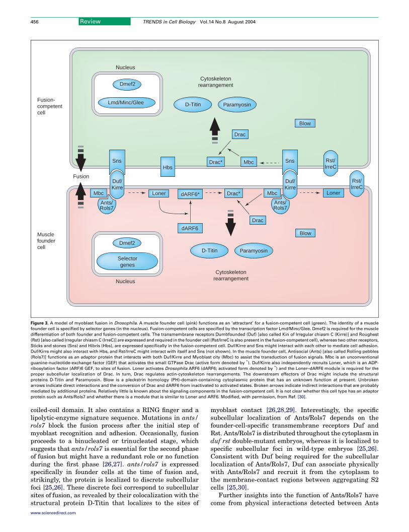
Figure 3. A model of myoblast fusion in Drosophila. A muscle founder cell (pink) functions as an ‘attractant’ for a fusion-competent cell (green). The identity of a muscle
founder cell is specified by selector genes (in the nucleus). Fusion-competent cells are specified by the transcription factor Lmd/Minc/Glee. Dmef2 is required for the muscle
differentiation of both founder and fusion-competent cells. The transmembrane receptors Dumbfounded (Duf) [also called Kin of Irregular chiasm C (Kirre)] and Roughest
(Rst) [also called Irregular chiasmC (IrreC)] are expressed and required in the founder cell (Rst/IrreC is also present in the fusion-competent cell), whereas two other receptors,
Sticks and stones (Sns) and Hibris (Hbs), are expressed specifically in the fusion-competent cell. Duf/Kirre and Sns might interact with each other to mediate cell adhesion.
Duf/Kirre might also interact with Hbs, and Rst/IrreC might interact with itself and Sns (not shown). In the muscle founder cell, Antisocial (Ants) [also called Rolling pebbles
(Rols7)] functions as an adaptor protein that interacts with both Duf/Kirre and Myoblast city (Mbc) to assist the transduction of fusion signals. Mbc is an unconventional
guanine-nucleotide-exchange factor (GEF) that activates the small GTPase Drac (active form denoted by *). Duf/Kirre also independently recruits Loner, which is an ADP-
ribosylation factor (ARF)6 GEF, to sites of fusion. Loner activates Drosophila ARF6 (dARF6; activated form denoted by *) and the Loner–dARF6 module is required for the
proper subcellular localization of Drac. In turn, Drac regulates actin-cytoskeleton rearrangements. The downstream effectors of Drac might include the structural
proteins D-Titin and Paramyosin. Blow is a pleckstrin homology (PH)-domain-containing cytoplasmic protein that has an unknown function at present. Unbroken
arrows indicate direct interactions and the conversion of Drac and dARF6 from inactivated to activated states. Broken arrows indicate indirect interactions that are probably
mediated by additional proteins. Relatively little is known about the signaling components in the fusion-competent cell. It is not clear whether this cell type has an adaptor
protein such as Ants/Rols7 and whether there is a module that is similar to Loner and ARF6. Modified, with permission, from Ref. [30].
Review TRENDS in Cell Biology Vol.14 No.8 August 2004456
coiled-coil domain. It also contains a RING finger and alipolytic-enzyme signature sequence. Mutations in ants/rols7 block the fusion process after the initial step ofmyoblast recognition and adhesion. Occasionally, fusionproceeds to a binucleated or trinucleated stage, whichsuggests that ants/rols7 is essential for the second phaseof fusion but might have a redundant role or no functionduring the first phase [26,27]. ants/rols7 is expressedspecifically in founder cells at the time of fusion and,strikingly, the protein is localized to discrete subcellularfoci [25,26]. These discrete foci correspond to subcellularsites of fusion, as revealed by their colocalization with thestructural protein D-Titin that localizes to the sites of
www.sciencedirect.com
myoblast contact [26,28,29]. Interestingly, the specificsubcellular localization of Ants/Rols7 depends on thefounder-cell-specific transmembrane receptors Duf andRst. Ants/Rols7 is distributed throughout the cytoplasm induf rst double-mutant embryos, whereas it is localized tospecific subcellular foci in wild-type embryos [25,26].Consistent with Duf being required for the subcellularlocalization of Ants/Rols7, Duf can associate physicallywith Ants/Rols7 and recruit it from the cytoplasm tothe membrane-contact regions between aggregating S2cells [25,30].
Further insights into the function of Ants/Rols7 havecome from physical interactions detected between Ants

Review TRENDS in Cell Biology Vol.14 No.8 August 2004 457
and Myoblast city (Mbc) [25], which is another essentialcomponent of the myoblast-fusion process [31,32]. Droso-phila Mbc belongs to the CDM family of proteins that alsoincludes Caenorhabditis elegans Ced-5, and mammalianDock180 and Dock2 [33]. CDM proteins in C. elegans andmammalian cells are involved in an evolutionarilyconserved signaling pathway (Ced-2, Ced-12, Ced-5 andCed-10 in C. elegans and CrkII, ELMO, Dock180 and Racin mammals) that modulates the small GTPase Rac, whichis a crucial regulator of cytoskeletal dynamics [34–36].This pathway mediates cytoskeletal rearrangementsduring the phagocytosis of apoptotic cells and during cellmovements [37]. It has been suggested that Dock180forms an unconventional two-part guanine-nucleotide-exchange factor (GEF) for Rac with the ELMO protein[38]. It is conceivable that Drosophila Mbc also regulatesthe activity of the small GTPase Drac during myoblastfusion, although the signaling mechanisms of Mbc areunderstood less well. Consistent with this hypothesis,Drac1 and Drac2 are required for myoblast fusion inDrosophila [39]. The physical interactions between Antsand Mbc and between Ants and Duf suggest that Antscould function as an intermediary protein that relays thefusion signal from the cell-surface receptor Duf to thecytoskeleton through the regulation of Mbc and Dracactivity [25]. It remains to be determined whether Ants/Rols7 regulates the GEF activity or the subcellularlocalization of Mbc. Furthermore, it will be interesting toinvestigate whether the homologs of CrkII and ELMO areinvolved in myoblast fusion in Drosophila [40].
Loner: a guanine-nucleotide-exchange factor that
regulates the ARF6 small GTPase duringmyoblast fusion
The recent characterization of the fusion-defective mutantloner has provided a new element to the understanding ofthe signaling cascade that regulates cytoskeletalrearrangement during Drosophila myoblast fusion(Figure 3 and Table 1). The loner gene encodes a putativeGEF that contains a Sec7 domain and an adjacentpleckstrin homology (PH) domain [30]. The Sec7 domainis found in GEFs for the ARF family of small GTPases [41],whereas PH domains have been implicated in binding tophospholipids in the plasma membrane [42]. Rescueexperiments have demonstrated that both of thesedomains are essential for the function of Loner in vivo[30]. Loner is expressed in founder cells, in which it islocalized in discrete subcellular foci (as is the case forAnts/Rols7). However, Loner is colocalized only partiallywith Ants, which suggests that only a portion of the Lonerprotein is localized to the sites of fusion. The transmem-brane receptor Duf is required for the proper subcellularlocalization of Loner in founder cells, which is also the casefor Ants. Furthermore, Duf can recruit Loner from thecytoplasm to the membrane-contact regions betweenaggregating S2 cells. However, the subcellular localizationof Loner is not dependent on that of Ants and vice versa.Thus, it seems that Ants and Loner are recruitedindependently to sites of fusion by the transmembranereceptor Duf [30].
How does Loner mediate myoblast fusion? The presenceof a Sec7 domain suggests that Loner might function as a
www.sciencedirect.com
GEF for the ARF family of small GTPases. In vitro, thepurified Sec7 domain of Loner displays specific GEFactivity towards Drosophila ARF6 (dARF6), whichsuggests that dARF6 might be a physiological target ofLoner [30]. Consistent with this hypothesis, the over-expression of a dominant negative form of dARF6 infounder cells blocks myoblast fusion [30]. Together, theseobservations reveal a novel Loner–dARF6-mediated sig-naling module that has an essential role in myoblastfusion. However, loss-of-function mutations of dARF6 will,ultimately, be required to strengthen this conclusion.
The relationships between the small GTPases dARF6 and
Drac1
The identification of dARF6 and Drac1 as essentialcomponents of myoblast fusion raises important questionsregarding the relationships between these two smallGTPases during the fusion process. Studies in culturedmammalian cells have implicated ARF6 in membranetrafficking and actin-cytoskeleton rearrangements, whichare two processes that have potential relevance tomyoblast fusion [43]. In particular, there is evidence thatARF6 regulates cytoskeletal rearrangement by controllingthe subcellular localization of Rac1 [44]. In Drosophilamuscle founder cells, the Loner–dARF6 module seems tocontrol the subcellular localization of Drac1. In loner-mutant embryos, Drac1 is distributed throughout thecytoplasm rather than being concentrated to the sites offusion, as is seen in wild-type embryos [30]. Thus, similarto what occurs in mammalian cells, the Loner–dARF6module could signal to the actin cytoskeleton through theregulation of Drac1 (Figure 3 and Table 1). However,considering the widespread roles for ARF6 in diverseprocesses, such as its regulation of the enzymes that areresponsible for lipid modification and its involvement inregulated secretion events, it remains to be determinedwhether these other functions of ARF6 also contribute tomyoblast fusion.
The downstream effectors of Drac
Considering the pivotal role of Drac in Drosophilamyoblast fusion, it would be interesting to determine thedownstream effectors of Drac during actin-cytoskeletonrearrangement. The characterization of the structuralproteins D-Titin and Paramyosin in muscle developmentmight help to do this. D-Titin and Paramyosin wereidentified initially as sarcomeric proteins. However, recentstudies have revealed unexpected functions for themduring myoblast fusion [28,45]. Both proteins are presentat myoblast-contact sites during fusion and are important,although not essential, for the fusion process [28,29,45]. Inaddition, the proper localization of D-Titin is dependent onAnts/Rols7 (the adaptor protein that is associated with theputative Drac1 GEF Mbc) [27]. These studies, togetherwith the interactions between D-Titin and the actincytoskeleton and between Paramyosin and the actincytoskeleton, have led to suggestions that the twostructural proteins have a role in the organization of theactin-cytoskeleton elements that are required for fusion[28,29,45] and that they might be among the manydownstream effectors of Drac (Figure 3 and Table 1).

Review TRENDS in Cell Biology Vol.14 No.8 August 2004458
Questions outstanding
Studies of Drosophila myoblast fusion are beginning toreveal a signaling pathway in muscle founder cells thattransduces signals from fusion receptors into changes inthe cytoskeleton. Meanwhile, these studies raise newquestions for future investigations, as highlighted next.
Identification of components of a ‘fusion complex’
The presence of multiple potential protein–protein-inter-action motifs in Ants/Rols7, combined with the obser-vation that Duf recruits both Ants/Rols7 and Loner to sitesof fusion, suggests that Duf and Ants/Rols7 might functionwithin a scaffold to anchor multiple proteins to the sites offusion, where a multiprotein ‘fusion complex’ mediates thecellular changes that accompany myoblast fusion. Theidentification of additional components of this fusioncomplex, through both genetic and biochemicalapproaches, is likely to provide important insights intomyoblast fusion. It will also be important to examinethe subcellular localization of the fusion complex atthe EM level to determine how the fusion complexrelates to the distinct ultrastructural entities thathave been observed during myoblast fusion, such aspaired vesicles and plaques.
How do juxtaposed membranes fuse with each other?
Cytoskeletal rearrangement is a prerequisite for themembrane merger of two apposing cells. It is requiredfor the two membranes to align effectively so that theirlipid bilayers are closely juxtaposed for fusion to proceed.Little is known about the actual fusion process. Forexample, it is unclear how the two membranes aredestabilized, how fusion pores form and which moleculesare involved in these events. During virus–cell fusion, ahydrophobic peptide in the fusogenic viral glycoproteinmediates the juxtaposition and fusion of two membranes[2,46], although no fusogen-like sequences have beenidentified in the known proteins that are involved inmyoblast fusion. However, the founder-cell adaptor pro-tein Ants/Rols7 contains a lipolytic-enzyme signaturesequence that is often present in lipases that are involvedin the modification of the lipid bilayer [47]. An isoform ofAnts/Rols7 that lacks the N-terminal region that includesthe lipolytic-enzyme signature sequence can no longerrescue the myoblast-fusion phenotype in ants/rols7-mutant embryos [26]. It will be interesting to determinethe specific contribution of this lipolytic-enzyme signaturesequence to membrane dynamics during myoblast fusion.
Signal transduction in fusion-competent cells
Little is known about how fusion signals are transduced infusion-competent cells. The cytoplasmic region of thetransmembrane receptor Sns, which is specific to fusion-competent cells, contains proline-rich sequences, potentialphosphorylation sites for various kinases and stretches ofevolutionarily conserved sequences that have unknownphysiological functions [22]. Mbc, which regulates thecytoskeleton in founder cells, is also present in fusion-competent cells and might provide a similar function byregulating Drac and cytoskeletal rearrangements duringfusion [32]. It will be interesting to determine whether
www.sciencedirect.com
Drosophila homologs of CrkII and ELMO, in addition toMbc and Drac, are required in fusion-competent cells. Itwill also be interesting to find out whether there is anadaptor protein in fusion-competent cells that is equival-ent to Ants/Rols7 in founder cells and that links the Snsreceptor to the cytoskeleton. Ongoing genetic screens inDrosophila might identify these and other potentialcomponents of fusion-competent cells and shed light onthe signal-transduction pathway that is employed in thiscell type.
Drosophila myoblast fusion: relevance to mammalian
myogenesis
Given the conserved cellular events that are involved inDrosophila myoblast fusion and mammalian myogenesis,it is conceivable that the genes that are required forDrosophila myoblast fusion might have conserved roles inmammalian myogenesis. Curiously, the mammalianhomologs of the Ig-domain-containing transmembranereceptors Duf, Rst, Sns and Hbs are not expressed in thedeveloping mesoderm. In fact, the mouse homolog of dufand rst (SC-1) is expressed predominantly in the nervoussystem [48]. In addition, the mouse homolog of Sns andHbs (nephrin) has been implicated in kidney development[49]. Thus, it seems that the initial recognition andadhesion between myoblasts during vertebrate myoblastfusion might use a different set of transmembranereceptors. This might reflect the differences in themolecular events that lead to the specification of myo-blasts in flies and vertebrates. However, preliminarystudies suggest that the intracellular components of themyoblast-fusion network might be conserved betweenDrosophila and vertebrates after a fusion signal hastriggered the recognition and adhesion of myoblasts. Oneof the mouse orthologs of ants, mants1, is expressed in avariety of mesodermal tissues, including somites, limbbuds and body-wall muscles [25]. The transient expressionof mants1 coincides with muscle differentiation, whichsuggests that it might have a role in muscle differentiationand myoblast fusion. The Loner–ARF6 module might alsohave a role in mammalian myogenesis because a dominantnegative form of ARF6 blocks MyoD-induced myotubeformation in a cell-culture model [30]. Future experimentsinvolving knockout or transgenic mice should addressdefinitively whether these fusion genes have conservedroles in mammalian myoblast fusion.
Myoblast fusion and muscle disease
Most studies of human muscle disease have focused ongenes such as dystrophin that affect the sarcolemma [47].Because embryonic myogenesis requires myoblast fusionto occur, complete loss-of-function mutations in fusiongenes are likely to cause embryonic lethality. However,hypomorphic alleles of these genes might result incongenital or postnatal muscle diseases. In fact, bothcentronuclear myopathy and myotonic dystrophy arecharacterized by minute myofibers, which suggests thatmyoblast fusion might be defective in these musclediseases [50,51]. In addition to its role during myogenesis,myoblast fusion is also required for muscle growth andrepair during exercise and muscle injury. For example,

Review TRENDS in Cell Biology Vol.14 No.8 August 2004 459
satellite cells can proliferate and fuse with existingmyotubes during exercise or they can fuse with injuredmuscle fibers to repair lesions. It is conceivable thatsimilar molecular mechanisms might be involved in adultsatellite-cell fusion and in myoblast fusion during embry-ogenesis. Therefore, certain types of adult myopathiesmight be associated with defects in the genes that arerequired for myoblast fusion during myogenesis. Theelucidation of the molecular and cellular mechanisms ofmyoblast fusion might provide insights into this intri-guing cell biology phenomenon and lead to an under-standing of and, ultimately, therapeutic interventions inhuman muscle diseases.
Concluding remarks
Recent studies in the fruit fly Drosophila have providednovel insights into the molecular mechanisms that controlmyoblast fusion during myogenesis. However, it is likelythat only the tip of the iceberg has been uncovered so far.Future studies that combine genetics with biochemical,cell biology and genomic approaches will, undoubtedly,provide this area of investigation with more excitingdiscoveries. In addition, the combination of insights fromstudies in both Drosophila and vertebrates will facilitateour understanding of this fascinating biological process.
Acknowledgements
E.H.C. thanks Duojia Pan for insightful discussions and comments on themanuscript. We thank Alisha Tizenor for help with the graphics. E.H.C.was supported by a postdoctoral fellowship from the Helen Hay WhitneyFoundation. E.N.O. was supported by the National Institutes of Healthand the D.W. Reynolds Center for Clinical Cardiovascular Research.
References
1 Blumenthal, R. et al. (2003) Membrane fusion. Chem. Rev. 103, 53–692 Hernandez, L.D. et al. (1996) Virus–cell and cell–cell fusion. Annu.
Rev. Cell Dev. Biol. 12, 627–6613 Wakelam, M.J. (1985) The fusion of myoblasts. Biochem. J. 228, 1–124 Knudsen, K.A. (1992) Fusion of myoblasts. In Membrane Fusion
(Wilschut, J. and Hoekstra, D. eds), pp. 601–626, Marcel Decker5 Abmayr, S.M. et al. (2003) Cell and molecular biology of myoblast
fusion. Int. Rev. Cytol. 225, 33–896 Horsley, V. and Pavlath, G.K. (2004) Forming a multinucleated cell:
molecules that regulate myoblast fusion. Cells Tissues Organs 176,67–78
7 Doberstein, S.K. et al. (1997) Genetic analysis of myoblast fusion:blown fuse is required for progression beyond the prefusion complex.J. Cell Biol. 136, 1249–1261
8 Bate, M. (1993) The mesoderm and its derivatives. In The Develop-ment of Drosophila melanogaster (Bate, M. and Martinez Ariazs, A.eds), pp. 1013–1090, Cold Spring Harbor Laboratory Press
9 Buckingham, M. (2001) Skeletal muscle formation in vertebrates.Curr. Opin. Genet. Dev. 11, 440–448
10 Bischoff, R. (1994) The satellite cell and muscle regeneration. InMyogenesis (Engel, A.G. and Franszini-Armstrong, C. eds), pp. 97–118,McGraw-Hill
11 Seale, P. et al. (2001) The potential of muscle stem cells. Dev. Cell 1,333–342
12 Frasch, M. (1999) Controls in patterning and diversification of somaticmuscles during Drosophila embryogenesis. Curr. Opin. Genet. Dev. 9,522–529
13 Baylies, M.K. et al. (1998) Myogenesis: a view from Drosophila. Cell93, 921–927
14 Bate, M. (1990) The embryonic development of larval muscles inDrosophila. Development 110, 791–804
15 Dworak, H.A. and Sink, H. (2002) Myoblast fusion in Drosophila.Bioessays 24, 591–601
www.sciencedirect.com
16 Duan, H. et al. (2001) Drosophila Lame duck, a novel member ofthe Gli superfamily, acts as a key regulator of myogenesis bycontrolling fusion-competent myoblast development. Development128, 4489–4500
17 Furlong, E.E. et al. (2001) Patterns of gene expression duringDrosophila mesoderm development. Science 293, 1629–1633
18 Ruiz-Gomez, M. et al. (2002) myoblasts incompetent encodes a zincfinger transcription factor required to specify fusion-competentmyoblasts in Drosophila. Development 129, 133–141
19 Paululat, A. et al. (1999) Essential genes for myoblast fusion inDrosophila embryogenesis. Mech. Dev. 83, 17–26
20 Ruiz-Gomez, M. et al. (2000) Drosophila dumbfounded: a myoblastattractant essential for fusion. Cell 102, 189–198
21 Strunkelnberg, M. et al. (2001) rst and its paralogue kirre actredundantly during embryonic muscle development in Drosophila.Development 128, 4229–4239
22 Bour, B.A. et al. (2000) Drosophila SNS, a member of the immunoglo-bulin superfamily that is essential for myoblast fusion. Genes Dev. 14,1498–1511
23 Dworak, H.A. et al. (2001) Characterization of Drosophila hibris, agene related to human nephrin. Development 128, 4265–4276
24 Artero, R.D. et al. (2001) The immunoglobulin-like protein Hibrisfunctions as a dose-dependent regulator of myoblast fusion and isdifferentially controlled by Ras and Notch signaling. Development 128,4251–4264
25 Chen, E.H. and Olson, E.N. (2001) Antisocial, an intracellular adaptorprotein, is required for myoblast fusion in Drosophila. Dev. Cell 1,705–715
26 Menon, S.D. and Chia, W. (2001) Drosophila rolling pebbles: amultidomain protein required for myoblast fusion that recruitsD-Titin in response to the myoblast attractant Dumbfounded. Dev.Cell 1, 691–703
27 Rau, A. et al. (2001) rolling pebbles (rols) is required in Drosophilamuscle precursors for recruitment of myoblasts for fusion. Develop-ment 128, 5061–5073
28 Zhang, Y. et al. (2000) Drosophila D-titin is required for myoblastfusion and skeletal muscle striation. J. Cell Sci. 113, 3103–3115
29 Machado, C. and Andrew, D.J. (2000) D-Titin: a giant protein withdual roles in chromosomes and muscles. J. Cell Biol. 151, 639–652
30 Chen, E.H. et al. (2003) Control of myoblast fusion by a guaninenucleotide exchange factor, Loner, and its effector ARF6. Cell 114,751–762
31 Rushton, E. et al. (1995) Mutations in a novel gene, myoblast city,provide evidence in support of the founder cell hypothesis forDrosophila muscle development. Development 121, 1979–1988
32 Erickson, M.R. et al. (1997) Drosophila myoblast city encodes aconserved protein that is essential for myoblast fusion, dorsal closure,and cytoskeletal organization. J. Cell Biol. 138, 589–603
33 Nolan, K.M. et al. (1998) Myoblast city, the Drosophila homolog ofDOCK180/CED-5, is required in a Rac signaling pathway utilized formultiple developmental processes. Genes Dev. 12, 3337–3342
34 Hall, A. (1998) Rho GTPases and the actin cytoskeleton. Science 279,509–514
35 Van Aelst, L. and D’Souza-Schorey, C. (1997) Rho GTPases andsignaling networks. Genes Dev. 11, 2295–2322
36 Ridley, A.J. (2001) Rho family proteins: coordinating cell responses.Trends Cell Biol. 11, 471–477
37 Grimsley, C. and Ravichandran, K.S. (2003) Cues for apoptotic cellengulfment: eat-me, don’t eat-me and come-get-me signals. TrendsCell Biol. 13, 648–656
38 Brugnera, E. et al. (2002) Unconventional Rac–GEF activity is mediatedthrough the Dock180–ELMO complex. Nat. Cell Biol. 4, 574–582
39 Hakeda-Suzuki, S. et al. (2002) Rac function and regulation duringDrosophila development. Nature 416, 438–442
40 Galletta, B.J. et al. (1999) Identification of a Drosophila homologue tovertebrate Crk by interaction with MBC. Gene 228, 243–252
41 Donaldson, J.G. and Jackson, C.L. (2000) Regulators and effectors ofthe ARF GTPases. Curr. Opin. Cell Biol. 12, 475–482
42 Chardin, P. et al. (1996) A human exchange factor for ARF containsSec7- and pleckstrin-homology domains. Nature 384, 481–484
43 Chavrier, P. and Goud, B. (1999) The role of ARF and Rab GTPases inmembrane transport. Curr. Opin. Cell Biol. 11, 466–475

Review TRENDS in Cell Biology Vol.14 No.8 August 2004460
44 Radhakrishna, H. et al. (1999) ARF6 requirement for Rac rufflingsuggests a role for membrane trafficking in cortical actin rearrange-ments. J. Cell Sci. 112, 855–866
45 Liu, H. et al. (2003) Drosophila paramyosin is important for myoblastfusion and essential for myofibril formation. J. Cell Biol. 160, 899–908
46 Jahn, R. et al. (2003) Membrane fusion. Cell 112, 519–53347 Upton, C. and Buckley, J.T. (1995) A new family of lipolytic enzymes?
Trends Biochem. Sci. 20, 178–17948 Fournier-Thibault, C. et al. (1999) BEN/SC1/DM-GRASP expression
during neuromuscular development: a cell adhesion molecule regu-lated by innervation. J. Neurosci. 19, 1382–1392
Have you contributed to a
Did you know that you are entitle
A 30% discount is available to ALL Elsevier book and journal contr
from us.
To take advantage of your discount:
1. Choose your book(s) from www.elsevier.com or www.books.elsev
2. Place your order
Americas:
TEL: +1 800 782 4927 for US customers
TEL: +1 800 460 3110 for Canada, South & Central America cu
FAX: +1 314 453 4898
E-MAIL: [email protected]
All other countries:
TEL: +44 1865 474 010
FAX: +44 1865 474 011
E-MAIL: [email protected]
You’ll need to provide the name of the Elsevier book or journa
orders within the US, Canada, and the UK.
If you are faxing your order, please enclose a copy of this pag
3. Make your payment
This discount is only available on prepaid orders. Please note
Elsevier Health Sciences products.
www.books.el
www.sciencedirect.com
49 Lenkkeri, U. et al. (1999) Structure of the gene for congenitalnephrotic syndrome of the finnish type (NPHS1) and characterizationof mutations. Am. J. Hum. Genet. 64, 51–61
50 Farkas-Bargeton, E. et al. (1988) Immaturity of muscle fibers in thecongenital form of myotonic dystrophy: its consequences and itsorigin. J. Neurol. Sci. 83, 145–159
51 Wockel, L. et al. (1998) Abundant minute myotubes in a patient who
later developed centronuclear myopathy. Acta Neuropathol. (Berl.) 95,547–551
n Elsevier publication?
d to a 30% discount on books?
ibutors when ordering books or stand-alone CD-ROMs directly
ier.com
stomers
l to which you have contributed. Shipping is FREE on pre-paid
e.
that this offer does not apply to multi-volume reference works or
sevier.com