
Toward a Cognitive Neural Prosthesis Using Focused Ultrasound · dry primate biscuits, as well as...
16
ORIGINAL RESEARCH published: 15 November 2017 doi: 10.3389/fnins.2017.00607 Frontiers in Neuroscience | www.frontiersin.org 1 November 2017 | Volume 11 | Article 607 Edited by: Mikhail Lebedev, Duke University, United States Reviewed by: Robert A. Gaunt, University of Pittsburgh, United States Didem Gokcay, Middle East Technical University, Turkey *Correspondence: Vincent P. Ferrera [email protected] Specialty section: This article was submitted to Neuroprosthetics, a section of the journal Frontiers in Neuroscience Received: 07 April 2017 Accepted: 17 October 2017 Published: 15 November 2017 Citation: Downs ME, Teichert T, Buch A, Karakatsani ME, Sierra C, Chen S, Konofagou EE and Ferrera VP (2017) Toward a Cognitive Neural Prosthesis Using Focused Ultrasound. Front. Neurosci. 11:607. doi: 10.3389/fnins.2017.00607 Toward a Cognitive Neural Prosthesis Using Focused Ultrasound Matthew E. Downs 1 , Tobias Teichert 2 , Amanda Buch 1 , Maria E. Karakatsani 1 , Carlos Sierra 1 , Shangshang Chen 2 , Elisa E. Konofagou 1, 3 and Vincent P. Ferrera 2, 4 * 1 Department of Biomedical Engineering, Columbia University, New York, NY, United States, 2 Department of Neuroscience, Columbia University, New York, NY, United States, 3 Department of Radiology, Columbia University, New York, NY, United States, 4 Department of Psychiatry, Columbia University, New York, NY, United States Non-invasive brain stimulation using focused ultrasound has many potential applications as a research and clinical tool, including its incorporation as either an extracorporeal or implantable neural prosthetic. To this end, we investigated the effect of focused ultrasound (FUS) combined with systemically administered microbubbles on visual-motor decision-making behavior in monkeys. We applied FUS to the putamen in one hemisphere to open the blood-brain barrier (BBB), and then tested behavioral performance 3–4h later. On days when the monkeys were treated with FUS, their decisions were faster and more accurate than days without sonication. The performance improvement suggested both a shift in the decision criterion and an enhancement of the use of sensory evidence in the decision process. FUS also interacted with the effect of a low dose of haloperidol. The findings indicate that a two-minute application of FUS can have a sustained impact on performance of complex cognitive tasks, and may increase the efficacy of psychoactive medications. The results lend further support to the idea that the dorsal striatum plays an integral role in evidence- and reward-based decision-making, and provide motivation for incorporating FUS into cognitive neural prosthetic devices. Keywords: blood-brain barrier, focused ultrasound stimulation, decision making, NHP model, drug delivery INTRODUCTION Brain stimulation is an essential tool for investigating causal brain-behavior relationships, mapping brain circuits, and treating neurological disorders. Established stimulation methods are either invasive (electrical or chemical stimulation, optogenetics), or have limited penetrability (TMS) or localizability (TDCS) (Miller, 1965; Dubuisson and Dennis, 1977; Kobayashi and Pascual- Leone, 2003; Nitsche et al., 2003; Calvo and Coimbra, 2006; Borchers et al., 2012). Focused ultrasound (FUS) is emerging as a non-invasive technology for neuromodulation that is capable of penetrating the skull and meninges to deliver mechanical energy to deep brain structures. FUS with systemically administered microbubbles has been shown to open the blood-brain barrier (BBB) in various animal models, and may also directly modulate neural activity (Hynynen et al., 2001; McDannold et al., 2005, 2012; Tung et al., 2011; Marquet et al., 2014; Chu et al., 2015; Downs et al., 2015a). The basic mechanisms underlying these effects are beginning to become clear (Sassaroli and Vykhodtseva, 2016). The non-invasive nature of FUS makes it an attractive option for human neuroprosthetics. Here, we first present results detailing cognitive improvement following application of FUS with microbubbles to the dorsal striatum, then discuss prospects for refining the delivery of FUS.
Transcript of Toward a Cognitive Neural Prosthesis Using Focused Ultrasound · dry primate biscuits, as well as...
ORIGINAL RESEARCHpublished: 15 November 2017doi: 10.3389/fnins.2017.00607
Frontiers in Neuroscience | www.frontiersin.org 1 November 2017 | Volume 11 | Article 607
Edited by:
Mikhail Lebedev,
Duke University, United States
Reviewed by:
Robert A. Gaunt,
University of Pittsburgh, United States
Didem Gokcay,
Middle East Technical University,
Turkey
*Correspondence:
Vincent P. Ferrera
Specialty section:
This article was submitted to
Neuroprosthetics,
a section of the journal
Frontiers in Neuroscience
Received: 07 April 2017
Accepted: 17 October 2017
Published: 15 November 2017
Citation:
Downs ME, Teichert T, Buch A,
Karakatsani ME, Sierra C, Chen S,
Konofagou EE and Ferrera VP (2017)
Toward a Cognitive Neural Prosthesis
Using Focused Ultrasound.
Front. Neurosci. 11:607.
doi: 10.3389/fnins.2017.00607
Toward a Cognitive Neural ProsthesisUsing Focused Ultrasound
Matthew E. Downs 1, Tobias Teichert 2, Amanda Buch 1, Maria E. Karakatsani 1,
Carlos Sierra 1, Shangshang Chen 2, Elisa E. Konofagou 1, 3 and Vincent P. Ferrera 2, 4*
1Department of Biomedical Engineering, Columbia University, New York, NY, United States, 2Department of Neuroscience,
Columbia University, New York, NY, United States, 3Department of Radiology, Columbia University, New York, NY,
United States, 4Department of Psychiatry, Columbia University, New York, NY, United States
Non-invasive brain stimulation using focused ultrasound has many potential applications
as a research and clinical tool, including its incorporation as either an extracorporeal
or implantable neural prosthetic. To this end, we investigated the effect of focused
ultrasound (FUS) combined with systemically administeredmicrobubbles on visual-motor
decision-making behavior in monkeys. We applied FUS to the putamen in one
hemisphere to open the blood-brain barrier (BBB), and then tested behavioral
performance 3–4 h later. On days when the monkeys were treated with FUS, their
decisions were faster and more accurate than days without sonication. The performance
improvement suggested both a shift in the decision criterion and an enhancement of the
use of sensory evidence in the decision process. FUS also interacted with the effect of a
low dose of haloperidol. The findings indicate that a two-minute application of FUS can
have a sustained impact on performance of complex cognitive tasks, and may increase
the efficacy of psychoactive medications. The results lend further support to the idea that
the dorsal striatum plays an integral role in evidence- and reward-based decision-making,
and provide motivation for incorporating FUS into cognitive neural prosthetic devices.
Keywords: blood-brain barrier, focused ultrasound stimulation, decision making, NHP model, drug delivery
INTRODUCTION
Brain stimulation is an essential tool for investigating causal brain-behavior relationships, mappingbrain circuits, and treating neurological disorders. Established stimulation methods are eitherinvasive (electrical or chemical stimulation, optogenetics), or have limited penetrability (TMS)or localizability (TDCS) (Miller, 1965; Dubuisson and Dennis, 1977; Kobayashi and Pascual-Leone, 2003; Nitsche et al., 2003; Calvo and Coimbra, 2006; Borchers et al., 2012). Focusedultrasound (FUS) is emerging as a non-invasive technology for neuromodulation that is capableof penetrating the skull and meninges to deliver mechanical energy to deep brain structures. FUSwith systemically administered microbubbles has been shown to open the blood-brain barrier(BBB) in various animal models, and may also directly modulate neural activity (Hynynen et al.,2001; McDannold et al., 2005, 2012; Tung et al., 2011; Marquet et al., 2014; Chu et al., 2015;Downs et al., 2015a). The basic mechanisms underlying these effects are beginning to become clear(Sassaroli andVykhodtseva, 2016). The non-invasive nature of FUSmakes it an attractive option forhuman neuroprosthetics. Here, we first present results detailing cognitive improvement followingapplication of FUS with microbubbles to the dorsal striatum, then discuss prospects for refining thedelivery of FUS.
Downs et al. Cognitive Effects of Focused Ultrasound
Recent studies in monkeys and humans have providedevidence that FUS alone can modify perception and behavior(Bystritsky et al., 2011; Deffieux et al., 2013; Hameroff et al.,2013; Legon et al., 2014; Lee et al., 2016). Deffieux et al. foundthat FUS can increase the latency of antisaccades in monkeys.Tactile discrimination was enhanced during FUS stimulationof the somatosensory cortex in human subjects, while overallmood improvedwhen the frontal-temporal cortex was stimulatedwith FUS (Hameroff et al., 2013; Legon et al., 2014). Lee et al.(2016) were able to evoke visual phosphenes and concomitantEEG activity. Further investigation using different species, braintargets, behavioral tasks, and FUS methodologies is warrantedto establish the effectiveness and range of applications for thisapproach. As a step toward determining the efficacy of FUS,we tested whether FUS with microbubbles had an effect on theperformance of a complex cognitive task 3–4 h after treatment.
FUS with microbubbles can increase the permeability ofthe BBB, which remains open for up to 48 h after treatment(Marquet et al., 2014), raising the possibility that cognitive orbehavioral changes might occur during this time period. Whilethe exact mechanisms of the BBB opening are unknown, acousticcavitation of the microbubbles in the focal area of the FUShas been determined as a major factor (Abbott, 2013). Thisacoustic cavitation causes the microbubbles to oscillate exertingmechanical forces on the surrounding vascular walls (Arvanitiset al., 2013). It is postulated that these mechanical forces stretchthe gap junctions between the endothelial cells and ‘open’ theBBB. Microbubbles are required for opening of the BBB viaFUS safely as they reduce the required acoustic intensity neededto open the BBB (Meairs and Alonso, 2007). While FUS hasbeen demonstrated to open the BBB without microbubbles, theacoustic intensities needed are near or at the range of tissueablation (Bakay et al., 1956; Vykhodtseva et al., 1995). Thus, forsafe BBB opening, a combination of microbubbles and FUS isrequired. While it has been shown that the opening of the BBB issafe (Marquet et al., 2014; Downs et al., 2015b), it is unknown ifthe opening of the BBB affects the neural functions of the openedregions of the brain.
In the current study, FUS was applied to the putamen, a partof the basal ganglia involved in cognition, reward, andmovementcontrol.We sought to devise a behavioral paradigm that would besensitive to changes in perception, motor performance, decision-making and motivation due to the opening of the BBB, andto the administration of threshold doses of D2-antagonists. Wetherefore trained monkeys to perform a perceptual decision-making task using a touchpanel display. The task involvedthe detection of coherent visual motion (Lappin and Bell,1976; Hanks and Shadlen, 2006) and also included a rewardmanipulation to test motivation. Electrophysiological studiespoint to a critical role of the striatum (caudate and putamen) insimilar tasks (Ding and Gold, 2013).
The objective of this study was to determine the effects of FUSwith microbubbles on decision making and motor performanceof NHP engaged in a coherent motion detection task. Rhesusmonkeys were treated with FUS and intravenous microbubblesto open the BBB and then tested behaviorally 3–4 h later todetermine if the opening modulated behavioral responses. The
current study also investigated the interaction of FUS with alow dose of the D2 dopamine antagonist haloperidol, as thistechnique could be used to non-invasively facilitate drug effectswhile minimizing side effects, or to deliver drugs that cannotcross the intact BBB. Additionally, the effects of haloperidol+ FUS were compared to both FUS and haloperidol alone todetermine if the BBB opening can enhance neurological effectsof drug delivery to the brain. The results indicate that FUSwith microbubbles can be used alone or in combination withpsychoactive drugs to modify performance on complex tasks.
METHODS
All procedures with monkeys were approved by the InstitutionalAnimal Care and Use Committees (IACUC) of ColumbiaUniversity and the New York State Psychiatric Institute (NYSPI).Two adult male Macaca mulatta (N, O) were used in thesedated sonication experiments (9 and 20 years old, 5.5 and9.5 kg). These monkeys were surgically naïve and underwent noprocedures during the course of these experiments other thanthose described below. Two adult male Macaca fascicularis (A,Z)were used in the awake sonication experiments (14, 18 yearsold, 5.3 and 5.6 kg.) These monkeys underwent surgery for theimplantation of a head post for head fixation during sonication.All monkeys were provided daily rations of vitamin enricheddry primate biscuits, as well as enrichment toys and allowedaccess to play modules. Monkeys were trained using operantconditioning to perform a visual-motor decision-making taskusing a touchpanel display. Prior to data collection, monkeyswere trained for several months until they reached asymptoticperformance. On behavioral testing days, monkeys performedthe task for fluid reward until satiated. After behavioral testing,Monkeys were given a fruit treat (banana, apple, or orange). Ondays when behavioral testing was not conducted, monkeys weregiven a liter of water.
Focused Ultrasound and Drug DeliveryFor sedates sonications, on selected days, monkeys were treatedwith FUS with microbubble 3–4 h prior to behavioral testing. Forthe FUS procedures, subjects were sedated with ketamine (10mg/kg) and atropine (0.04 mg/kg) for initial placement of anIV line into the saphenous vein and insertion of the intubationtube. Once the intubation tube had been secured, the NHPwere placed under general anesthesia (isoflurane 1–2%) andplaced into a stereotaxic positioning frame to ensure accuratetargeting. Microbubbles (4–5 um, in-house prepared as describedin Feshitan et al., 2009) were administered intravenously at theonset of the FUS application. FUS was applied transcraniallythrough a single-element transducer (H-107, Sonic Concepts,WA, USA). The transducer was driven by a pair of functiongenerators (Keysight 33220A, CA, USA) to generate 10msduration pulses of a 500 kHz sine wave with a duty cycle of 2Hzand total duration of 120 s. This waveform was passed through apower amp and impedance matching network (Sonic Concepts,WA, USA) to drive the transducer at 400 kPa (Feshitan et al.,2009).
Frontiers in Neuroscience | www.frontiersin.org 2 November 2017 | Volume 11 | Article 607
Downs et al. Cognitive Effects of Focused Ultrasound
For awake sonications, the same procedure was used exceptthat monkeys were not given isoflurane. They received alight dose of ketamine (5 mg/kg) for implantation of an IVcatheter in the saphenous vein for delivery of microbubbles.Animals performed the behavioral task for an hour beforebeginning the FUS procedure. The FUS was applied at theonset of MB injection, which was through surgical tubingattached to the catheter extending from the work booth. Afterthe FUS procedure finished NHP were allowed to work untilsatiated.
The putamen region of the basal ganglia was targeted for allexperiments. Targeting of the FUS was optimized with an in-house developed targeting pipeline to minimize loss of energythrough skull and maximize the area of the putamen withinthe focal area resulting in ∼30% of the putamen targeted.Throughout the procedure, vital signs were continuouslymonitored (heart rate, SPO2, mean arterial pressure, respiratoryrate and end tidal CO2). After the FUS procedure there was a3 to 4 h recovery period allowing the monkeys to fully recoverfrom anesthesia as prior studies conducted within our labdemonstrated that time to have minimal effects of anesthesia(both ketamine and isoflurane) (Downs et al., 2015a). Afterthe recovery period they showed normal alertness, appetite andmobility as evidenced by their ability to walk, climb and consumefood.
Haloperidol, a D2 dopamine receptor antagonist (R&DSystems, Inc., Minneapolis, MN), was used during some sessionsto augment neuromodulation. Haloperidol powder was dissolvedin saline and titrated to the concentration of 0.01 mg/kg. Onselected days, before the task began, monkeys were administeredeither saline or haloperidol (0.01 mg/kg) intramuscularly. Theinjection was given 5min prior to the start of behavioraltesting. The threshold dose of haloperidol and administrationtime was determined as the maximum dose at a timepointthat had a minimal effect on behavioral results when the BBBwas intact. The timing of events during the FUS procedure,recovery, drug injection and behavioral testing is shown inFigure 1A.
MRI AnalysisOne day after the FUS procedure, BBB opening and safetywas verified with contrast enhanced T1-weighted as well asT2-weighted MRI and susceptibility-weighted imaging scansrespectively. All MRI scans (3T, Philips Medical Systems, MA,USA) were acquired 36 h after the FUS procedure. T2-weighted(TR = 10ms, TE = 27ms, flip angle = 90◦, spatial resolution =
400 × 400µm2, slice thickness = 2mm with no interslice gap)and susceptibility-weighted image (TR= 19ms, TE= 27ms, flipangle = 15◦, spatial resolution = 400 × 400µm2, slice thickness= 1mm with no interslice gap) scans were used to verify thesafety of the procedure. Contrast enhanced T1-weighted (TR= 19ms, TE = 27ms, flip angle = 15◦, spatial resolution =
400 × 400µm2, slice thickness = 1mm with no interslicegap) scans were acquired 30min after IV administration of 0.2ml/kg gadodiamide (Omniscan R©, 573.66 DA, GE, Healthcare,Princeton, NY, USA). Gadodiamide was used as the contrastagent as it does not cross the intact BBB. All acquired scans
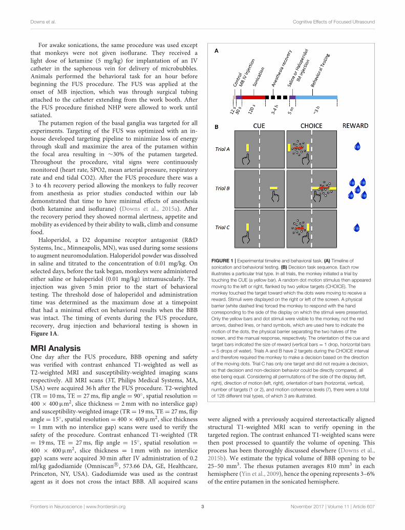
FIGURE 1 | Experimental timeline and behavioral task. (A) Timeline of
sonication and behavioral testing. (B) Decision task sequence. Each row
illustrates a particular trial type. In all trials, the monkey initiated a trial by
touching the CUE (a yellow bar). A random dot motion stimulus then appeared
moving to the left or right, flanked by two yellow targets (CHOICE). The
monkey touched the target toward which the dots were moving to receive a
reward. Stimuli were displayed on the right or left of the screen. A physical
barrier (white dashed line) forced the monkey to respond with the hand
corresponding to the side of the display on which the stimuli were presented.
Only the yellow bars and dot stimuli were visible to the monkey, not the red
arrows, dashed lines, or hand symbols, which are used here to indicate the
motion of the dots, the physical barrier separating the two halves of the
screen, and the manual response, respectively. The orientation of the cue and
target bars indicated the size of reward (vertical bars = 1 drop, horizontal bars
= 5 drops of water). Trials A and B have 2 targets during the CHOICE interval
and therefore required the monkey to make a decision based on the direction
of the moving dots. Trial C has only one target and did not require a decision,
so that decision and non-decision behavior could be directly compared, all
else being equal. Considering all permutations of the side of the display (left,
right), direction of motion (left, right), orientation of bars (horizontal, vertical),
number of targets (1 or 2), and motion coherence levels (7), there were a total
of 128 different trial types, of which 3 are illustrated.
were aligned with a previously acquired stereotactically alignedstructural T1-weighted MRI scan to verify opening in thetargeted region. The contrast enhanced T1-weighted scans werethen post processed to quantify the volume of opening. Thisprocess has been thoroughly discussed elsewhere (Downs et al.,2015b). We estimate the typical volume of BBB opening to be25–50 mm3. The rhesus putamen averages 810 mm3 in eachhemisphere (Yin et al., 2009), hence the opening represents 3–6%of the entire putamen in the sonicated hemisphere.
Frontiers in Neuroscience | www.frontiersin.org 3 November 2017 | Volume 11 | Article 607
Downs et al. Cognitive Effects of Focused Ultrasound
Behavioral TestingMonkeys sat in a custom-made polycarbonate primate chair thatallowed them to reach out to visual stimuli presented on a 20-inchLCD touchscreen monitor (NEC 2010x with 3M SC4 resistivetouchscreen) placed directly in front of the chair. The resolutionof the LCD was 1,280 horizontal × 1,024 vertical pixels (55.4 ×
45.4 deg. visual angle at 14 in viewing distance) with a refreshrate of 60Hz. The touchscreen device had a resolution of 1,024× 1,024 pixels and a sampling rate of 60Hz. The primate chairincorporated a polycarbonate midline divider so that stimulipresented on the right side of the display could only be reachedby the right hand, and likewise for the left side. Behavior wasreinforced with drops of fluid delivered by a juice tube mountedon the chair.
The behavioral task was presented as discrete trials lastingroughly 5 s each. Different behavioral tasks were used forsedate and awake sonications. For sedate sonications, monkeysperformed a motion detection with unequal rewards. For awakesonication, the monkeys performed a simple reaching task withunequal rewards. The tasks were the same except that the latterdid not have a motion detection component. For the motiondetection task, each trial began with a visual cue stimuluspresented on the left or right side of the monitor (Figure 1B,“CUE”). The cue was a vertically or horizontally oriented yellowbar (1× 3 deg, 43.8 cd/m2 luminance). The monkey touched thecue with the corresponding hand to initiate the trial. After a shortdelay, the cue was replaced by a random dot motion stimulus(Figure 1B, “CHOICE.”) The motion stimulus consisted of 100dots (each dot was 0.17 deg square, luminance 71.6 cd/m2)moving within a circular aperture of 10 deg diameter. Some of thedots moved in random directions while others moved coherentlyin a single direction (dot lifetime was 2 frames). The coherentdirection, either leftward or rightward, varied from trial to trial.The strength of the motion stimulus (aka motion coherence)varied from 0 to 0.7 in steps of 0.1. A particular coherencelevel was selected randomly for each trial and the coherencewas constant for the duration of the trial. The motion stimuluswas flanked on either side by two target stimuli that appearedsimultaneously with the motion stimulus. The target stimuli wereyellow bars that had the same orientation, size and luminanceas the cue. The direction of the coherent dots indicated whichtarget would be rewarded. The monkey was reinforced withdrops of water for touching the appropriate target (Figure 1B,“REWARD”). There was no punishment for incorrect responsesor failures to respond. No signal instructed the monkeys when torespond; rather, they were allowed to touch at any time after themotion stimulus and targets appeared.
To test motivation, the experiment included two rewardsizes, one of which was chosen randomly on each trial: smalloffered reward (1 drop of water, 0.03ml) and large offeredreward (5 drops, 0.15ml). Offered reward level on each trialwas signaled by the orientation of the cue and target stimuli.Horizontal orientation indicated large reward, vertical indicatedsmall reward.
One seventh of the trials were controls that were identical tothe other trials except that the target for the incorrect responsewas not presented. On these trials, the monkey could ignore the
motion stimulus and simply touch the correct target to receivea reward. The purpose of these trials was to assess movementaccuracy and response time when no decision was required.
This experimental behavioral paradigm controls for all thevariables of interest: display side (left or right), cue/targetorientation (vertical or horizontal, corresponding to small andlarge reward), motion direction (left or right), motion coherence(0.0 to 0.7), and number of targets (1 or 2). This resulted in abalanced design comprising 128 conditions per block of trials.All conditions were randomly interleaved within each behavioralsession.
For the reaching task with unequal rewards (awakesonications), everything was the same as the motion detectiontask except that no motion stimulus was presented (see Downset al., 2015a for further details). No decision was required in thistask as there was only one target to reach for at any given time.
StatisticsQuantitative analyses were performed using Matlab 8.3 with theStatistics 9.0 toolbox (Mathworks, Natick MA). The statisticalequations follow the Mathworks format. Response times wereanalyzed with multivariate ANOVA and generalized linear modelregression (using the glmfit function in the Matlab Statisticstoolbox). The GLMmodel equation was:
RT = β0 + β1x1 + . . . + βnxn
where the xi are the explanatory variables described below andthe βi are the regression coefficients. RT distributions werenormalized by log transformation.
Performance accuracy or outcome (correct, incorrect) wasanalyzed withmultivariate ANOVA and logistic regression (usingthe mnrfit function in the Matlab Statistics toolbox). The logisticregression equation was:
ln[p/(1− p)] = β0 + β1x1 + . . . + βnxn
Where p is the probability of a correct outcome, the xi are theexplanatory variables, and the βi are the regression coefficients.The explanatory variables used in all analyses were: subject (N,O), motion coherence (0 to 0.7, 8 levels), offered reward (1 or 5drops), presence of sonication, sonicated hemisphere (ispsilateralor contralateral to responding hand), and drug treatment (salineor haloperidol).
RESULTS
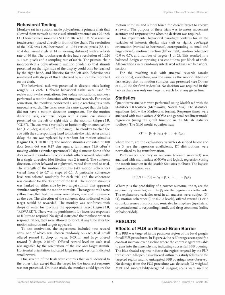
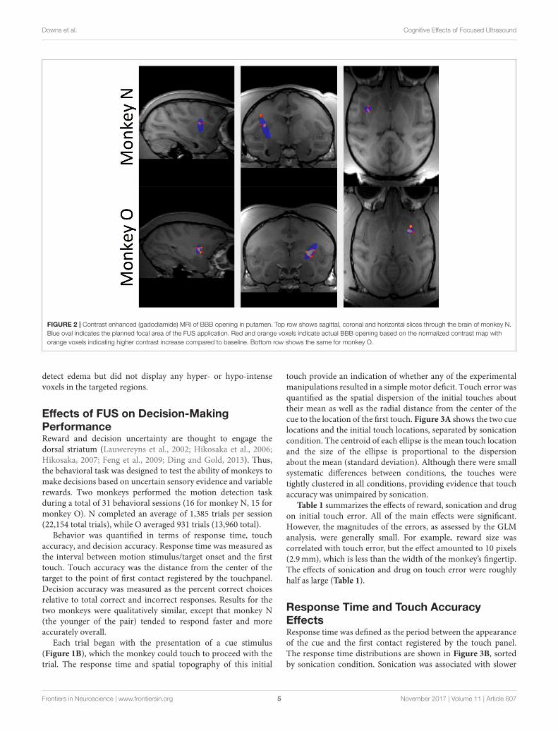
Effects of FUS on Blood-Brain BarrierThe BBB was targeted in the putamen region of the basal gangliafor all FUS procedures. In Figure 2, the red/orange areas specify acontrast increase over baseline where the contrast agent was ableto pass into the parenchyma, indicating successful BBB opening.The blue shaded regions indicate the region targeted by the FUStransducer. All openings achieved within this study fell inside thetargeted region and no untargeted BBB openings were observed.No damage from the FUS procedure was detected; T2-weightedMRI and susceptibility-weighted imaging scans were used to
Frontiers in Neuroscience | www.frontiersin.org 4 November 2017 | Volume 11 | Article 607
Downs et al. Cognitive Effects of Focused Ultrasound
FIGURE 2 | Contrast enhanced (gadodiamide) MRI of BBB opening in putamen. Top row shows sagittal, coronal and horizontal slices through the brain of monkey N.
Blue oval indicates the planned focal area of the FUS application. Red and orange voxels indicate actual BBB opening based on the normalized contrast map with
orange voxels indicating higher contrast increase compared to baseline. Bottom row shows the same for monkey O.
detect edema but did not display any hyper- or hypo-intensevoxels in the targeted regions.
Effects of FUS on Decision-MakingPerformanceReward and decision uncertainty are thought to engage thedorsal striatum (Lauwereyns et al., 2002; Hikosaka et al., 2006;Hikosaka, 2007; Feng et al., 2009; Ding and Gold, 2013). Thus,the behavioral task was designed to test the ability of monkeys tomake decisions based on uncertain sensory evidence and variablerewards. Two monkeys performed the motion detection taskduring a total of 31 behavioral sessions (16 for monkey N, 15 formonkey O). N completed an average of 1,385 trials per session(22,154 total trials), while O averaged 931 trials (13,960 total).
Behavior was quantified in terms of response time, touchaccuracy, and decision accuracy. Response time was measured asthe interval between motion stimulus/target onset and the firsttouch. Touch accuracy was the distance from the center of thetarget to the point of first contact registered by the touchpanel.Decision accuracy was measured as the percent correct choicesrelative to total correct and incorrect responses. Results for thetwo monkeys were qualitatively similar, except that monkey N(the younger of the pair) tended to respond faster and moreaccurately overall.
Each trial began with the presentation of a cue stimulus(Figure 1B), which the monkey could touch to proceed with thetrial. The response time and spatial topography of this initial
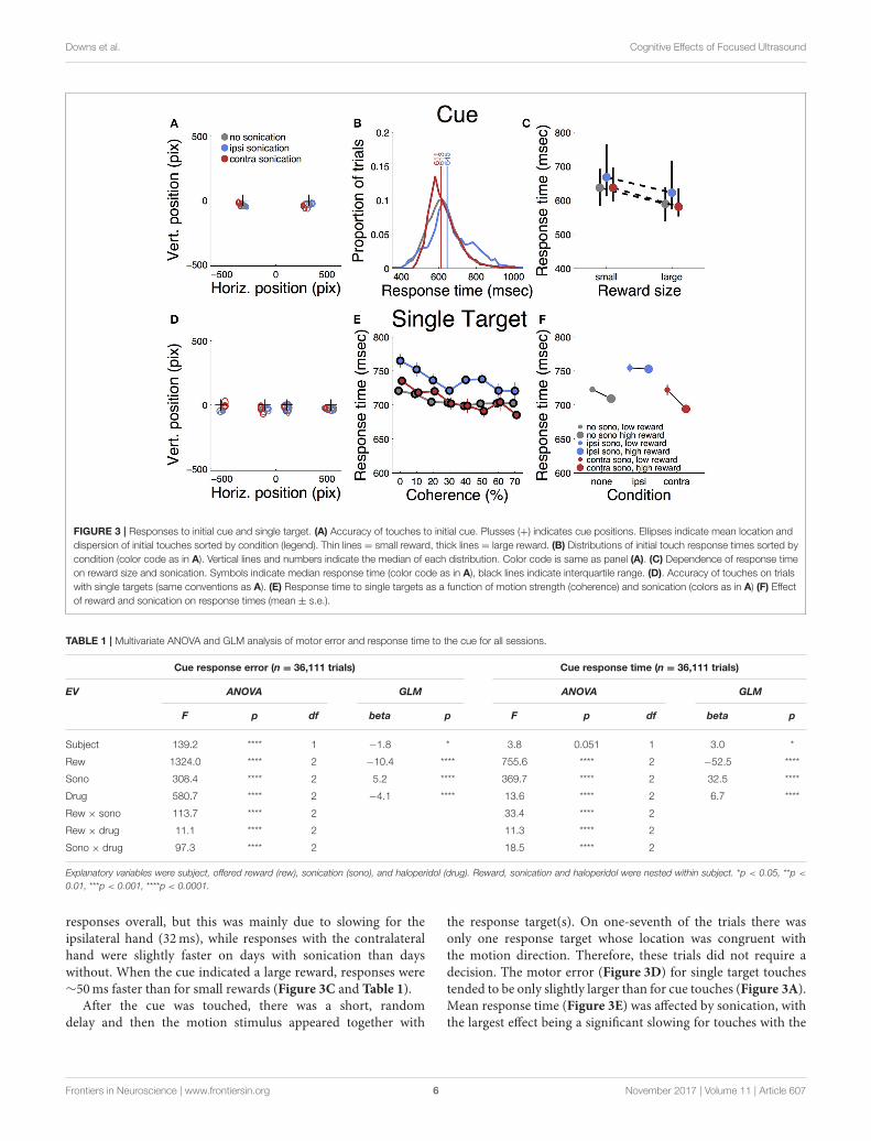
touch provide an indication of whether any of the experimentalmanipulations resulted in a simplemotor deficit. Touch error wasquantified as the spatial dispersion of the initial touches abouttheir mean as well as the radial distance from the center of thecue to the location of the first touch. Figure 3A shows the two cuelocations and the initial touch locations, separated by sonicationcondition. The centroid of each ellipse is the mean touch locationand the size of the ellipse is proportional to the dispersionabout the mean (standard deviation). Although there were smallsystematic differences between conditions, the touches weretightly clustered in all conditions, providing evidence that touchaccuracy was unimpaired by sonication.
Table 1 summarizes the effects of reward, sonication and drugon initial touch error. All of the main effects were significant.However, the magnitudes of the errors, as assessed by the GLManalysis, were generally small. For example, reward size wascorrelated with touch error, but the effect amounted to 10 pixels(2.9mm), which is less than the width of the monkey’s fingertip.The effects of sonication and drug on touch error were roughlyhalf as large (Table 1).
Response Time and Touch AccuracyEffectsResponse time was defined as the period between the appearanceof the cue and the first contact registered by the touch panel.The response time distributions are shown in Figure 3B, sortedby sonication condition. Sonication was associated with slower
Frontiers in Neuroscience | www.frontiersin.org 5 November 2017 | Volume 11 | Article 607
Downs et al. Cognitive Effects of Focused Ultrasound
FIGURE 3 | Responses to initial cue and single target. (A) Accuracy of touches to initial cue. Plusses (+) indicates cue positions. Ellipses indicate mean location and
dispersion of initial touches sorted by condition (legend). Thin lines = small reward, thick lines = large reward. (B) Distributions of initial touch response times sorted by
condition (color code as in A). Vertical lines and numbers indicate the median of each distribution. Color code is same as panel (A). (C) Dependence of response time
on reward size and sonication. Symbols indicate median response time (color code as in A), black lines indicate interquartile range. (D). Accuracy of touches on trials
with single targets (same conventions as A). (E) Response time to single targets as a function of motion strength (coherence) and sonication (colors as in A) (F) Effect
of reward and sonication on response times (mean ± s.e.).
TABLE 1 | Multivariate ANOVA and GLM analysis of motor error and response time to the cue for all sessions.
Cue response error (n = 36,111 trials) Cue response time (n = 36,111 trials)
EV ANOVA GLM ANOVA GLM
F p df beta p F p df beta p
Subject 139.2 **** 1 −1.8 * 3.8 0.051 1 3.0 *
Rew 1324.0 **** 2 −10.4 **** 755.6 **** 2 −52.5 ****
Sono 308.4 **** 2 5.2 **** 369.7 **** 2 32.5 ****
Drug 580.7 **** 2 −4.1 **** 13.6 **** 2 6.7 ****
Rew × sono 113.7 **** 2 33.4 **** 2
Rew × drug 11.1 **** 2 11.3 **** 2
Sono × drug 97.3 **** 2 18.5 **** 2
Explanatory variables were subject, offered reward (rew), sonication (sono), and haloperidol (drug). Reward, sonication and haloperidol were nested within subject. *p < 0.05, **p <
0.01, ***p < 0.001, ****p < 0.0001.
responses overall, but this was mainly due to slowing for theipsilateral hand (32ms), while responses with the contralateralhand were slightly faster on days with sonication than dayswithout. When the cue indicated a large reward, responses were∼50ms faster than for small rewards (Figure 3C and Table 1).
After the cue was touched, there was a short, randomdelay and then the motion stimulus appeared together with
the response target(s). On one-seventh of the trials there wasonly one response target whose location was congruent withthe motion direction. Therefore, these trials did not require adecision. The motor error (Figure 3D) for single target touchestended to be only slightly larger than for cue touches (Figure 3A).Mean response time (Figure 3E) was affected by sonication, withthe largest effect being a significant slowing for touches with the
Frontiers in Neuroscience | www.frontiersin.org 6 November 2017 | Volume 11 | Article 607
Downs et al. Cognitive Effects of Focused Ultrasound
hand ipsilateral to the sonicated hemisphere. With sonication,the average response time with the contralateral hand (735ms,n = 1,093,) was significantly faster than with the ipsilateralhand (772ms, n = 1,103, t-test p < 0.0001). Responses werealso significantly faster when a large reward was available andthis effect interacted significantly with sonication (Figure 3F).Statistical results (ANOVA and GLM) are given in Table 2.
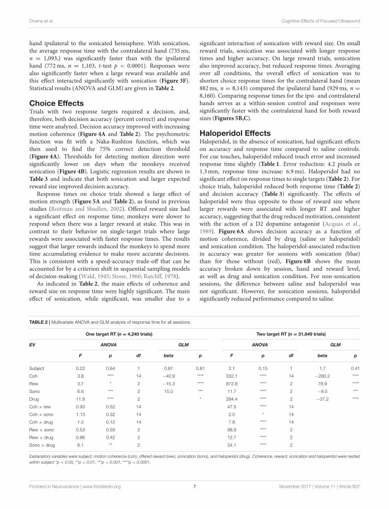
Choice EffectsTrials with two response targets required a decision, and,therefore, both decision accuracy (percent correct) and responsetime were analyzed. Decision accuracy improved with increasingmotion coherence (Figure 4A and Table 2). The psychometricfunction was fit with a Naka-Rushton function, which wasthen used to find the 75% correct detection threshold(Figure 4A). Thresholds for detecting motion direction weresignificantly lower on days when the monkeys receivedsonication (Figure 4B). Logistic regression results are shown inTable 3 and indicate that both sonication and larger expectedreward size improved decision accuracy.
Response times on choice trials showed a large effect ofmotion strength (Figure 5A and Table 2), as found in previousstudies (Roitman and Shadlen, 2002). Offered reward size hada significant effect on response time; monkeys were slower torespond when there was a larger reward at stake. This was incontrast to their behavior on single-target trials where largerewards were associated with faster response times. The resultssuggest that larger rewards induced the monkeys to spend moretime accumulating evidence to make more accurate decisions.This is consistent with a speed-accuracy trade-off that can beaccounted for by a criterion shift in sequential sampling modelsof decision-making (Wald, 1945; Stone, 1960; Ratcliff, 1978).
As indicated in Table 2, the main effects of coherence andreward size on response time were highly significant. The maineffect of sonication, while significant, was smaller due to a
significant interaction of sonication with reward size. On smallreward trials, sonication was associated with longer responsetimes and higher accuracy. On large reward trials, sonicationalso improved accuracy, but reduced response times. Averagingover all conditions, the overall effect of sonication was toshorten choice response times for the contralateral hand (mean882ms, n = 8,143) compared the ipsilateral hand (929ms, n =
8,160). Comparing response times for the ipsi- and contralateralhands serves as a within-session control and responses weresignificantly faster with the contralateral hand for both rewardsizes (Figures 5B,C).
Haloperidol EffectsHaloperidol, in the absence of sonication, had significant effectson accuracy and response time compared to saline controls.For cue touches, haloperidol reduced touch error and increasedresponse time slightly (Table 1. Error reduction: 4.2 pixels or1.3mm, response time increase: 6.9ms). Haloperidol had nosignificant effect on response times to single targets (Table 2). Forchoice trials, haloperidol reduced both response time (Table 2)and decision accuracy (Table 3) significantly. The effects ofhaloperidol were thus opposite to those of reward size wherelarger rewards were associated with longer RT and higheraccuracy, suggesting that the drug reducedmotivation, consistentwith the action of a D2 dopamine antagonist (Acquas et al.,1989). Figure 6A shows decision accuracy as a function ofmotion coherence, divided by drug (saline or haloperidol)and sonication condition. The haloperidol-associated reductionin accuracy was greater for sessions with sonication (blue)than for those without (red), Figure 6B shows the meanaccuracy broken down by session, hand and reward level,as well as drug and sonication condition. For non-sonicationsessions, the difference between saline and haloperidol wasnot significant. However, for sonication sessions, haloperidolsignificantly reduced performance compared to saline.
TABLE 2 | Multivariate ANOVA and GLM analysis of response time for all sessions.
One target RT (n = 4,240 trials) Two target RT (n = 31,849 trials)
EV ANOVA GLM ANOVA GLM
F p df beta p F p df beta p
Subject 0.22 0.64 1 0.81 0.81 2.1 0.15 1 1.7 0.41
Coh 3.8 **** 14 −40.9 **** 332.1 **** 14 −280.2 ****
Rew 3.7 * 2 −15.3 **** 872.8 **** 2 78.9 ****
Sono 6.6 *** 2 15.0 *** 11.7 **** 2 −8.5 ***
Drug 11.8 **** 2 * 284.4 **** 2 −37.2 ****
Coh × rew 0.93 0.52 14 47.5 **** 14
Coh × sono 1.13 0.32 14 2.0 * 14
Coh × drug 1.5 0.12 14 7.8 **** 14
Rew × sono 0.53 0.59 2 98.9 **** 2
Rew × drug 0.86 0.42 2 12.7 **** 2
Sono × drug 6.1 ** 2 34.1 **** 2
Explanatory variables were subject, motion coherence (coh), offered reward (rew), sonication (sono), and haloperidol (drug). Coherence, reward, sonication and haloperidol were nested
within subject *p < 0.05, **p < 0.01, ***p < 0.001, ****p < 0.0001.
Frontiers in Neuroscience | www.frontiersin.org 7 November 2017 | Volume 11 | Article 607
Downs et al. Cognitive Effects of Focused Ultrasound
FIGURE 4 | Effects of sonication on decision accuracy. (A) Accuracy (percent
correct) vs. coherence for sonicated and non-sonicated sessions. Solid
vertical lines are average performance ±1 s.d. estimated by bootstrap. Solid
curves are fits of Naka-Rushton functions. Dashed horizontal line indicates
75% correct level. Dotted vertical lines are coherence thresholds for 75%
correct performance. (B) Thresholds (75% correct) sorted by sonication
condition. Small black dots are individual sessions, large colored dots are
mean threshold across sessions. Note that the thresholds estimated from the
aggregated data in (A) are not expected to precisely match the means of the
individual session thresholds in (B) due to non-linearities in the fitting process.
Effects of AnesthesiaKetamine lengthens choice reaction time and reduces accuracyin humans (Micallef et al., 2002) and monkeys (Stoet andSnyder, 2006). In a previous study (Downs et al., 2015a),we measured the effect of low dose ketamine (5 mg/kg, IM)on reaching performance in two cynomolgous monkeys (M.fascicularis). These monkeys performed a reaching task withunequal rewards but without a motion stimulus. This task didnot require a decision as there was only one reach target atany given time. Behavior was tested immediately after ketamine
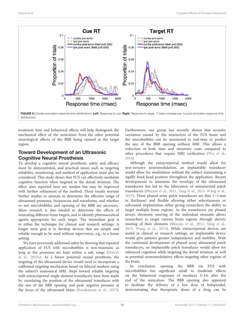
administration. Those data are reproduced in Figure 7 andshow that there was an initial slowing of response times thatreturned to baseline after 30–60min. Figure 8 shows the sameexperiment but with FUS+microbubbles administered duringbehavioral testing (indicated by the vertical black line). In thiscase, baseline (pre-sonication) response times were stable, butstarted to decrease after the sonication (data re-analyzed fromDowns et al., 2015a). This decrease could have been due tothe sonication with microbubbles alone or an interaction withketamine. The latter seems unlikely, as the baseline performancedid not show the same pattern of response time elevation as whenketamine was given without sonication (Figure 7). For sonicationsessions, response time distributions pre- and post-sonicationare shown for the contra- and ipsilateral hands (Figure 9). Pre-sonication, responses were slower with the contralateral hand.Post-sonication, all responses were faster, and responses with thecontralateral hand were as fast or faster than the ipsilateral hand.
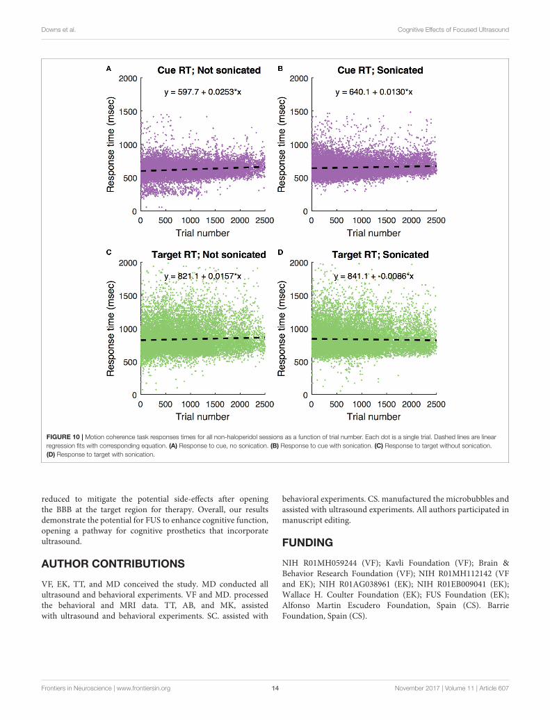
These data show that ketamine alone at the doses used hereprolongs response time with recovery within about an hour.To determine if this pattern was evident in the current study,Figure 10 shows response times in the motion coherence task asa function of trial number for all sonication and non-sonicationsessions. There is no evidence that response times became fasterover the course of a session for either sonicated or non-sonicatedsessions.
DISCUSSION
Cognitive Effects of Focused Ultrasoundwith Microbubbles in the Dorsal StriatumWe targeted the putamen in monkeys with FUS plus intravenousmicrobubbles to open the BBB. Because the BBB remains openfor up to 48 h (Marquet et al., 2014), we were able to test ifthere are subtle cognitive or behavioral changes subsequent tothe procedure. Decision-making in monkeys has been studiedpreviously with random dot motion tasks very similar to thatused in the current study (Roitman and Shadlen, 2002; Fenget al., 2009). Electrophysiological evidence suggests that thedorsal striatum (caudate and putamen) plays a role in such tasks(Ding and Gold, 2013), thus motivating us to use a variation ofthe task that could reveal changes in perception, motor control,decision-making and motivation.
Sessions without sonication were used to establish a behavioralbaseline. We confirmed previous work showing that responsetimes vary inversely with the strength of the motion signal(Roitman and Shadlen, 2002). The lengthening of responsetimes (RT) is an effective strategy to optimize accuracy, astemporal integration of weak motion signals improves decisionaccuracy. Manipulating the relative reward size for the tworesponse alternatives can introduce a response bias (Fenget al., 2009; Teichert and Ferrera, 2010; Teichert et al.,2014). Here we found that manipulating reward size forcorrect responses induced animals to trade response speed foraccuracy, but did not introduce a response bias as there wasnever any incentive to choose the incorrect target. We foundthat when a larger reward was offered, monkeys responded
Frontiers in Neuroscience | www.frontiersin.org 8 November 2017 | Volume 11 | Article 607
Downs et al. Cognitive Effects of Focused Ultrasound
TABLE 3 | Logistic regression analysis of decision accuracy.
EV All sessions Sessions without haloperidol Sessions without sonication
beta p beta p beta p
TWO TARGETS
Subject −0.60 **** −0.66 **** −0.74 ****
Coh 6.24 **** 6.31 **** 5.44 ****
Rew 0.26 **** 0.21 **** 0.32 ****
Sono 0.17 **** 0.16 ****
Drug −0.11 *** −0.11 *
Explanatory variables were subject, motion coherence (coh), offered reward (rew), sonication (sono), and haloperidol (drug). Coherence, reward, sonication and haloperidol were nested
within subject. *p < 0.05, **p < 0.01, ***p < 0.001, ****p < 0.0001.
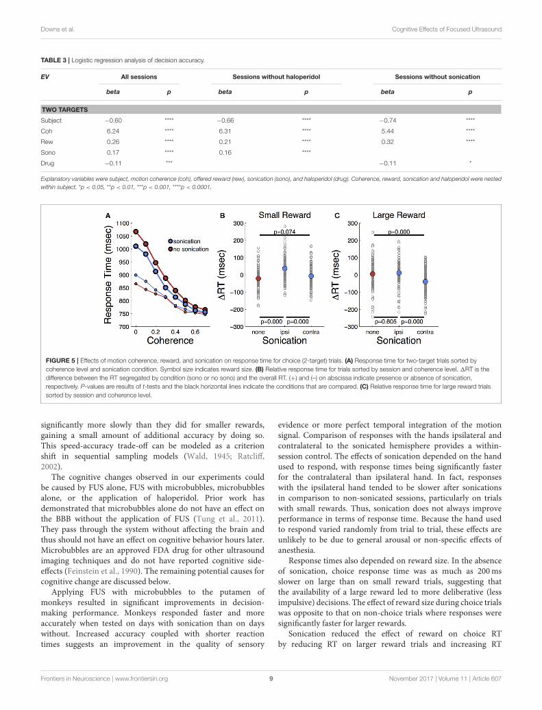
FIGURE 5 | Effects of motion coherence, reward, and sonication on response time for choice (2-target) trials. (A) Response time for two-target trials sorted by
coherence level and sonication condition. Symbol size indicates reward size. (B) Relative response time for trials sorted by session and coherence level. 1RT is the
difference between the RT segregated by condition (sono or no sono) and the overall RT. (+) and (–) on abscissa indicate presence or absence of sonication,
respectively. P-values are results of t-tests and the black horizontal lines indicate the conditions that are compared. (C) Relative response time for large reward trials
sorted by session and coherence level.
significantly more slowly than they did for smaller rewards,gaining a small amount of additional accuracy by doing so.This speed-accuracy trade-off can be modeled as a criterionshift in sequential sampling models (Wald, 1945; Ratcliff,2002).
The cognitive changes observed in our experiments couldbe caused by FUS alone, FUS with microbubbles, microbubblesalone, or the application of haloperidol. Prior work hasdemonstrated that microbubbles alone do not have an effect onthe BBB without the application of FUS (Tung et al., 2011).They pass through the system without affecting the brain andthus should not have an effect on cognitive behavior hours later.Microbubbles are an approved FDA drug for other ultrasoundimaging techniques and do not have reported cognitive side-effects (Feinstein et al., 1990). The remaining potential causes forcognitive change are discussed below.
Applying FUS with microbubbles to the putamen ofmonkeys resulted in significant improvements in decision-making performance. Monkeys responded faster and moreaccurately when tested on days with sonication than on dayswithout. Increased accuracy coupled with shorter reactiontimes suggests an improvement in the quality of sensory
evidence or more perfect temporal integration of the motionsignal. Comparison of responses with the hands ipsilateral andcontralateral to the sonicated hemisphere provides a within-session control. The effects of sonication depended on the handused to respond, with response times being significantly fasterfor the contralateral than ipsilateral hand. In fact, responseswith the ipsilateral hand tended to be slower after sonicationsin comparison to non-sonicated sessions, particularly on trialswith small rewards. Thus, sonication does not always improveperformance in terms of response time. Because the hand usedto respond varied randomly from trial to trial, these effects areunlikely to be due to general arousal or non-specific effects ofanesthesia.
Response times also depended on reward size. In the absenceof sonication, choice response time was as much as 200msslower on large than on small reward trials, suggesting thatthe availability of a large reward led to more deliberative (lessimpulsive) decisions. The effect of reward size during choice trialswas opposite to that on non-choice trials where responses weresignificantly faster for larger rewards.
Sonication reduced the effect of reward on choice RTby reducing RT on larger reward trials and increasing RT
Frontiers in Neuroscience | www.frontiersin.org 9 November 2017 | Volume 11 | Article 607

Downs et al. Cognitive Effects of Focused Ultrasound
FIGURE 6 | Effects of sonication and haloperidol on performance. (A) Psychometric functions for all sessions, divided by drug (saline/haloperidol) and presence of
sonication. Circles are mean pct correct, curves are Naka-Rushton fits. Dashed horizontal line indicates 75% correct threshold level. Vertical lines indicate motion
coherence corresponding to 75% threshold. (B) Average pct correct. Each small symbol is the mean for a given session, hand, and reward level. Large symbols are
means over all sessions for a given drug and sonication condition. S, saline; H, haloperidol.
on small reward trials, all while increasing accuracy. Themagnitude of the changes in RT were equal or greater thanthose reported for intracranial electrical stimulation of parietalcortex in monkeys performing a similar task, albeit with aneye movement rather than reaching response (Hanks andShadlen, 2006). Thus, for large rewards, sonication appears toimprove the efficiency of decision-making, possibly by improvingthe quality of the sensory signal or the rate of evidenceaccumulation. In other words, improved decision efficiencyafter sonication might result from greater signal-to-noise orby reducing the leakiness of the integrator. These findingsprovide new evidence that the dorsal striatum (caudate andputamen) is involved in sensory evidence accumulation and thusplays an integral role in the decision process (Ding and Gold,2013).
Decision-making performance improvements were foundeven though animals were tested 3–4 h after sonication,suggesting that there may be a persistent effect on the activity orresponsiveness of putamen neurons, which, in turn, may be dueto a direct effect of ultrasound or an indirect effect of opening theBBB. It is likely that BBB opening alters the local extracellularmilieu, possibly by enriching the parenchymal concentrationof oxygen and glucose. Ultrasound may also directly affect thepermeability of mechanically or thermally sensitive ion channels(Yoo et al., 2011). Further experiments are needed to ascertainthe temporal window within which performance improvementsare obtained.
Control experiments show that ketamine alone slowsresponse times, but the effect only lasts about 1 h, whereasbehavioral performance in this study occurred 3–4 h after thecessation of anesthesia. Response times showed no evidenceof slowing at the beginning of each session. It is possible
that there are more subtle effects of anesthesia alone or incombination with BBB opening, but it would be extraordinaryif these led to improvements in performance. Studies of thepostoperative cognitive effects of general anesthetics, includingisoflurane, suggest that there may be little or no cognitiveimpairment (Bryson and Wyand, 2006). We were unableto find any studies suggesting that isoflurance improvescognition.
Recently, McDannold et al. (2015) showed that opening theBBB facilitated the blockade of neural activity by GABA insomatosensory cortex of rats. Here, we found that sonicationinteracted with a low dose of haloperidol, a D2 dopamineantagonist, that was injected 5min prior to behavioral testing.Lower levels of striatal D2 dopamine receptors are associatedwith reduced motivation and increased impulsivity (Trifilieff andMartinez, 2014). Previous studies of the effects of haloperidolon response times have reported mixed results depending onspecies, task and dosage (Brockel and Fowler, 1995; Kernet al., 1998; Blokland and Honig, 1999). In the current study,low dose haloperidol tended to shorten response time andreduce decision accuracy. Hence, the effects of haloperidolwere opposite to those of increasing reward size, consistentwith the idea that the behavioral effects of reward may bedue to reduced motivation, mediated by striatal D2 dopaminereceptors. Haloperidol may inhibit signaling through theindirect basal ganglia pathway, allowing the direct pathway toproduce shorter latency movements (Albin et al., 1989; DeLong,1990).
The effects of haloperidol showed an interaction withsonication. This result indicates that FUS can be used incombination with dopaminergic medications to modulatecognitive performance. The results also suggest that the
Frontiers in Neuroscience | www.frontiersin.org 10 November 2017 | Volume 11 | Article 607

Downs et al. Cognitive Effects of Focused Ultrasound
FIGURE 7 | Timecourse of the behavioral effect of ketamine without sonication. (A) Response time for reaches to the cue presentation. (B) Responses to the target.
Each dot is a single trial. The solid curve shows the median response time in a 4-min window. Solid black lines are fits of the exponential function whose equation is
given at the top of each panel.
systemic dose of a drug necessary to achieve a desiredpharmacological effect may be reduced by increasing BBBpermeability through the application of FUS to a targetedbrain region, even if the drug in question readily crossesthe BBB. This would allow for smaller systemic doses, andthus reduction of potential side effects of currently availabledrugs for therapies to treat neurological and psychiatricdisorders.
There are a few previous studies investigating the effectof FUS without BBB opening on alert subjects performingbehavioral tasks. Deffieux and colleagues applied FUS tomonkeysperforming an antisaccade task by targeting the left frontal eyefield (FEF) and the premotor cortex (Deffieux et al., 2013).Ipsilateral antisaccade latencies were significantly slowed whiletargeting the FEF but not the premotor cortex. Two othergroups investigated the effects of FUS on human subjects(Hameroff et al., 2013; Legon et al., 2014). Subjects tested byLegon et al. exhibited enhanced sensitivity to the frequencyof air puffs and improved two-point tactile discrimination
while FUS was applied to their somatosensory cortex. FUSwas applied to the frontal-temporal cortex in subjects ofthe Hameroff et al. study and unlike the other two studieswith simultaneous/immediate behavioral testing, results weredetermined 10 and 40min after application. Subjects reporteda significant improvement on the Global Affect test, as wellas slightly reduced pain levels 40min after the application ofFUS. These studies demonstrate that FUS is capable of affectingthe function of the brain depending on the targeting area,while the Hameroff et al. study shows the effects could be timesensitive. Two key differences from the current study is that inthe aforementioned studies the BBB remained undisrupted in thetargeted region to the knowledge of the experimenters. Thereare also differences in the timeline of behavioral assessment.In the prior NHP study conducted by Deffieux et al. theyonly observed an increase in antisaccade latency during the100ms application of the FUS, not after it had ceased. Hameroffet al. observed effects only within 40min after applicationof the FUS. Thus, this suggests to different mechanisms of
Frontiers in Neuroscience | www.frontiersin.org 11 November 2017 | Volume 11 | Article 607
Downs et al. Cognitive Effects of Focused Ultrasound
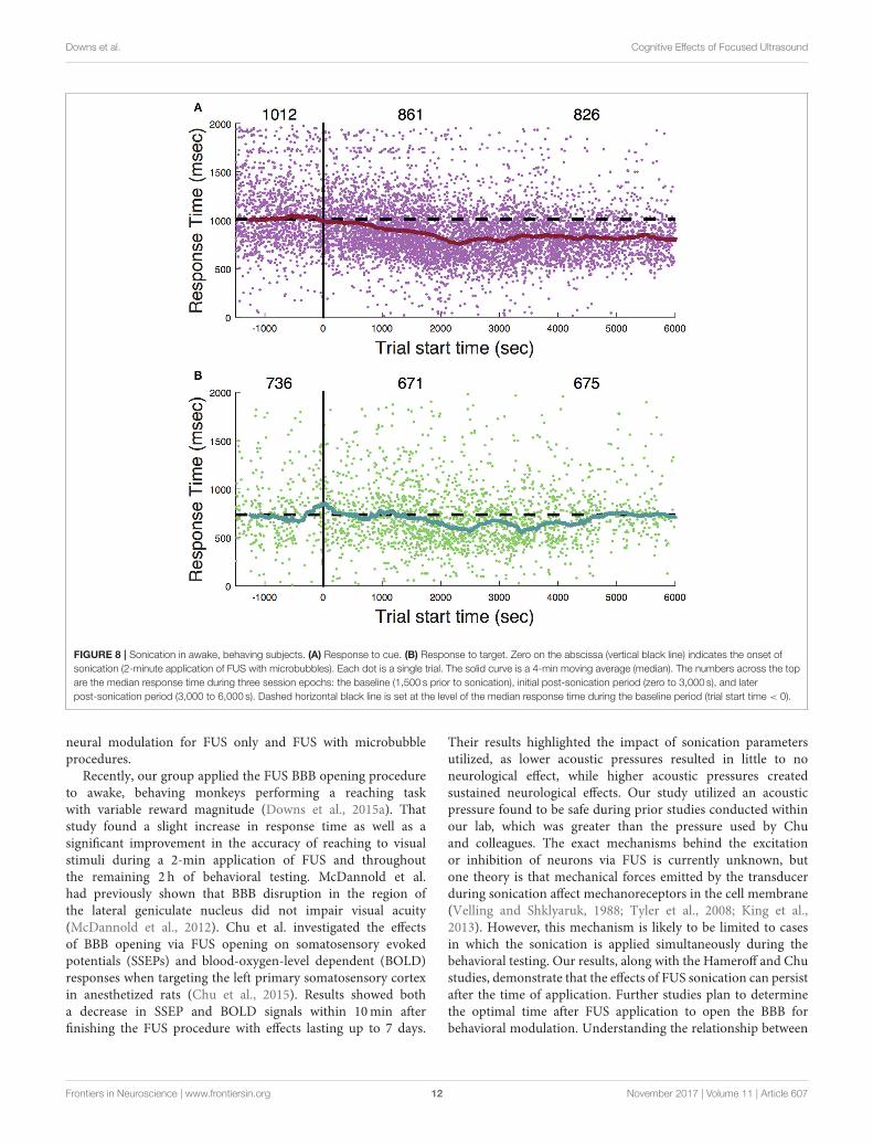
FIGURE 8 | Sonication in awake, behaving subjects. (A) Response to cue. (B) Response to target. Zero on the abscissa (vertical black line) indicates the onset of
sonication (2-minute application of FUS with microbubbles). Each dot is a single trial. The solid curve is a 4-min moving average (median). The numbers across the top
are the median response time during three session epochs: the baseline (1,500 s prior to sonication), initial post-sonication period (zero to 3,000 s), and later
post-sonication period (3,000 to 6,000 s). Dashed horizontal black line is set at the level of the median response time during the baseline period (trial start time < 0).
neural modulation for FUS only and FUS with microbubbleprocedures.
Recently, our group applied the FUS BBB opening procedureto awake, behaving monkeys performing a reaching taskwith variable reward magnitude (Downs et al., 2015a). Thatstudy found a slight increase in response time as well as asignificant improvement in the accuracy of reaching to visualstimuli during a 2-min application of FUS and throughoutthe remaining 2 h of behavioral testing. McDannold et al.had previously shown that BBB disruption in the region ofthe lateral geniculate nucleus did not impair visual acuity(McDannold et al., 2012). Chu et al. investigated the effectsof BBB opening via FUS opening on somatosensory evokedpotentials (SSEPs) and blood-oxygen-level dependent (BOLD)responses when targeting the left primary somatosensory cortexin anesthetized rats (Chu et al., 2015). Results showed botha decrease in SSEP and BOLD signals within 10min afterfinishing the FUS procedure with effects lasting up to 7 days.
Their results highlighted the impact of sonication parametersutilized, as lower acoustic pressures resulted in little to noneurological effect, while higher acoustic pressures createdsustained neurological effects. Our study utilized an acousticpressure found to be safe during prior studies conducted withinour lab, which was greater than the pressure used by Chuand colleagues. The exact mechanisms behind the excitationor inhibition of neurons via FUS is currently unknown, butone theory is that mechanical forces emitted by the transducerduring sonication affect mechanoreceptors in the cell membrane(Velling and Shklyaruk, 1988; Tyler et al., 2008; King et al.,2013). However, this mechanism is likely to be limited to casesin which the sonication is applied simultaneously during thebehavioral testing. Our results, along with the Hameroff and Chustudies, demonstrate that the effects of FUS sonication can persistafter the time of application. Further studies plan to determinethe optimal time after FUS application to open the BBB forbehavioral modulation. Understanding the relationship between
Frontiers in Neuroscience | www.frontiersin.org 12 November 2017 | Volume 11 | Article 607
Downs et al. Cognitive Effects of Focused Ultrasound
FIGURE 9 | Awake sonication response time distributions. Left: Response to cue. Right: Response to target. T-tests compare pre- to post-sonication response time
distributions.
treatment time and behavioral effects will help distinguish themechanical effect of the sonication from the other potentialneurological effects of the BBB being opened at the targetregion.
Toward Development of an UltrasonicCognitive Neural ProsthesisTo develop a cognitive neural prosthesis, safety and efficacymust be demonstrated, and practical issues such as targetingreliability, monitoring, and method of application must also beconsidered. This study shows that FUS can effectively modulatecognitive function when targeted to the dorsal striatum. Theeffect sizes reported here are modest but may be improvedwith further refinement of the method. These results warrantfurther studies in animals to determine the effective range ofultrasound pressures, frequencies and waveforms, and whetheror not microbubbles and opening of the BBB are necessary.More research is also needed to determine the effects ofsonicating different brain targets, and to identify pharmaceuticalagents appropriate for each target. The immediate goal isto refine the technique for clinical and research settings. Alonger term goal is to develop devices that are simple andreliable enough to be used without supervision, e.g., in a homesetting.
We have previously addressed safety by showing that repeatedapplication of FUS with microbubbles is non-traumatic aslong as the pressures are kept within a safe range (Downset al., 2015a). As a future potential neural prosthesis, thetargeting of the ultrasound device would need to incorporate acalibrated targeting mechanism based on fiducial markers usingthe subject’s anatomical MRI. Steps toward reliable targetingwith extracorporeal single element transducers have been madeby correlating the position of the ultrasound transducer withthe size of the BBB opening and peak negative pressure atthe focus of the ultrasound beam (Karakatsani et al., 2017).
Furthermore, our group has recently shown that acousticcavitation caused by the interaction of the FUS beam andthe microbubbles can be monitored in real-time to predictthe size of the BBB opening without MRI. This allows areduction of both time and monetary costs compared toother procedures that require MRI verification (Wu et al.,2016).
Although the extracorporeal method would allow fornon-invasive neuromodulation, an implantable transducerwould allow for modulation without the subject maintaining arigidly fixed head position throughout the application. Recentdevelopments to minimize the envelope of the ultrasoundtransducers has led to the fabrication of miniaturized patchtransducers (Bhuyan et al., 2011; Yang et al., 2013; Wang et al.,2015). These phased array patch transducers are thin (∼1mmin thickness) and flexible allowing either subcutaneous orsubcranial implantation either giving researchers the ability totarget multiple brain regions. As the transducers are phasedarrays, electronic steering of the individual elements allowsresearchers to target various brain regions through electricsteering of their elements (Bhuyan et al., 2011; Yang et al.,2013; Wang et al., 2015). While extracorporeal devices areuseful in clinical or research settings, an implantable devicewould give patients greater independence and mobility. Withthe continued development of phased array ultrasound patchtransducers, an implantable patch transducer would allow forenhanced cognition while targeting the dorsal striatum, as wellas potential neuromodulatory effects targeting other regions ofthe brain.
In conclusion, opening the BBB via FUS withmicrobubbles has significant small to moderate effectson the behavioral responses of monkeys 3–4 h after theend of the sonication. The BBB opening also appearedto facilitate the delivery of a low dose of haloperidol,demonstrating that therapeutic doses of a drug can be
Frontiers in Neuroscience | www.frontiersin.org 13 November 2017 | Volume 11 | Article 607
Downs et al. Cognitive Effects of Focused Ultrasound
FIGURE 10 | Motion coherence task responses times for all non-haloperidol sessions as a function of trial number. Each dot is a single trial. Dashed lines are linear
regression fits with corresponding equation. (A) Response to cue, no sonication. (B) Response to cue with sonication. (C) Response to target without sonication.
(D) Response to target with sonication.
reduced to mitigate the potential side-effects after openingthe BBB at the target region for therapy. Overall, our resultsdemonstrate the potential for FUS to enhance cognitive function,opening a pathway for cognitive prosthetics that incorporateultrasound.
AUTHOR CONTRIBUTIONS
VF, EK, TT, and MD conceived the study. MD conducted allultrasound and behavioral experiments. VF and MD. processedthe behavioral and MRI data. TT, AB, and MK, assistedwith ultrasound and behavioral experiments. SC. assisted with
behavioral experiments. CS. manufactured the microbubbles andassisted with ultrasound experiments. All authors participated inmanuscript editing.
FUNDING
NIH R01MH059244 (VF); Kavli Foundation (VF); Brain &Behavior Research Foundation (VF); NIH R01MH112142 (VFand EK); NIH R01AG038961 (EK); NIH R01EB009041 (EK);Wallace H. Coulter Foundation (EK); FUS Foundation (EK);Alfonso Martin Escudero Foundation, Spain (CS). BarrieFoundation, Spain (CS).
Frontiers in Neuroscience | www.frontiersin.org 14 November 2017 | Volume 11 | Article 607
Downs et al. Cognitive Effects of Focused Ultrasound
REFERENCES
Abbott, N. J. (2013). Blood–brain barrier structure and function and thechallenges for CNS drug delivery. J. Inherit. Metab. Dis. 36, 437–449.doi: 10.1007/s10545-013-9608-0
Acquas, E., Carboni, C. P., and DiChiara, G. (1989). SCH23390 blocksdrug-conditioned place-preference and place aversion: Anhedonia (lack ofreward) or apathy (lack of motivation) after dopamine receptor blockade.Psychopharmacology 99, 151–155. doi: 10.1007/BF00442800
Albin, R. L., Young, A. B., and Penney, J. B. (1989). The functional anatomy of basalganglia disorders. Trends Neurosci. 12, 366–375. doi: 10.1016/0166-2236(89)90074-X
Arvanitis, C. D., Livingstone, M. S., and McDannold, N. (2013). Combinedultrasound andMR imaging to guide focused ultrasound therapies in the brain.Phys. Med. Biol. 58, 4749–4761. doi: 10.1088/0031-9155/58/14/4749
Bakay, L., Hueter, T. F., Ballantine, H. T., and Sosa, D. (1956). Ultrasonicallyproduced changes in the blood-brain barrier. AMA Arch. Neurol. Psychol. 76,457–467. doi: 10.1001/archneurpsyc.1956.02330290001001
Bhuyan, A., Choe, B. C., Lee, P., Cristman, Ö., Oralkan, B. T., and Khuri-Yakub(2011). “Miniaturized, wearable, ultrasound probe for on-demand ultrasoundscreening,” in EEE Ultrasonics Symposium (Orlando, FL), 1060–1063.doi: 10.1109/ULTSYM.2011.0260
Blokland, A., and Honig, W. (1999). Intra-striatal haloperidol and scopolamineinjections: effects on choice reaction time performance in rats. Eur.
Neuropsychopharmacol. 9, 523–531. doi: 10.1016/S0924-977X(99)00036-XBorchers, S., Himmelbach, M., Logothetis, N., and Karnath, H.-O. (2012). Direct
electrical stimulation of human cortex - the gold standard for mapping brainfunctions? Nat. Rev. Neurosci. 13, 63–70. doi: 10.1038/nrn3140
Brockel, B. J., and Fowler, S. C. (1995). Effects of chronic haloperidol on reactiontime and errors in a sustained attention task: partial reversal by anticholinergicsand by amphetamine. J. Pharmacol. Exp. Ther. 275, 1090–1098.
Bryson, G. L., and Wyand, A. (2006). Evidence-based clinical update: generalanesthesia and the risk of delirium and postoperative cognitive dysfunction.Can. J. Anesth. 53, 669–677. doi: 10.1007/BF03021625
Bystritsky, A., Korb, A. S., Douglass, P. K., Cohen, M. S., Melega, W. P.,Mulgaonkar, A. P., et al. (2011). A review of low-intensity focused ultrasoundpulsation. Brain Stimul. 4, 125–136. doi: 10.1016/j.brs.2011.03.007
Calvo, F., and Coimbra, N. C. (2006). Interactions between opioid-peptides-containing pathways and GABAA-receptors-mediated systemsmodulate panic-like-induced behaviors elicited by electric and chemicalstimulation of the inferior colliculus. Brain Res. 1104, 92–102.doi: 10.1016/j.brainres.2006.05.056
Chu, P.-C., Liu, H. L., Lai, H. Y., Lin, C. Y., Tsai, H. C., and Pei, Y. C.(2015). Neuromodulation accompanying focused ultrasound-induced blood-brain barrier opening. Sci. Rep. 5:15477. doi: 10.1038/srep15477
Deffieux, T., Younan, Y.,Wattiez, N., Tanter,M., Pouget, P., andAubry, J. F. (2013).Low-intensity focused ultrasound modulates monkey visuomotor behavior.Curr. Biol. 23, 2430–2433. doi: 10.1016/j.cub.2013.10.029
DeLong, M. R. (1990). Primate models of movement disorders of basal gangliaorigin. Trends Neurosci. 13, 281–285. doi: 10.1016/0166-2236(90)90110-V
Ding, L., and Gold, J. I. (2013). The basal ganglia’s contributions to perceptualdecision making. Neuron 79, 640–649. doi: 10.1016/j.neuron.2013.07.042
Downs, M. E., Buch, A., Karakatsani, M. E., Konofagou, E. E., and Ferrera, V.P. (2015b). Blood-brain barrier opening in behaving non-human primatesvia focused ultrasound with systemically administered microbubbles. Sci. Rep.5:15076. doi: 10.1038/srep15076
Downs, M. E., Buch, A., Sierra, C., Karakatasani, M. E., Teichert, T., Chen, S., et al.(2015a). Long-term safety of repeated blood-brain barrier opening via focusedultrasound with microbubbles in non-human primates performing a cognitivetask. PLoS ONE 10:e25911. doi: 10.1371/journal.pone.0125911
Dubuisson, D., and Dennis, S. G. (1977). The formalin test: a quantitativestudy of the analgesic effects of morphine, meperidine, and brain stemstimulation in rats and cats. Pain 4, 161–174. doi: 10.1016/0304-3959(77)90130-0
Feinstein, S. B., Cheirif, J., Ten Cate, F. J., Silverman, P. R., Heidenreich, P. A.,Dick, C., et al. (1990). Safety and efficacy of a new transpulmonary ultrasoundcontrast agent: initial multicenter clinical results. J. Am. Coll. Cardiol. 16,316–324. doi: 10.1016/0735-1097(90)90580-I
Feng, S., Holmes, P., Rorie, A., and Newsome, W. T. (2009). Can Monkeys chooseoptimally when faced with noisy stimuli and unequal rewards? PLoS Comput.
Biol. 5:e00284. doi: 10.1371/journal.pcbi.1000284Feshitan, J. A., Chen, C. C., Kwan, J. J., and Borden, M. A. (2009). Microbubble size
isolation by differential centrifugation. J. Colloid Interface Sci. 329, 316–324.doi: 10.1016/j.jcis.2008.09.066
Hameroff, S., Trakas, M., Duffield, C., Annabi, E., Gerace, M. B., Boyle, P., et al.(2013). Transcranial ultrasound (TUS) effects on mental states: a pilot study.Brain Stimul. 6, 409–415. doi: 10.1016/j.brs.2012.05.002
Hanks, T., and Shadlen, M. N. (2006). Microstimulation of area LIP affectsdecision-making in a motion discrimination task. Nat. Neurosci. 9, 682–689.doi: 10.1038/nn1683
Hikosaka, O. (2007). Basal ganglia mechanisms of reward-oriented eye movement.Ann. N.Y. Acad. Sci. 1104, 229–249. doi: 10.1196/annals.1390.012
Hikosaka, O., Nakamura, K., and Nakahara, H. (2006). Basal gangliaorient eyes to reward. J. Neurophysiol. 95, 567–584. doi: 10.1152/jn.00458.2005
Hynynen, K., McDannold, N., Vykhodtseva, N., and Jolesz, F. A. (2001).Noninvasive MR imaging-guided focal opening of the blood-brain barrier inrabbits. Radiology 22, 640–646. doi: 10.1148/radiol.2202001804
Karakatsani, M. E., Samiotaki, G., Downs, M., Ferrera, V., and Konofagou,E. (2017). Targeting effects on the volume of the focused ultrasoundinduced blood-brain barrier opening in non-human primates in
vivo. IEEE Trans. Ultrason. Ferroelectr. Freq. Control. 64, 798–810.doi: 10.1109/TUFFC.2017.2681695
Kern, R. S., Green, M. F., Marshall, B. D. Jr., Wirshing, W. C., Wirshing, D.,McGurk, S., et al. (1998). Risperidone vs. haloperidol on reaction time, manualdexterity, and motor learning in treatment-resistant schizophrenia patients.Biol. Psychiatry 44, 726–732. doi: 10.1016/S0006-3223(98)00088-2
King, R. L., Brown, J. R., Newsome, W. T., and Pauly, K. B. (2013). Effectiveparameters for ultrasound-induced in vivo neurostimulation. Ultrasound Med.
Biol. 39, 312–331. doi: 10.1016/j.ultrasmedbio.2012.09.009Kobayashi, M., and Pascual-Leone, A. (2003). Transcranial magnetic stimulation
in neurology. Lancet Neurol. 2, 145–156. doi: 10.1016/S1474-4422(03)00321-1Lappin, J. S., and Bell, H. H. (1976). The detection of coherence in moving random
dot patents. Vis. Res. 16, 161–168. doi: 10.1016/0042-6989(76)90093-6Lauwereyns, J., Watanabe, K., Coe, B., and Hikosaka, O. (2002). A neural
correlate of response bias in monkey caudate nucleus. Nature 418, 413–417.doi: 10.1038/nature00892
Lee, W., Kim, H. C., Jung, Y., Chung, Y. A., Song, I. U., Lee, J. H., et al. (2016).Transcranial focused ultrasound stimulation of human primary visual cortex.Sci. Rep. 6:34026 doi: 10.1038/srep34026
Legon, W., Sato, T. F., Opitz, A., Mueller, J., Barbour, A., Williams, A.,et al. (2014). Transcranial focused ultrasound modulates the activity ofprimary somatosensory cortex in humans. Nat. Neurosci. 17, 322–329.doi: 10.1038/nn.3620
Marquet, F., Teichert, T., Wu, S.-W., Tung, Y.-S., Downs, M., Wang,S., et al. (2014). Real-time, transcranial monitoring of safe blood-brain barrier opening in non-human primates. PLoS ONE 9:e84310.doi: 10.1371/journal.pone.0084310
McDannold, N., Arvanitis, C. D., Vykhodtseva, N., and Livingstone, M. S. (2012).Temporary disruption of the blood-brain barrier by use of ultrasound andmicrobubbles: safety and efficacy evaluation in rhesus macaques. Cancer Res.72, 3652–3663. doi: 10.1158/0008-5472.CAN-12-0128
McDannold, N., Vykhodtseva, N., Raymond, S., Jolesz, F. A., and Hynynen,K. (2005). MRI-guided targeted blood-brain barrier disruption with focusedultrasound: histological findings in rabbits. Ultrasound Med. Biol. 31,1527–1537. doi: 10.1016/j.ultrasmedbio.2005.07.010
McDannold, N., Zhang, Y., Power, C., Arvanitis, C. D., Vykhodtseva, N., andLivingstone, M. (2015). Targeted, noninvasive blockade of cortical neuronalactivity. Sci. Rep. 5:16253. doi: 10.1038/srep16253
Meairs, S., and Alonso, A. (2007). Ultrasound, microbubbles andthe blood–brain barrier. Prog. Biophys. Mol. Biol. 93, 354–362.doi: 10.1016/j.pbiomolbio.2006.07.019
Micallef, J., Guillermain, Y., Tardieu, S., Hasbroucq, T., Possamai, C., Jouve,E., et al. (2002). Effects of subanesthetic doses of ketamine on sensorimotorinformation processing in healthy subjects.Clin. Neuropharmacol. 35, 101–106.doi: 10.1097/00002826-200203000-00008
Frontiers in Neuroscience | www.frontiersin.org 15 November 2017 | Volume 11 | Article 607

Downs et al. Cognitive Effects of Focused Ultrasound
Miller, N. E. (1965). Chemical coding of behavior in the brain. Science 148,328–338. doi: 10.1126/science.148.3668.328
Nitsche, M., Schauenburg, A., Lang, N., Liebetanz, D., Exner, C., Paulus, W.,et al. (2003). Facilitation of implicit motor learning by weak transcranialdirect current stimulation of the primary motor cortex in the human. J. Cogn.Neurosci. 15, 619–626. doi: 10.1162/089892903321662994
Ratcliff, R. (1978). A theory of memory retrieval. Psychol. Rev. 85, 59–108.doi: 10.1037/0033-295X.85.2.59
Ratcliff, R. (2002). A diffusion model account of response time and accuracyin a brightness discrimination task: fitting real data and failing to fitfake but plausible data. Psychon. Bull. Rev. 9, 278–291. doi: 10.3758/BF03196283
Roitman, J. D., and Shadlen, M. N. (2002). Response of neurons in the lateralintraparietal area during a combined visual discrimination reaction time task.J. Neurosci. 22, 9475–9489.
Sassaroli, E., and Vykhodtseva, N. (2016). Acoustic neuromodulation from a basicscience perspective. J. Ther. Ultrasound 4:17. doi: 10.1186/s40349-016-0061-z
Stoet, G., and Snyder, L. H. (2006). Effects of the NMDA antagonist ketamineon task-switching performance: evidence for specific impairmentsof executive control. Neuropsychopharmacologia 31, 1675–1681.doi: 10.1038/sj.npp.1300930
Stone, M. (1960). Models for choice reaction time. Psychometrika 25, 251–260.doi: 10.1007/BF02289729
Teichert, T., and Ferrera, V. P. (2010). Suboptimal integration of rewardmagnitude and prior reward likelihood in categorical decisions by monkeys.Front. Neurosci. 4:186. doi: 10.3389/fnins.2010.00186
Teichert, T., Yu, D., and Ferrera, V. P. (2014). Performancemonitoring in monkey frontal eye field. J. Neurosci. 34, 1657–1671.doi: 10.1523/JNEUROSCI.3694-13.2014
Trifilieff, P., and Martinez, D. (2014). Imaging addiction: D2 receptorsand dopamine signaling in the striatum as biomarkers for impulsivity.Neuropharmacology 76, 498–509. doi: 10.1016/j.neuropharm.2013.06.031
Tung, Y.-S., Marquet, F., Teichert, T., Ferrera, V., and Konofagou, E. E. (2011).Feasibility of noninvasive cavitation-guided blood-brain barrier opening usingfocused ultrasound and microbubbles in nonhuman primates. Appl. Phys. Lett.98:163704. doi: 10.1063/1.3580763
Tyler, W. J., Tufail, Y., Finsterwald, M., Tauchmann, M. L., Olson, E.J., and Majestic, C. (2008). Remote excitation of neuronal circuits
using low-intensity, low-frequency ultrasound. PLoS ONE 3:e3511.doi: 10.1371/journal.pone.0003511
Velling, V. A., and Shklyaruk, S. P. (1988). Modulation of the functional state ofthe brain with the aid of focused ultrasound action. Neurosci. Behav. Physiol.18, 369–375. doi: 10.1007/BF01193880
Vykhodtseva, N. I., Hynynen, K., and Damianou, C. (1995). Histologiceffects of high intensity pulsed ultrasound exposure with subharmonicemission in rabbit brain in vivo. Ultrasound Med. Biol. 21, 969–979.doi: 10.1016/0301-5629(95)00038-S
Wald, A. (1945). Sequential tests of statistical hypotheses. Ann. Math. Stat. 16,117–186.
Wang, Z., Xue, Y. Q., Chen, Y., Shu, H., Tian, Y., Yang, D., et al. (2015). Aflexible ultrasound transducer array with micro-machined bulk PZT. Sensors15, 2538–2547. doi: 10.3390/s150202538
Wu, S.-Y., Sanchez, C. S., Samiotaki, G., Buch, A., Ferrera, V. P., and Konofagou,E. E. (2016). Characterizing focused-ultrasound mediated drug delivery to theheterogeneous primate brain in vivo with acoustic monitoring. Nat. Sci. Rep.6:37094. doi: 10.1038/srep37094
Yang, Y. H., Tian, B., Yan, H., Sun, C., Wu, Y., Shu, L. G., et al. (2013). Aflexible piezoelectricmicromachined ultrasound transducer.RSCAdv. 3, 24900.doi: 10.1039/c3ra44619k
Yin, D., Valles, F. E., Fiandaca, M. S., Forsayeth, J., Larson, P., Starr, P., et al.(2009). Striatal volume differences between non-human and human primates.J. Neurosci. Methods 176, 200–205. doi: 10.1016/j.jneumeth.2008.08.027
Yoo, S. S., Bystritsky, A., Lee, J. H., Zhang, Y., Fischer, K., Min, B. K., et al. (2011).Focused ultrasound modulates region-specific brain activity. NeuroImage 56,1267–1275. doi: 10.1016/j.neuroimage.2011.02.058
Conflict of Interest Statement: The authors declare that the research wasconducted in the absence of any commercial or financial relationships that couldbe construed as a potential conflict of interest.
Copyright © 2017 Downs, Teichert, Buch, Karakatsani, Sierra, Chen, Konofagou and
Ferrera. This is an open-access article distributed under the terms of the Creative
Commons Attribution License (CC BY). The use, distribution or reproduction in
other forums is permitted, provided the original author(s) or licensor are credited
and that the original publication in this journal is cited, in accordance with accepted
academic practice. No use, distribution or reproduction is permitted which does not
comply with these terms.
Frontiers in Neuroscience | www.frontiersin.org 16 November 2017 | Volume 11 | Article 607


