TOTAL ELBOW REPLACEMENT IN DOGS - … · Unlike previous and newer elbow prostheses, the TATE is a...
5
TOTAL ELBOW REPLACEMENT IN DOGS Loïc M. Déjardin, DVM, MS, DACVS, DECVS Reunan P. Guillou, Doc. Vét. College of Veterinary Medicine Michigan State University, East Lansing – Michigan Key Points Unlike previous and newer elbow prostheses, the TATE is a cementless, resurfacing press-fit design implanted as a single unit via limited surgical approaches. Subjective evaluation suggests that long term functional recovery and severe complication rates compare favorably to those of stemmed cemented designs. Prospective experimental and clinical evaluations as well as retrieval analysis are ongoing and may provide some needed objective data in the near future. New total and hemi prostheses have been developed and are currently in pre-clinical trials. Introduction: In recent years, new total and hemi elbow replacement prostheses (TER & HER) have been designed (Conzemius, Acker – Van Der Muellen and Innes [TER] / Wendelburg – Tepic [HER]) for the treatment of intractable end-stage canine elbow osteoarthritis (OA) as well as early so called “medial compartment disease”. While most systems remain cemented, unlinked, semi-constrained, stemmed designs, a novel TER system (TATE Elbow™) was recently devised by Acker and Van Der Meulen (Fig. 1). This proceeding which focuses on the TATE Elbow™ prosthesis, describes recent modifications in implant design and surgical technique and summarizes clinical results and complications. TATE design and surgical procedure: In addition to being an unlinked, semi-constrained design, the TATE is a cementless implant designed to use a novel resurfacing concept, as well as minimally invasive surgery (MIS). These fundamental differences may explain the improved, time-matched, clinical outcomes seen with the TATE prosthesis in particular with regards to lateral luxation and humeral/ulnar fractures, complications seen with cemented stemmed designs. Figure 1: Schematics of a 1 st generation TATE prosthesis. The TATE is a cementless, stemless press-fit system. The prosthesis is pre-assembled and implanted as a single unit (cartridge). Components are maintained together via a set plate, which once removed allow for joint range of motion. 705
-
Upload
truongkhanh -
Category
Documents
-
view
215 -
download
0
Transcript of TOTAL ELBOW REPLACEMENT IN DOGS - … · Unlike previous and newer elbow prostheses, the TATE is a...
TOTAL ELBOW REPLACEMENT IN DOGS Loïc M. Déjardin, DVM, MS, DACVS, DECVS
Reunan P. Guillou, Doc. Vét. College of Veterinary Medicine
Michigan State University, East Lansing – Michigan Key Points
Unlike previous and newer elbow prostheses, the TATE is a cementless, resurfacing press-fit design implanted as a single unit via limited surgical approaches.
Subjective evaluation suggests that long term functional recovery and severe complication rates compare favorably to those of stemmed cemented designs.
Prospective experimental and clinical evaluations as well as retrieval analysis are ongoing and may provide some needed objective data in the near future.
New total and hemi prostheses have been developed and are currently in pre-clinical trials.
Introduction:
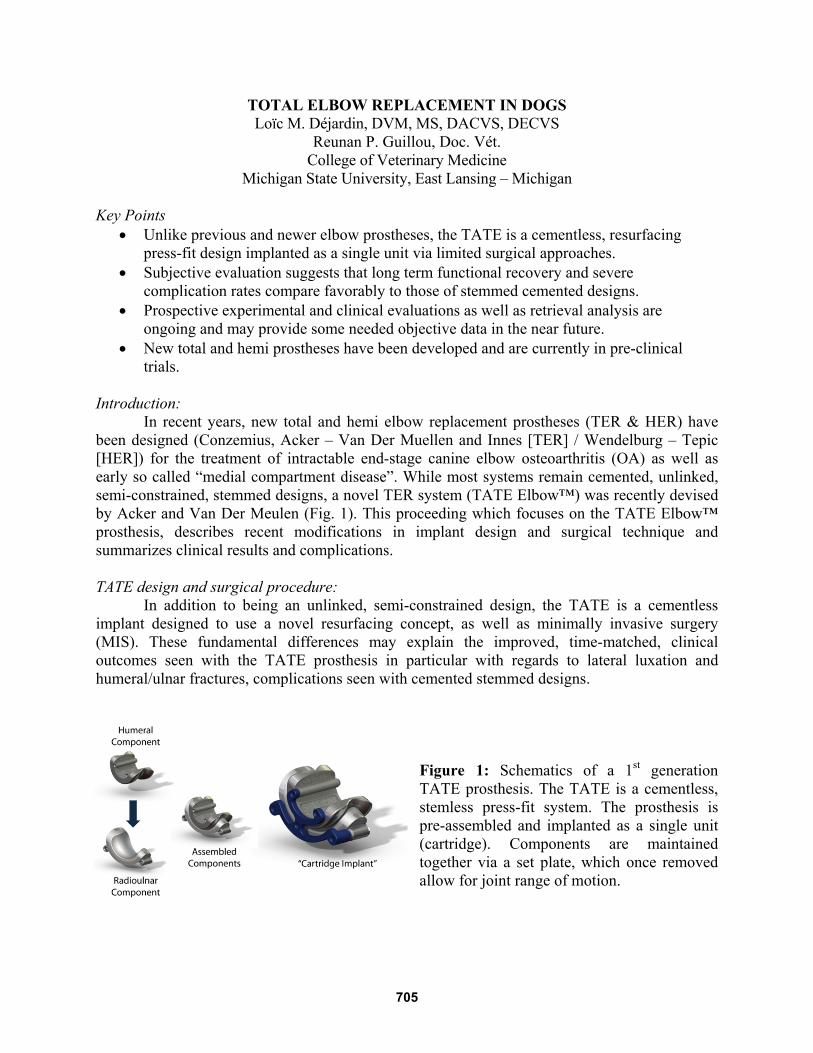
In recent years, new total and hemi elbow replacement prostheses (TER & HER) have been designed (Conzemius, Acker – Van Der Muellen and Innes [TER] / Wendelburg – Tepic [HER]) for the treatment of intractable end-stage canine elbow osteoarthritis (OA) as well as early so called “medial compartment disease”. While most systems remain cemented, unlinked, semi-constrained, stemmed designs, a novel TER system (TATE Elbow™) was recently devised by Acker and Van Der Meulen (Fig. 1). This proceeding which focuses on the TATE Elbow™ prosthesis, describes recent modifications in implant design and surgical technique and summarizes clinical results and complications.
TATE design and surgical procedure: In addition to being an unlinked, semi-constrained design, the TATE is a cementless implant designed to use a novel resurfacing concept, as well as minimally invasive surgery (MIS). These fundamental differences may explain the improved, time-matched, clinical outcomes seen with the TATE prosthesis in particular with regards to lateral luxation and humeral/ulnar fractures, complications seen with cemented stemmed designs.
Figure 1: Schematics of a 1st generation TATE prosthesis. The TATE is a cementless, stemless press-fit system. The prosthesis is pre-assembled and implanted as a single unit (cartridge). Components are maintained together via a set plate, which once removed allow for joint range of motion.
705
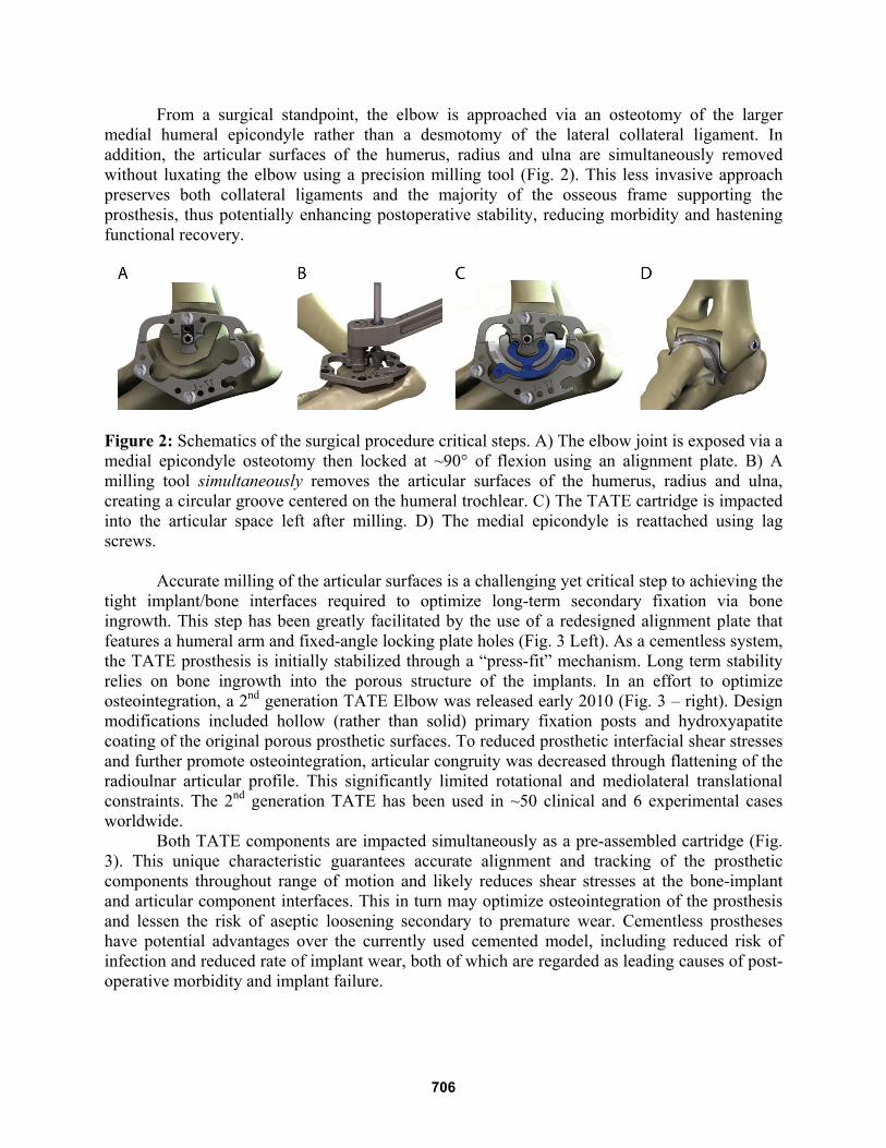
From a surgical standpoint, the elbow is approached via an osteotomy of the larger medial humeral epicondyle rather than a desmotomy of the lateral collateral ligament. In addition, the articular surfaces of the humerus, radius and ulna are simultaneously removed without luxating the elbow using a precision milling tool (Fig. 2). This less invasive approach preserves both collateral ligaments and the majority of the osseous frame supporting the prosthesis, thus potentially enhancing postoperative stability, reducing morbidity and hastening functional recovery.
Figure 2: Schematics of the surgical procedure critical steps. A) The elbow joint is exposed via a medial epicondyle osteotomy then locked at ~90° of flexion using an alignment plate. B) A milling tool simultaneously removes the articular surfaces of the humerus, radius and ulna, creating a circular groove centered on the humeral trochlear. C) The TATE cartridge is impacted into the articular space left after milling. D) The medial epicondyle is reattached using lag screws.
Accurate milling of the articular surfaces is a challenging yet critical step to achieving the tight implant/bone interfaces required to optimize long-term secondary fixation via bone ingrowth. This step has been greatly facilitated by the use of a redesigned alignment plate that features a humeral arm and fixed-angle locking plate holes (Fig. 3 Left). As a cementless system, the TATE prosthesis is initially stabilized through a “press-fit” mechanism. Long term stability relies on bone ingrowth into the porous structure of the implants. In an effort to optimize osteointegration, a 2nd generation TATE Elbow was released early 2010 (Fig. 3 – right). Design modifications included hollow (rather than solid) primary fixation posts and hydroxyapatite coating of the original porous prosthetic surfaces. To reduced prosthetic interfacial shear stresses and further promote osteointegration, articular congruity was decreased through flattening of the radioulnar articular profile. This significantly limited rotational and mediolateral translational constraints. The 2nd generation TATE has been used in ~50 clinical and 6 experimental cases worldwide.
Both TATE components are impacted simultaneously as a pre-assembled cartridge (Fig. 3). This unique characteristic guarantees accurate alignment and tracking of the prosthetic components throughout range of motion and likely reduces shear stresses at the bone-implant and articular component interfaces. This in turn may optimize osteointegration of the prosthesis and lessen the risk of aseptic loosening secondary to premature wear. Cementless prostheses have potential advantages over the currently used cemented model, including reduced risk of infection and reduced rate of implant wear, both of which are regarded as leading causes of post-operative morbidity and implant failure.
706

Clinical outcome: While clinical and experimental studies are ongoing at Michigan State University, no
objective data is available on the clinical outcome of the TATE Elbow system. The following is a compilation of subjective and objective data obtained from 2 clinical centers (Sun Valley Animal Center [Acker – 25 cases] and MSU [Authors – 14 cases]). We emphasize that this information is mostly subjective in nature and therefore should be assessed cautiously. The TATE prosthesis has been implanted in approximately 180 to 200 cases worldwide since July 2007 (~5 years ago).
Figure 3: Left: Schematics of the new alignment plate. To improve stability during milling a humeral arm and locking plate holes were incorporated in this new design. Right: Schematics of the 2nd generation TATE (left to right: the humeral interface, TATE cartridge 3-D view and RU interface). To optimize osteointegration, design refinements include open primary fixation posts (red arrows) and HA coating of the porous surfaces of the prosthetic components (blue arrows). In addition, articular congruity was reduced to limit interfacial sheer stresses.
Subjective clinical evaluation and feedback from dog owners suggest that limb function improves up to 1 year after surgery following a typical aggravation of the lameness between 6 and 12 weeks. Although dogs appear pain free and show improved range of motion, mainly in extension, subtle to mild lameness may persist.
Severe complications (n=3) consisting of two ulnar fractures and one implant loosening were recorded, all within 8 weeks post-operatively. One acute fracture occurred 7 days postoperatively following a fall. Subsequent deep infection due to intense licking of the wound developed over the ensuing week and warranted amputation of the limb. The second ulnar fracture occurred 2 months after surgery and was successfully revised using a combination of a plate and tension band. In one case, excessive periprosthetic osteolysis around the radioulnar component was documented 6 weeks post-operatively. While the dog’s lameness improved for 6 months after surgery, aseptic loosening continued to progress over the following year. Recent lameness aggravation will likely result in explantation and elbow arthrodesis. The rate of severe complications, up to 4 years after surgery, was ~7.7% prior to revision and ~5.5% following successful revision.
Minor complications (n = 3 – ~7.7%) included clinically inconsequential pin migration (1), screw loosening (1), superficial local wound dehiscence (1). All were successfully revised.
Iatrogenic intra-operative complication (n = 1 – ~2.6%) consisting of iatrogenic transection of the ulnar nerve was described by Acker in one dog who remains ambulatory three years post-operatively.
707

Objective force plate analysis was conducted on 6 patients at Michigan State University up to 32 months after implantation of a TATE system (ongoing prospective clinical study). In all cases, pre-operative peak vertical force of the affected limb was significantly lower than normal reported range of 105% to 125% BW at the trot. By 6 to 12 months after surgery, the peak vertical force of the operated limb was greater than that of the contralateral side. Continued improvement was seen at 2 years, as the peak vertical force of the operated limbs had returned to a normal reported value of ~115% BW. One dog underwent bilateral elbow replacement. That dog received a 2nd generation TATE prosthesis 2 years after successful implantation of a 1st generation implant (Fig. 4).
Figure 4: Force plate analysis of a dog that underwent bilateral TATE elbow replacement. Peak vertical force as a percentage of body weight is on the y-axis; time in month is on the x-axis. Please see text for details.
Prospective experimental and clinical evaluations of the TATE system, as well as retrieval analysis are ongoing and may provide needed objective data in the near future (Fig. 5).
708
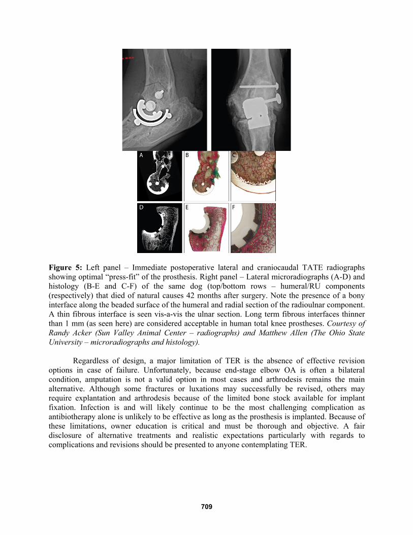
Figure 5: Left panel – Immediate postoperative lateral and craniocaudal TATE radiographs showing optimal “press-fit” of the prosthesis. Right panel – Lateral microradiographs (A-D) and histology (B-E and C-F) of the same dog (top/bottom rows – humeral/RU components (respectively) that died of natural causes 42 months after surgery. Note the presence of a bony interface along the beaded surface of the humeral and radial section of the radioulnar component. A thin fibrous interface is seen vis-a-vis the ulnar section. Long term fibrous interfaces thinner than 1 mm (as seen here) are considered acceptable in human total knee prostheses. Courtesy of Randy Acker (Sun Valley Animal Center – radiographs) and Matthew Allen (The Ohio State University – microradiographs and histology).
Regardless of design, a major limitation of TER is the absence of effective revision options in case of failure. Unfortunately, because end-stage elbow OA is often a bilateral condition, amputation is not a valid option in most cases and arthrodesis remains the main alternative. Although some fractures or luxations may successfully be revised, others may require explantation and arthrodesis because of the limited bone stock available for implant fixation. Infection is and will likely continue to be the most challenging complication as antibiotherapy alone is unlikely to be effective as long as the prosthesis is implanted. Because of these limitations, owner education is critical and must be thorough and objective. A fair disclosure of alternative treatments and realistic expectations particularly with regards to complications and revisions should be presented to anyone contemplating TER.
709